


 , Pedro López-Sanchez 2, Diego Lezama-Martínez 1, Erik Pérez-García 1, M. Fernanda Skat Montoya-Hernández 1, Alberto Aranda-Fraustro 3, Jazmin Flores-Monroy 1,*
1 Laboratorio de Farmacología del Miocardio, Facultad de Estudios Superiores Cuautitlán, Universidad Nacional Autónoma de México, 54740 Estado de México, Mexico
2 Laboratorio de Farmacología Molecular, Escuela Superior de Medicina, Instituto Politécnico Nacional, 11340 Ciudad de México, Mexico
3 Laboratorio de Patología, Instituto Nacional de Cardiología Dr. Ignacio Chávez. CDMX. Tlalpan, 14080 Ciudad de México, Mexico
Abstract
Background: Cardiovascular diseases (CVDs) are the leading cause of
women’s mortality, linked to aging and reduced estrogen during menopause.
Estrogen replacement therapy (ERT) is suggested for CVDs prevention. Yet, its
timing initiation remains contentious. Thus, we aimed to evaluate the effect of
early and late estrogen therapy on cardiac function and lipid metabolism in
ovariectomized old female Wistar rats. Methods: Fifty randomized female
Wistar rats were included in 5 groups (n = 10, 18 months old): (1) Sham, (2) 10
weeks post ovariectomy (Ovx-10 w), (3) 10 weeks post Ovx + early estrogen
replacement therapy (Ovx 10 w-early ERT), (4) 20 weeks post Ovx (Ovx-20 w) and
(5) Ovx 20 w-late ERT. Three days (early ERT) or 10 weeks (late ERT) after
surgery 17-
Keywords
- estrogen-replacement-therapy
- ovariectomy
- aging
- echocardiography
- electrocardiography
- fibrosis
- lipid-metabolism
Cardiovascular diseases (CVDs) are the leading cause of death worldwide. For decades, they have been underestimated in women; consequently, the lack of studies has caused misunderstandings in diagnosis and treatment [1]. Therisk factors for CVD in women are; preeclampsia, gestational diabetes, polycystic ovary syndrome, menopause, hysterectomy, and ovariectomy [2, 3], most of which are related to decreased estrogen levels.
Estrogens mediate actions in the cardiovascular system through different
mechanisms, including the modulation of NO production [4, 5, 6, 7], anti-inflammatory
effects [4, 8], and regulation of mitochondrial function and biogenesis [9].
Furthermore, during menopause, estrogen depletion promotes chronic activation of
the renin-angiotensin system (RAS). The RAS plays an important role in heart
remodeling and contributes to the development of left ventricular hypertrophy
(LVH) and myocardial fibrosis through paracrine and autocrine signaling [10].
Dysregulated RAS activity leads to volume overload and vasoconstriction, causing
increased left ventricular diastolic filling pressures. RAS is also involved in
myocardial and vascular inflammation, mediated by the activation of different
cell types and the secretion of cytokines and chemokines [10]. This inflammation
contributes to myocardial fibrosis, altering left ventricular structure and
geometry [10], decreasing ventricular relaxation [11] and leading to diastolic
dysfunction. In addition, ovariectomy has been related to metabolic disorders
such as dyslipidemia [12]. Dyslipidemia is characterized by an increase in
hepatic triglyceride (TG), low-density lipoprotein (LDL), and cholesterol levels,
and a decrease in high-density lipoprotein (HDL) levels. As a consequence,
atherosclerotic cardiovascular disease risk increases, leading to cardiac
dysfunction [13, 14]. In rodents, ovarian failure induced by bilateral
ovariectomy contributes to the development of heart failure with preserved
ejection fraction (HFpEF) [15, 16]. HFpEF is a complex clinical syndrome with
symptomatic left ventricular dysfunction. It results from impaired ventricular
function or structural abnormalities, leading to inadequate cardiac output
despite a preserved ejection fraction (EF) of
The connection between ovarian hormone depletion and the increase in the incidence of CVD has led to the idea that estrogen replacement therapy (ERT) can protect the cardiovascular system. Therefore, clinical studies have been conducted to clarify these effects [19]. However, the results are controversial. Some studies revealed significant adverse effects on the cardiovascular system [20, 21, 22], while others confirmed the cardioprotective effects of this hormone [23]. The reason for these differences was explained by the population included in the clinical trial, as mainly young women were included. In relation to this, aging is closely related to menopause, leading to molecular, functional, and morphological changes, including progressive inflammation. Aging increases the expression of cell adhesion molecules in the endothelium and other inflammatory biomarkers, that have been correlated with higher cardiovascular risk. Another factor affected by aging is an increase in left ventricle wall thickness, which decreases its dimensions and, consequently, the blood volume, and is accompanied by diastolic dysfunction [24].
The Women’s Health Initiative (WHI) was one of the first organizations to test the cardioprotective effects of ERT in women with hysterectomy, and another study to test this was the Heart and Estrogen/Progestin Replacement Study, which was conducted in women with pre existing CVD. However, the results of both studies failed to corroborate the beneficial effects of ERT in the prevention of CVD [25]. In 2001, Alexander et al. [26] demonstrated that ERT increases the incidence of cardiovascular events when therapy begins after myocardial infarction. Thus, it was postulated that hormones may have adverse cardiovascular effects if therapy is administered to women over 60 years old with pre existing CVD. Additionally, the WHI determined that ERT is not able to prevent CVD in women who initiate therapy long after the beginning of menopause, but is effective when started early. However, this study only evaluated the short-term effects of ERT [27]. Furthermore, in 2012, Schierbeck et al. [28] showed that menopausal women, after 10 years with ERT, the risk of mortality and incidence of heart failure or myocardial infarction was significantly reduced, without an apparent increase in the risk of breast cancer or venous thromboembolism. However, at the neuronal level, López et al. [29] showed that early but not late ERT has beneficial effects on oxidative stress, glucose reuptake, and metabolic profiles. Therefore, one of the most important factors to consider in the use of ERT is the time from hormonal loss to the beginning of therapy administration. Despite this, the role of early estrogen intervention in preserving left ventricle function in postmenopausal women has received little attention. In animal models, ovariectomy leads to remodeling of the left ventricle, which can be avoided by ERT through G-protein-coupled estrogen receptor (GPER) activation [30, 31]. For those reasons, in this work we aim to determine the effect of early and late ERT on cardiac function and lipid metabolism in old female Wistar rats.
Fifty aged female Wistar rats (18 months old) were obtained from Facultad de Estudios Superiores Cuautitlán UNAM animal facility. The animals were kept at 24 °C under 12 h of darkness and 12 h of light, with free access to water and food. All procedures were carried out following the guidelines of the Federal Regulation for the experimentation and care of animals (SAGARPA, NOM-062-ZOO, 1999, Mexico) and National Institutes of Health Guide for the Care and Use of Laboratory Animals (NIH Publications No. 8023, revised 1978, USA). This project was conducted under the authorization of the Internal Committee for the Care of Experimental Animals of FES Cuautitlán (C19_13).
The animals were randomized in to 5 groups as follows: (1) Sham; (2) 10 weeks
post ovariectomy (Ovx 10 w); (3) Ovx 10 w + Early estrogen replacement therapy
(Ovx 10 w-early ERT); (4) 20 weeks post-ovariectomy (Ovx 20 w); (5) Ovx 20 w +
Late estrogen replacement therapy (Ovx 20 w-late ERT). The animals were
anesthetized and underwent to bilateral ovariectomy, as we have previously
described [32]. 17-
A non-invasive technique, which was performed under anesthesia with ketamine-xylazine (40 mg/kg and 15 mg/kg). The rat was placed in dorsal decubitus position and the chest was shaved. The heart is monitored by a high frequency linear transducer on the skin (16 Mhz) connected to a Sonoscape X3V ultrasound equipment in M mode and B mode from parasternal long and short axis, as Zacchigna et al. [33] previous reported.
To assessed hemodynamics, the rat was placed on a dissection table. Subsequently, a tracheostomy was performed, and the right carotid artery of the muscle and vagus nerve was dissected. In the carotid artery a catheter (PE-10 previously heparinized) connected to a pressure transducer was placed (model MP100WSW; Biopac Systems Inc, Santa Barbara, CA). The catheter was inserted into the left ventricle to measure left ventricular systolic pressure (LVSP), left ventricular diastolic pressure (LVPD), maximum range of isovolumetric pressure development (+dP/dt max) and –dP/dt max, as well as heart rate (HR). The register was obtained using the AcqKnowledge 1.8 software (Biopac Systems Inc) [34].
After cardiac catheterization, electrocardiography was conducted. Electrodes were connected above the skin, left hind leg, left front leg and chest to obtain the standard limb lead II surface electrocardiogram (ECG100C, Biopac Systems, CA, USA). The data was recorded with a data acquisition system (MP100, Biopac Inc, CA, USA). To calculate the corrected QT interval, the normalized Bazett’s formula was used (QTcn-B = QT / (RR / f )1/2) as Kmecova et al. [35] previously reported.
This parameter was estimated by the weight index of the heart. It was calculated using the following formula: Hwi = Hw/Bw; where Hwi = heart weight index; Hw = heart weight; Bw = body weight of the rat, as we have previoulsly reported [34].
The heart was horizontally cut into three slices and fixed in 10% formaldehyde (pH 7.4). The tissue was processed by Hematoxylin-Eosin and Masson’s trichrome staining. To evaluate the cardiomyocytes area and collagen deposition, Motic Images Plus version 3.0 Software (Schertz, TX, USA) was used, the images were captured with a camera attached to the Motic Series B1-253SP optical microscope (San Antonio, TX, USA) and AmScope software 4.11.20131.20220108 (Irvine, CA,USA).
Cholesterol, HDL, LDL and TG were determined in serum by spectrophotometry at
the end of treatment in all experimental groups. The assay was conducted according to the supplier instructions (CT series, TG 1780111, HDL 1220231, LDL 1220220, Chol 1400060 Wiener Lab, Rosario, Argentina). 17
All data is presented as mean
To confirm the effectiveness of estrogen replacement therapy we measured
17-
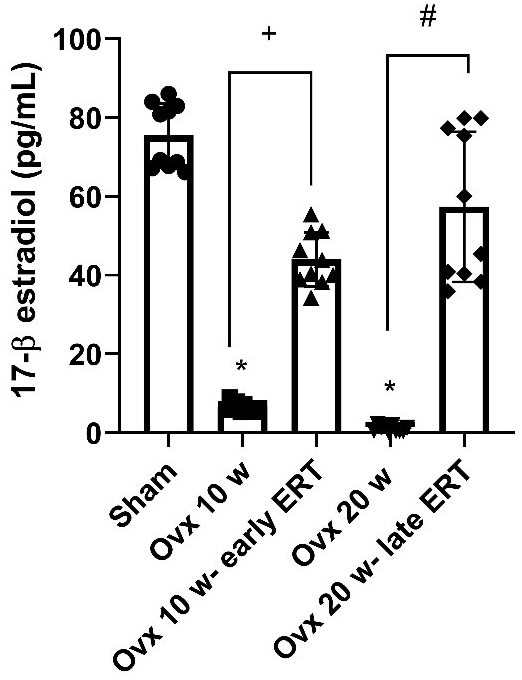 Fig. 1.
17-
To confirm the effectiveness of ovariectomy surgery, the uterus weight index was determined (Table 1). This index estimates whether the uterus presents atrophy because of ovarian surgical removal. Table 1 shows that all groups with bilateral ovariectomy had a lower weight index than the Sham group. However, among the ovariectomy groups, the Ovx 10 w- early ERT group had the highest weight index. Aditionally, body weight was measured at the end of each treatment, and Table 1 shows that the body weights were higher in the Ovx 10 w and Ovx 20 w groups than in the Sham group. Regarding cardiac weight index, no significant differences were found, although there is a trend towards an increase in the Ovx 10 w and Ovx 20 w-late ERT groups compared to the Sham group.
| Group | Uterus weight index (g/g) | Body weight (g) | Heart Weight Index (g/g) |
| Sham | 0.38 |
369 |
0.31 |
| Ovx 10 w | 0.20 |
410 |
0.40 |
| Ovx 10 w-early ERT | 0.28 |
389 |
0.34 |
| Ovx 20 w | 0.15 |
455 |
0.31 |
| Ovx 20 w-late ERT | 0.16 |
376 |
0.43 |
These results are the mean
The uterus weight index decreased in all groups compared to the. Sham
group (*p
Echocardiography was used to assess cardiac function (Fig. 2). The ejection fraction (EF) (Fig. 2A) was significantly reduced in the Ovx 20 w group compared to the Sham group, while in the rest of the groups no changes were observed. Furthermore, the shortening fraction (Fig. 2B) was decreased in all ovariectomized groups compared to the Sham group. In contrast, the left ventricle internal dimension at end-systole (LVIDS) (Fig. 2C) was significantly increased in all groups compared to the Sham group. Likewise, the end systolic volume (ESV) (Fig. 2D) was significantly higher in the Ovx 20 w group than in the Sham group. Finally, left ventricle (LV) mass (LVM) (Fig. 2E) was significantly lower in the Ovx 20 w than in the Ovx 10 w group. No changes were found in the rest of the echocardiographic parameters (Table 2).
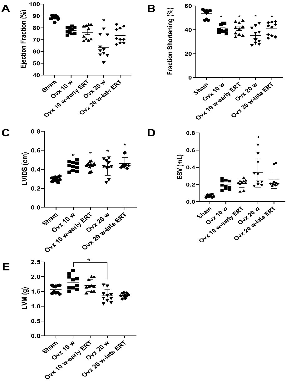 Fig. 2.
Modifications of cardiac function in Wistar rat with early and
late estrogen replacement therapy. (A) The ejection fraction was decreased in
the Ovx 20 w group compared to the Sham group (*p = 0.032). (B) All Ovx
groups presented lower shortening fraction values than the Sham group (p = 0.03). (C) LVIDS was higher in all Ovx groups than in the Sham group
(*p = 0.020). (D) The ESV was increased in the Ovx 20 w group
(*p = 0.049) compared to the Sham group. (E) Finally, LVM was decreased
in the Ovx 20 w group than in the Ovx 10 w group (*p
| Parameter | Sham | Ovx 10 w | Ovx 10 w-early ERT | Ovx 20 w | Ovx 20 w-late ERT |
| IVSd (cm) | 0.24 |
0.24 |
0.20 |
0.19 |
0.16 |
| LVIDd (cm) | 0.64 |
0.74 |
0.75 |
0.77 |
0.77 |
| LVPWd (cm) | 0.25 |
0.26 |
0.25 |
0.18 |
0.19 |
| IVSs (cm) | 0.40 |
0.36 |
0.31 |
0.41 |
0.28 |
| LVIDs (cm) | 0.30 |
0.44 |
0.45 |
0.40 |
0.47 |
| LVPWs (cm) | 0.31 |
0.34 |
0.33 |
0.54 |
0.26 |
| EDV (mL) | 0.60 |
0.90 |
0.96 |
0.79 |
1.03 |
| ESV (mL) | 0.07 |
0.21 |
0.22 |
0.34 |
0.26 |
| EF (%) | 89 |
76 |
76 |
68 |
75 |
| SV (mL) | 0.54 |
0.69 |
0.73 |
0.52 |
0.77 |
| FS (%) | 54 |
40 |
40 |
35 |
40 |
| LVM (g) | 1.6 |
1.8 |
1.7 |
1.4 |
1.4 |
The EF was decreased in the Ovx 20 w groups compared to the Sham group
(*p = 0.032. All Ovx groups presented lower FS values than the Sham
group (*p = 0.030). LVIDS was higher in all Ovx groups than in the Sham
group (*p = 0.020). The ESV was increased in the Ovx 20 w group
(*p = 0.049) than in the Sham group. Finally, LVM was lower in the Ovx
20 w group than in the Ovx 10 w group (
These results are the mean
Regarding, the electrical activity of the heart, the P wave amplitude was lower in the Ovx 20 w group than in the. Sham group (Table 3). However, the QRS complex amplitude was higher in the Ovx 20 w and Ovx 20 w-late ERT groups than in the Ovx 10 w and Ovx 10 w-early ERT groups respectively.
| Parameter | Sham | Ovx 10 w | Ovx 10 w-early ERT | Ovx 20 w | Ovx 20 w-late ERT |
| RR (ms) | 309.3 |
325.0 |
326.3 |
282.5 |
361.0 |
| HR (bpm) | 195.8 |
203.7 |
192.5 |
233.2 |
172.9 |
| P wave (ms) | 24.2 |
20.0 |
25.0 |
18.8 |
24.0 |
| PR (ms) | 83.3 |
73.8 |
87.5 |
90.0 |
86.0 |
| QRS (ms) | 28.3 |
21.3 |
23.0 |
32.5 |
31.0 |
| QT (ms) | 119.2 |
103.8 |
118.8 |
91.3 |
93.0 |
| QTc (ms) | 83.3 |
73.0 |
80.7 |
66.8 |
62.4 |
| ST (ms) | 5.8 |
13.8 |
10 |
5.0 |
11.0 |
The P wave amplitude was lower in the Ovx 20 w group than in the Sham group
(*p = 0.013). The QRS complex amplitude was increased in the Ovx 20 w
and Ovx 20 w-late ERT groups compared to the Ovx 10 w and Ovx 10 w-early ERT
groups respectively (
For the rest of the electrocardiographic measurements, no significant differences were found (Table 3). However, Fig. 3 shows the most important alterations found in all experimental groups. To compare our results with a normal ECG record, we show the record from a young Sham rat young record (12 weeks old) as a normal ECG. Right axis deviation with deep S wave was observed in some records in the Sham group (Fig. 3B). ST segment depression with an ascending T wave was observed in the Ovx 10 w group (Fig. 3C). Downsloping ST depression was observed in one subject in the Ovx 10 w-early ERT group (Fig. 3D) and two subjects had atrial fibrillation (Fig. 3E). On the other hand, in the Ovx 20 w group, the ST segment elevation was observedin 7 subjects (Fig. 3F–H), right axis deviation with a deep S wave (Fig. 3G) and the inversion of the P wave was observed in two subjects (Fig. 3H), and QRS fragmentation with an “M” pattern was observed in 4 subjects (Fig. 3I). Finally, in the Ovx 20 w-late ERT group, inverted P waves were also observed in 2 subjects with (Fig. 3J), and ST elevation accompanied by the elevation of the P wave and its elongation were found in 4 subjects (Fig. 3K).
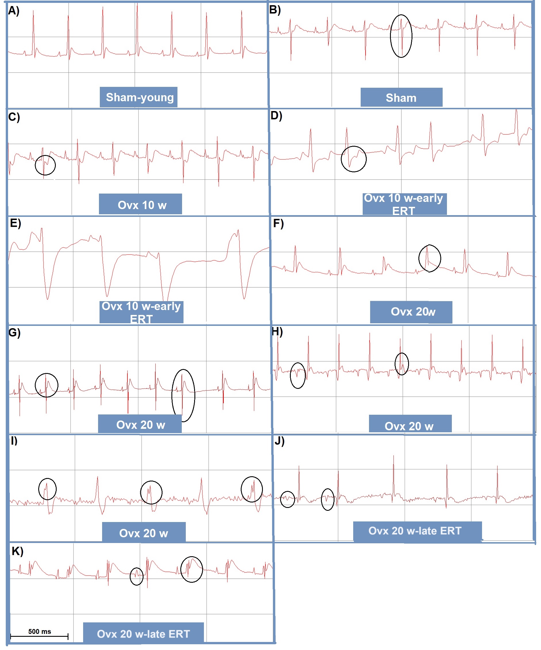 Fig. 3.
Alterations in ECG morphology in female Wistar rats with early and late ERT. Representative electrocardiographic records with relevant alterations of each experimental group. (A) A young rat of the Sham group (12 weeks old). (B) Sham: deep S wave. (C) Ovx 10 w: ST-segment depression. (D) Ovx 10 w-early ERT: ST-segment depression. (E) Ovx 10 w-early ERT: atrial fibrillation. (F) Ovx 20 w: ST elevation. (G) Ovx 20 w: ST elevation and deep S wave. (H) Ovx 20 w: inversion of P wave with ST elevation. (I) Ovx 20 w: fragmented QRS complex with an “M” pattern. (J) Ovx 20 w-late ERT: inversion of P wave. (K) Ovx 20 w-late ERT: elevation of P and T wave, ST elevation and fragmented QRS complex.
Subsequently, arterial blood pressure and heart rate (HR) were measured in all experimental groups, and changes were observed. However, these changes were not due to estrogen therapy but to the postovariectomy time (Table 4). A significant increase in diastolic blood pressure (DBP) was observed in the 20-week ovariectomy groups, with and without ERT. Although we found no significant differences, mean arterial pressure (MAP) tendeds to be lower in the Ovx 10 w and Ovx 10 w-early ERT groups than in the Sham group. Likewise, systolic blood pressure (SBP) was decreased in the Ovx 10 w and Ovx 10 w-early ERT groups compared to the Sham group. However, HR tended to decrease in all groups, except the Ovx 20 w-late ERT group, compared to the Sham group.
| Parameter | Sham | Ovx-10 w | Ovx-20 w | Ovx-10 w early ERT | Ovx-late ERT |
| MAP (mmHg) | 92 |
88 |
94 |
87 |
93 |
| SBP (mmHg) | 120 |
112 |
120 |
114 |
117 |
| DBP (mmHg) | 76 |
78 |
84 |
76 |
85 |
| HR (lat/min) | 373 |
350 |
357 |
353 |
383 |
DBP was increased after 20 weeks postovariectomy with and without ERT groups
compared to the Sham group (*p = 0.047). MAP, mean arterial pressure;
SBP, systolic blood pressure; DBP, diastolic blood pressure; HR, heart rate.
These results are the mean
Despite the changes observed in the ECG, echocardiogram, and arterial blood pressure, in the ventricular hemodynamic parameters the only significant difference was found in the isovolumetric index of relaxation (–dP/dt), where it was decreased in the Ovx 20 w and Ovx 10 w groups compared to the Sham group (Fig. 4D).
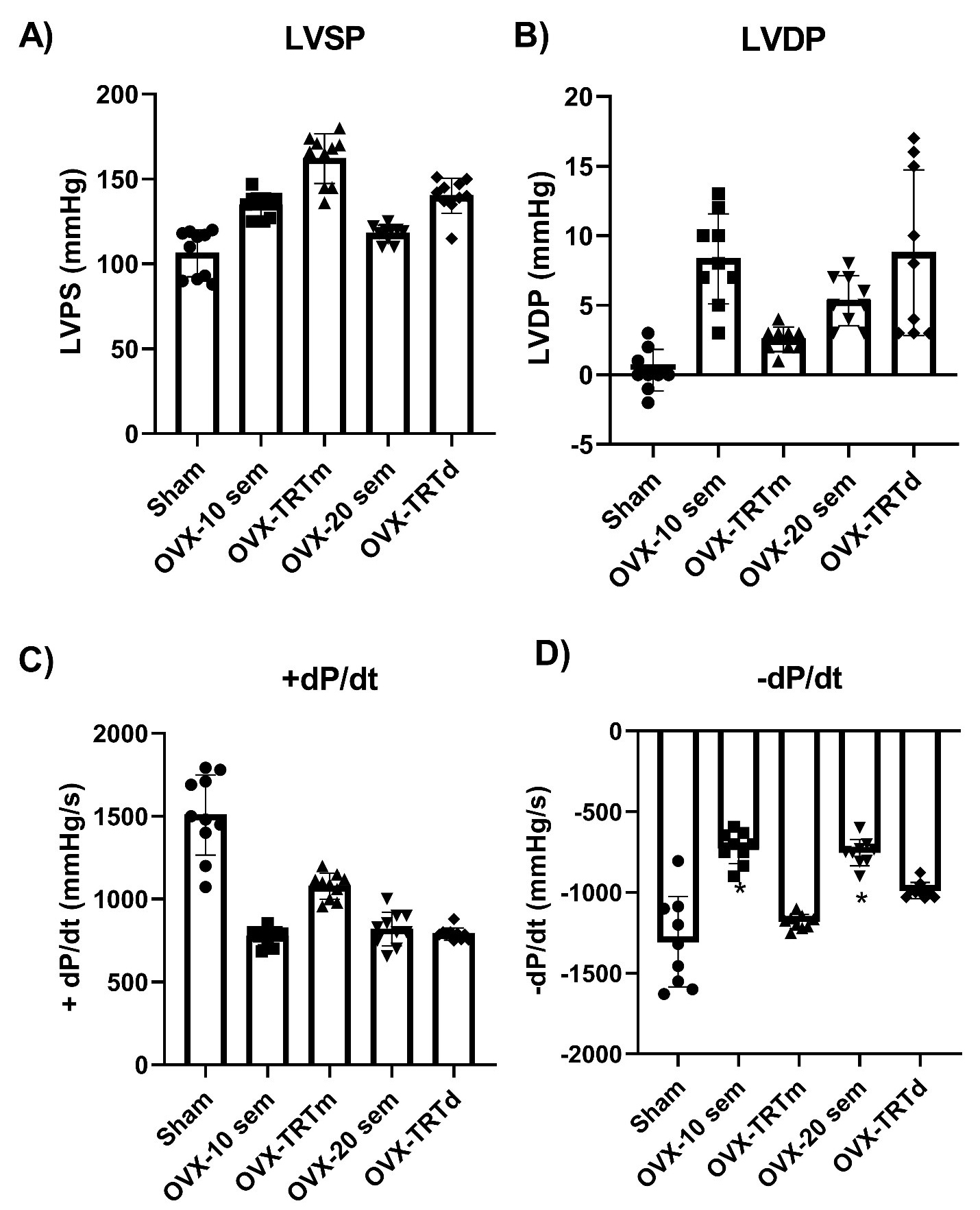 Fig. 4.
Hemodynamic parameters in Wistar rats with early and late
estrogen replacement therapy. Left ventricle hemodynamic
parameters in ovariectomized female rats with early and late ERT. (A) Left
ventricle systolic pressure (LVSP). (B) Left ventricle diastolic pressure (LVDP).
(C) Isovolumetric index of contraction (+dP/dt). (D) Isovolumetric index of
relaxation (–dP/dt), Ovx 20 w and Ovx 10 w groups decreased –dP/dt vs
Sham (*p = 0.034). These results are the mean
For the study of cardiac morphology, the cardiomyocyte area and the percentage of collagen deposition in the LV were determined. In addition, a qualitative histological analysis was performed. Fig. 5A shows that the Ovx 20 w group had smaller cardiomyocytes than the Ovx 10 w group, which had similar values to those of the Sham group. No significant differences were found among the rest of the groups.
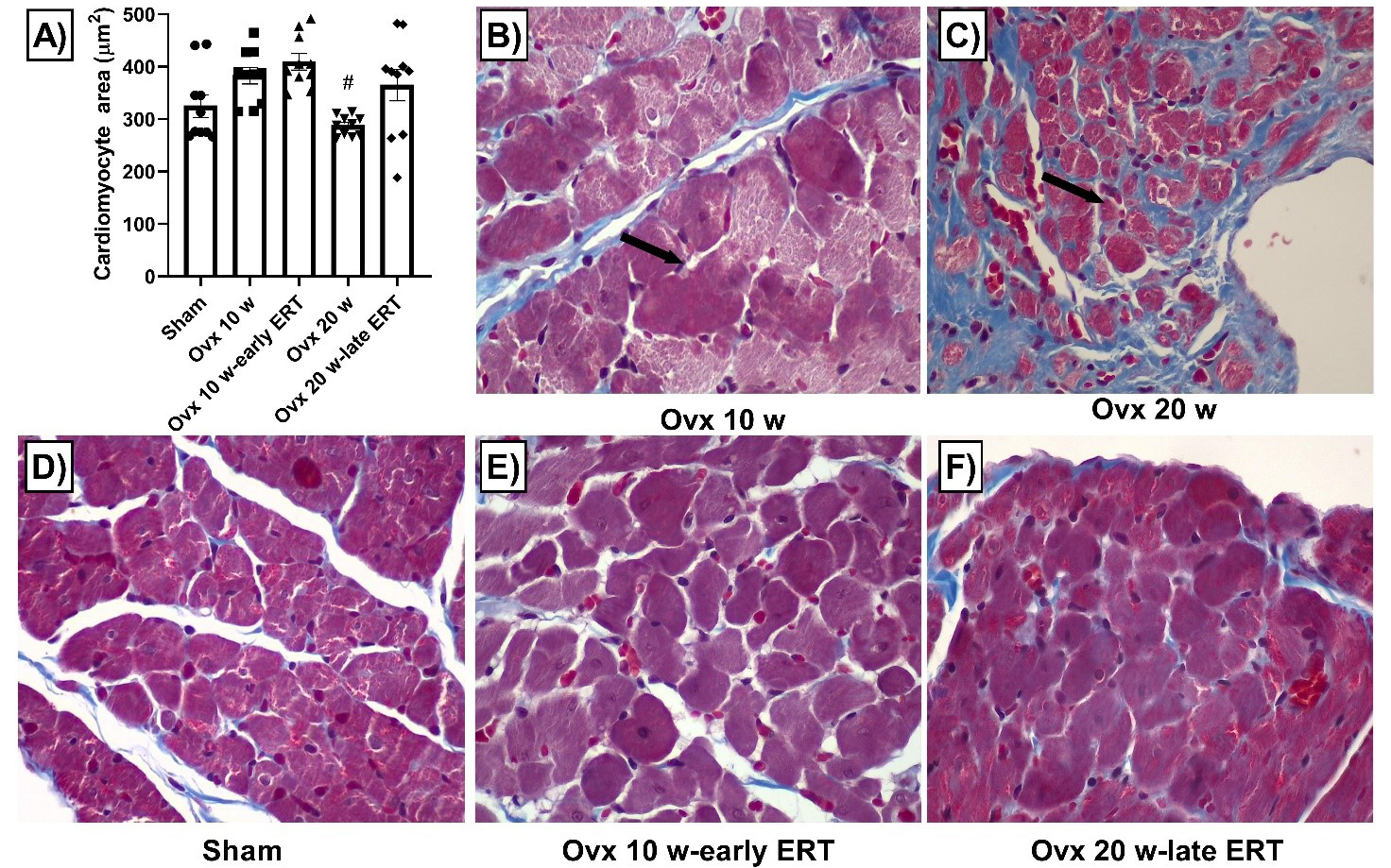 Fig. 5.
Cardiomyocyte area from ovariectomized rats with early and late
ERT. Masson stain, 400X, left ventricle. (A) Cardiomyocyte area. Ovx 20 w had a
lower cardiomyocyte area vs Ovx 10 w (
Collagen deposition (Fig. 6A), was significantly increased only in the 10 weeks post ovariectomy group compared to the Sham group. In addition, in Fig. 6, we show representative photographs of each group, where a greater amount of collagen can be observed in the Ovx 10 w group.
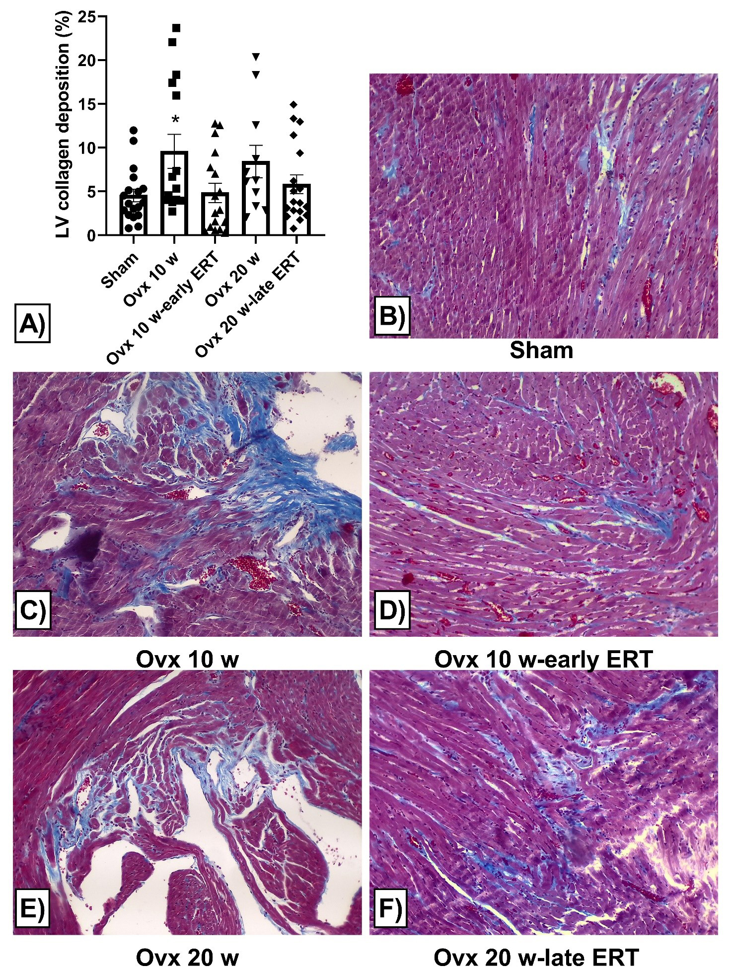 Fig. 6.
Alterations in left ventricle (LV) collagen deposition of ovariectomized rats
with early and late ERT. Masson stain, 100x, left ventricle. (A) LV collagen
deposition (%). LV was increased in the Ovx 10 w group compared to the Sham
group (*p
To study the morphological abnormalities that may occur in the myocardium due to ovariectomy, a histopathological analysis was performed. The most important findings are shown in Fig. 7.
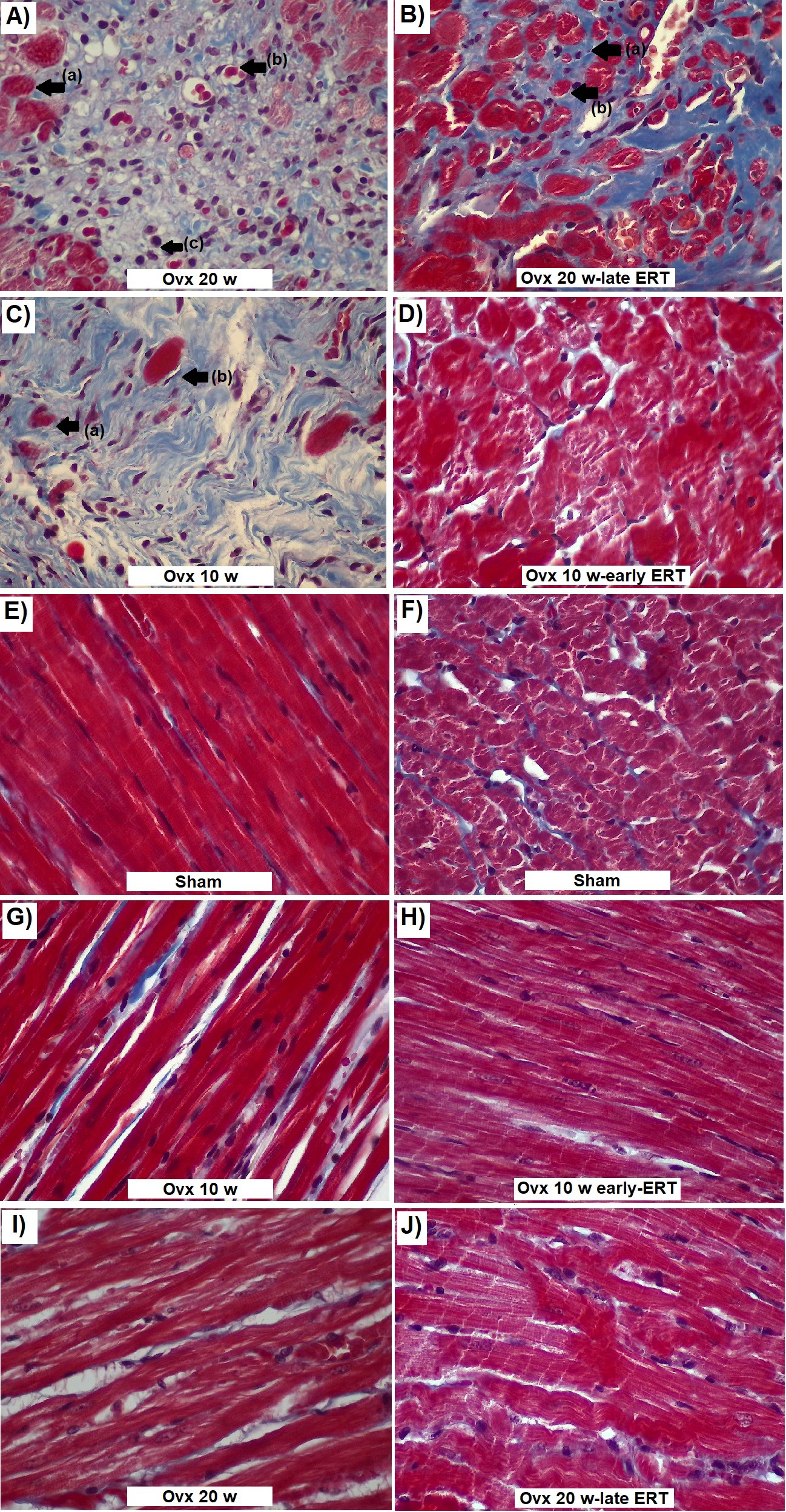 Fig. 7.
LV abnormalities after ovariectomy with early and late ERT. Masson stain 400X, LV free wall. (A) Ovx 20 w myocytes. Scar tissue with collagen deposition, myocytes without nucleus (a), apoptotic vacuoles (b), infiltration of inflammatory cells (c). (B) Ovx 20 w-late ERT myocytes. Interstitial collagen deposition (a), atrophied myocytes (b). (C) Ovx 10 w myocytes. Scar tissue with collagen deposition, myocytes without nucleus (a), atrophied myocytes (b). (D) Ovx 10 w-early ERT increase in myocyte size. (E) Sham cardiac fibers. Normal morphology of cardiac fibers in aged females (F) Sham myocytes. Normal morphology of myocytes in aged females. (G) Ovx 10 w cardiac fibers. Fiber derangement and collagen deposition. (H) Ovx 10 w-early ERT. Disarray of cardiac fibers with striations. (I) Ovx 20 cardiac fibers. Fiber thinning and derangement. (J) Ovx 20 w-late ERT cardiac fibers. cardiac fibers with strations.
In Fig. 7, representative photographs of each group are shown. In the Sham group (Fig. 7E,F) we observed deposition of interstitial collagen in the LV and normal morphology of cardiac fibers in aged females, which displayed slight alterations in the arrangement of the cardiac fibers (Fig. 7E). In the 10 weeks postovariectomy, disarray of cardiac fibers with striations (Fig. 7G), loss of nuclei (Fig. 7C,a), deposition of collagen in scar tissue indicative of ischemic processes (Fig. 7C) and atrophied myocytes (Fig. 7C,b) were observed. In the Ovx 10 w-early ERT group, there seemed to be an increase in myocyte size (Fig. 7D). Some areas were observed with a pale tone and absence of defined myocardial fibers, however, most of the cardiac fibers had a similar appearance to those in the Sham group.
In the Ovx 20 w group (Fig. 7A), we observed areas with scar tissue characterized by collagen deposition and large ischemic areas with myocytes without nuclei (a), apoptotic vacuoles (b) and leukocyte infiltration (c). Ischemic areas were mainly present in the LV free wall and papillary muscles and were found in all subjects in the Ovx 20 w group. Fiber derangement, thinning and the absence of nuclei, which are characteristics of necrotic bands, were observed (Fig. 7I). Finally, in the Ovx 20 w-late ERT group, narrowing of the cardiac fibers left a greater space between them (Fig. 7I), and contracted bands with loss of nucleus were observed (Fig. 7I, Regarding collagen deposition, an increase in interstitial and replacement tissue was found (Fig. 7A).
As alterations in lipid metabolism are one of the major risk factors that contribute to CVD, the serum levels of cholesterol, triglycerides, HDL and LDL were determined (Fig. 8).
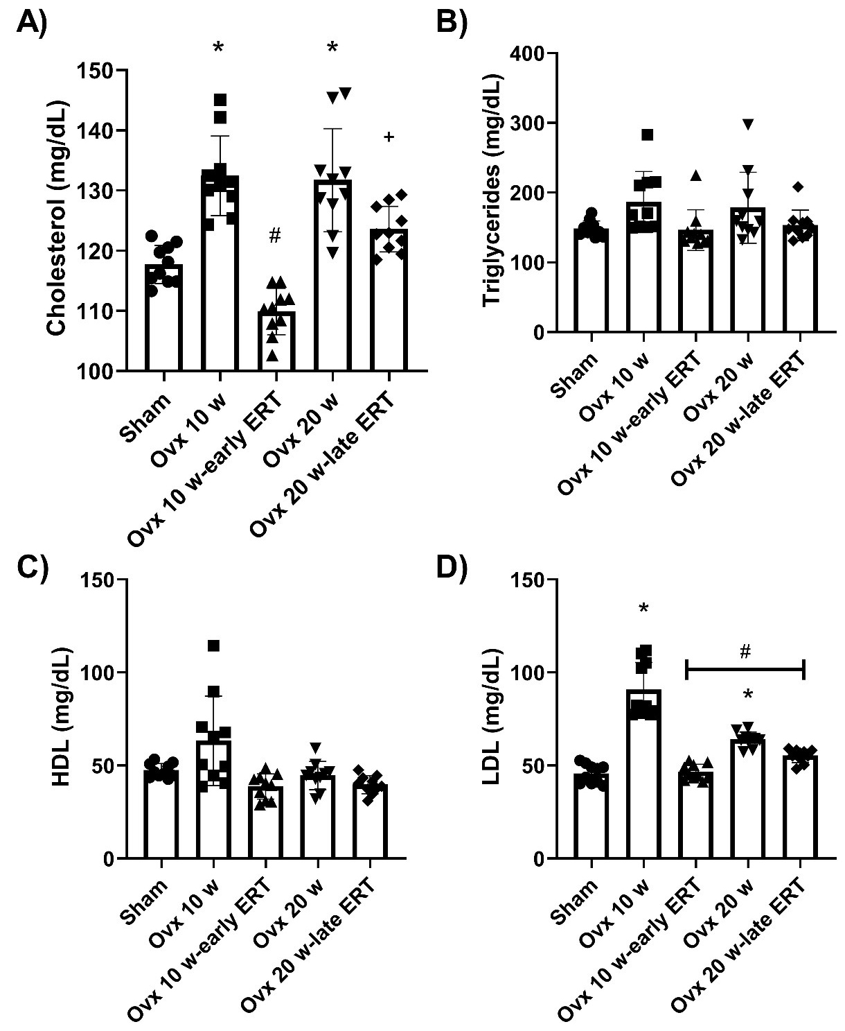 Fig. 8.
Alterations in lipid metabolism of ovariectomized rats with
early and late ERT. Serum levels of cholesterol, triglycerides, HDL and LDL. (A)
Cholesterol; Ovx 10 w (*p = 0.008) and Ovx 20 (*p = 0.019)
increased vs Sham. Ovx 10 w-early ERT had lower cholesterol levels than
Ovx 10 w (
Triglyceride and HDL levels were not significantly different, although triglyceride levels tended to increase in the Ovx 10 w and Ovx 20 w groups (Fig. 8B). The highest levels of cholesterol (Fig. 8A) were observed in the Ovx 10 w and Ovx 20 w groups. However, the serum cholesterol were lower in the group with early ERT than in. their control group (Ovx 10 w) or in the late ERT group (Ovx 20 w-late ERT). Finally, the Ovx 10 w group showed the highest LDL levels, and these levels were significantly increased in the Ovx 20 w group compared to the Sham group (Fig. 8D).
Our study shows that estrogen depletion causes an increase in body weight and promotes dyslipidemia. These metabolic alterations cause an imbalance in the heart energy demand that promotes ischemia and myocardial infarction; this was confirmed in our results by ST elevation and QRS complex fragmentation, leading to cardiac remodeling. This cardiac adaptive response, increased perivascular, interstitial and replacement fibrosis leading to cardiomyocyte hypertrophy. At 20 weeks postovariectomy cardiomyocytes returned to their normal size, with thinning of cardiac fibers. All these changes promote myocardial contractile dysfunction, reflected in EF and FS decreases, as well as in the diastolic function noticed by the increase in DBP and decrease in –dP/dt.
Our results showed that both early and late ERT prevented the decrease in –dP/dt and the increase in LDL. However, only early ERT prevented the increase in cholesterol and collagen deposition and inhibited the development of ischemic events and myocardial fiber derangement. Despite these positive effects, we observed some electrocardiographic alterations indicating ischemic processes in the treated groups, but in a smaller proportion in the early ERT group. In line with our outcomes, the timing hypothesis proposes that ERT should be initiated in women under 60 or within the first 10 years of menopause to achieve cardiovascular benefits [36]. Clinical trials, including the Kronos Early Estrogen Prevention Study (KEEPS) [37, 38] and the Early Versus Late Intervention Trial (ELITE) [39] tested this theory. KEEPS failed to demonstrate that ERT slows atherosclerosis progression over four years [38, 40]. In contrast, ELITE revealed that early ERT slowed atherosclerosis, while late initiation had limited effect [39]. A posttrial ELITE analysis supported this, showing that higher estrogen levels reduced atherosclerosis progression after early ERT but increased it after late initiation [41]. Our results showed that early ERT, at a dose of 5 µg/kg/day, can reduce the severity of lipid metabolism, cardiac function and morphology alterations caused by bilateral ovariectomy; however, it is not able to prevent all cardiometabolic changes.
In this study, a menopause model was established in 18-month-old rats through bilateral ovariectomy. Ovariectomized rats show important changes such as increased body weight, uterine atrophy and low estrogen levels [42] as confirmed in our results (Fig. 1, Table 1). These changes return to values similar to those of the sham group with the administration of ERT.
Menopause is associated with alterations in cardiac function [43]. In our findings, EF was lower in the Ovx 20 w group. Despite this significant decrease, this parameter was within the normal range (54 to 74%) [44, 45]. However, this result should be noted since recent studies have highlighted that EF should be interpreted with caution for the diagnosis of HF in women [43]. Women do not experience a significant decrease in EF with age [43], and generally have higher values throughout their lives compared to men [43]. Therefore, considering female EF as reduced or preserved cannot be based on the parameters established in the male sex [46]. Taking this into account, the decrease in EF after 20 weeks postovariectomy could indicate the onset of ventricular systolic dysfunction, even though it remains within the normal range.
The decrease in EF observed in the ovariectomy groups is associated with a reduction in the shortening fraction (FS). Thus, the reduction in FS is related to the decrease in EF and therefore to increase in LVIDS, as confirmed in our results (Fig. 2). The increase in LVIDS is related to a lower ejected stroke volume (SV). However, in our results, the SV seems to increase, which could be attributed to a volume overload [47]. Yuhan Wang et al. [42] demonstrated that EF and FS decrease after 16 weeks post-ovariectomy, while LVIDS increases. However, this is not observed at 4 or 8 weeks after surgery. This confirms our results and indicates that changes in cardiac function due to ovariectomy are time dependent.
In this study, we also observed an elevation in diastolic blood pressure (DBP) 20 weeks postovariectomy with or without ERT (Table 3). Some studies have shown that ovariectomy leads to a time-dependent increase in blood pressure (BP) [48] which could explain the absence of changes after 10 weeks postovariectomy. Regarding ventricular parameters, we found significant changes only in the isovolumetric relaxation index (–dP/dt), which is associated with a nonsignificant increase in left ventricular diastolic pressure (LVDP) in ovariectomy groups without ERT. Increased LVDP is often considered an early indicator of HF with diastolic dysfunction and is reflected by decreased –dP/dt [49]. Our findings suggest that ovariectomy directly affects left ventricular relaxation, impairing the ability of the heart to relax before ventricular filling.
Electrocardiographic (ECG) alterations were observed in all experimental groups. In aged females, an increase in the depth of the S wave was observed, as a possible indicator of lung damage [50]. In the Ovx 10 w group, downsloping ST depression was observed, which is commonly associated with subendocardial ischemic events [51, 52]. In Ovx 10 w-early ERT, we observed one subject with ST-segment depression and two with atrial fibrillation. The association between estrogen levels and atrial fibrillation has not been conclusively demonstrated, with some studies showing no correlation in women [53]. However, Tsai et al. [54] showed that equine conjugated estrogens have been associated with a higher incidence of atrial fibrillation and ischemic events in Taiwanese women.
At 20 weeks postovariectomy with or without ERT, more significant alterations were observed, including ST segment elevation, P-wave inversion and QRS fragmentation (fQRS). All these changes are directly or indirectly related to ischemic processes. In agreement with our results, Dhote et al. [55] demonstrated that females with ovariectomy and ischemia show ST-segment elevation until reperfusion. fQRS (Fig. 3I), characterized by additional peaks in the QRS complex [56, 57], indicates myocardial scars and is prognostic of ventricular dysfunction [58]. Additionally, the increase in QRS complex amplitude in the Ovx 20 w groups (Table 3) is associated with delayed electrical impulse propagation due to interstitial fibrosis [59].
Finally, only in the Ovx 20 w-late ERT group, was an increase in the P wave amplitude observed, resulting in a wide P wave (Fig. 3K). Abnormalities in atrial activation, are often associated with left atrial hypertrophy usually due to mitral stenosis [60].
In our study, ovariectomy exacerbated fibrosis by increasing collagen deposition (Fig. 6A). Interstitial fibrosis involves the diffuse deposition of collagen around cardiomyocytes and was observed mainly in the Ovx 10 w and Ovx 20 w groups as a response to the increase in LV work [61]. Perivascular fibrosis was observed in the Ovx 20 w and late ERT groups, which involves the accumulation of collagen fibers in the adventitious layer of coronary arteries. This lead to reduced oxygen distribution and promotes cardiac ischemia [61]. Finally, replacement fibrosis occurs during the repair of necrotic cardiomyocytes after conditions such as myocardial infarction [62]. In our study, ovariectomy led to ischemic events and cell death in the heart, as indicated by large scar areas characterized by replacement fibrosis, although to a lesser extent in the early ERT group (Fig. 7). Previous research by Lee et al. [63] demonstrated that ovariectomy activates Fas-dependent apoptosis.
The fewer ischemic areas observed in our early ERT group, may be attributed to
the cardioprotective role of estradiol. In support of our results Liu et
al. [64] showed that the activation of GPER can attenuate fibrosis and the
inflammatory response by inhibiting the TGF
As a part of a the remodeling process, we found infiltration of immune cells in
the Ovx 20 w group (Fig. 7A), indicating an inflammatory process. Chronic
low-grade inflammation plays a role in promoting fibrosis in heart failure [61].
In connection with these findings, Stice et al. [66], reported that
ovariectomy in aged rats increases the expression of TNF
Our results suggest that ovariectomy after 10 weeks leads to slight
cardiomyocyte hypertrophy (Table 1, Fig. 2, Fig. 5). In aged females, ovariectomy
leads to an increase in LV mass increase through the overexpression of
Ovariectomy has been associated with dyslipidemia, a process characterized by abnormal levels of cholesterol, triglycerides (TGs), low-density lipoproteins (LDLs) and high-density lipoproteins (HDLs) [13, 70]. In our results, we observed such changes in cholesterol and LDL levels, both dependent on postovariectomy time (Fig. 8). In relation to the cholesterol elevation (Fig. 8A), it has been demonstrated that ovariectomy promotes cholesterol accumulation in the liver and arterial deposition mediated by LDL [71]. This, may be due to the imbalance between the activity and synthesis of LDL and HDL, effects that were reversed with ERT.
The main function attributed to HDL is the removal and transport of free cholesterol from cells and other lipoproteins to the liver, preventing the formation of atherosclerotic plaques [72]. In our results, total HDL levels remained unchanged (Fig. 8C). Additionally, Argeri et al. [73] showed that there are no changes in HDL levels after 24 weeks postovariectomy, However, HDL alterations are complex and dependent on postmenopausal time, with changes in the proportions of HDL subclasses [72].
The importance of LDL increase in ovariectomized groups without ERT (Fig. 8D) is related to the effects of these molecules when oxidized. Oxidized LDL causes macrophages activation in the arteries, leading to the formation of atherosclerotic plaques. The rupture of this plaque is the main cause of myocardial infarctions and ischemic processes [13]. Ngo et al. [74] reported that LDL increases can be avoided with high doses of estradiol (36 µg/kg/day). However, in our results, the increase in LDL after 10 weeks postovariectomy, was prevented with a lower dose of ERT in the Ovx 10 w-early ERT group (5 µg/kg/day) (Fig. 8D). However, in the groups without ERT, after 20 weeks postovariectomy LDL values were decreased compared to those at 10 weeks. In agreement with our results, Teixeira et al. [75] found temporal variations in LDL, cholesterol, and HDL after ovariectomy. Thus, our results confirm that ovariectomy induces time-dependent lipid metabolic alterations.
While this study provides valuable insights into the effects of early and late estrogen replacement therapy (ERT) on blood glucose/insulin balance and vascular reactivity in ovariectomized Wistar rats, there are several limitations that need to be considered when applying this information to humans due to differences in biology. In comparison with ovariectomy, menopause involves a more complex gradual hormonal decline, involving multiple organs. The dose and timing of ERT used in the study may not match typical human therapy protocols, making it challenging to determine the appropriate and effective dose for hormone replacement in humans. Additionally, in preclinical trials we might not fully mirror the complexity of human responses to ERT, based on genetics, lifestyle, and health conditions.
Ovariectomy causes time-dependent alterations in lipid metabolism, morphology, electrical activity, and heart contractile function. Early but not late ERT prevents some of these effects.
Raw data were generated at UNAM. Derived data supporting the findings of this study are available from the corresponding author JFM on request.
JFM, DRH, and PLS designed the research study. DRH, MFSMH and EPG performed the research. AAF provided help and advice on histology analysis. JFM, DRH, EPG, MFSMH and DLM analyzed the data. DRH, JFM and PLS wrote the manuscript. All authors contributed to editorial changes in the manuscript. All authors read and approved the final manuscript. All authors have significantly contributed to this work and agreed to be accountable for all its aspects.
All animal procedures were conducted according to the Federal Regulation for Animal Experimentation and Care (SAGARPA, NOM-062-ZOO, 1999, Mexico), Universidad Nacional Autonoma de Mexico ethics committee number C19_13 (CICUAE- FESC) and National Institutes of Health Guide for the Care and Use of Laboratory Animals (NIH Publications No. 8023, revised 1978, USA).
The authors thank Crisoforo Mercado Marquez from Facultad de Estudios Superiores Cuautitlán, Animal facilities for providing the experimental animals. Olga Lidia Pérez Reyes from Laboratorio de Patología, Instituto Nacional de Cardiología Ignacio Chávez for the technical support processing tissues for histology.
This research was funded by UNAM-PAPIIT grants numbers IN202022 and IN217122I. FES Cuautitlán grants numbers CI2211 and CI2259. CONACYT grant number A1-S-8958.
The authors declare no conflict of interest.
References
Publisher’s Note: IMR Press stays neutral with regard to jurisdictional claims in published maps and institutional affiliations.