1. Introduction
Seaweed is a group of marine macroalgae species and is rich in dietary fiber,
which suppresses the rapid rise in postprandial blood sugar [1]. Edible seaweed
is an important ingredient in Asian counties, especially Japan [2]. For example,
kombu (Saccharina japonica) is used in soup stock, and hijiki
(Sargassum fusiformis) is mainly served as a boiled dish. Edible
seaweeds are important in Japanese cuisine. Recently, interest in healthy foods
in Europe and the United States has been growing [2]. Thus, the use of edible
seaweed as a food ingredient is increasing and is often called “sea vegetable”
[3]. The red alga Neopyropia spp. (formerly Porphyra spp.) is
one of the most commercially available edible seaweeds because it is farmed in
Japan, Korea, and China. Commercially available dried Neopyropia
products are called purple laver (Europe and the United States), nori (Japan),
zicai (China), and kim (Korea) [4].
According to our previous studies on plant-based foods with high vitamin
B (B) contents (Fig. 1), among commercially available edible
seaweed products, only dried purple laver (nori) products contain substantial
amounts of B, which is the sole vitamin not found in plant-based food
sources [5]. This finding suggests that nori is the B source suitable for
vegetarians. B and folate are involved in the biosynthesis of methionine
and nucleic acid in mammals [6] (Fig. 2). B and folate deficiencies
disrupt this metabolic pathway to accumulate homocysteine [7], which is known as
a risk factor for cardiovascular and cerebrovascular diseases such as Alzheimer’s
disease, because the accumulated homocysteine induces the formation of reactive
oxygen species [8].
 Fig. 1.
Structural formula of vitamin B and partial structures of
pseudovitamin B. (1) Vitamin B and (2) pseudovitamin B.
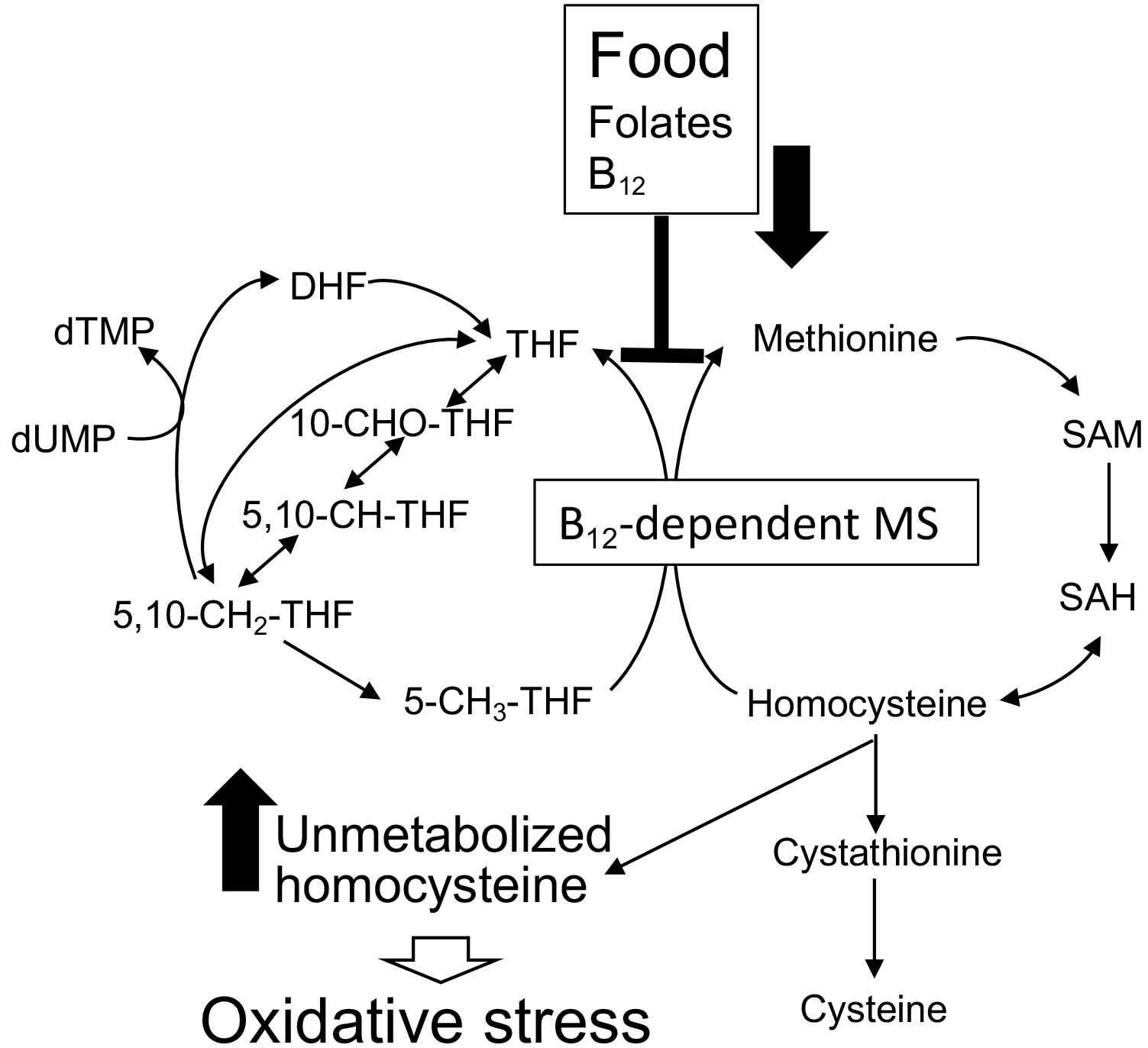 Fig. 2.
Physiological effects of folate and B on
methionine metabolism in humans. DHF, dihydrofolate; dTMP, deoxythymidine
monophosphate; dUMP, deoxyuridine monophosphate; MS, methionine synthase; SAH,
S-adenosylhomosysteine; SAM, S-adenosylmethionine; THF,
tetrahydrofolate.
Folate compounds found in naturally occurring foods are present in reduced forms
such as tetrahydrofolate (THF) (Fig. 3) and methyl, methylene, methenyl, formyl,
or formimino-group that binds to the N5 or N10 (or both) of the
molecules [9]. Moreover, the length of the poly--glutamate chain of THF
varies depending on the food [10]. Accurate quantification of folate compounds in
foods is important in determining the nutritional value of folate. The
poly--glutamate chain of THF must be treated with folate conjugase to
be converted into the mono--glutamate form before being analyzed using
both microbiological assay and high-performance liquid chromatography (HPLC)
[11].
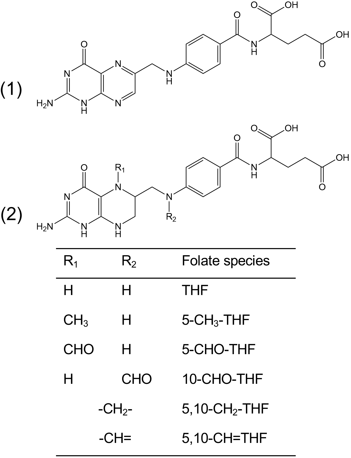 Fig. 3.
Structure of folate compounds. (1) Folic acid or pteroyl
glutamate and (2) reduced folate compounds.
HPLC has also been widely used to determine folate compounds in foods [12].
However, purification is required when using HPLC to determine food extracts.
Various solid-phase extraction procedures with commercially available disposable
cartridges such as strong anion exchange cartridges have been reported [12, 13].
Affinity chromatography with folate-binding protein (FBP; from cow’s milk)
attached to agarose gel removes impurities more effectively [12, 14]. Affinity
columns are not available commercially; thus, they must be prepared by the
investigator. Because such process of food folate assay is laborious, little
information is available on the folate contents of edible seaweeds, especially
purple laver products. If commercially available dried purple laver products
contain substantial amounts of folate compounds, they would be good sources of
both B and folate compounds in humans. Thus, we developed a simple method
for purifying folate compounds from food extracts using FBP and a commercially
available centrifugal ultrafiltration unit. Accordingly, this study aimed to
determine and characterize folate compounds from commercially available dried
purple laver products using HPLC with FBP purification.
2. Materials and Methods
2.1 Materials
Folic acid and FBP (from bovine milk) were obtained from Sigma-Aldrich (St.
Louis, MO, USA). (6R, S)-5,10-Methenyl-5,6,7,8-tetrahydrofolic
acid (5,10-CH=THF) chloride, (6R, S)-5,6,7,8-tetrahydrofolic
acid (THF) trihydrochloride, (6R, S)-5-formyl-5,6,7,8-tetrahydrofolic acid (5-CHO-THF) calcium salt, and
(6S)-5-methyl-5,6,7,8-tetrahydrofolic acid (5-CH-THF) calcium salt
were purchased from Schircks Laboratories (Zurich, Switzerland) and used as
folate standard compounds. 10-CHO-THF was prepared according to the method
published previously [15]. Type II porcine kidney acetone powder (Sigma-Aldorich)
was used as folate conjugases. -amylase (from Aspergillus oryzae), protease (type XIV, from Streptomyces griseus), and
certified reference material BCR-485 (from mixed vegetables) were obtained from
Sigma-Aldrich. Ultracel-10K (Amicon
Ultra-2 mL) was purchased from Merck Millipore Ltd. (Tullagreen, Ireland) and
used for centrifugal filter units for separation from FBP to folate compounds.
Purple laver (Neopyropia yezoensis, previously Porphyra yezoensis) products (dried, toasted, and seasoned and toasted), dried
kombu (Saccharina japonica) products, dried wakame (Undaria
pinnatifida) products, and boiled and dried hijiki (Sargassum
fusiformis) products were obtained from local markets in Tottori City, Japan, on
October 25, 2022.
2.2 Preparation of Folate Conjugase
To remove the endogenous folate compounds, porcine kidney (0.12 g) powder was
dissolved in 20 mL of 0.1 mol/L sodium phosphate buffer (pH 6.1), treated with
activated charcoal powder (2.0 g), stirred for 1 h at 4 °C, and
centrifuged at 900 g for 10 min at 4 °C. The
supernatant fraction was treated with a membrane filter (25AS020AS;
ADVANTEC Tokyo Roshi Kaisha Ltd., Tokyo, Japan) and used as
the folate conjugase.
2.3 Extraction of Folate Compounds and Tri-Enzyme Treatments
Total folate compounds were extracted from commercially available dried purple
laver products according to the tri-enzyme method [11]. Five grams of the laver
products were homogenized with a mortar and pestle. An aliquot (0.2 g) of the
homogenate was dissolved in 2.0 mL of 0.1 mol/L sodium phosphate buffer (pH 6.1)
containing 2% (v/v) ascorbic acid and 0.2% (v/v) 2-mercaptoethanol and diluted
with distilled water to a final volume of 5.0 mL. Octanol (100 L)
was then added, and the homogenates were autoclaved at 100 °C for 10
min. After cooling to room temperature (25 °C), 1 mL of 0.1 mol/L sodium
phosphate buffer (pH 6.1) containing 2% (v/v) ascorbic acid and 0.2% (v/v)
2-mercaptoethanol and 100 L of protease solution
(7.0 10 U) were added to each homogenate, and the homogenates
were then incubated at 37 °C for 3 h. To stop the protease enzyme
reaction, the homogenates were heated at 100 °C for 3 min. After cooling
with ice, they were treated with 100 L of -amylase.
solution (0.3 U) for 2 h at 37 °C. Thereafter the homogenates were
further treated with 400 L of porcine kidney folate conjugase for
16 h at 37 °C. To stop the enzyme reactions, the treated homogenates
were heated at 100 °C for 3 min and then cooled to room temperature (25
°C). The homogenate was diluted with distilled water to a final volume
of 10 mL, filtered through filler paper (type 2, 90 mm,
ADVANTEC®), and used as a food folate extract.
2.4 Microbiological Assay of Total Folate Compounds
Folate assays were performed using a polypropylene tube (13 100 mm,
Bio-Rad Laboratories, Hercules, CA, USA), which contain the food extract
(50 L), 0.1 mol/L sodium phosphate buffer (pH 7.0,
200 L), and Lactobacillus rhamnosus folate assay medium
(1 mL). The prepared assay mixture was diluted with distilled water to give a
final volume of 2.0 mL and then autoclaved at 121 °C for 5 min. After
cooling to room temperature (25 °C), the tube was inoculated aseptically
with 50 L of L. rhamnosus ATCC 7469 pre-cultured in
Lactobacilli inoculum broth as described above, washed three times with 5 mL of
saline buffer, and dissolved in saline buffer to achieve 92% light transmittance
at 660 nm. After incubating the tube at 37 °C for 22 h, its optical
density at 660 nm was determined using a UV-2550 Spectrophotometer (Shimadzu
Corporation, Kyoto, Japan). Each food sample was assayed for folate contents in
triplicates, and this was repeated at least thrice.
2.5 Purification of Folate Compounds Using FBP and
Ultracel-10K Centrifugal Filter Unit
An Ultracel-10K centrifugal filter unit was treated with 1
mL of 25% (v/v) methanol solution and centrifuged at 7000 g
for 10 min to wash its membrane. Then, 1 mL of 50 mmol/L potassium phosphate
buffer containing 2% (v/v) ascorbic acid and 0.2% (v/v) 2-mercaptoethanol was
added to the filter unit, which was centrifuged under the same conditions.
Aliquots (100 g) of lyophilized FBP was dissolved in 0.5 mL of 50
mmol/L potassium phosphate buffer containing 2% (v/v) ascorbic acid and 0.2%
(v/v) 2-mercaptoethanol. The FBP solution was added to each solution (0.5 mL) of
standard folate compounds (100 ng/mL) in a microtube, mixed well, and incubated
on ice for 30 min. Then, the mixture was transferred into the washed
Ultracel-10K centrifugal filter unit and centrifuged at
7000 g for 10 min at 4 °C (Supplementary
Fig. 1). The centrifugal filter unit was washed twice with 1 mL of 50 mmol/L
potassium phosphate buffer containing 2% (v/v) ascorbic acid and 0.2% (v/v)
2-mercaptoethanol to purify the formed FBP–folate complex. Folate compounds were
liberated from the FBP complex by the denaturation of FBP and treated with 300
L of 40 mmol/L trifluoroacetic acid containing 2% (v/v) ascorbic acid and
0.2% (v/v) 2-mercaptoethanol, removed by centrifugation at 7000 g for 10 min at 4 °C. The remaining folate compounds on the
membrane of the filter unit was recovered to wash twice with 400 L of 25%
(v/v) methanol solution containing 2% (%) ascorbic acid and 0.2% (v/v)
2-mercaptoethanol. The filtrate fractions were combined and used as HPLC samples.
After the denatured FBP was recovered with 50 mmol/L potassium phosphate buffer
from the filter unit, it was treated in 50 mmol/L potassium phosphate buffer for
24 h at 4 °C until the denatured FBP was completely converted to the
renatured form. FBP could be reused approximately 10 times to purify folate
compounds in this system.
2.6 Determination of Folate Compounds Using HPLC
After the pH of the purple laver extracts prepared as described above was
adjusted to pH 7.4 by the addition of 1 mol/L NaOH, the treated extracts were
diluted 1.5 times with distilled water. An aliquot (1.5 mL) of the diluted purple
laver extracts was mixed with 1.0 mL of 200 g/mL FBP solution at 4
°C for 20 min to purify folate compounds. Then, the solution was then
transferred to an Ultracel-10K centrifugal filter unit and
subjected to centrifugation at 7000 g at 4 °C for
15 min. Subsequently, the FBP–folate complex was washed twice with 1 mL of 50
mmol/L potassium phosphate buffer (pH 7.4) containing 2% (%) ascorbic acid and
0.2% (v/v) 2-mercaptoethanol.
After the washed FBP–folate complex was treated with 500 L of 40 mmol/L
trifluoroacetic acid solution containing 2% (%) ascorbic acid and 0.2% (v/v)
2-mercaptoethanol to denature FBP, folate compounds were recovered by
centrifugation under the same conditions. After the
Ultracel-10K unit was washed with 400 L of 25%
(v/v) methanol solution containing 2% (%) ascorbic acid and 0.2% (v/v)
2-mercaptoethanol, the filtrate and washing fraction were combined and used as an
HPLC sample.
The prepared sample was analyzed by HPLC using a Shimazu HPLC apparatus (SCL10A
system controller, LC-10Ai pump, CT-20A column oven, fluorescence detector
Shimazu RF-530, and electrochemical detector GL Science ED 723) and CDS version 5
chromato-data processing system (LAsoft, Ltd., Chiba, Japan)
(Supplementary Fig. 2). A 500-L aliquot of each
sample was placed on an IntertSusuain AQ-C18 HPLC column (5 m,
4.6 100 mm, GL Sciences) at 40 °C and eluted equilibrated
with 50 mmol/L potassium dihydrogen phosphate solution (pH 2.0) containing 7%
(v/v) acetonitrile at a flow rate of 1.0 mL/min. Folate compounds were
detected at 292-nm excitation/362-nm emission (fluorescence detector) and at
1000 mV versus Ag/AgCl (electrochemical detector). Authentic THF, 5-CH-THF,
and 5-CHO-THF were detected during monitoring at fluorescence detection, and the
retention times were 6.6 min, 7.7 min, and 17.5 min, respectively
(Supplementary Fig. 3). Authentic 5,10-CH=THF (with a retention
time of 10.9 min) and folic acid (with a retention time of 21.4 min) were
completely separated from other compounds contained in the sample such as
ascorbic acid and 2-mercaptoethanol during monitoring at electrochemical
detection; however, THF, 5-CH-THF, and 5-CHO-THF could not. Since
10-CHO-THF and 5,10-CH=THF could not be separated under these conditions, the
peak fraction with a retention time of 10.9 min represents the sum values of
10-CHO-THF and 5,10-CH=THF.
2.7 Extraction and Determination of B
Each sample (2 g) of the commercially available dried purple laver products was
homogenized using a mortar and pestle. Total B compounds were extracted
from each homogenate by boiling with 40 mL of distilled water and 10 mL of 0.57
mol/L acetate buffer (pH 4.5) containing 0.05% (w/v) KCN for 30 min in
the dark to convert B compounds into the CN forms. The extraction
procedures were performed in a draft chamber (Dalton Co., Tokyo, Japan). An
aliquot (20 mL) of each extract (100 mL) prepared above was placed on a Sep-Pak
® plus C18 cartridge (Waters Corp., Milford, MA, USA) that was
activated with 5 mL of 100% ethanol and equilibrated with 10 mL of distilled
water. B compounds were eluted from the C18 cartridge with 2 mL of 75%
(v/v) ethanol. After the eluate was filtered with a DISMIC-25JP membrane filter
(Toyo Roshi Kaisya, Ltd, Tokyo, Japan), the remaining B compounds on the filter
were recovered with 1 mL of 75% (v/v) ethanol. The filtrates were combined and
evaporated to dryness under reduced pressure. The residual fraction was dissolved
in 1.0 mL of distilled water. B compounds were purified from the solution
using an immunoaffinity column (EASI-EXTRACT B P80; 8.0
60 mm, R-Biopharm, Darmstadt, Germany) according to the manufacturer’s
protocol. The elute was evaporated to dryness under pressure is reduced,
dissolved in 100 L of Milli-Q water, and used as an HPLC sample, as
described previously [16]. The HPLC apparatus (SPD-10AV UV-Vis detector, SCL-10A
VP system controller, DGU-20A degasser, LC-10Ai liquid chromatograph,
CTO-20A column oven) and a reversed-phase HPLC column (Wakosil-II 5C18RS,
4.6 150 mm; FUJIFILM Wako Pure Chemical Corp., Osaka,
Japan) were used. B compounds were isocratically eluted with 20% (v/v)
methanol containing 1% (v/v) acetic acid at a flow rate of 1.0 mL/min at 40
°C and monitored by measuring the absorbance at 361 nm.
3. Results and Discussion
3.1 Content of Total B and Folate Compounds in Commercially
Available Edible Seaweed Products
Total B was extracted from various edible seaweed products and determined
using HPLC. Dried purple laver (nori) products contained substantial
amounts (approximately 30–60 g B/100 g dry weight) of
B. These values were much higher than those in other edible seaweeds
(0.5 g/100 g dry weight) (Table 1). Moreover, nori products did
not contain pseudovitamin B (Fig. 1) mostly found in edible cyanobacteria
because only a single peak of B was detected during HPLC (Fig. 4)
(Supplementary Fig. 4). These results coincide with previously reported values
determined using a microbiological assay method [17].
Table 1.Total B and folate contents of commercially available
edible seaweed products.
|
Total folate compounds |
Total B |
| (µg/100 g) |
(µg/100 g) |
| Dried purple laver (N. yezoensis) |
1309.0 53.4 |
59.7 18.2 |
| Toasted purple laver (N. yezoensis) |
1259.6 46.2 |
58.4 18.0 |
| Seasoned and toasted purple laver (N. yezoensis) |
876.8 136.4 |
28.9 11.6 |
| Dried kombu (S. japonica) |
230.3 23.4 |
0.1 0.1 |
| Boiled and dried hijiki (S. fusiformis) |
149.0 30.0 |
ND |
| Dried wakame (U. pinnatifida) |
66.5 27.0 |
0.5 0.1 |
| After each of the commercially available edible seaweed products described in
the table was extracted and treated with tri-enzymes, total folate compounds were
determined using the microbiological method. B was extracted from seaweed
products, purified with a B-immunoaffinity column, and determined using
HPLC. Data are represented as mean SD (n = 3). ND, not detected. |
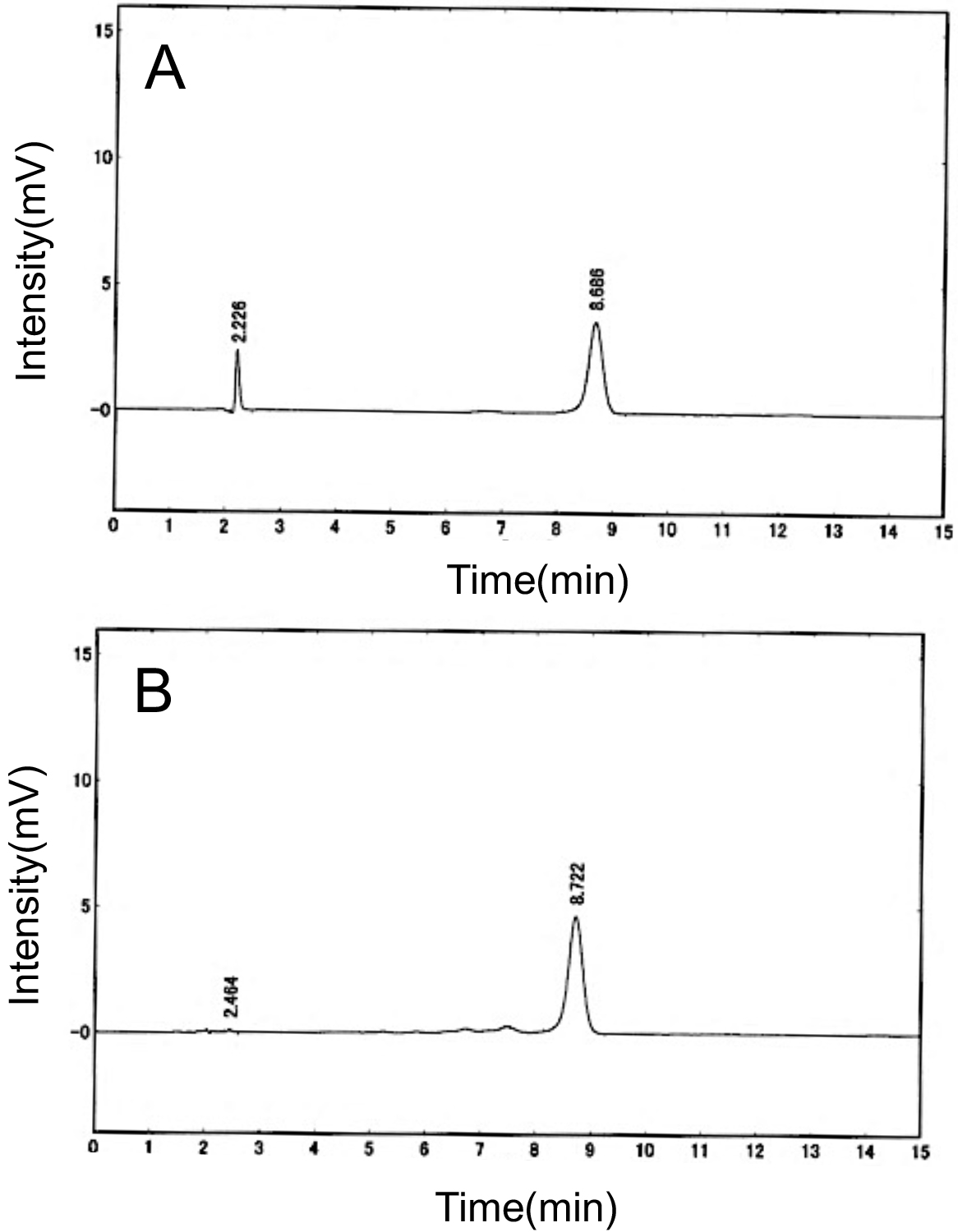 Fig. 4.
HPLC chromatograms for authentic B and corrinoid
compounds present in dried purple laver products. (A) Authentic B. (B)
Corrinoid compounds present in dried purple laver products. These are typical
HPLC chromatograms of authentic B and corrinoid compounds present in dried
purple laver products for three independent experiments.
Miyamoto et al. [18] reported that the B contents of the
seasoned and toasted purple laver products were reportedly about half of the
values of the dried purple laver products. Similar results were obtained in this
study (Table 1). No B content was reduced in dried purple laver products
during the toasting process [18], suggesting that the decreased B contents
in the seasoned and toasted laver products may be due to B destruction
caused by the interaction of various seasonings rather than the toasting process.
Total folate compounds were extracted, treated with the tri-enzyme (proteinase,
-amylase, and conjugase) method, and determined using microbiological assay. The
total folate content was much higher in dried purple laver (nori)
products (approximately 880–1300 g/100 g dry weight) than in other
edible seaweeds (230 g/100 g dry weight). These values
determined from the tri-enzyme-treated extracts of seaweed products were similar
to those from the extracts treated with di-enzymes (proteinase and conjugase)
that were adopted as an official method of food composition analysis in Japan
[19]. These results indicated that dried purple laver (nori) products contain
high levels of B and folate compounds compared with other seaweed products
tested.
3.2 Determination of Folate Compounds Found in Some Purple Laver
(Nori) Products Using HPLC after FBP Purification
Before the HPLC analysis, folate compounds from food extracts should be
purified. Affinity chromatography with FBP attached to agarose gel has been used
to remove impurities more effectively. The preparation of affinity columns is
time-consuming, and the folate-binding capacity of FBP is slightly lost during
the preparation. Thus, we developed a simple method for purifying folate
compounds from food extracts using FBP and Ultracel®-10K
centrifugal filter unit (Supplementary Fig. 1). To evaluate the recovery
rates of folate compounds during purification, various standard compounds such as
THF, 5-CHO-THF, 5,10-CH=THF, and 5-CHO-THF were used. As shown in
Supplementary Table 1, more than 85% of the recovery rate were obtained
at each folate compound. To evaluate whether this method can be applied into food
folate analysis, certified reference material BCR-485 (from mixed vegetables) was
used as a sample. Our HPLC analysis indicated that 5-CH-THF (229
g/100 g) is the predominant folate compound in BCR-485, which
contained 244 g of total folate compounds per 100 g weight
(Supplementary Table 2). These values coincided those reported
previously, indicating that this method can be applicable to food folate
analysis.
This HPLC method was performed to determine folate compounds found in some
purple laver (nori) products. Fig. 5A,B showed the elution profiles of the
FBP-purified compounds of a dried purple laver product in fluorescence and
electrochemical detectors, respectively. The retention times of peaks 1 (6.7 min)
and 2 (7.7 min) were identical to those of authentic THF and 5-CH-THF,
respectively, in fluorescence detection. Whereas in electrochemical detection,
the retention times of peaks 1 (6.7 min), 3 (11.7 min), 4 (16.6 min), and 5 (20.1
min) were identical to those of authentic THF, 5,10-CH=THF (or 10-CHO-THF or
both), 5-CHO-THF, and folic acid (FA), respectively. Similar elution patterns of FBP-purified
compounds were found in all purple laver products. Table 2 summarizes the levels
of folate compounds determined from commercially available purple laver products.
5-CH-THF (163–253 g/100 g dry weight) was the major folate
compound in all purple laver products. 5-CHO-THF was found in the similar level
(approximately 73–175 g/100 g dry weight). 5,10-CH=THF and
10-CHO-THF (approximately 57–123 g/100 g dry weight), THF
(approximately 17–28 g/100 g dry weight), and FA (9.8–52
g/100 g dry weight) were the minor folate compounds found in purple
laver products. FA would be due to the oxidation of the reduced folate compounds
such as 5-CH-THF during food processing and storage. The sum of these
folate compounds identified in HPLC was approximately 40% of total folate
compounds determined by the microbiological assay in each purple laver product,
suggesting that some of the unidentified peaks may be derived from certain folate
compounds. However, no information is available on whether these unknown 1–3
fractions contain some folate compounds because the antioxidant reagent
2-mercaptoethanol was eluted in the fraction of unknown peak 1. Although
information on the levels of reduced folate compounds in edible seaweeds is very
limited, Porphyra spp. (100 g, dry weight) reportedly contained
approximately 61 g of folate compounds, which consist of
5-CH-THF (approximately 34 g/100 g, dry weight) and FA
(approximately 36 g/100 g, dry weight) [20].
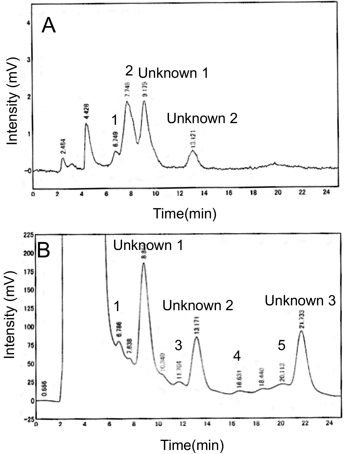 Fig. 5.
HPLC chromatograms for folate compounds present in dried purple
laver products. (A) Fluorescence detector; and (B) electrochemical detector. The
retention times of peaks 1, 2, 3, 4, and 5 were identical to those of authentic
THF, 5-CH-THF, and 5,10-CH = THF or 10-CHO-THF, 5-CHO-THF, and folic acid (FA),
respectively.
Table 2.Levels of folate compounds of commercially available dried
purple laver products.
|
Dried purple laver |
Toasted purple laver |
Seasoned and toasted purple laver |
| (µg/100 g dry weight) |
| THF |
27.2 1.9 |
27.8 7.4 |
16.8 1.6 |
| 5-CH-THF |
253.7 24.4 |
202.7 45.0 |
163.3 19.5 |
| 5,10-CH=THF + |
123.1 26.0 |
82.2 11.2 |
56.6 8.3 |
| 10-CHO-THF |
| 5-CHO-THF |
127.1 3.5 |
175.6 93.0 |
73.3 9.7 |
| FA |
52.0 17.1 |
25.7 11.7 |
9.8 8.0 |
| Total folate compounds |
581.1 51.2 |
514.0 130.5 |
319.8 26.6 |
| Folate compounds were purified from the tri-enzyme-treated extract of purple
laver products using FBP and then determined using HPLC. Data are represented as
mean SD (n = 3). |
Because the weight of one sheet (20 20 cm) of dried purple laver
products is approximately 3 g [21], two sheets (approximately 6 g) of dried
purple laver products would be sufficient to meet the recommended dietary
allowance (RAD) of adults for B (2.4 g/day). In RAD for
folates (400 g/day), ingestion of two sheets of the products would
provide approximately 80 g of folates per day [22].
It is concerning that the excessive consumption of edible seaweed products may
lead to the ingestion of harmful amounts of iodine because substantial amounts of
iodine (approximately 200 mg/100 g dry weight) have been found in dried kombu
products [23, 24]. However, dried purple laver products reportedly contained less
iodine (approximately 4–8 mg/100 g dry weight) [21]. Therefore, consuming two
sheets (approximately 6 g/day) of dried purple laver products would not lead to
an excessive intake of iodine (approximately 0.2–0.5 mg/day).


