


1 Department of Burns and Plastic Surgery, the First Affiliated Hospital of Hunan University of Chinese Medicine, 410007 Changsha, Hunan, China
2 Department of Endocrinology, the First Affiliated Hospital of Hunan University of Chinese Medicine, 410007 Changsha, Hunan, China
3 Department of Scientific Research, Hunan Brain Hospital, 410007 Changsha, Hunan, China
4 Clinical Medical School of Hunan University of Chinese Medicine, 410007 Changsha, Hunan, China
5 Department of Anesthesiology, the First Affiliated Hospital of Hunan University of Chinese Medicine, 410007 Changsha, Hunan, China
6 College of Integrated Traditional Chinese and Western Medicine, Hunan University of Chinese Medicine, 410208 Changsha, Hunan, China
Abstract
Objective: We explore the effects of endothelial progenitor cell
(EPC)-derived exosomes (EPCexos) and of astragaloside IV (ASIV)-stimulated
EPCexos (ASIV-EPCexos) on type I diabetic-wound healing, and determine the basic
molecular mechanisms of action. Methods: EPCs were exposed to different
concentrations of ASIV to generate ASIV-EPCexos. A chronic-wound healing model
involving streptozotocin-stimulated diabetic rats was established. These rats
were treated with EPCexos, ASIV-EPCexos, rapamycin, and wortmannin. Wound healing
was evaluated by direct photographic observation, hematoxylin and eosin staining,
and Masson’s trichrome staining. Results: ASIV treatment increased the abilities
of EPCs (e.g., proliferation), as well as exosome secretion. EPCexo showed a “cup
holder” like structure. Treatment with ASIV-EPCexos increased the wound-healing
rate, collagen-deposition area, bromodeoxyuridine uptake, VEGF expression, and
the number of CD31- and
Keywords
- astragaloside IV
- PI3K/AKT/mTOR
- endothelial progenitor cells
- exosome
- angiogenesis
The delayed and prolonged healing of diabetic wounds is a major challenge for healthcare providers worldwide [1]. Presently, typical clinical treatments for patients with chronic wounds are the dressing care of local ulcer wounds and repeated debridement of necrotic tissue [2, 3]. These dressings include preservatives, antioxidants, growth factors, and analgesics [4]. However, the treatment effects are inadequate.
Exosomes, with diameters ranging from 30 to 200 nm, are extracellular vesicles originating from the endosome and contain a wide range of substances, including proteins, DNA, lipids, and metabolites [5]. The process of exosome biosynthesis includes the double inward folding of the cell membrane and the creation of intracellular multivesicular bodies (MVBs) that contain intraluminal vesicles (ILVs). These MVBs can then either be degraded through fusion with lysosomes or autophagic vesicles or release ILVs as exosomes [5, 6]. Exosomes are crucial in various physiological and pathological processes via cellular communication, such as immune response, tissue repair, and cancer progression [5, 7, 8]. For instance, mesenchymal stem cell (MSC)-derived exosomes (MSCexo) promote the growth of endothelial cells and skin fibroblasts to accelerate the repair of skin wounds [9]. Fibroblast proliferation and endothelial-cell angiogenesis are the salient features of diabetic-wound healing [10]. Endothelial progenitor cells (EPCs) foster the regeneration of endothelial cells by secreting exosomes instead of self-differentiating into mature endothelial cells [11]. EPC-derived exosomes (EPCexos) can appreciably promote the vitality and angiogenesis of rat aortic endothelial cells [12]. Therefore, we speculate that EPCexos may be effective as a potential treatment for promoting diabetic-wound healing.
The decoction made from astragalus root is called “astragalus” and is widely used in traditional Chinese medicine to treat viral and bacterial infections, inflammation, and cancers. Astragaloside IV (ASIV) is one of the main active ingredients in the aqueous extract of astragalus [13]. ASIV has various pharmacological effects through multiple pathways, including anti-inflammatory, antifibrotic, antioxidative stress, antiasthma, antidiabetic, immunological, and cardioprotective [14]. Therefore, ASIV effectively protects against focal cerebral ischemia, liver fibrosis, cancer, diabetes, and cardiovascular diseases [15]. After myocardial infarction, ASIV exerts angiogenesis and cardioprotective effects via the PTEN/PI3K/AKT pathway [16]. Our previous studies have also shown that ASIV promotes the secretion of EPCexos [17]. However, the effects of ASIV-stimulated EPCexos (ASIV-EPCexos) on diabetic-wound healing have not yet been explored.
PI3K is important in mitosis, survival, differentiation, cytoskeleton configuration and remodeling, angiogenesis, glucose transport regulation, and cyst transport [18]. PI3K catalyzes the formation of phosphatidylinositol triphosphate (PIP3), which binds to the PH domain of 3-phosphoinositide-dependent protein kinase 1 (PDK1) to activate AKT [19]. The PI3K/AKT/mTOR pathway modulates cell growth, survival, metabolism, and immunity [20, 21]. Activation of the PI3K/AKT/mTOR pathway increases the expression of VEGF and stimulates angiogenesis [22]. The AKT/eNOS pathway can regulate angiogenesis and tissue repair [23]. We speculate that ASIV could promote EPCexo secretion and diabetic-wound healing via the PI3K/AKT pathway.
In the present study, animal experiments were conducted to explore the effects of ASIV-EPCexos on type I diabetic-wound healing, and determine the underlying mechanism of action.
This study was approved by the Ethics Committee of the First Hospital of Hunan University of Chinese Medicine (NO.HN-LL-KY-2020-013-01). All experiments were performed strictly in accordance with the Declaration of Helsinki, and informed consent was obtained from all the participants.
Umbilical-cord blood from healthy, full-term newborns was obtained from the
Obstetrics and Gynecology Department of the First Hospital, Hunan University of
Chinese Medicine, Changsha, China. Heparin (20 U/mL) was added to the cord blood
for anticoagulation. The isolated cord blood mononuclear cells were resuspended
in DMEM (D5796, Sigma, Saint Louis, MO, USA) with 10% FCS (04-001-1ACS, Gibco,
Carlsbad, CA, USA). The cell suspension (3
Male, 8-week-old Sprague-Dawley rats (n = 96) weighing 250–300 g were purchased
from Hunan SJA Laboratory Animal Co., Ltd. (Changsha, Hunan, China). The
experimental protocol was approved by the Animal Experimentation Ethics Committee
of the First Hospital of Hunan University of Chinese Medicine (NO.
ZYFY20201018-2). Rats were housed alone and exposed to a 12/12-h light/dark cycle
at 22–24 °C and with ad libitum access to food and water. One week
later, they were divided into control (n = 12) and diabetic (n = 84) groups.
After fasting for 12–16 h, rats in the diabetic group were given a single
intraperitoneal injection of streptozotocin (STZ) solution (65 mg/kg body weight,
in 0.1 M citrate buffer, pH = 4.5, S0130, Sigma-Aldrich, Saint Louis, State of
Missouri, USA) to induce a type I diabetes model [25, 26]. Blood glucose levels
were randomly monitored daily after the first 72 h. When three consecutive random
blood-glucose concentrations were more than 16.7 mmol/L, a successful model of
diabetes was considered to have been established. After 14 d, rats in the control
and diabetic groups were anesthetized and shaved. The dorsal skin of the rats was
disinfected, and four pieces of 1.5
The expression of the surface markers CD31 (+), CD34 (+), CD45 (–), and CD133 (+) of EPCs was evaluated using flow cytometry. After approximately 12 d of subculture, cells were obtained. The resuspended cells were precipitated with 100 µL of 0.5% BSA-PBS. Subsequently, 2 µL CD34-FITC (bs-0646R-FITC, Bioss, Beijing, China), 1 µL CD133-FITC (bs-0209R-FITC, Bioss, Beijing, China), 3 µL CD31-FITC (bs-0195R-FITC, Bioss, Beijing, China), and 1 µL CD45-FITC (bs-0522R-FITC, Bioss, Beijing, China) antibodies were incubated with the cells at 37 °C in the dark for 30 min. After washing and centrifugation, the cells were analyzed by flow cytometry (A00-1-1102, Beckman, Brea, California, USA).
The expression of CD34 and VEGF receptor 2 (VEGFR2) of EPCs was detected using
IF staining. IF staining was used to evaluate the expression of CD31,
Following treatment of EPCs with ASIV for 24 h, the medium was removed, discarded, and replaced with 100 µL medium containing 10% CCK8 (NU679, Dojindo, Kumamoto, Japan). The absorbance was measured at 450 nm using a microplate reader (MB-530, HEALES, Shenzhen, Guangdong, China).
Intervention EPCs were hydrolyzed into single cells. A cell suspension, prepared
in a serum-free basal medium (1
Corning Matrigel Basement Membrane Matrix (356234, BD Biosciences, Franklin
Lakes, NJ, USA) was added to each well of a 48-well plate until the wells
were evenly covered without bubbles. The 48-well plates were then incubated at 37
°C for 1 h. Cells were digested with 0.25% trypsin and suspended before
the addition of 7.5
An ExoQuantTM Overall Exosome Capture and Quantification Assay Kit (#K1201-100, Biovision, San Francisco, CA, USA) was used for quantitative analysis of exosomes. All procedures were carried out following the guidelines. The absorbance was obtained at 450 nm. The concentration of the exosomes was calculated after plotting a standard curve.
A 30 µg sample of denatured protein was added to each well of the gel.
Samples were electrophoresed for 130 min at a constant voltage of 75 V. Target
proteins were transferred from the gel to a nitrocellulose (NC) membrane at a
constant current of 300 mA. Following the transfer process, the NC membrane was
subjected to blocking. The primary antibodies against CD63 (25682-1-AP), CD9
(20597-1-AP), CD81 (66866-1-Ig), mTOR (66888-1-Ig), ras homolog enriched in brain
(Rheb, 15924-1-AP), PI3K (67071-1-Ig), AKT (10176-2-AP), PIP3 (17552-1-AP), PDK1
(10026-1-AP), and
HE staining was used to observe the morphological changes in rat skin. Paraffin-embedded rat-skin tissue samples were cut into 4-µm-thick sections using a slicer. The sections were dewaxed and rehydrated using xylene and ethanol and stained with HE. The sections were examined under a microscope (BA210T, Motic, Xiamen, Fujian, China).
Paraffin-embedded rat skin tissues were cut, dewaxed, and rehydrated. The sections were incubated with hematoxylin solution, rinsed with distilled water, and stained with acid fuchsin solution. Next, the sections were then incubated with 1% phosphomolybdic acid and blue aniline solutions for 5 min. The samples were examined under a microscope.
Total RNA was extracted, and the cDNA was obtained by reverse transcription
(CW2569, Beijing CWBIO Co., Ltd., Beijing, China). The primer sequences for the
target genes are presented in Table 1. Primers were synthesized by Shanghai
Sangon Biotech. A PCR system containing the fluorescent dye UltraSYBR Mixture
(CW2601, Beijing CWBIO Co., Ltd., Beijing, China) was prepared, and the reaction
was performed with QuantStudio1 (Thermo, Waltham, Massachusetts, USA). The
fluorescence signal was monitored in real time, and the internal reference was
| Gene | Sequences (5 |
| VEGFa | F: GGGAGCAGAAAGCCCATGAA |
| R: GCTGGCTTTGGTGAGGTTTG | |
| VEGFb | F: GTGGTCAAACAACTCGTGCC |
| R: CTGGGGCTGTCTGGCTTC | |
| VEGFc | F: AACCTCCATGTGTGTCCGTC |
| R: TGCTGAGGTAACCTGTGCTG | |
| FGF | F: ACACCACGGACAAAGAAATTGAGG |
| R: CCCGATAGAATTACCCGCCAAGCA | |
| Ang-1 | F: ACATCCCGTCTTGAAATCCAAC |
| R: TGTCCAGCTCTTCCTTGTGT | |
| mTOR | F: AGAACCAATTATACTCGCTCCCT |
| R: GCAACCTCAAAGCAGTCCCC | |
| Rheb | F: GGACCTGCATATGGAAAGGGT |
| R: CATCACCGAGCACGAAGACT | |
| eNOS | F: GTTGACCAAGGCAAACCACC |
| R: GCTGACTCCCTCCCAGTCTA | |
| PI3K | F: AGCCACAGATCCACTTAACCC |
| R: CTTGCTGTCCCCACTTTACTGA | |
| AKT | F: GTCACCTCTGAGACCGACACC |
| R: GCCTCCGTTCACTGTCCAC | |
| PDK1 | F: CGCCTCTATGCACAGTACTTCCAG |
| R: CGTCAGCCTCGTGGTTGGTTC | |
| TSC2 | F: TACCCCTGAGAAGGACAAGTT |
| R: CAAGCTGGCACTGGTAAGAGA | |
| PIP3 | F: GCAGTTTGAACCCAAAGCCC |
| R: AAGCGCATTCCTTTCCGTTG | |
| IL-6 | F: TCACTATGAGGTCTACTCGG |
| R: CATATTGCCAGTTCTTCGTA | |
| IL-1 |
F: CAGCAGCATCTCGACAAGAG |
| R: AAAGAAGGTGCTTGGGTCCT | |
| F: ACCCTGAAGTACCCCATCGAG | |
| R: AGCACAGCCTGGATAGCAAC |
Statistical analyses were conducted using GraphPad Prism 8.0.1 (GraphPad
Software, Inc., San Diego, CA, USA), and the data were expressed as mean
EPCs were identified using flow cytometry and IF staining. The expression of CD31, CD34, and CD133 was positive, but the expression of CD45 was negative, indicating that the endothelial cells were successfully extracted (Supplementary Fig. 1). As depicted in Fig. 1A, the viability of EPCs in the 50, 100, and 200 mg/L groups exhibited a substantial enhancement in comparison to the control group. The cell migration and tube-formation abilities of EPCs in the 50, 100, and 200 mg/L groups were appreciably improved (Fig. 1B,C). EPCs in the 100 mg/L group showed optimal ability. Therefore, ASIV promotes the progress of EPCs, and a concentration of 100 mg/L exhibited the optimal effect.
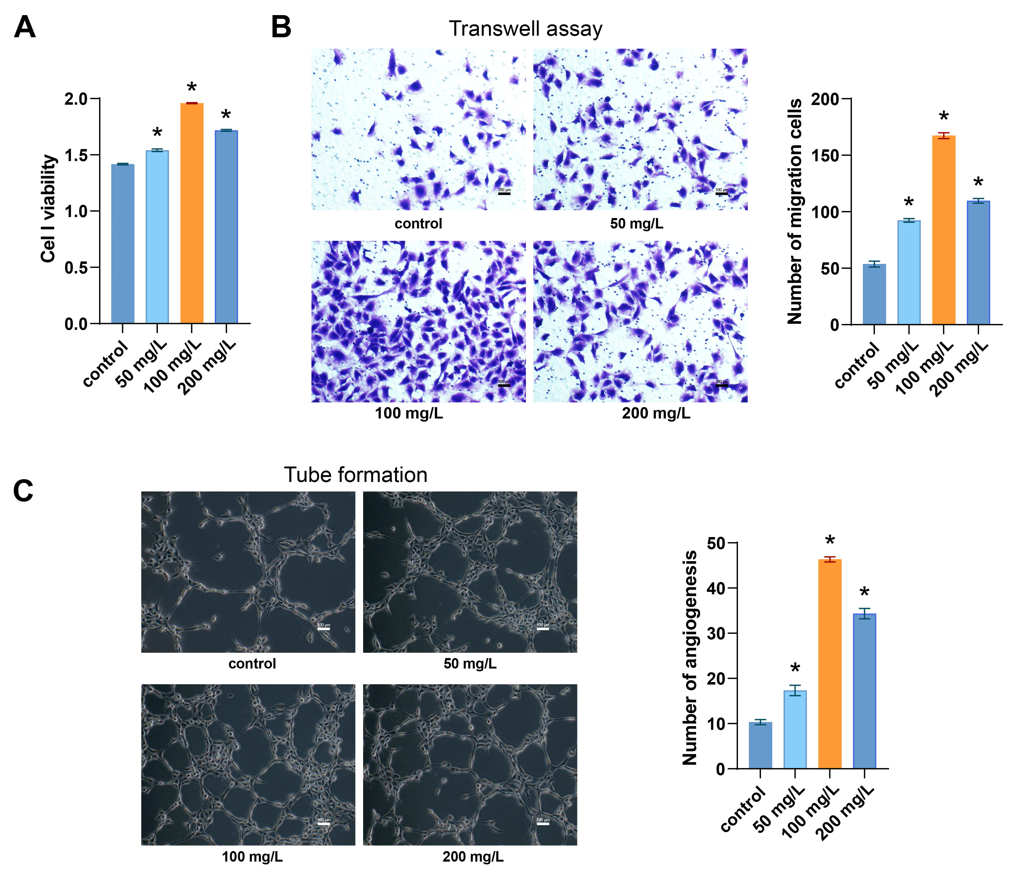 Fig. 1.
ASIV promotes EPC progress. (A) Representative graphs of CCK-8
showing cell viability. (B) Representative light micrographs and graphs showing
cell migration by Transwell assay. (C) Representative light micrographs and
graphs of tube formation assay revealing the tube formation ability. *p
We next examined the effect of ASIV on EPCexo secretion. The EPCexo characteristic markers CD9, CD63, and CD81 levels were detected [17]. The expression of those markers was positive in all the groups (Fig. 2A), indicating the successful extraction of EPCexos. In TEM, EPCexos showed a “cup holder” structure in the four groups (Fig. 2B). EPCexos were quantified using ELISA. As shown in Fig. 2C, the concentration of EPCexos was appreciably higher in the 50, 100, and 200 mg/L groups than in the control group, with the highest concentration observed in the 100 mg/L group. These findings indicate that ASIV facilitates EPCexo secretion, with the optimal effect observed at a concentration of 100 mg/L.
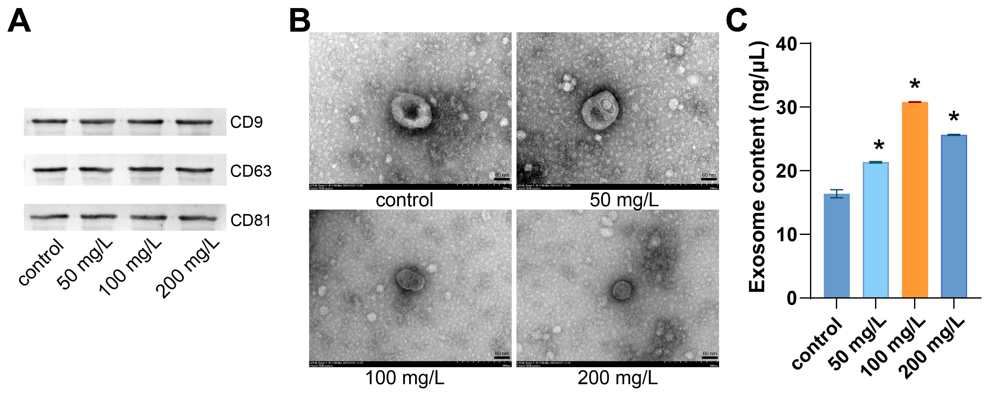 Fig. 2.
ASIV promotes EPCexo secretion. (A) The WB analysis of shows
the expression of EPCexo characteristic markers CD9, CD63, and CD81. (B)
Representative micrographs of EPCexo morphology were obtained via TEM. Scale bar
= 60 nm. (C) Comparison of EPCexo content via ELISA in the different groups.
*p
The chronic-wound model in STZ-stimulated diabetic rats was used to investigate
whether ASIV-EPCexos promote type I diabetic-wound healing. The EPCexo group
exhibited a higher rate of wound healing compared to the model group (Fig. 3A,B).
After ASIV-EPCexo treatment, wound healing rates in the low-, mid-, and
hi-ASIV-EPCexo groups were substantially higher than in the EPCexo group, and the
hi-ASIV-EPCexo group exhibited optimal healing. As opposed to the model group,
the positive staining rates of BrdU and VEGF were substantially raised in the
EPCexo group (Fig. 3C–E). Moreover, the low-, mid- and hi-ASIV-EPCexo groups
exhibited stronger positive staining for BrdU and VEGF compared to the EPCexo
group, and the hi-ASIV-EPCexo group demonstrated the highest staining intensity.
Granulation tissue formation, epithelial reformation, collagen deposition,
fibroblast proliferation, and angiogenesis are hallmarks of wound healing [29].
In contrast to the model group, the epidermal thickness in the EPCexo and low-,
mid-, and hi-ASIV-EPCexo groups gradually decreased. The collagen area was higher
in the low-, mid-, and hi-ASIV-EPCexo groups than in the model group (Fig. 4A–C). IF staining results revealed that the number (No.) of CD31 and
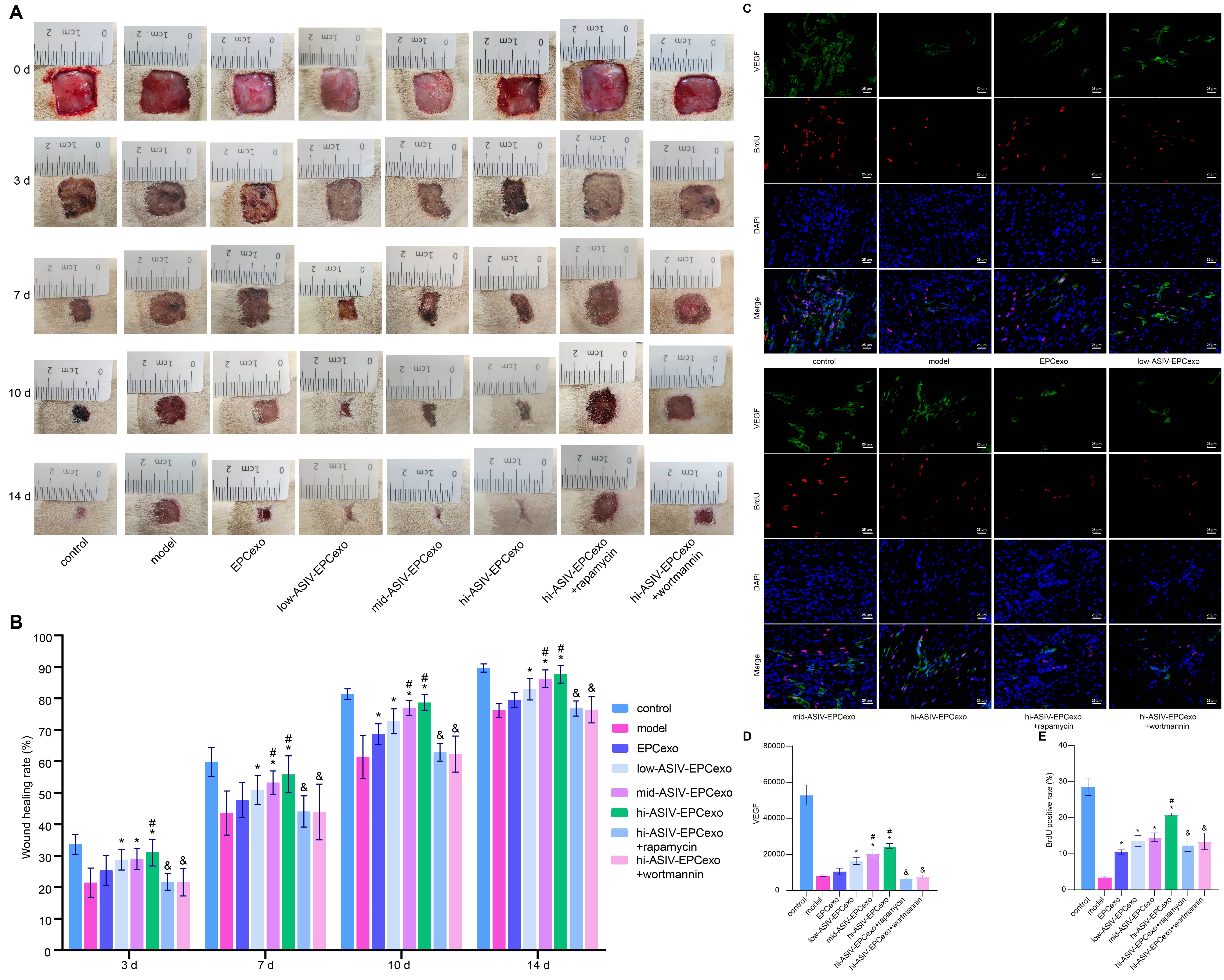 Fig. 3.
ASIV-EPCexos may promote type I diabetic-wound healing via
facilitating BrdU and VEGF expression. (A) Representative images of the diabetic
wounds of rat skin at different time points. (B) Comparison of wound healing rate
of rat skin at different time points. (C) Representative micrographs of IF
staining showing the expression of BrdU and VEGF. (D,E) Statistical analysis of
the expression of BrdU and VEGF. *p
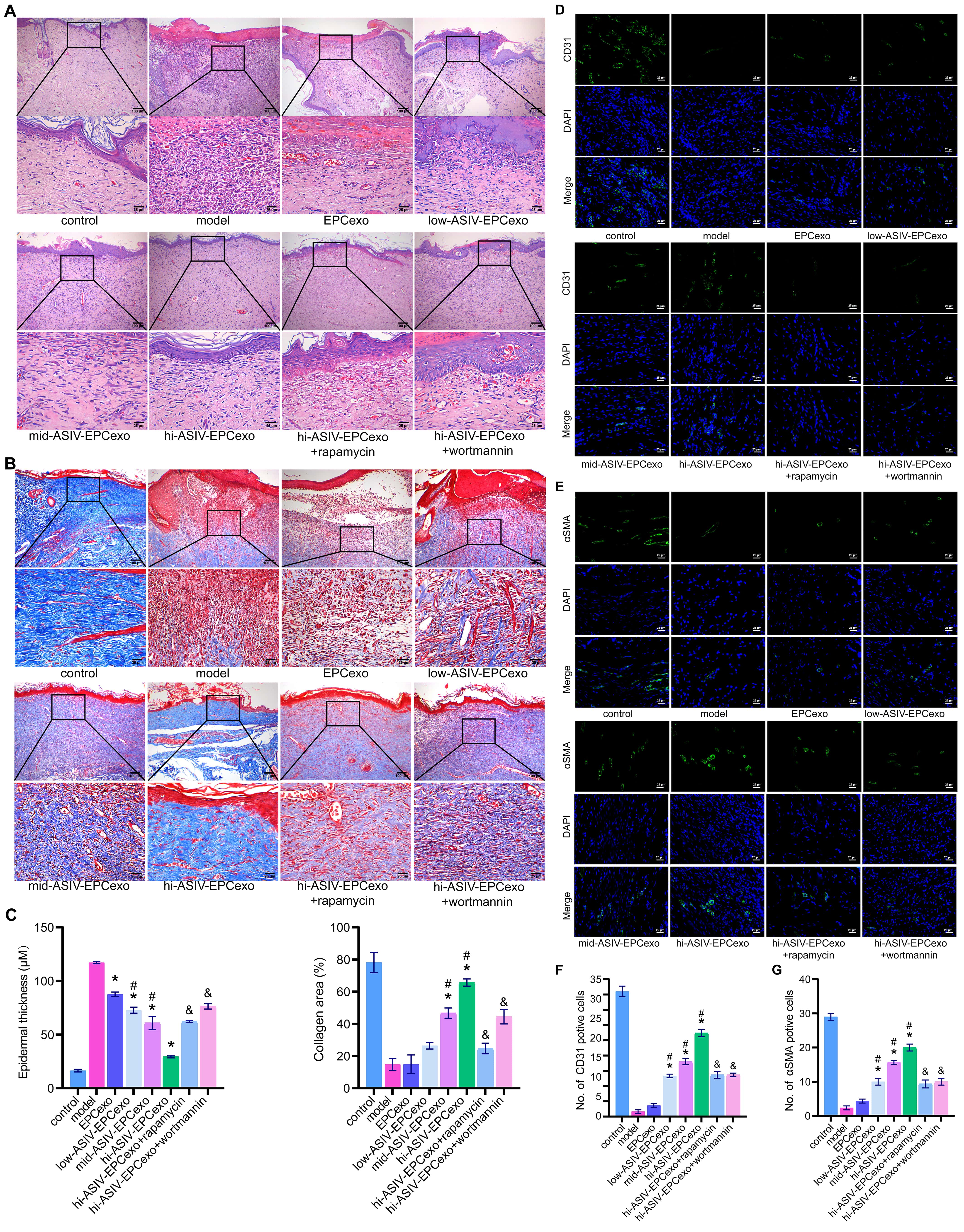 Fig. 4.
ASIV-EPCexos may promote type I diabetic-wound healing via
modulating CD31 and
To study the mechanism by which ASIV-EPCexos accelerate type I diabetic wound healing, we analyzed the changes in skin tissue at the molecular level. The expression levels related to vascular growth (VEGFa, VEGFb, VEGFc, FGF, and Ang-1) in the EPCexo group were higher than in the model group. These expression levels were further increased following ASIV-EPCexo treatment. Compared to the hi-ASIV-EPCexo group, these expression levels in the hi-ASIV-EPCexo + rapamycin and hi-ASIV-EPCexo + wortmannin groups were substantially decreased (Fig. 5A,B). We then examined the expression levels of the pathway proteins and genes in the samples. In terms of the model group, the expression levels of p-mTOR, mTOR, p-mTOR/mTOR, Rheb, eNOS, PI3K, AKT, PIP3, PDK1, and TSC2 in the EPCexo group were raised (Fig. 5C–F), although several of them were not substantially different. In addition, the expression levels of genes and proteins related to these pathways were higher in the low-, mid-, and hi-ASIV-EPCexo groups than in the EPCexo group. However, the expression of these genes and proteins decreased in the hi-ASIV-EPCexo + rapamycin and hi-ASIV-EPCexo + wortmannin groups. The results suggest that ASIV-EPCexos may activate the PI3K/AKT/mTOR pathway, thereby promoting angiogenesis.
 Fig. 5.
ASIV-EPCexos promote angiogenesis via the PI3K/AKT/mTOR pathway
in diabetic wounds. (A) Representative graphs of RT-qPCR showing the relative mRNA expression of VEGFa,
VEGFb, VEGFc, FGF, and Ang-1. (B) The WB
analysis shows the protein expression of VEGFa, VEGFb, VEGFc, FGF, and Ang-1.
(C,D) Representative graphs of RT-qPCR showing the expression of
mTOR, Rheb, eNOS, PI3K, AKT,
PIP3, PDK1, and TSC2. (E) The WB analysis
shows the protein expression of mTOR and p-mTOR. (F) The WB analysis shows the
protein expression of Rheb, eNOS, PI3K, AKT, PIP3, PDK1, and TSC2. *p
Interleukin (IL)-1
 Fig. 6.
ASIV-EPCexos inhibit inflammation via the PI3K/AKT/mTOR pathway
in type I diabetic-wound healing. (A) Representative graphs of RT-qPCR showing
the mRNA expression of IL-6 and IL-1
The present work found that ASIV-EPCexos promoted type I diabetic-wound healing,
which could be blocked by the mTOR- and PI3K- specific inhibitors, rapamycin, and
wortmannin. We found that ASIV-EPCexos promoted type I diabetic wound healing by
activating the PI3K/AKT/mTOR pathway. Our earlier study only demonstrated that
ASIV could stimulate human EPCs to secrete exosomes [17], whereas the present
study examined the effects of ASIV-EPCexos on wound healing in type I diabetic
rats. We established a type I diabetic rat model with skin wounds and treated
with EPCexos or with low-, mid-, or high-ASIV-EPCexos. Photographs of the wound
healing at different time points showed all of these treatments promoted the
healing of diabetic wounds. Following treatment with EPCexos or ASIV-EPCexos, the
expression of inflammatory factors decreased, the levels of VEGF, BrdU, CD31, and
The growth factor VEGF has important angiogenic activity that promotes mitosis
and inhibits apoptosis of endothelial cells, as well as promoting vascular
permeability and cell migration [32]. ASIV enhances the expression of VEGF,
thereby promoting angiogenesis in wound tissues [33]. CD31 is involved in immune
regulation and angiogenesis [34]. The downregulation of miR-126-3p in parathyroid
tumors promotes endothelial cell transition to the
FGF regulates cell fate, angiogenesis, immunity, and metabolism via its receptors FGFR1, FGFR2, FGFR3, and FGFR4 [37]. Ang-1 plays a regulatory role in processes associated with proliferation, inflammation, vascular fibrosis, and remodeling [38]. We found that ASIV-EPCexos increased the expression of genes and proteins related to blood-vessel growth (VEGFa, VEGFb, VEGFc, FGF, and Ang-1), which could explain their ability to promote angiogenesis in diabetic wounds. Many substances regulate the proliferation, migration, and tube-forming abilities of EPCs via the PI3K/AKT pathway, including naringin [39]. AKT-dependent TSC2 phosphorylation promotes RHEB-mTORC1 [40]. TSC2/Rheb mediates extracellular signal-regulated, kinase-dependent regulation of mTORC1 activity in C2C12 myoblasts [41]. Downstream targets of the PI3K/AKT pathway, mTOR and eNOS, can increase VEGF expression and promote angiogenesis [42, 43]. The current study found that the expression of genes and proteins related to the PI3K/AKT/mTOR pathway was significantly increased following treatment with ASIV-EPCexos, and this was reversed by rapamycin and wortmannin. In short, our results indicate that ASIV-EPCexos can activate the PI3K/AKT/mTOR pathway and promote angiogenesis at the wound surface by targeting mTOR and eNOS in endothelial cells.
Chronic, unresolved inflammation is a hallmark of non-healing wounds and
adversely affects the wound-healing process [44]. The pro-inflammatory cytokines
IL-6 and IL-1
Type I diabetes mellitus (TIDM) is an autoimmune disease in which the immune
system mistakenly attacks and destroys insulin-producing
Diabetic foot ulcers (DFU) are a significant and grave complication of diabetes.
They are characterized by impaired blood supply to the feet caused by neuropathy
or vascular disease, which subsequently leads to ulceration, infection, and a
range of other complications [52]. DFU has emerged as a global public health
concern with serious implications for human well-being due to its unfavorable
prognosis, ultimately increasing the risk of ulceration, amputation, and even
mortality [52, 53]. DFU primarily occurs in adults with TIIDM but can also occur
in adult patients with TIDM [54, 55, 56]. For example, a retrospective study by
Rasmussen et al. [54] reported that out of 5640 adult patients with
TIDM, 255 developed DFU. Among 6953 adult patients with TIIDM, 310 developed DFU
in a specialty hospital in Denmark during 2001–2014. In a prospective clinical
study, among 31 adult patients diagnosed with DFU, 14 were caused by TIDM [56].
There are also a number of studies focusing on wound healing in TIDM [31, 57, 58]. For example, Liu et al. [31] found that neutrophil extracellular
traps (NETs) contribute to NLRP3 inflammasome activation and sustained
inflammatory responses in type I diabetic wounds. Costa et al. [57]
revealed that xanthohumol effectively regulates inflammation, oxidative stress,
and angiogenesis in the process of cutaneous wound healing in rats with TIDM.
White et al. [58] conducted a study to investigate the efficacy of
combination therapies involving various growth factors in promoting wound healing
using a mouse model of type I diabetic wounds. Their findings showed that triple
therapy was highly effective [58]. This study aimed to investigate the effects
and potential mechanisms of ASIV-EPCexos on a rat model of type I diabetic-wound
healing. After skin excision, we performed HE, MT, and IF staining (VEGF, BrdU,
CD31,
There are similarities in the underlying causes of impaired and delayed wound
healing in patients with TIDM and TIIDM. Chronic hyperglycemia, a key common
factor, leads to a persistent state of inflammation that disrupts the natural
progression of inflammation, repair, and regeneration during wound healing [57, 59, 60]. Additionally, compromised immune function in patients with TIDM and
TIIDM increases the vulnerability of wounds to infections and elongates the
healing process. Notably, the dysfunction of immune cells and foot neuropathy
contributes to the formation of DFU and further hinder the healing process
[59, 60, 61]. In the current study, the expression levels of the IL-6 and
IL-1
Several cell types, such as MSCs, fibroblasts, macrophages, and EPCs, have been reported to play roles in wound healing [10, 62, 63, 64, 65]. For example, MSCs or MSCexos can promote wound healing [62, 63]. Fibroblast proliferation and endothelial cell angiogenesis are prominent features of diabetic wound healing [10]. Our previous studies have demonstrated that ASIV has the potential to enhance the secretion of exosomes from EPCs. This was determined by comparing the mass concentration of exosomes secreted by EPCs in both the control group and the group treated with ASIV [17]. EPCs possess remarkable migratory capacity and the ability to differentiate into endothelial cells, making them crucial in neovascularization, tissue regeneration, and wound healing processes [65]. Huang et al. [66] reported that ASIV-treated EPCs had a positive impact on angiogenesis and wound healing. EPCs promote endothelial cell regeneration by the secretion of exosomes [11]. Specifically, ASIV-EPCexos have been observed to appreciably enhance the proliferation, migration, and angiogenesis of rat aortic endothelial cells [12]. Therefore, ASIV-EPCexos were chosen as the focus of the study.
Exosomes contain various biologically active components, including proteins, lipids, and RNA. Multiple mechanisms may be responsible for mediating the therapeutic effects of exosomes. The proangiogenic and anti-inflammatory effects of the active components of EPCexos remain unclear, and a future goal should be to identify these molecules.
The current study confirms the stimulatory effect of ASIV on EPCexo secretion. ASIV-EPCexos promote diabetic-wound healing by activating the PI3K/AKT/mTOR pathway. These results may provide novel therapeutic options for the clinical treatment of diabetic wounds.
The datasets used and analyzed during the current study are available from the corresponding author on reasonable request.
WX and XZo designed the research study. WX, XB, XZh, HL, HX, LZ, and YX performed the research. QY analyzed the data. WX wrote the manuscript. All authors contributed to editorial changes in the manuscript. All authors read and approved the final manuscript.
This study was approved by the Ethics Committee of the First Hospital of Hunan University of Chinese Medicine (NO.HN-LL-KY-2020-013-01). All experiments were performed strictly in accordance with the Declaration of Helsinki, and informed consent was obtained from all the participants.
Not applicable.
This research was funded by the Clinical Medical Technology Innovation Guidance Project of Hunan Provincial Science and Technology Department (No. 2021SK51412), the Hunan Natural Science Foundation Youth Fund Project (No. 2019J50460), the National Natural Science Foundation of China Youth Science Fund Project (No. 81904217), and Science and Health Joint Project of Hunan Natural Science Foundation (No.2021JJ70033).
The authors declare no conflict of interest.
References
Publisher’s Note: IMR Press stays neutral with regard to jurisdictional claims in published maps and institutional affiliations.