IMR Press / FBL / Volume 28 / Issue 10 / DOI: 10.31083/j.fbl2810274
Open Access Review
Mechanical Signaling in Dental Pulp Stem Cells
Jiahe Zhao1,†Wen Du2,†Daimo Guo3Siyi Wang1Wei Du4,*
Show Less
1 West China School of Stomatology, Sichuan University, 610041 Chengdu, Sichuan, China
2 State Key Laboratory of Oral Diseases, National Clinical Research Center for Oral Diseases, Department of Prosthodontics, West China Hospital of Stomatology, Sichuan University, 610041 Chengdu, Sichuan, China
3 State Key Laboratory of Oral Diseases, National Clinical Research Center for Oral Diseases, West China Hospital of Stomatology, Sichuan University, 610041 Chengdu, Sichuan, China
4 State Key Laboratory of Oral Diseases, National Clinical Research Center for Oral Diseases, Department of Cariology and Endodontics, West China Hospital of Stomatology, Sichuan University, 610041 Chengdu, Sichuan, China
*Correspondence: weidu@scu.edu.cn (Wei Du)
These authors contributed equally.
Front. Biosci. (Landmark Ed) 2023, 28(10), 274; https://doi.org/10.31083/j.fbl2810274
Submitted: 31 May 2023 | Revised: 11 July 2023 | Accepted: 20 July 2023 | Published: 31 October 2023
Copyright: © 2023 The Author(s). Published by IMR Press.
This is an open access article under the CC BY 4.0 license.
Abstract

Dental pulp stem cells (DPSCs) are a type of mesenchymal stem cells derived from dental pulp that serves as an important model for investigating biological regeneration. DPSCs have a multipotent differentiation capacity and can promote different biological processes, including osteogenesis, odontogenesis, chondrogenesis, and angiogenesis. These biological processes are regulated by an extensive range of intra- and extra-cellular factors. Further, biomechanical cues, such as substrate stiffness, physical stress, and cell spreading, have been highlighted as particularly important modulators of DPSC function. This review sought to discuss various related signaling components involved in biomechanical cues and their respective roles in cellular and tissue responses in DPSCs, summarize current findings, and provide an outlook on the potential applications of biomechanics in regenerative medicine and tissue engineering.

Keywords
dental pulp stem cells
mechanical signaling
cell fate determination
1. Introduction

Dental pulp stem cells (DPSCs), a type of mesenchymal stem cells derived from dental pulp [1], can differentiate into a variety of cells, such as dental pulp, dentin, and osteoblasts, and serve as important models for investigating biological regeneration [2, 3, 4]. Biomechanics, an interdisciplinary field that investigates the effects of forces and motion on biological systems, focuses on mechanical stimuli and their impact on cell behavior [5, 6, 7]. Over the past decades, mechanical stimuli, such as tissue stiffness, physical forces, and cell spreading, have led to significant advances in the fields of regenerative medicine and tissue engineering [8, 9, 10]. Understanding the underlying mechanisms of DPSC response to mechanical stimuli can provide multiple ideas for the regeneration and immune regulation of various cells or tissues, such as dentin, bone, and cartilage. Here, we summarize these findings and provide a perspective on the prospects and potential applications of DPSCs in bioengineering and regenerative medicine.

2. Dental Pulp Stem Cells (DPSCs)

Mesenchymal stem cells (MSCs) are heterogeneous stem cells that exhibit self-renewal and multilineage differentiation ability [11, 12]. MSCs can be isolated from different tissues, such as bone marrow, adipose tissue, and umbilical cord tissue [13, 14, 15]. In the oral cavity, MSCs can be classified into several distinct subtypes, including DPSCs [1, 16], stem cells from human exfoliated deciduous teeth (SHEDs) [17], stem cells from apical papilla (SCAPs) [18, 19], periodontal ligament stem cells (PDLSCs) [20], dental follicle stem cells (DFSCs) [21], gingival-derived mesenchymal stem cells (GMSCs) [22], and buccal fat pad-derived stem cells (BFPSCs) [23]. DPSCs, which were first isolated from the dental pulp of third molars [1, 24, 25], are characterized by high self-renewal and differentiation capacities [1, 26, 27]. Similar to MSCs, DPSCs express stem cell markers, such as CD29, CD44, CD73, CD90, CD105, CD146, CD271, and STRO-1 [1, 28], whereas the hematopoietic markers including CD14, CD34, CD45, and CD117, are absent or expressed at very low levels [29, 30]. DPSCs also express the vascular endothelial growth factors, TLR4 and TLR5 [31, 32, 33]. Therefore, owing to their heterogeneity, DPSCs could be a great choice for regenerative medicine and clinical therapy.

Several studies have revealed that DPSCs can differentiate into odontoblasts, osteoblasts, neural cells, chondrocytes, and adipocytes [3, 4]. Odontoblasts are specialized cells that produce the dentin matrix and are responsible for mineral deposition [34]. Similarly, osteoblasts are specialized cells that produce the bone matrix and regulate bone metabolism [35]. Odonto/osteogenesis is the most important differentiation potential of DPSCs. Many studies have explored the effects of different culture conditions [1, 36, 37], signaling molecules [38, 39, 40, 41], and chemicals [42, 43, 44, 45] on the odonto/osteogenic differentiation of DPSCs. For instance, BMP and TGF-β signaling have been reported to be involved in the differentiation of DPSCs into odontoblast-like cells [46, 47]. However, the specific mechanisms underlying the regulation of the odonto/osteogenic differentiation have not been fully elucidated. During the complex process of odonto/osteogenic differentiation, an array of odonto/osteogenic-associated markers, such as alkaline phosphatase (ALP), collagen type 1 (COL1), osteocalcin (OCN), osteopontin (OPN), dentin stromal acidic phosphoprotein 1 (DMP1), stromal extracellular phosphoglycoprotein (MEPE), and dentin salivary phosphoprotein (DSPP) [48], are exclusively expressed. DPSCs can express specific neural markers, such as nestin, β-III tubulin, glial fibrillary acidic protein, synaptophysin, and S100 protein [49]. Under different conditions, DPSCs can differentiate into various types of neuronal cells, such as oligodendrocyte progenitors [50], stellate neuron-like phenotype [51], neuronal and Schwann glial lineage cells [52], and spiral ganglion neuron-like cells [53]. DPSCs also exhibit other differentiation capabilities and promote various biological processes, including chondrogenesis [54], adipogenesis [55], hepatogenesis [56, 57, 58], and myogenesis [59].

Owing to the multilineage differentiation potential and availability of DPSCs, they are attractive candidates for tissue engineering and regenerative medicine. DPSCs have been demonstrated to regenerate the pulp-dentin complex [1], which provides an immense step toward endodontic treatment and enables dentin-pulp regeneration. DPSCs can also stimulate endothelial [60] and immune cells [61], indicating a potential role for DPSCs in angiogenesis, wound healing, and immune regulation. As DPSCs originate from the neural crest, they exhibit neuroregenerative properties. Although emerging evidence has revealed that DPSC-based therapy is promising, more studies on the biological attributes of DPSCs are warranted.

3. Mechanical Signaling and Related Signal Components

The complex process of mechanical signal perception in cells is primarily mediated by specialized mechanosensors that convert these signals into biochemical signals, triggering a cascade of biological effects. Mechanosensors consist of various molecules, including cadherin-catenin junctions and cell adhesions complexes, which mediate cell-cell adhesion and cell-extracellular matrix (ECM) adhesion, respectively, and then transduce mechanical forces into intracellular signaling (mechanotransduction) and mediate cellular processes, such as gene expression, cell proliferation, and differentiation [62, 63, 64, 65] (Fig. 1). Below, we opted to discuss various signaling components involved in biomechanical stress and their respective roles in cellular and tissue responses, with a particular focus on stem cells.
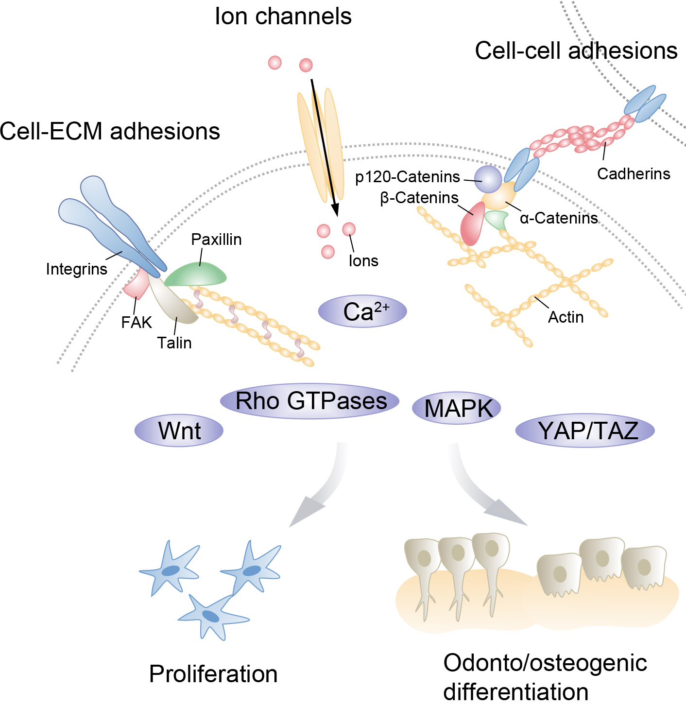
Fig. 1.

Interconnected mechanical network in dental pulp stem cells (DPSCs). Various mechanical cues from the cell and the extracellular environment activate mechano-sensing factors on the cell surface. These factors, including cell-extracellular matrix (ECM) adhesion complexes such as focal adhesions and cell-cell adhesion such as cadherin–catenin junctions and ion channels, regulate specific mechanotransduction pathways, which affect DPSC proliferation and odonto/osteogenesis.

3.1 Cell-Cell Adhesion

Adherens junctions (AJs), one of the major mechanosensory cell-cell junction structures, allow cells to be precisely interconnected, ensuring the structural stability of the multicellular layer of specific tissues, such as the epithelium and pulp [66, 67, 68]. The transmission of mechanical signals between cells varies with cell density [69]. Cells can sense compressive or tensile forces at intercellular contacts and respond by modifying the biomedical properties of adhesion receptors, constituting mechanochemical feedback loops that eventually affect biological behaviors, including cell proliferation [70]. Proper intercellular mechanical signal exchanges are critical for tissue homeostasis.

The most widely studied adhesion receptors for modifying cell-cell junctions and mediating mechanotransduction are cadherins [71, 72]. Various cadherin members are specifically expressed in tissues. E- and P-cadherin are most prevalent in the epithelia, whereas N-cadherin is more frequently present in non-epithelial cells. Further, only blood vessels express VE-cadherin. Notably, these molecules are structure equivalent [73]. Classic cadherins phenotypically interact with adjacent cell surfaces and contribute to AJ formation. Classic cadherins consist of a single-pass transmembrane domain, an N-terminal extracellular domain with five cadherin domains that create adhesive contacts, and a cytoplasmic domain associated with actin-binding proteins, including α-catenin, β-catenin, and p120-catenin [74, 75]. Different binding interfaces between the N-terminal domains, which have diverse mechanical functions, determine the bonding strength of AJs. Cadherin binding can be classified into strand dimers, X-dimers, and lateral dimers based on the different structures of cadherin-cadherin adhesion [76].

Endogenous or exogenous stimuli can be converted into biochemical signals by AJs. However, the cadherin-catenin core complex, which links cadherin to the actin cytoskeleton, regulates the cytoplasmic mechanical signaling chain and directly coordinates the dynamic organization of the cytoskeleton [77]. p120-catenin (which binds to the membrane-proximal region of the cytoplasmic domain of cadherins) can modify cell cohesion by decreasing cadherin renewal in the membrane [78]. βcatenin binds to the membrane-distal region of the cytoplasmic domain of cadherins and recruits α-catenin. α-catenin is a major molecule involved in cadherin mechanotransduction. Notably, this molecule undergoes force-dependent conformation and interacts with actin filaments or alternative actin-binding proteins, such as vinculin, ZO-1, afadin, α-actinin, formin-1, and EPLIN, to trigger the junction-base response [79, 80]. Vinculin is proposed to serve as a junction stabilizer as it can recruit α-catenin under internal contractility and external tension [81]. Vinculin may modify the actin-binding strength or recruit actin regulators in response to mechanical forces [82]. Finally, the cadherin-catenin complex mediates Rho-GTPase signaling (including RhoA, Rac1, and Cdc42) to regulate the cytoskeletal structure [83].

3.2 Cell-ECM Adhesion

Cell-ECM interactions are generally mediated by focal adhesions (FAs), which have been demonstrated to physically connect the ECM to the cytoskeleton [84]. Cells are enclosed in their local ECM, and can detect mechanical changes caused by varying ECM components [85]. Mechanochemical feedback loops also exist, whereby cells release synthesizing or degrading enzymes that eventually modify the mechanical properties of the ECM [86]. Cell morphology and motility respond quickly to mechanical variations, influencing tissue and organ development, wound repair, and immunological response [87, 88, 89].

The ECM, which mainly consists of collagen fibers, elastin, proteoglycans, and glycoproteins, contains a large amount of biological information [90]. Integrin, a key mechanoreceptor or FA, may identify certain ECM ligands and transmit mechanical signals intracellularly by binding to kindlin, talin, fibronectin, or vinculin, which are eventually linked to the actomyosin cytoskeleton [91]. The quantity and intensity of FAs vary in mechanically induced conformations of integrin-ligand complexes [92]. Talin and vinculin are F-actin-binding proteins. The head and tail domains of vinculin bind talin and actin, respectively. Vinculin enhances Talin-F-actin links by recruiting more F-actin [93]. Focal adhesion kinase (FAK), paxillin, Arp2/3, and β-PIX also contribute to actomyosin bundle assembly by activating Rac1 GTPase [94, 95, 96, 97].

As FAs and AJs are intracellularly related to the actin cytoskeleton, they share a common collection of receptor proteins and signaling molecules, including the Rho family of GTPases and vinculin, forming a cross-regulation network [98, 99]. FAs and AJs communicate and collaborate to maintain the mechanochemical signal ecology and achieve tensional homeostasis within tissues.

3.3 Ion Channel

Mechanosensitive ion channels, including Piezo and transient receptor potential (TRP) channels, are vital clusters of operative proteins that convert physical stimuli into intracellular biological signals [100]. These channels, located in the cell membrane, open and close in response to mechanical tension, voltage, and ligand binding, thereby mediating the influx of cations, such as Ca2+, Na+, K+, and Mg2+, across the cell membrane. For example, mechanical stretch and shear stress trigger cells via Piezo family proteins (Piezo1 and Piezo2) to activate Ca2+-dependent downstream molecules [101, 102], such as ERK1/2, thereby promoting cells to undergo mitosis [103, 104]. Cell compression maintains tissue homeostasis via cell extrusion by controlling Piezo activity [105]. Studies have demonstrated that the force generated by actin and microtubule cytoskeleton-related motors leads to the activation of Piezo channels [106, 107]. Although Piezo channels induce downstream signaling molecules mainly by modifying Ca2+ influx [104, 108], the mechanism by which Piezo and other signaling molecules coordinate to elicit cell responses must be further explored [109].

Similar to Piezo channels, TRP channels serve as signal transducers by modifying the intracellular Ca2+ concentration. Generally, TRP channels are classified as the ankyrin subfamily (TRPA), canonical subfamily (TRPC), melastatin subfamily (TRPM), mucolypin subfamily (TRPML), polycystin subfamily (TRPP), and vanilloid subfamily (TRPV) [110, 111, 112]. Several studies have revealed that TRP channels can be mediated by direct force activation via signaling cascades [113, 114, 115], and the G protein-coupled receptor (GPCR) is one example. Mechanical stimuli activate GPCRs to couple with the heterotrimeric G-protein (Gq) and recruit PLC, subsequently activating TRP channels. Actin cytoskeleton has been demonstrated to be an essential transduction component that regulates the mechanosensitivity of TRP channels. Together, these studies revealed that mechanosensitive ion channels can modify intracellular ion concentrations, thereby transforming mechanical signals into biochemical signals.

3.4 Rho GTPases

Rho GTPases are a family of small G proteins [116] that have been found to regulate a variety of fundamental cellular processes, including morphogenesis, cell migration, cell division, and gene expression [117]. The link between mechanical cues and Rho GTPases has been widely recognized [118, 119, 120]. One of the major mechanical cues regulating Rho GTPases is the ECM, where integrins and fatty acids are the main mediators. For instance, ECM stiffness modulates the activity of Rho GTPases through focal adhesion kinases and integrins, leading to actin cytoskeleton assembly via the LIMK-cofilin pathway [121, 122]. Integrin α6β4, and integrin α5, which are mediated by the stiffness sensor epidermal growth factor receptor (EGFR), can also induce Rho activation. Studies have revealed that tensile stress can induce the phosphorylation of FAK/Src, leading to RhoA activation, whereas shear stress can block Rac1 by inhibiting paxillin at FAs [118, 119]. However, how integrin-mediated compression modulates Rho GTPases requires further investigation.

Ion channel activation stimulates Rho GTPases by inducing Ca2+ influx. For example, Piezo and TRP activated by stress or force can trigger Ca2+ influx, leading to the activation of the RhoA and Rac signaling pathways [117, 118]. As the mechanisms by which other ion channels participate in the regulation of Rho GTPase activity remain unclear, this will be a future direction for exploration.

3.5 Yes-Associated Protein (YAP)/Transcriptional Co-Activator with PDZ-Binding Motif (TAZ)

YAP/TAZ is a transcriptional co-activator initially discovered downstream of the Hippo signaling pathway. YAP/TAZ comprises a cascade signaling module of two pairs of kinases: MST1/2 and LATS1/2 [123]. MST1/2 and LATS1/2 function as core protein kinases that phosphorylate YAP/TAZ, leading to the inhibition of nuclear localization and transcriptional coactivation [124]. Although the cytoplasmic restriction of YAP/TAZ promotes their degradation or regulates other signaling pathways, the accumulation of YAP/TAZ in the nucleus drives their interaction with DNA-binding transcription factors, such as TEADs, thereby regulating cell proliferation and differentiation [125].

The ability of YAP and TAZ to respond to diverse mechanical inputs underscores their importance in the regulation of mechanotransduction. The status of YAP/TAZ activity is based on their cellular localization [126, 127, 128, 129]. For example, different ECM elasticities and cell spreading can modulate YAP/TAZ localization. While stiff substrates and large cell spreading promote YAP/TAZ nuclear shift, contact inhibition, which involves cell geometry remodeling, inhibits YAP/TAZ activity. Thus, changes in cell-ECM contacts, which are typically affected by FAs, and cell-cell contacts, which are mediated by cadherin adhesion sites, are assumed to be strongly related to YAP/TAZ activity [130, 131]. The angiomotin (AMOT) complex at tight junctions directly restricts YAP/TAZ in the cytoplasm and/or induces LATS1/2-mediated YAP/TAZ phosphorylation. E-cadherin and α-catenin, which are adherens junction elements, have been demonstrated to inhibit YAP/TAZ nuclear accumulation. Notably, the presence of the actin cytoskeleton and actin contractility are required for YAP/TAZ nuclear localization [124, 131, 132]. Mechanical signals induced by ECM rigidity and cell shape induce YAP/TAZ activation and nuclear accumulation via Rho/Rho kinase-dependent actin rearrangement [131]. As diverse signals converge on actin cytoskeletal tension, the regulation of YAP/TAZ has been demonstrated to be multifaceted; thus, more studies are warranted to validate the regulation of YAP/TAZ by mechanical signals.

3.6 Wnt/β-Catenin

The Wnt/β-catenin signaling pathway has been identified as a crucial component of mechanotransduction pathways. Wnt ligands bind to a receptor complex including a member of the Frizzled family and low-density lipoprotein-related receptor 5 (Lrp5) or Lrp6. When a Wnt signal is present, the cytoplasmic domains of Lrp5 or Lrp6 are phosphorylated, which eventually raises the level of β-catenin in the cytoplasm, allowing it to enter the nucleus and activate transcriptional activity [133]. Based on prior evidence, mechanical forces can upregulate the expression of the target genes of the Wnt/β-catenin signaling pathway [134]. Wnt/β-catenin signaling activation can further increase the sensitivity of cells to mechanical forces [135]. Moreover, oscillatory fluid flow induces β-catenin accumulation in the nucleus, promotes TCF/LEF-associated gene transcription, and upregulates the expression of Wnt-related proteins [136]. Another study found that estradiol (E2) had a sensitizing effect on the expression of mechanically induced cyclooxygenase-2 (Cox-2). This mechanosensitizing effect of E2 may be ligand-specific as it can be inhibited by the anti-estrogen, ICI 182,780. However, mechanical strain reduces the sensitizing effect of E2 and the stimulatory effect of Wnt in the presence of Wnt signaling activators. Additionally, mechanical stretching has been demonstrated to stimulate the expansion of SOX9+ progenitors by activating Wnt/β-catenin signaling [137]. A recent study [138] revealed that different extracellular mechanical inputs (such as mechanical compression, matrix rigidity, osmotic pressure, and stretch) affect intracellular crowding in different manners, thereby impacting the mechanism by which Wnt/β-catenin is activated through the regulation of LRP6 signalosome.

3.7 Mitogen-Activated Protein Kinase (MAPK)

There are four main MAPK signaling pathways in mammalian cells: extracellular signal-regulated protein kinase (ERK)1/2, c-Jun amino-terminal kinase (JNK), p38 MAPK, and ERK5. Therefore, the MAPK signaling pathway plays a crucial role in cytoskeletal regulation [139, 140]. ERK1/2 modulates cytoskeletal signaling by activating myosin light chain kinase (MLCK), which phosphorylates the light chain regulatory sequence of myosin and induces microfilament contraction [141, 142]. ERK1/2 can affect cytoskeletal signaling by phosphorylating calpain, a calcium-dependent protease that cleaves structural proteins, leading to the breakage of cell adhesion sites. ERK1/2 can also phosphorylate kinases at the local adhesion site, which regulates the activity of integral proteins and prevent their polymerization with piled proteins, thereby facilitating the depolymerization of integral proteins from the ECM. These pathways, which are regulated by ERK1/2 signaling, are critical for maintaining the balance of cytoskeletal dynamics.

The p38 MAPK pathway is considered a stress-activated signaling pathway (SAPK) as it is activated by various environmental stresses [143, 144]. The p38 MAPK signaling pathway is related to anti-proliferation and apoptosis [145], which differs from the function of ERK1/2; thus, an interaction occurs between these two pathways. Understanding the intricate interplay between different MAPK signaling pathways is essential for gaining insights into the mechanisms underlying cell fate determination and tissue homeostasis.

4. Role of Mechanical Signals in DPSCs

The effects of mechanical stimulation on DPSCs are multifaceted. Multiple factors that control cell fate determination of DPSCs have been identified. Below, we focused on the role of mechanical signaling components in the proliferation and differentiation of DPSCs.

4.1 Mechanoregulation of DPSCs Proliferation

Integrin is crucial for DPSC proliferation in the cell-ECM interplay. For instance, the inhibition of integrin-α5 (ITGA5) expression was found to prevent the migration and proliferation of DPSCs but enhance odontogenic differentiation [146, 147]. When ITGA5 expression is suppressed, the levels of pFAK, pERK1/2, and pAKT are upregulated. Furthermore, the expression of the Wnt/β-catenin regulators, DKK1 and SFRP1, has been shown to increase, indicating a crosstalk between cell-ECM-mediated signaling and cell-cell adhesion-related signaling. Integrin-α6 (ITGA6) plays a similar role in promoting pluripotency maintenance and the proliferation of DPSCs while inhibiting their odonto/osteogenic differentiation through the Rho/ROCK signaling pathway [148].

The role of ion channels in DPSC proliferation has been extensively studied. The activation of Piezo1 by Yoda1 induces ATP release, which promotes DPSC migration, subsequently activating the P2 receptor purinergic signaling pathway and the downstream PYK2 and MEK/ERK signaling pathways [149]. The proliferation of DPSCs is affected by the activation of MAPK/ERK1/2 signaling after 24 h of low-intensity pulsed ultrasound (LIPUS) stimulation [150]. In contrast, the presence of the piezo blocker, ruthenium red (RR), inhibits the proliferation of LIPUS-stimulated DPSCs [151]. These results demonstrate that Piezo positively modulates DPSC proliferation. The TRPC1 channel inhibitor, SKF96365, inhibits the proliferation of DPSCs in a dose-dependent manner [152], whereas TRPM4 is essential for the proliferation and survival of DPSCs by mediating Ca2+ signaling [153]. Notably, the inhibition of TRPM7 suppresses the proliferation and migration of DPSCs under conditions of induced osteogenic differentiation [154]. Further research is required to explore the specific mechanisms by which ion channels interact with other signaling pathways to control cell proliferation.

Rho GTPases, particularly Rac1, have been shown to participate in the regulation of DPSC proliferation. Rac1 silencing suppresses the pro-apoptotic effect of miR-224 in DPSCs. However, whether other Rho GTPases are involved in the regulation of DPSC proliferation remains to be investigated [155].

Based on growing evidence, YAP/TAZ plays a role in the proliferation process. DPSCs seeded in a static magnetic field accumulate nuclear YAP/TAZ has been demonstrated to promote cell proliferation [156]. In addition, the inhibition of TAZ in DPSCs downregulates the regulation of CTGF and Cyr6, and suppresses cellular proliferation and migration through the TGF-β-dependent signaling pathway [157]. Consistently, increased TAZ expression mediated by miR-584 promotes the proliferation and migration of DPSCs via the PI3K/AKT pathway [158]. As YAP/TAZ signaling has been extensively shown to be involved in the regulation of cell proliferation in other stem cells [159, 160], further research is necessary to clarify the specific mechanism by which YAP/TAZ cooperates in concert with other mechanical components to promote DPSC proliferation.

Wnt signaling is another signaling pathway that is essential for the growth of DPSCs. The promotion of DPSC stemness by the Wnt signaling pathway is related to its effects on oxidative metabolism upon activation. In particular, metabolic remodeling is accompanied by enhanced glycolysis and mitochondrial tricarboxylic acid cycle (TCA) activity [161]. When Wnt/β-catenin expression is inhibited, the calcium hydroxide-induced proliferation and migration of DPSCs are abolished [162]. Wnt signaling promotes DPSC stemness by coordinating with the Notch signaling pathway [163]. Overall, Wnt signaling promotes the proliferation of DPSCs.

Increasing evidence highlights the role of the MAPK signaling pathway in the mechanically stimulated proliferation of DPSCs. When uniaxial stretching is applied to DPSCs, the expression of phosphorylated Akt, ERK1/2, and p38 MAPK is induced, which promotes DPSC proliferation. In contrast, stretch-induced proliferation of DPSCs is abolished after inhibition of the MAPK/ERK pathway. Interestingly, osteocalcin and osteopontin are significantly inhibited by stretching, indicating that stretching inhibits osteogenic differentiation of DPSCs [164]. As previously mentioned, LIPUS stimulation activates MAPK/ERK1/2 signaling in DPSCs to increase their proliferation. Using selective ERK1/2 inhibitors before ultrasound exposure abolished the stimulatory effect on DPSC proliferation, whereas the inhibition of p38 and JNK had no effect [148]. Further, vibrations induce G0/G1 arrest in DPSCs, inhibiting their proliferation [165]. These data emphasize the unique role of ERK1/2 in the MAPK pathway in the presence of vibrations during DPSC proliferation [150]. Atypical physical stimuli may contribute to the proliferation of DPSCs via MAPK signaling. For example, the application of 0.4-Telsa static magnetic fields (SMFs) on DPSCs significantly triggers p38 MAPK and promotes the proliferation of DPSCs. During this process, the cytoskeleton and cell morphology are reorganized [166]. Cells treated with a 0.4-Telsa SMF also exhibit a higher regeneration potential capacity for pulp repair by regulating MAPK signaling. Furthermore, the promotion of proliferation and migration is inhibited by the p38 inhibitor, SB203580 [167].

4.2 Mechanoregulation of DPSCs Odonto/Osteogenic Differentiation

The role of mechanical ion channels in the process of odonto/osteogenic differentiation of DPSCs has been demonstrated. TRPM7 in DPSCs reduces the expression of specific odontoblast markers, such as ALP, DSPP, BSP, RUNX2, and OSX, indicating that TRPM7 plays an important role in the osteogenic differentiation of DPSCs [154]. Odonto/osteogenic differentiation induced by low-level light-emitting diodes has been shown to be mediated by TRPV1. Specifically, capsazepine, a selective TRPV1 inhibitor, inhibits odonto/osteogenesis in DPSCs [168]. However, the piezo-dependent mechanism mediating DPSC differentiation remains unclear.

Rho GTPases and the downstream effector protein, ROCK, promote the differentiation of DPSCs. Treatment with the C3 exoenzyme, a RhoA/ROCK signaling pathway inhibitor, was found to suppress the expression of RUNX2. In particular, the presence of the C3 exoenzyme has been demonstrated to significantly promote odontoblast differentiation of DPSCs in the late stage without affecting the early stage. An interesting research direction would be to determine whether other Rho GTPases can mediate mechanical signals to regulate DPSC differentiation [169].

The mechanoregulation of YAP/TAZ during DPSC differentiation has been reported previously. SMFs rearrange the cytoskeleton of DPSCs and recruit YAP/TAZ to the nucleus, which upregulates the corresponding genes, CTGF and ANKRD1, finally promoting DPSC mineralization [156]. Consistently, the roughness and pore sizes associated with the scaffold topographic cues affect the nuclear localization of YAP by altering the arrangement and morphology of cellular F-actin, thereby promoting the odonto/osteogenic differentiation of DPSCs [170]. The topographical factors of PGLA membranes promote the nuclear translocation of β-catenin in DPSCs, suggesting YAP/TAZ interacts with Wnt/β-catenin and promotes the odontogenic differentiation process of DPSCs under mechanical stimulation [171]. A recent study revealed that hyaluronans promote odonto/osteogenesis by activating YAP/TAZ in DPSCs [172]. The subcellular location of YAP/TAZ was found to remain the same on polydimethylsiloxane (PDMS) substrates; however, the relationship between YAP/TAZ and mechanical cues requires further investigation.

Emerging evidence connects DPSC differentiation to mechanically induced Wnt signaling. Pulsed electromagnetic fields (PEMFs), such as DSPP, DMP1, and RUNX2, induce a significant increase in odontogenic markers, and GSK-3β/β-catenin signaling is involved in this process [173]. Wnt10a, which acts as an upstream regulatory molecule of DSPP, is induced by cell-matrix interaction during DPSC odontoblastic differentiation [174]. When DPSCs were cultured on more rigid PDMS, the odontogenic differentiation ability increased with upregulation. In contrast, the expression of GSK-3β, a negative regulator of β-catenin in the Wnt signaling pathway, was inhibited [175]. Therefore, the odonto/osteogenic differentiation of DPSCs is regulated by matrix stiffness through the typical Wnt/β-catenin signaling pathway. The Wnt/β-catenin pathway is activated by SATB2-mediated DKK1, which ultimately promotes odonto/osteogenesis in DPSCs [176]. Interestingly, a previous study revealed that the typical Wnt signaling pathway plays a negative role in the regulation of odonto/osteogenetic differentiation of DPSCs. Wnt1 inhibits ALP activity, mineralizes nodule formation, and induces OPN expression in DPSCs. In addition, the overexpression of β-catenin inhibits the differentiation and mineralization of DPSCs [177]. Similarly, short-term activation of Wnt signaling by Wnt3a is reversible in the default osteoblastic lineage pre-differentiation phenotype of DPSCs [178]. Thus, different Wnt signaling components can participate in the positive and negative regulatory mechanisms of DPSC differentiation.

Several studies have supported the role of the MAPK pathway in DPSC differentiation. Exposure to medium-magnitude sonic vibration was found to enhance the odontogenic differentiation of DPSCs, which was accompanied by an increase in the expression of osteogenic markers (osteocalcin, BMP-2, and ALP). These studies suggest that mechanical vibrations are related to MAPK signaling during DPSC differentiation. Nevertheless, 0.4-Telsa SMF exposure promoted a significant increase in the expression of DSPP and DMP-1, whereas DPSC differentiation was significantly reduced in the presence of the p38 inhibitors, SB203580 and SMF [167]. Another study revealed that the odontoblastic differentiation of DPSCs under mechanical compression is mediated via the MAPK pathway by ERK1/2 and p38 rather than by JNK, as the phosphorylation level of JNK remains the same. In addition, coordination between the MAPK and Wnt signaling pathways has been demonstrated to play a role in promoting DPSC differentiation [179].

5. Conclusions

Recently, there has been a significant interest in the role of mechanical stimuli in tissue homeostasis. DPSCs, which exhibit self-renewal and multilineage potential, play a critical role in pulp homeostasis and restorative dentin formation. This review provides further insights into the biomechanical properties of DPSCs. Notably, the diversity of mechanical stimuli increases the potential intricate interaction in DPSCs, thereby providing a greater opportunity to explore the interplay between each mechanical stimulation factor in determining DPSC fate. Thus, mechanical cues may offer innovative biomaterial platforms or biochemical-mechanical strategies that regulate the differentiation and proliferation of DPSCs by interconnecting cell microenvironments, biomaterials, and cell behaviors. Overall, this study provides a theoretical basis for DPSC applications in tissue engineering and regenerative medicine.

Author Contributions

JZ, WenD and WeiD conceived the study conception, design and manuscript preparation. JZ and SW preformed literature review. DG contributed to figure preparation. All authors contributed to editorial changes in the manuscript. All authors read and approved the final manuscript.

Ethics Approval and Consent to Participate

Not applicable.

Acknowledgment

Not applicable.

Funding

This work was supported by NSFC grant 82201003, Natural Science Foundation of Sichuan (No. 2022NSFSC0612), and the Research Funding from West China Hospital of Stomatology Sichuan University (RCDWJS2023-11).

Conflict of Interest

The authors declare no conflict of interest.

References
[1]
Gronthos S, Mankani M, Brahim J, Robey PG, Shi S. Postnatal human dental pulp stem cells (DPSCs) in vitro and in vivo. Proceedings of the National Academy of Sciences of the United States of America. 2000; 97: 13625–13630.
[2]
Peng L, Ye L, Zhou XD. Mesenchymal stem cells and tooth engineering. International Journal of Oral Science. 2009; 1: 6–12.
[3]
Roato I, Chinigò G, Genova T, Munaron L, Mussano F. Oral Cavity as a Source of Mesenchymal Stem Cells Useful for Regenerative Medicine in Dentistry. Biomedicines. 2021; 9: 1085.
[4]
Li B, Ouchi T, Cao Y, Zhao Z, Men Y. Dental-Derived Mesenchymal Stem Cells: State of the Art. Frontiers in Cell and Developmental Biology. 2021; 9: 654559.
[5]
Hayward MK, Muncie JM, Weaver VM. Tissue mechanics in stem cell fate, development, and cancer. Developmental Cell. 2021; 56: 1833–1847.
[6]
Sutlive J, Xiu H, Chen Y, Gou K, Xiong F, Guo M, et al. Generation, Transmission, and Regulation of Mechanical Forces in Embryonic Morphogenesis. Small (Weinheim an Der Bergstrasse, Germany). 2022; 18: e2103466.
[7]
Song Y, Soto J, Chen B, Yang L, Li S. Cell engineering: Biophysical regulation of the nucleus. Biomaterials. 2020; 234: 119743.
[8]
Ning W, Muroyama A, Li H, Lechler T. Differentiated Daughter Cells Regulate Stem Cell Proliferation and Fate through Intra-tissue Tension. Cell Stem Cell. 2021; 28: 436–452.e5.
[9]
Salar Amoli M, Anand R, EzEldeen M, Amorim PA, Geris L, Jacobs R, et al. The development of a 3D printable chitosan-based copolymer with tunable properties for dentoalveolar regeneration. Carbohydrate Polymers. 2022; 289: 119441.
[10]
Chen YY, He ST, Yan FH, Zhou PF, Luo K, Zhang YD, et al. Dental pulp stem cells express tendon markers under mechanical loading and are a potential cell source for tissue engineering of tendon-like tissue. International Journal of Oral Science. 2016; 8: 213–222.
[11]
Weng Z, Wang Y, Ouchi T, Liu H, Qiao X, Wu C, et al. Mesenchymal Stem/Stromal Cell Senescence: Hallmarks, Mechanisms, and Combating Strategies. Stem Cells Translational Medicine. 2022; 11: 356–371.
[12]
Williams AR, Hare JM. Mesenchymal stem cells: biology, pathophysiology, translational findings, and therapeutic implications for cardiac disease. Circulation Research. 2011; 109: 923–940.
[13]
Majka M, Sułkowski M, Badyra B, Musiałek P. Concise Review: Mesenchymal Stem Cells in Cardiovascular Regeneration: Emerging Research Directions and Clinical Applications. Stem Cells Translational Medicine. 2017; 6: 1859–1867.
[14]
Berebichez-Fridman R, Gómez-García R, Granados-Montiel J, Berebichez-Fastlicht E, Olivos-Meza A, Granados J, et al. The Holy Grail of Orthopedic Surgery: Mesenchymal Stem Cells-Their Current Uses and Potential Applications. Stem Cells International. 2017; 2017: 2638305.
[15]
Liechty KW, MacKenzie TC, Shaaban AF, Radu A, Moseley AM, Deans R, et al. Human mesenchymal stem cells engraft and demonstrate site-specific differentiation after in utero transplantation in sheep. Nature Medicine. 2000; 6: 1282–1286.
[16]
Sedgley CM, Botero TM. Dental stem cells and their sources. Dental Clinics of North America. 2012; 56: 549–561.
[17]
Miura M, Gronthos S, Zhao M, Lu B, Fisher LW, Robey PG, et al. SHED: stem cells from human exfoliated deciduous teeth. Proceedings of the National Academy of Sciences of the United States of America. 2003; 100: 5807–5812.
[18]
Sonoyama W, Liu Y, Fang D, Yamaza T, Seo BM, Zhang C, et al. Mesenchymal stem cell-mediated functional tooth regeneration in swine. PLoS ONE. 2006; 1: e79.
[19]
Sonoyama W, Liu Y, Yamaza T, Tuan RS, Wang S, Shi S, et al. Characterization of the apical papilla and its residing stem cells from human immature permanent teeth: a pilot study. Journal of Endodontics. 2008; 34: 166–171.
[20]
Seo BM, Miura M, Gronthos S, Bartold PM, Batouli S, Brahim J, et al. Investigation of multipotent postnatal stem cells from human periodontal ligament. Lancet (London, England). 2004; 364: 149–155.
[21]
Morsczeck C, Götz W, Schierholz J, Zeilhofer F, Kühn U, Möhl C, et al. Isolation of precursor cells (PCs) from human dental follicle of wisdom teeth. Matrix Biology: Journal of the International Society for Matrix Biology. 2005; 24: 155–165.
[22]
Kim D, Lee AE, Xu Q, Zhang Q, Le AD. Gingiva-Derived Mesenchymal Stem Cells: Potential Application in Tissue Engineering and Regenerative Medicine - A Comprehensive Review. Frontiers in Immunology. 2021; 12: 667221.
[23]
Farré-Guasch E, Martí-Pagè C, Hernádez-Alfaro F, Klein-Nulend J, Casals N. Buccal fat pad, an oral access source of human adipose stem cells with potential for osteochondral tissue engineering: an in vitro study. Tissue Engineering. Part C, Methods. 2010; 16: 1083–1094.
[24]
Han J, Menicanin D, Gronthos S, Bartold PM. Stem cells, tissue engineering and periodontal regeneration. Australian Dental Journal. 2014; 59: 117–130.
[25]
Gronthos S, Arthur A, Bartold PM, Shi S. A method to isolate and culture expand human dental pulp stem cells. Methods in Molecular Biology (Clifton, N.J.). 2011; 698: 107–121.
[26]
Estrela C, Alencar AHGD, Kitten GT, Vencio EF, Gava E. Mesenchymal stem cells in the dental tissues: perspectives for tissue regeneration. Brazilian Dental Journal. 2011; 22: 91–98.
[27]
Gronthos S, Brahim J, Li W, Fisher LW, Cherman N, Boyde A, et al. Stem cell properties of human dental pulp stem cells. Journal of Dental Research. 2002; 81: 531–535.
[28]
Alliot-Licht B, Bluteau G, Magne D, Lopez-Cazaux S, Lieubeau B, Daculsi G, et al. Dexamethasone stimulates differentiation of odontoblast-like cells in human dental pulp cultures. Cell and Tissue Research. 2005; 321: 391–400.
[29]
Li S, Luo L, He Y, Li R, Xiang Y, Xing Z, et al. Dental pulp stem cell-derived exosomes alleviate cerebral ischaemia-reperfusion injury through suppressing inflammatory response. Cell Proliferation. 2021; 54: e13093.
[30]
Li H, Ghazanfari R, Zacharaki D, Lim HC, Scheding S. Isolation and characterization of primary bone marrow mesenchymal stromal cells. Annals of the New York Academy of Sciences. 2016; 1370: 109–118.
[31]
Beyer Nardi N, da Silva Meirelles L. Mesenchymal stem cells: isolation, in vitro expansion and characterization. Handbook of Experimental Pharmacology. 2006; 249–282.
[32]
Chen YK, Huang AHC, Chan AWS, Shieh TY, Lin LM. Human dental pulp stem cells derived from different cryopreservation methods of human dental pulp tissues of diseased teeth. Journal of Oral Pathology & Medicine: Official Publication of the International Association of Oral Pathologists and the American Academy of Oral Pathology. 2011; 40: 793–800.
[33]
Rosa V, Botero TM, Nör JE. Regenerative endodontics in light of the stem cell paradigm. International Dental Journal. 2011; 61: 23–28.
[34]
Kawashima N, Okiji T. Odontoblasts: Specialized hard-tissue-forming cells in the dentin-pulp complex. Congenital Anomalies. 2016; 56: 144–153.
[35]
Mackie EJ. Osteoblasts: novel roles in orchestration of skeletal architecture. The International Journal of Biochemistry & Cell Biology. 2003; 35: 1301–1305.
[36]
Langenbach F, Handschel J. Effects of dexamethasone, ascorbic acid and β-glycerophosphate on the osteogenic differentiation of stem cells in vitro. Stem Cell Research & Therapy. 2013; 4: 117.
[37]
Cui Y, Ji W, Gao Y, Xiao Y, Liu H, Chen Z. Single-cell characterization of monolayer cultured human dental pulp stem cells with enhanced differentiation capacity. International Journal of Oral Science. 2021; 13: 44.
[38]
Xu J, Yu B, Hong C, Wang CY. KDM6B epigenetically regulates odontogenic differentiation of dental mesenchymal stem cells. International Journal of Oral Science. 2013; 5: 200–205.
[39]
Cui D, Xiao J, Zhou Y, Zhou X, Liu Y, Peng Y, et al. Epiregulin enhances odontoblastic differentiation of dental pulp stem cells via activating MAPK signalling pathway. Cell Proliferation. 2019; 52: e12680.
[40]
Irfan M, Kim JH, Marzban H, Reed DA, George A, Cooper LF, et al. The role of complement C5a receptor in DPSC odontoblastic differentiation and in vivo reparative dentin formation. International Journal of Oral Science. 2022; 14: 7.
[41]
Zheng H, Wang N, Li L, Ge L, Jia H, Fan Z. miR-140-3p enhanced the osteo/odontogenic differentiation of DPSCs via inhibiting KMT5B under hypoxia condition. International Journal of Oral Science. 2021; 13: 41.
[42]
Maity J, Barthels D, Sarkar J, Prateeksha P, Deb M, Rolph D, et al. Ferutinin induces osteoblast differentiation of DPSCs via induction of KLF2 and autophagy/mitophagy. Cell Death & Disease. 2022; 13: 452.
[43]
Yu L, Zeng L, Zhang Z, Zhu G, Xu Z, Xia J, et al. Cannabidiol Rescues TNF-α-Inhibited Proliferation, Migration, and Osteogenic/Odontogenic Differentiation of Dental Pulp Stem Cells. Biomolecules. 2023; 13: 118.
[44]
Chan YH, Ho KN, Lee YC, Chou MJ, Lew WZ, Huang HM, et al. Melatonin enhances osteogenic differentiation of dental pulp mesenchymal stem cells by regulating MAPK pathways and promotes the efficiency of bone regeneration in calvarial bone defects. Stem Cell Research & Therapy. 2022; 13: 73.
[45]
Vukovic M, Lazarevic M, Mitic D, Jaksic Karisik M, Ilic B, Andric M, et al. Acetylsalicylic-acid (ASA) regulation of osteo/odontogenic differentiation and proliferation of human dental pulp stem cells (DPSCs) in vitro. Archives of Oral Biology. 2022; 144: 105564.
[46]
Nakashima M, Nagasawa H, Yamada Y, Reddi AH. Regulatory role of transforming growth factor-beta, bone morphogenetic protein-2, and protein-4 on gene expression of extracellular matrix proteins and differentiation of dental pulp cells. Developmental Biology. 1994; 162: 18–28.
[47]
Lorusso F, Inchingolo F, Dipalma G, Postiglione F, Fulle S, Scarano A. Synthetic Scaffold/Dental Pulp Stem Cell (DPSC) Tissue Engineering Constructs for Bone Defect Treatment: An Animal Studies Literature Review. International Journal of Molecular Sciences. 2020; 21: 9765.
[48]
Iezzi I, Cerqueni G, Licini C, Lucarini G, Mattioli Belmonte M. Dental pulp stem cells senescence and regenerative potential relationship. Journal of Cellular Physiology. 2019; 234: 7186–7197.
[49]
Martens W, Wolfs E, Struys T, Politis C, Bronckaers A, Lambrichts I. Expression pattern of basal markers in human dental pulp stem cells and tissue. Cells, Tissues, Organs. 2012; 196: 490–500.
[50]
Askari N, Yaghoobi MM, Shamsara M, Esmaeili-Mahani S. Human Dental Pulp Stem Cells Differentiate into Oligodendrocyte Progenitors Using the Expression of Olig2 Transcription Factor. Cells, Tissues, Organs. 2014; 200: 93–103.
[51]
Wang D, Wang Y, Tian W, Pan J. Advances of tooth-derived stem cells in neural diseases treatments and nerve tissue regeneration. Cell Proliferation. 2019; 52: e12572.
[52]
Luzuriaga J, Pineda JR, Irastorza I, Uribe-Etxebarria V, García-Gallastegui P, Encinas JM, et al. BDNF and NT3 Reprogram Human Ectomesenchymal Dental Pulp Stem Cells to Neurogenic and Gliogenic Neural Crest Progenitors Cultured in Serum-Free Medium. Cellular Physiology and Biochemistry: International Journal of Experimental Cellular Physiology, Biochemistry, and Pharmacology. 2019; 52: 1361–1380.
[53]
Gonmanee T, Thonabulsombat C, Vongsavan K, Sritanaudomchai H. Differentiation of stem cells from human deciduous and permanent teeth into spiral ganglion neuron-like cells. Archives of Oral Biology. 2018; 88: 34–41.
[54]
Huang GTJ, Gronthos S, Shi S. Mesenchymal stem cells derived from dental tissues vs. those from other sources: their biology and role in regenerative medicine. Journal of Dental Research. 2009; 88: 792–806.
[55]
Lee YM, Shin SY, Jue SS, Kwon IK, Cho EH, Cho ES, et al. The role of PIN1 on odontogenic and adipogenic differentiation in human dental pulp stem cells. Stem Cells and Development. 2014; 23: 618–630.
[56]
Ishkitiev N, Yaegaki K, Imai T, Tanaka T, Nakahara T, Ishikawa H, et al. High-purity hepatic lineage differentiated from dental pulp stem cells in serum-free medium. Journal of Endodontics. 2012; 38: 475–480.
[57]
Chen YK, Huang AHC, Chan AWS, Lin LM. Human dental pulp stem cells derived from cryopreserved dental pulp tissues of vital extracted teeth with disease demonstrate hepatic-like differentiation. Journal of Tissue Engineering and Regenerative Medicine. 2016; 10: 475–485.
[58]
Han YJ, Kang YH, Shivakumar SB, Bharti D, Son YB, Choi YH, et al. Stem Cells from Cryopreserved Human Dental Pulp Tissues Sequentially Differentiate into Definitive Endoderm and Hepatocyte-Like Cells in vitro. International Journal of Medical Sciences. 2017; 14: 1418–1429.
[59]
Song B, Jiang W, Alraies A, Liu Q, Gudla V, Oni J, et al. Bladder Smooth Muscle Cells Differentiation from Dental Pulp Stem Cells: Future Potential for Bladder Tissue Engineering. Stem Cells International. 2016; 2016: 6979368.
[60]
Bronckaers A, Hilkens P, Fanton Y, Struys T, Gervois P, Politis C, et al. Angiogenic properties of human dental pulp stem cells. PLoS ONE. 2013; 8: e71104.
[61]
Ji L, Bao L, Gu Z, Zhou Q, Liang Y, Zheng Y, et al. Comparison of immunomodulatory properties of exosomes derived from bone marrow mesenchymal stem cells and dental pulp stem cells. Immunologic Research. 2019; 67: 432–442.
[62]
Wang N, Butler JP, Ingber DE. Mechanotransduction across the cell surface and through the cytoskeleton. Science (New York, N.Y.). 1993; 260: 1124–1127.
[63]
Geiger B, Spatz JP, Bershadsky AD. Environmental sensing through focal adhesions. Nature Reviews. Molecular Cell Biology. 2009; 10: 21–33.
[64]
Humphrey JD, Dufresne ER, Schwartz MA. Mechanotransduction and extracellular matrix homeostasis. Nature Reviews. Molecular Cell Biology. 2014; 15: 802–812.
[65]
Borghi N, Sorokina M, Shcherbakova OG, Weis WI, Pruitt BL, Nelson WJ, et al. E-cadherin is under constitutive actomyosin-generated tension that is increased at cell-cell contacts upon externally applied stretch. Proceedings of the National Academy of Sciences of the United States of America. 2012; 109: 12568–12573.
[66]
Garcia MA, Nelson WJ, Chavez N. Cell-Cell Junctions Organize Structural and Signaling Networks. Cold Spring Harbor Perspectives in Biology. 2018; 10: a029181.
[67]
Xu J, Shao M, Pan H, Wang H, Cheng L, Yang H, et al. Novel role of zonula occludens-1: A tight junction protein closely associated with the odontoblast differentiation of human dental pulp cells. Cell Biology International. 2016; 40: 787–795.
[68]
Bazzoni G, Dejana E. Endothelial cell-to-cell junctions: molecular organization and role in vascular homeostasis. Physiological Reviews. 2004; 84: 869–901.
[69]
Angulo-Urarte A, van der Wal T, Huveneers S. Cell-cell junctions as sensors and transducers of mechanical forces. Biochimica et Biophysica Acta. Biomembranes. 2020; 1862: 183316.
[70]
Hannezo E, Heisenberg CP. Mechanochemical Feedback Loops in Development and Disease. Cell. 2019; 178: 12–25.
[71]
Halbleib JM, Nelson WJ. Cadherins in development: cell adhesion, sorting, and tissue morphogenesis. Genes & Development. 2006; 20: 3199–3214.
[72]
Smutny M, Yap AS. Neighborly relations: cadherins and mechanotransduction. The Journal of Cell Biology. 2010; 189: 1075–1077.
[73]
Gul IS, Hulpiau P, Saeys Y, van Roy F. Evolution and diversity of cadherins and catenins. Experimental Cell Research. 2017; 358: 3–9.
[74]
Ivanov DB, Philippova MP, Tkachuk VA. Structure and functions of classical cadherins. Biochemistry. Biokhimiia. 2001; 66: 1174–1186.
[75]
Pokutta S, Weis WI. Structure and mechanism of cadherins and catenins in cell-cell contacts. Annual Review of Cell and Developmental Biology. 2007; 23: 237–261.
[76]
Leckband DE, de Rooij J. Cadherin adhesion and mechanotransduction. Annual Review of Cell and Developmental Biology. 2014; 30: 291–315.
[77]
Clarke DN, Miller PW, Lowe CJ, Weis WI, Nelson WJ. Characterization of the Cadherin-Catenin Complex of the Sea Anemone Nematostella vectensis and Implications for the Evolution of Metazoan Cell-Cell Adhesion. Molecular Biology and Evolution. 2016; 33: 2016–2029.
[78]
Hatzfeld M. The p120 family of cell adhesion molecules. European Journal of Cell Biology. 2005; 84: 205–214.
[79]
Takeichi M. Multiple functions of α-catenin beyond cell adhesion regulation. Current Opinion in Cell Biology. 2018; 54: 24–29.
[80]
Wang A, Dunn AR, Weis WI. Mechanism of the cadherin-catenin F-actin catch bond interaction. eLife. 2022; 11: e80130.
[81]
Leerberg JM, Yap AS. Vinculin, cadherin mechanotransduction and homeostasis of cell-cell junctions. Protoplasma. 2013; 250: 817–829.
[82]
Shi B, Matsui T, Qian S, Weiss TM, Nicholl ID, Callaway DJE, et al. An ensemble of cadherin-catenin-vinculin complex employs vinculin as the major F-actin binding mode. Biophysical Journal. 2023; 122: 2456–2474.
[83]
Fukata M, Kaibuchi K. Rho-family GTPases in cadherin-mediated cell-cell adhesion. Nature Reviews. Molecular Cell Biology. 2001; 2: 887–897.
[84]
Rens EG, Merks RMH. Cell Shape and Durotaxis Explained from Cell-Extracellular Matrix Forces and Focal Adhesion Dynamics. IScience. 2020; 23: 101488.
[85]
Lukashev ME, Werb Z. ECM signalling: orchestrating cell behaviour and misbehaviour. Trends in Cell Biology. 1998; 8: 437–441.
[86]
Kanchanawong P, Calderwood DA. Organization, dynamics and mechanoregulation of integrin-mediated cell-ECM adhesions. Nature Reviews. Molecular Cell Biology. 2023; 24: 142–161.
[87]
Yang Y, Lin H, Shen H, Wang B, Lei G, Tuan RS. Mesenchymal stem cell-derived extracellular matrix enhances chondrogenic phenotype of and cartilage formation by encapsulated chondrocytes in vitro and in vivo. Acta Biomaterialia. 2018; 69: 71–82.
[88]
Haage A, Goodwin K, Whitewood A, Camp D, Bogutz A, Turner CT, et al. Talin Autoinhibition Regulates Cell-ECM Adhesion Dynamics and Wound Healing In Vivo. Cell Reports. 2018; 25: 2401–2416.e5.
[89]
Shimizu Y, Shaw S. Lymphocyte interactions with extracellular matrix. FASEB Journal: Official Publication of the Federation of American Societies for Experimental Biology. 1991; 5: 2292–2299.
[90]
Mouw JK, Ou G, Weaver VM. Extracellular matrix assembly: a multiscale deconstruction. Nature Reviews. Molecular Cell Biology. 2014; 15: 771–785.
[91]
Campbell ID, Humphries MJ. Integrin structure, activation, and interactions. Cold Spring Harbor Perspectives in Biology. 2011; 3: a004994.
[92]
Liu C, Qu L, Zhao C, Shou C. Extracellular gamma-synuclein promotes tumor cell motility by activating β1 integrin-focal adhesion kinase signaling pathway and increasing matrix metalloproteinase-24, -2 protein secretion. Journal of Experimental & Clinical Cancer Research: CR. 2018; 37: 117.
[93]
Wang Y, Yao M, Baker KB, Gough RE, Le S, Goult BT, et al. Force-Dependent Interactions between Talin and Full-Length Vinculin. Journal of the American Chemical Society. 2021; 143: 14726–14737.
[94]
Zhou C, Zhang D, Du W, Zou J, Li X, Xie J. Substrate mechanics dictate cell-cell communication by gap junctions in stem cells from human apical papilla. Acta Biomaterialia. 2020; 107: 178–193.
[95]
Zhou C, Zhang D, Zou J, Li X, Zou S, Xie J. Substrate Compliance Directs the Osteogenic Lineages of Stem Cells from the Human Apical Papilla via the Processes of Mechanosensing and Mechanotransduction. ACS Applied Materials & Interfaces. 2019; 11: 26448–26459.
[96]
Rotty JD, Brighton HE, Craig SL, Asokan SB, Cheng N, Ting JP, et al. Arp2/3 Complex Is Required for Macrophage Integrin Functions but Is Dispensable for FcR Phagocytosis and In Vivo Motility. Developmental Cell. 2017; 42: 498–513.e6.
[97]
DeMali KA, Barlow CA, Burridge K. Recruitment of the Arp2/3 complex to vinculin: coupling membrane protrusion to matrix adhesion. The Journal of Cell Biology. 2002; 159: 881–891.
[98]
Mui KL, Chen CS, Assoian RK. The mechanical regulation of integrin-cadherin crosstalk organizes cells, signaling and forces. Journal of Cell Science. 2016; 129: 1093–1100.
[99]
Jülich D, Cobb G, Melo AM, McMillen P, Lawton AK, Mochrie SGJ, et al. Cross-Scale Integrin Regulation Organizes ECM and Tissue Topology. Developmental Cell. 2015; 34: 33–44.
[100]
Kung C. A possible unifying principle for mechanosensation. Nature. 2005; 436: 647–654.
[101]
Fang XZ, Zhou T, Xu JQ, Wang YX, Sun MM, He YJ, et al. Structure, kinetic properties and biological function of mechanosensitive Piezo channels. Cell & Bioscience. 2021; 11: 13.
[102]
Szczot M, Nickolls AR, Lam RM, Chesler AT. The Form and Function of PIEZO2. Annual Review of Biochemistry. 2021; 90: 507–534.
[103]
Chen X, Wanggou S, Bodalia A, Zhu M, Dong W, Fan JJ, et al. A Feedforward Mechanism Mediated by Mechanosensitive Ion Channel PIEZO1 and Tissue Mechanics Promotes Glioma Aggression. Neuron. 2018; 100: 799–815.e7.
[104]
He L, Si G, Huang J, Samuel ADT, Perrimon N. Mechanical regulation of stem-cell differentiation by the stretch-activated Piezo channel. Nature. 2018; 555: 103–106.
[105]
Eisenhoffer GT, Loftus PD, Yoshigi M, Otsuna H, Chien CB, Morcos PA, et al. Crowding induces live cell extrusion to maintain homeostatic cell numbers in epithelia. Nature. 2012; 484: 546–549.
[106]
Wang J, Jiang J, Yang X, Zhou G, Wang L, Xiao B. Tethering Piezo channels to the actin cytoskeleton for mechanogating via the cadherin-β-catenin mechanotransduction complex. Cell Reports. 2022; 38: 110342.
[107]
Lee W, Nims RJ, Savadipour A, Zhang Q, Leddy HA, Liu F, et al. Inflammatory signaling sensitizes Piezo1 mechanotransduction in articular chondrocytes as a pathogenic feed-forward mechanism in osteoarthritis. Proceedings of the National Academy of Sciences of the United States of America. 2021; 118: e2001611118.
[108]
Miyazaki A, Sugimoto A, Yoshizaki K, Kawarabayashi K, Iwata K, Kurogoushi R, et al. Coordination of WNT signaling and ciliogenesis during odontogenesis by piezo type mechanosensitive ion channel component 1. Scientific Reports. 2019; 9: 14762.
[109]
Zhu B, Qian W, Han C, Bai T, Hou X. Piezo 1 activation facilitates cholangiocarcinoma metastasis via Hippo/YAP signaling axis. Molecular Therapy. Nucleic Acids. 2021; 24: 241–252.
[110]
Nilius B, Szallasi A. Transient receptor potential channels as drug targets: from the science of basic research to the art of medicine. Pharmacological Reviews. 2014; 66: 676–814.
[111]
Wu LJ, Sweet TB, Clapham DE. International Union of Basic and Clinical Pharmacology. LXXVI. Current progress in the mammalian TRP ion channel family. Pharmacological Reviews. 2010; 62: 381–404.
[112]
Nilius B, Owsianik G. The transient receptor potential family of ion channels. Genome Biology. 2011; 12: 218.
[113]
Nelson P, Ngoc Tran TD, Zhang H, Zolochevska O, Figueiredo M, Feng JM, et al. Transient receptor potential melastatin 4 channel controls calcium signals and dental follicle stem cell differentiation. Stem Cells (Dayton, Ohio). 2013; 31: 167–177.
[114]
Jin J, Wu LJ, Jun J, Cheng X, Xu H, Andrews NC, et al. The channel kinase, TRPM7, is required for early embryonic development. Proceedings of the National Academy of Sciences of the United States of America. 2012; 109: E225–E233.
[115]
Willard VP, Leddy HA, Palmer D, Wu CL, Liedtke W, Guilak F. Transient receptor potential vanilloid 4 as a regulator of induced pluripotent stem cell chondrogenesis. Stem Cells (Dayton, Ohio). 2021; 39: 1447–1456.
[116]
Madaule P, Axel R. A novel ras-related gene family. Cell. 1985; 41: 31–40.
[117]
Etienne-Manneville S, Hall A. Rho GTPases in cell biology. Nature. 2002; 420: 629–635.
[118]
McBeath R, Pirone DM, Nelson CM, Bhadriraju K, Chen CS. Cell shape, cytoskeletal tension, and RhoA regulate stem cell lineage commitment. Developmental Cell. 2004; 6: 483–495.
[119]
Burridge K, Monaghan-Benson E, Graham DM. Mechanotransduction: from the cell surface to the nucleus via RhoA. Philosophical Transactions of the Royal Society of London. Series B, Biological Sciences. 2019; 374: 20180229.
[120]
Keung AJ, de Juan-Pardo EM, Schaffer DV, Kumar S. Rho GTPases mediate the mechanosensitive lineage commitment of neural stem cells. Stem Cells (Dayton, Ohio). 2011; 29: 1886–1897.
[121]
Zhao XH, Laschinger C, Arora P, Szászi K, Kapus A, McCulloch CA. Force activates smooth muscle alpha-actin promoter activity through the Rho signaling pathway. Journal of Cell Science. 2007; 120: 1801–1809.
[122]
Yim EKF, Sheetz MP. Force-dependent cell signaling in stem cell differentiation. Stem Cell Research & Therapy. 2012; 3: 41.
[123]
Yu FX, Guan KL. The Hippo pathway: regulators and regulations. Genes & Development. 2013; 27: 355–371.
[124]
Zhao B, Wei X, Li W, Udan RS, Yang Q, Kim J, et al. Inactivation of YAP oncoprotein by the Hippo pathway is involved in cell contact inhibition and tissue growth control. Genes & Development. 2007; 21: 2747–2761.
[125]
Totaro A, Panciera T, Piccolo S. YAP/TAZ upstream signals and downstream responses. Nature Cell Biology. 2018; 20: 888–899.
[126]
Pan D. The hippo signaling pathway in development and cancer. Developmental Cell. 2010; 19: 491–505.
[127]
Yu FX, Zhao B, Guan KL. Hippo Pathway in Organ Size Control, Tissue Homeostasis, and Cancer. Cell. 2015; 163: 811–828.
[128]
Piccolo S, Dupont S, Cordenonsi M. The biology of YAP/TAZ: hippo signaling and beyond. Physiological Reviews. 2014; 94: 1287–1312.
[129]
Gaspar P, Tapon N. Sensing the local environment: actin architecture and Hippo signalling. Current Opinion in Cell Biology. 2014; 31: 74–83.
[130]
Aragona M, Panciera T, Manfrin A, Giulitti S, Michielin F, Elvassore N, et al. A mechanical checkpoint controls multicellular growth through YAP/TAZ regulation by actin-processing factors. Cell. 2013; 154: 1047–1059.
[131]
Dupont S, Morsut L, Aragona M, Enzo E, Giulitti S, Cordenonsi M, et al. Role of YAP/TAZ in mechanotransduction. Nature. 2011; 474: 179–183.
[132]
Wada KI, Itoga K, Okano T, Yonemura S, Sasaki H. Hippo pathway regulation by cell morphology and stress fibers. Development (Cambridge, England). 2011; 138: 3907–3914.
[133]
Liu J, Xiao Q, Xiao J, Niu C, Li Y, Zhang X, et al. Wnt/β-catenin signalling: function, biological mechanisms, and therapeutic opportunities. Signal Transduction and Targeted Therapy. 2022; 7: 3.
[134]
Choi RB, Robling AG. The Wnt pathway: An important control mechanism in bone’s response to mechanical loading. Bone. 2021; 153: 116087.
[135]
Robinson JA, Chatterjee-Kishore M, Yaworsky PJ, Cullen DM, Zhao W, Li C, et al. Wnt/beta-catenin signaling is a normal physiological response to mechanical loading in bone. The Journal of Biological Chemistry. 2006; 281: 31720–31728.
[136]
Arnsdorf EJ, Tummala P, Jacobs CR. Non-canonical Wnt signaling and N-cadherin related beta-catenin signaling play a role in mechanically induced osteogenic cell fate. PLoS ONE. 2009; 4: e5388.
[137]
Meng F, Shen C, Yang L, Ni C, Huang J, Lin K, et al. Mechanical stretching boosts expansion and regeneration of intestinal organoids through fueling stem cell self-renewal. Cell Regeneration (London, England). 2022; 11: 39.
[138]
Li Y, Chen M, Hu J, Sheng R, Lin Q, He X, et al. Volumetric Compression Induces Intracellular Crowding to Control Intestinal Organoid Growth via Wnt/β-Catenin Signaling. Cell Stem Cell. 2021; 28: 63–78.e7.
[139]
Sun Y, Meng GM, Guo ZL, Xu LH. Regulation of heat shock protein 27 phosphorylation during microcystin-LR-induced cytoskeletal reorganization in a human liver cell line. Toxicology Letters. 2011; 207: 270–277.
[140]
Cargnello M, Roux PP. Activation and function of the MAPKs and their substrates, the MAPK-activated protein kinases. Microbiology and Molecular Biology Reviews: MMBR. 2011; 75: 50–83.
[141]
Hunger-Glaser I, Salazar EP, Sinnett-Smith J, Rozengurt E. Bombesin, lysophosphatidic acid, and epidermal growth factor rapidly stimulate focal adhesion kinase phosphorylation at Ser-910: requirement for ERK activation. The Journal of Biological Chemistry. 2003; 278: 22631–22643.
[142]
Klemke RL, Cai S, Giannini AL, Gallagher PJ, de Lanerolle P, Cheresh DA. Regulation of cell motility by mitogen-activated protein kinase. The Journal of Cell Biology. 1997; 137: 481–492.
[143]
Keshet Y, Seger R. The MAP kinase signaling cascades: a system of hundreds of components regulates a diverse array of physiological functions. Methods in Molecular Biology (Clifton, N.J.). 2010; 661: 3–38.
[144]
Kyriakis JM, Avruch J. Mammalian mitogen-activated protein kinase signal transduction pathways activated by stress and inflammation. Physiological Reviews. 2001; 81: 807–869.
[145]
Wagner EF, Nebreda AR. Signal integration by JNK and p38 MAPK pathways in cancer development. Nature Reviews. Cancer. 2009; 9: 537–549.
[146]
Xu S, Cui L, Ma D, Sun W, Wu B. Effect of ITGA5 down-regulation on the migration capacity of human dental pulp stem cells. International Journal of Clinical and Experimental Pathology. 2015; 8: 14425–14432.
[147]
Wang H, Ning T, Song C, Luo X, Xu S, Zhang X, et al. Priming integrin α5 promotes human dental pulp stem cells odontogenic differentiation due to extracellular matrix deposition and amplified extracellular matrix-receptor activity. Journal of Cellular Physiology. 2019; 234: 12897–12909.
[148]
Zhang W, Shen J, Zhang S, Liu X, Pan S, Li Y, et al. Silencing integrin α6 enhances the pluripotency-differentiation transition in human dental pulp stem cells. Oral Diseases. 2022; 28: 711–722.
[149]
Mousawi F, Peng H, Li J, Ponnambalam S, Roger S, Zhao H, et al. Chemical activation of the Piezo1 channel drives mesenchymal stem cell migration via inducing ATP release and activation of P2 receptor purinergic signaling. Stem Cells (Dayton, Ohio). 2020; 38: 410–421.
[150]
Gao Q, Walmsley AD, Cooper PR, Scheven BA. Ultrasound Stimulation of Different Dental Stem Cell Populations: Role of Mitogen-activated Protein Kinase Signaling. Journal of Endodontics. 2016; 42: 425–431.
[151]
Gao Q, Cooper PR, Walmsley AD, Scheven BA. Role of Piezo Channels in Ultrasound-stimulated Dental Stem Cells. Journal of Endodontics. 2017; 43: 1130–1136.
[152]
Luo L, Zhang Y, Chen H, Hu F, Wang X, Xing Z, et al. Effects and mechanisms of basic fibroblast growth factor on the proliferation and regenerative profiles of cryopreserved dental pulp stem cells. Cell Proliferation. 2021; 54: e12969.
[153]
Ngoc Tran TD, Stovall KE, Suantawee T, Hu Y, Yao S, Yang LJ, et al. Transient receptor potential melastatin 4 channel is required for rat dental pulp stem cell proliferation and survival. Cell Proliferation. 2017; 50: e12360.
[154]
Cui L, Xu SM, Ma DD, Wu BL. The effect of TRPM7 suppression on the proliferation, migration and osteogenic differentiation of human dental pulp stem cells. International Endodontic Journal. 2014; 47: 583–593.
[155]
Qiao W, Li D, Shi Q, Wang H, Wang H, Guo J. miR-224-5p protects dental pulp stem cells from apoptosis by targeting Rac1. Experimental and Therapeutic Medicine. 2020; 19: 9–18.
[156]
Zheng L, Zhang L, Chen L, Jiang J, Zhou X, Wang M, et al. Static magnetic field regulates proliferation, migration, differentiation, and YAP/TAZ activation of human dental pulp stem cells. Journal of Tissue Engineering and Regenerative Medicine. 2018; 12: 2029–2040.
[157]
Tian S, Tian X, Liu Y, Dong F, Wang J, Liu X, et al. Effects of TAZ on human dental pulp stem cell proliferation and migration. Molecular Medicine Reports. 2017; 15: 4326–4332.
[158]
Tian S, Liu Y, Dong F, Dou Y, Li W, Wang J. Knockdown of microRNA-584 promotes dental pulp stem cells proliferation by targeting TAZ. Cell Cycle (Georgetown, Tex.). 2020; 19: 1048–1058.
[159]
Shi J, Farzaneh M, Khoshnam SE. Yes-Associated Protein and PDZ Binding Motif: A Critical Signaling Pathway in the Control of Human Pluripotent Stem Cells Self-Renewal and Differentiation. Cellular Reprogramming. 2020; 22: 55–61.
[160]
Hwang JH, Lee DH, Byun MR, Kim AR, Kim KM, Park JI, et al. Nanotopological plate stimulates osteogenic differentiation through TAZ activation. Scientific Reports. 2017; 7: 3632.
[161]
Uribe-Etxebarria V, Agliano A, Unda F, Ibarretxe G. Wnt signaling reprograms metabolism in dental pulp stem cells. Journal of Cellular Physiology. 2019; 234: 13068–13082.
[162]
Liu Y, Liu N, Na J, Li C, Yue G, Fan Y, et al. Wnt/β-catenin plays a dual function in calcium hydroxide induced proliferation, migration, osteogenic differentiation and mineralization in vitro human dental pulp stem cells. International Endodontic Journal. 2023; 56: 92–102.
[163]
Uribe-Etxebarria V, Luzuriaga J, García-Gallastegui P, Agliano A, Unda F, Ibarretxe G. Notch/Wnt cross-signalling regulates stemness of dental pulp stem cells through expression of neural crest and core pluripotency factors. European Cells & Materials. 2017; 34: 249–270.
[164]
Hata M, Naruse K, Ozawa S, Kobayashi Y, Nakamura N, Kojima N, et al. Mechanical stretch increases the proliferation while inhibiting the osteogenic differentiation in dental pulp stem cells. Tissue Engineering. Part a. 2013; 19: 625–633.
[165]
Lee W, Eo SR, Choi JH, Kim YM, Nam MH, Seo YK. The Osteogenic Differentiation of Human Dental Pulp Stem Cells through G0/G1 Arrest and the p-ERK/Runx-2 Pathway by Sonic Vibration. International Journal of Molecular Sciences. 2021; 22: 10167.
[166]
Lew WZ, Huang YC, Huang KY, Lin CT, Tsai MT, Huang HM. Static magnetic fields enhance dental pulp stem cell proliferation by activating the p38 mitogen-activated protein kinase pathway as its putative mechanism. Journal of Tissue Engineering and Regenerative Medicine. 2018; 12: 19–29.
[167]
Lew WZ, Feng SW, Lin CT, Huang HM. Use of 0.4-Tesla static magnetic field to promote reparative dentine formation of dental pulp stem cells through activation of p38 MAPK signalling pathway. International Endodontic Journal. 2019; 52: 28–43.
[168]
Chen J, Sang Y, Li J, Zhao T, Liu B, Xie S, et al. Low-level controllable blue LEDs irradiation enhances human dental pulp stem cells osteogenic differentiation via transient receptor potential vanilloid 1. Journal of Photochemistry and Photobiology. B, Biology. 2022; 233: 112472.
[169]
Huang X, Chen X, Chen H, Xu D, Lin C, Peng B. Rho/Rho-associated protein kinase signaling pathway-mediated downregulation of runt-related transcription factor 2 expression promotes the differentiation of dental pulp stem cells into odontoblasts. Experimental and Therapeutic Medicine. 2018; 15: 4457–4464.
[170]
Du Y, Montoya C, Orrego S, Wei X, Ling J, Lelkes PI, et al. Topographic cues of a novel bilayered scaffold modulate dental pulp stem cells differentiation by regulating YAP signalling through cytoskeleton adjustments. Cell Proliferation. 2019; 52: e12676.
[171]
Li G, Xu Z, Yang M, Ning Y, Ye L, Jiang H, et al. Topographic Cues of a PLGA Scaffold Promote Odontogenic Differentiation of Dental Pulp Stem Cells through the YAP/β-Catenin Signaling Axis. ACS Biomaterials Science & Engineering. 2023; 9: 1598–1607.
[172]
La Noce M, Stellavato A, Vassallo V, Cammarota M, Laino L, Desiderio V, et al. Hyaluronan-Based Gel Promotes Human Dental Pulp Stem Cells Bone Differentiation by Activating YAP/TAZ Pathway. Cells. 2021; 10: 2899.
[173]
Lim HM, Nam MH, Kim YM, Seo YK. Increasing Odontoblast-like Differentiation from Dental Pulp Stem Cells through Increase of β-Catenin/p-GSK-3β Expression by Low-Frequency Electromagnetic Field. Biomedicines. 2021; 9: 1049.
[174]
Yamashiro T, Zheng L, Shitaku Y, Saito M, Tsubakimoto T, Takada K, et al. Wnt10a regulates dentin sialophosphoprotein mRNA expression and possibly links odontoblast differentiation and tooth morphogenesis. Differentiation; Research in Biological Diversity. 2007; 75: 452–462.
[175]
Liu N, Zhou M, Zhang Q, Zhang T, Tian T, Ma Q, et al. Stiffness regulates the proliferation and osteogenic/odontogenic differentiation of human dental pulp stem cells via the WNT signalling pathway. Cell Proliferation. 2018; 51: e12435.
[176]
Xin T, Li Q, Bai R, Zhang T, Zhou Y, Zhang Y, et al. A novel mutation of SATB2 inhibits odontogenesis of human dental pulp stem cells through Wnt/β-catenin signaling pathway. Stem Cell Research & Therapy. 2021; 12: 595.
[177]
Uribe-Etxebarria V, García-Gallastegui P, Pérez-Garrastachu M, Casado-Andrés M, Irastorza I, Unda F, et al. Wnt-3a Induces Epigenetic Remodeling in Human Dental Pulp Stem Cells. Cells. 2020; 9: 652.
[178]
Scheller EL, Chang J, Wang CY. Wnt/beta-catenin inhibits dental pulp stem cell differentiation. Journal of Dental Research. 2008; 87: 126–130.
[179]
Miyashita S, Ahmed NEMB, Murakami M, Iohara K, Yamamoto T, Horibe H, et al. Mechanical forces induce odontoblastic differentiation of mesenchymal stem cells on three-dimensional biomimetic scaffolds. Journal of Tissue Engineering and Regenerative Medicine. 2017; 11: 434–446.

Publisher’s Note: IMR Press stays neutral with regard to jurisdictional claims in published maps and institutional affiliations.

Share
Back to top