


 , Xiaolong Zhou 1,*
1 Key Laboratory of Applied Technology on Green Eco-Healthy Animal Husbandry of Zhejiang Province, Zhejiang Provincial Engineering Laboratory for Animal Health Inspection & Internet Technology, College of Animal Science and Technology & College of Veterinary Medicine of Zhejiang A&F University, 311300 Hangzhou, Zhejiang, China
†These authors contributed equally.
Academic Editors: Simona Daniele and Rebecca Piccarducci
Abstract
Background: Epidemic encephalitis B is a common zoonosis that threatens both pigs and humans. Effective prevention and control of epidemic encephalitis B is difficult. The cellular defence mechanism is closely related to the body’s resistance to viral invasion. Long non-coding RNAs (lncRNAs) are involved in regulating various cellular activities. We previously found that lncRNA-SUSAJ1 could inhibit the proliferation of Japanese encephalitis virus (JEV). However, the mechanism underlying this suppression remains unclear. Methods: We performed Western blotting and quantitative reverse-transcription polymerase chain reaction (RT-qPCR) analyses, as well as mitochondrial membrane potential, flow cytometry, terminal deoxynucleotidyl transferase dUTP nick-end labelling (TUNEL), RNA pull-down, and RNA immunoprecipitation assays. Results: JC-1 cationic dye staining showed that lncRNA-SUSAJ1 promoted the depolarisation of mitochondrial membrane potential; H2DCFDA probe staining showed that lncRNA-SUSAJ1 enhanced the level of reactive oxygen species in PK15 porcine kidney cells. qRT-PCR and Western blotting revealed the expression levels of associated mRNAs and proteins, and the TUNEL and flow cytometry assays detected cell apoptosis. Their results showed that lncRNA-SUSAJ1 promoted the expression of pro-apoptotic genes and inhibited the expression of anti-apoptotic genes. RNA pull-down experiments using biotin-labelled lncRNA-SUSAJ1 showed colocalisation between lncRNA-SUSAJ1 and the 70 kDa heat shock protein (Hsp70). lncRNA-SUSAJ1 also activated unfolded protein response-related pathways, regulated protein degradation, and promoted apoptosis via the endoplasmic reticulum stress response, thereby inhibiting viral replication. Conclusions: The findings of this study provide insight into the specific molecular mechanism of lncRNA-SUSAJ1 resistance to viral proliferation by promoting cell apoptosis, clarify the antiviral effect of lncRNA-SUSAJ1 on JEV.
Keywords
- lncRNA-SUSAJ1
- JEV
- endoplasmic reticulum stress response
- apoptosis
Japanese encephalitis is usually caused by Japanese encephalitis virus (JEV), a common zoonosis. It poses a major threat to pigs and humans [1]. JEV belongs to the Flaviviridae family. It is a single-stranded RNA virus with neurotoxic and neuroinvasive characteristics. Functional proteins encoded during JEV replication [2, 3] include the NS3 protein, which has three distinct enzyme activities that contribute to viral replication and assembly [4]. Infection with JEV results in acute central nervous system diseases in humans. JEV infection outcomes in pigs do not vary by breed or sex [5]. JEV infection occurs frequently in pigs in summer, and leads to nerve injury and reproductive disorders that cause substantial economic losses for the pig industry [6].
In the human genome, approximately 93% of DNA can be transcribed into RNA; 98% of RNAs are long non-coding RNAs (lncRNAs). LncRNAs are RNA molecules with lengths of 200–100,000 nt and no ability to translate protein [7]. Additionally, lncRNAs regulate various cellular activities [8, 9]. Recently, lncRNAs have been shown to play important roles in the host response and natural immunity caused by viral infection [10, 11]; these findings have attracted considerable attention in the context of disease resistance breeding [12, 13]. However, the relationship between lncRNAs and JEV infection has been unclear.
LncRNAs play important roles in many processes regulating gene expression. For
example, they may interfere with the nuclear factor-kappa binding
(NF-
Apoptosis, a type of programmed cell death, is a mechanism that allows cells to self-destruct in the presence of appropriate stimuli. Distinct morphological changes during apoptosis include cell contraction, nuclear condensation, and nuclear pyknosis, along with biochemical phenomena (e.g., DNA cleavage between nucleosomes). Some studies [18, 19, 20, 21] have shown that lncRNAs can effectively regulate the energy required for viral proliferation from host cells, by regulating the apoptosis of virus-infected cells; this promotes resistance to viral infection.
There is a close relationship between mitochondrial health and apoptosis. Apoptosis can be caused by activation of the mitochondrial pathway (endogenous pathway), which involves caspase-9. A change in mitochondrial membrane potential displaces the pro-apoptotic protein Bax, releasing cytochrome C into the cytoplasm, activating caspases, and leading to programmed cell death. Apoptosis is also closely related to the levels of reactive oxygen species (ROS) [20, 22, 23]; mitochondria are sensitive to changes in ROS within cells. Importantly, mitochondrial status directly determines cellular fate [24].
The endoplasmic reticulum (ER) is the main site of eukaryotic protein synthesis and processing. During viral replication, many viral proteins must be synthesized. When ER proteins cannot be correctly folded and modified, the ER stress response and unfolded protein response (UPR) are triggered to clear unfolded proteins and restore ER homeostasis [25]. The ER stress response contributes to viral infection; it can inhibit viral replication by regulating autophagy, thereby degrading proteins [26, 27]. However, the molecular mechanism underlying the regulation of viral replication has not yet been elucidated.
In this study, we investigated the antiviral effect of lncRNA-SUSAJ1 on JEV infection, with the aim of better understanding the role of lncRNA in JEV infection and providing new insights into the clinical prevention and treatment of JEV infection.
PK15 porcine kidney cells and BHK-21 hamster kidney cells were purchased from
the American Type Culture Collection (ATCC, USA). PK15 and BHK-21 cells were both
grown in high-glucose Dulbecco’s modified Eagle medium (hgDMEM; Gibco, USA)
containing 10% foetal bovine serum; they were incubated at 37 °C (5% CO
JEV strain SA14-14-2 was amplified in BHK-21 cells. All infections were carried out by incubating the cells with virus at the MOI = 1, then the inoculum was removed, the cells were washed three times with PBS and fresh medium was added. The infection was performed and the infected PK15 cells were maintained in DMEM supplemented with 2% FBS without penicillin/streptomycin mixtures.
For Western blotting and/or immunofluorescence, primary antibodies were
purchased with specificity for the following proteins: eIF2
PK15 cells were seeded on plates for 24 h. When the cell growth density reached
80–90%, using Lipofectamine 3000 (Invitrogen, USA), 2
PK15 cells were seeded on 12-well plates for 24 h, then transfected using
Lipofectamine. After 6 h of transfection, JEV infection was conducted. At 24 and
48 h after infection, 195
Apoptosis of JEV-infected cells was detected by flow cytometry. Cells were stained using a fluorescein isothiocyanate annexin V apoptosis detection kit (CA1020; Solarbio, China) in accordance with the manufacturer’s instructions. The cells were analysed using a BD-FACS Canto-II flow cytometer (BD Biosciences, USA) and the percentage of apoptotic cells was quantified. Annexin V-fluorescein isothiocyanate-positive cells were classified as early apoptotic cells; PI-positive cells were classified as necrotic cells. Cells with both markers were classified as late apoptotic cells. All data were analysed by FlowJo software (TreeStar FlowJo 10.4.0, USA).
RNA was extracted from PK15 cells using TRIzol (Invitrogen); RNA quality was evaluated by spectrophotometry (Nano-300; Hangzhou Allsheng, China). RNA was reverse-transcribed to cDNA using PrimeScript RT Master Mix (Perfect Real Time; TaKaRa, Japan), in accordance with the manufacturer’s instructions. Glyceraldehyde 3-phosphate dehydrogenase was used as the internal control for both lncRNAs and mRNA. SYBR Green was used for RT-qPCR on a real-time fluorescent thermocycler (CFX96 Touch; Bio-Rad, USA). The primer sequences used in this study are listed in Supplementary Table 1.
A commercial one-step terminal deoxynucleotidyl transferase (TdT)-mediated dUTP
nick end labelling (TUNEL) apoptosis detection kit (C1088; Beyotime, China) was
used for TUNEL staining in accordance with the manufacturer’s instructions. Cells
grown in 12-well plates were incubated with 50
A mitochondrial membrane potential detection kit (JC-1 cationic dye; M8650; Solarbio) was used to measure the mitochondrial membrane potential of PK15 cells, in accordance with the manufacturer’s instructions. The following fluorescence values were detected using the Synergy-4 multi-functional enzyme reader (Synergy H1; BioTek, USA): 490 nm excitation wavelength/530 nm emission wavelength, and 525 nm excitation wavelength/590 nm emission wavelength. The mitochondrial membrane potential was determined on the basis of the red/green fluorescence ratio. Red and green fluorescence intensities were analysed to determine the mitochondrial membrane potential.
The ROS-sensitive probe 2’,7’-dichlorodihydrofluorescein diacetate
(H
Synthesis of lncRNA-SUSAJ1 was conducted using a MAXIscript Kit (K0441; Thermo Fisher Scientific, USA) in accordance with the manufacturer’s instructions. The interaction between lncRNA-SUSAJ1 and unknown proteins was examined using the Pierce Magnetic RNA-Protein Pull-Down Kit (20164; Thermo Fisher Scientific, USA), in accordance with the manufacturer’s instructions.
The SimpleCh (R) Kit (Cell Signaling Technology) was used for RNA immunoprecipitation (RIP) assay, to assess the binding between the Hsp70 protein and lncRNA-SUSAJ1. Dithiothreitol (Beyotime) was added to the lysate to prevent RNA degradation; TRIzol was used to extract total RNA. Finally, RT-qPCR was performed on the Hsp70 protein-containing RNA mixture after adsorption with BeyoMag™ Protein A+G Magnetic Beads (P2108; Beyotime); this confirmed that the target lncRNA-SUSAJ1 was present in the pull-down mixture.
SPSS (mac 26.0.0.2; SPSS Inc., USA) was used for data analysis. All data are
presented as means
In a previous study, we found that lncRNA-SUSAJ1 resisted the proliferation of JEV [28], although the molecular mechanism remained unclear. ER stress is an important pathway for regulating virus assembly and proliferation [29, 30, 31, 32], and leads to the activation of the UPR pathway, which includes three main signalling pathways: protein kinase RNA-like ER kinase (PERK), IRE1, and activating transcription factor 6 (ATF6).
To investigate the effect of lncRNA-SUSAJ1 on ER stress, we performed Western
blotting analysis. PK15 cells were transfected with plasmid for 6 h, and then
infected with JEV for 24 and 48 h; total protein samples were extracted for
Western blotting. LncRNA-SUSAJ1 overexpression promoted the expression of the ER
stress marker protein GRP78 and induced phosphorylation of the eIF2
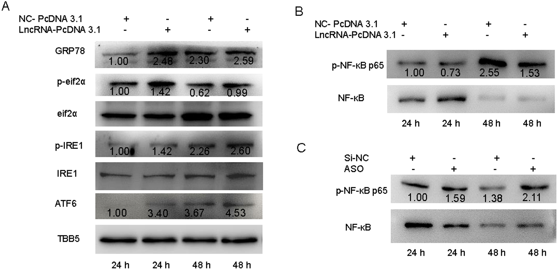 Fig. 1.
The effect of lncRNA-SUSAJ1 on ER stress. (A) Overexpression of
lncRNA-SUSAJ1 affects the expression level of ER stress-related proteins.
Overexpression of lncRNA-SUSAJ1 in PK15 cells by pcDNA3.1. Whole-cell lysates
were harvested from the treatment groups as indicated. Immunoblotting was
performed to detect ER stress related signal protein levels using TBB5,
eIF2
HSP70 inhibits cell apoptosis by inhibiting pro-apoptosis factors [36, 37, 38]. In this study, we investigated the relationship between lncRNA-SUSAJ1 and HSP70. An RNA pull-down assay was performed to explore interactions in PK15 cells infected with JEV for 36 h. In JEV-infected PK15 cells, the amount of Hsp70 protein pulled down by lncRNA-SUSAJ1 was higher than that in non-infected JEV cells (Fig. 2A). RNA samples from the experimental Hsp70-RNA complex group and control IgG-RNA complex group were isolated and purified using the RIP assay, and then verified by qRT-PCR. The level of lncRNA-SUSAJ1 expression was significantly higher in the Hsp70 group than in the IgG group (Fig. 2B). Then the qRT-PCR product was diluted 100-fold and used for nucleic acid electrophoresis to confirm the presence of lncRNA-SUSAJ1 (Fig. 2C).
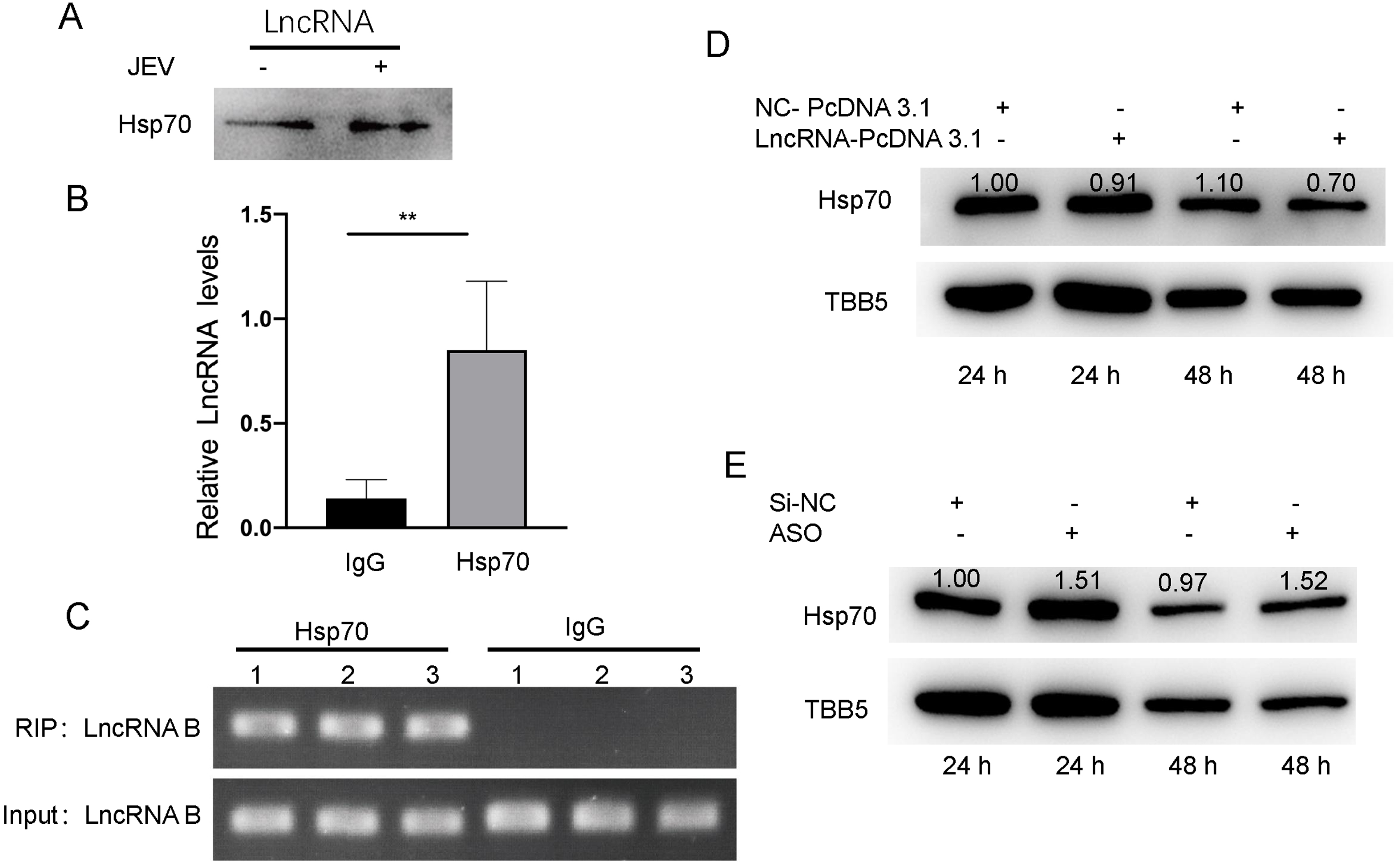 Fig. 2.
The interaction between lncRNA-SUSAJ1 and HSP70. (A) The
interaction of sense lcnRNA-SUSAJ1 with HSP70 was determined by RNA-pull down and
western blot. (B) The interaction between HSP70 and lcnRNA-SUSAJ1 was determined
by RNA-RIP. IgG was used as a negative control. (C) The agarose gel of
lncRNA-SUSAJ1 after RNA-rip by HSP70. (D) Overexpression of lncRNA-SUSAJ1 in PK15
cells by pcDNA3.1. Whole-cell lysates were harvested from the treatment groups as
indicated. Immunoblotting was performed to detect HSP70 protein levels using TBB5
as an internal control. *p
PK15 cells were transfected with plasmid for 6 h, and then infected with JEV for 24 and 48 h; total protein samples were extracted for Western blotting. At 24 and 48 h after infection, pcDNA 3.1 lncRNA-SUSAJ1 inhibited Hsp70 protein expression compared to the control group NC, whereas lncRNA-SUSAJ1 knockdown promoted its expression (Fig. 2D,E). These results show that Hsp70 protein interacted with lncRNA-SUSAJ1 and that lncRNA-SUSAJ1 inhibited the expression of Hsp70 protein. Therefore, we conclude that lncRNA-SUSAJ1 may facilitate apoptosis in PK15 cells.
To study the effects of lncRNA-SUSAJ1 on apoptosis in JEV-infected PK15 cells, we transfected the pcDNA 3.1 lncRNA-SUSAJ1 and NC into the cells according to the manufacturer’s instructions. After 6 h of transfection, JEV infection was conducted for 24 and 48 h. qRT-PCR was performed to detect the mRNA expression levels of apoptosis-related genes. After the transfection of pcDNA 3.1 lncRNA-SUSAJ1, the expression level of lncRNA-SUSAJ1 was significantly higher than that of NC (Fig. 3A). At 48 h after infection, the expression levels of the pro-apoptotic genes caspase-3, caspase-8, caspase-9, BCL2L11, Bax, and TP53 were significantly increased (Fig. 3B–F,H); at 24 h after infection, the expression level of the anti-apoptotic gene BCL2 was significantly decreased (Fig. 3G).
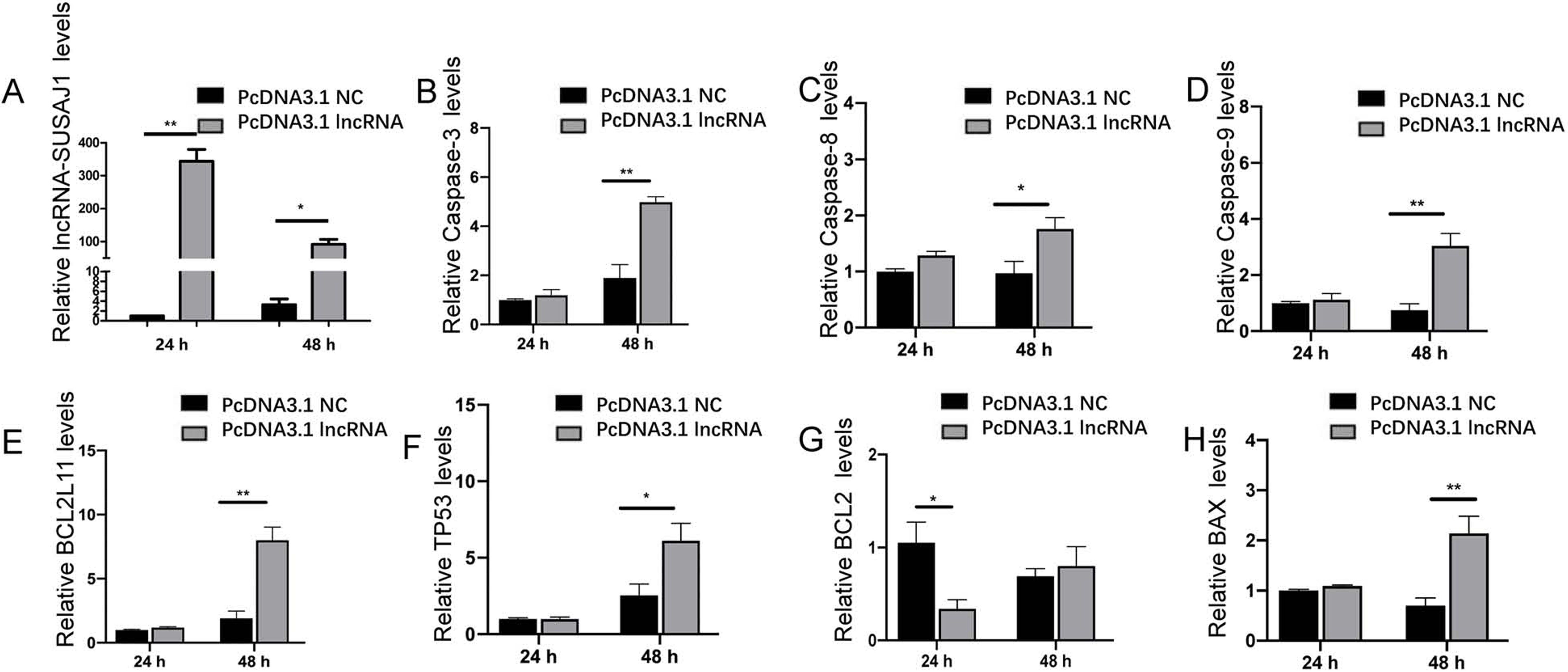 Fig. 3.
lncRNA-SUSAJ1 overexpression elevates the mRNA levels of
apoptosis-related genes. (A–H) PK15 cells were transfected with pcDNA3.1
lncRNA-SUSAJ1. Time course of lncRNA-SUSAJ1, Caspase-8, Caspase-9 et al.
mRNA levels were determined by RT-qPCR after JEV infection (n = 3 per group).
* p
Mitochondrial membrane potential was measured using the JC-1 cationic dye staining method. After transfection with lncRNA-SUSAJ1 overexpression and NC plasmids for 6 h, cells were infected with JEV for 24 and 48 h; they were then subjected to JC-1 cationic dye staining analysis.
When the mitochondrial membrane potential is normal, direct observation using a fluorescence-inverted microscope shows accumulation of JC-1 cationic dye in the mitochondrial matrix, forming a polymer that emits strong red fluorescence; when depolarised, the dye exists in the cytoplasm as a monomer that produces green fluorescence. At 24 and 48 h after infection, red and green fluorescence intensities indicated promotion of mitochondrial membrane potential depolarisation (Fig. 4A).
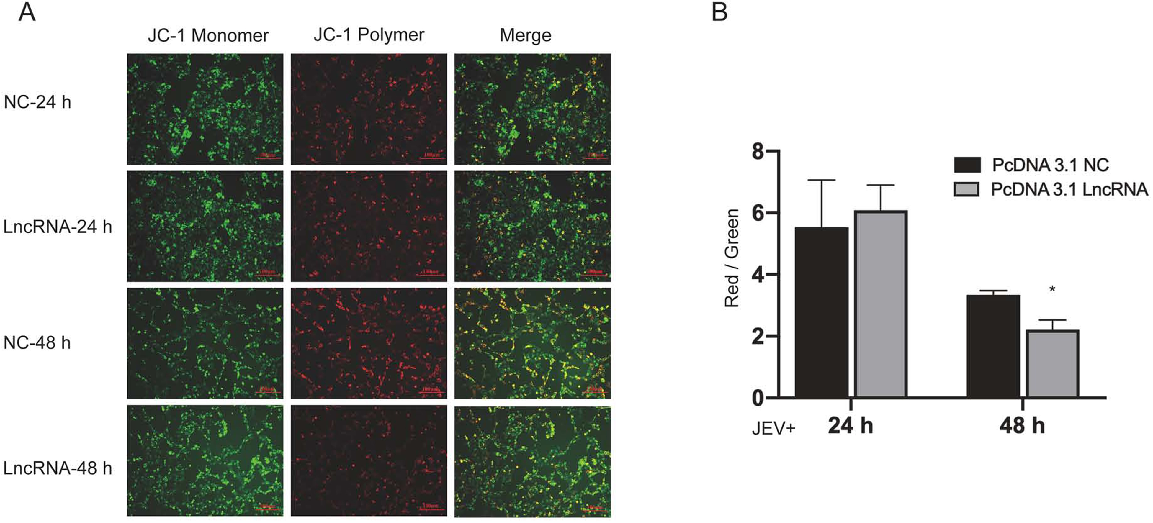 Fig. 4.
Effects of lncRNA-SUSAJ1 overexpression on mitochondrial
membrane potential (MMP). (A) PK15 cells were transfected with pcDNA3.1
lncRNA-SUSAJ1 and stained by JC-1 after JEV infected. Time course of MMP were
stained by JC-1 dye, red bar = 100
Enzyme labelling analysis showed that the degree of mitochondrial membrane potential depolarisation at 48 h after infection was higher in the lncRNA-SUSAJ1 overexpression group than NC control group (Fig. 4B).
After lncRNA-SUSAJ1 overexpression, ROS levels in JEV-infected PK15 cells were examined by H2DCFDA and Hoechst 33342 staining. Blue-green fluorescence intensity was analysed in merged images. Compared with the uninfected group, the green fluorescence signal at 48 h was significantly increased in the infected group (Fig. 5A), indicating that lncRNA-SUSAJ1 overexpression significantly increased ROS levels in PK15 cells after JEV infection.
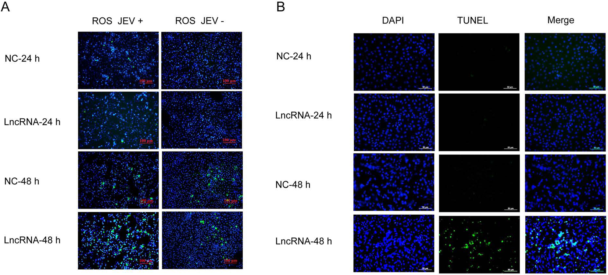 Fig. 5.
Effects of lncRNA-SUSAJ1 overexpression on ROS level and cell
apoptosis. (A) PK15 cells were transfected with pcDNA3.1 lncRNA-SUSAJ1 and
stained by the ROS reagent after JEV infected. Time course of ROS levels were
determined by immunofluorescence, red bar = 100
TUNEL analysis was used to identify late apoptotic cells. After lncRNA-SUSAJ1 overexpression and 48 h of JEV infection, the proportion of late apoptotic PK15 cells significantly increased (Fig. 5B). The results of TUNEL analysis were consistent with the those of flow cytometry analysis. With increasing JEV infection time, the proportion of late apoptotic cells increased among PK15 cells with lncRNA-SUSAJ1 overexpression.
Apoptosis was investigated by flow cytometry analysis. Among cells
overexpressing lncRNA-SUSAJ1, after 24 h of JEV infection, the proportion of
viable cells decreased by 11.5% (p
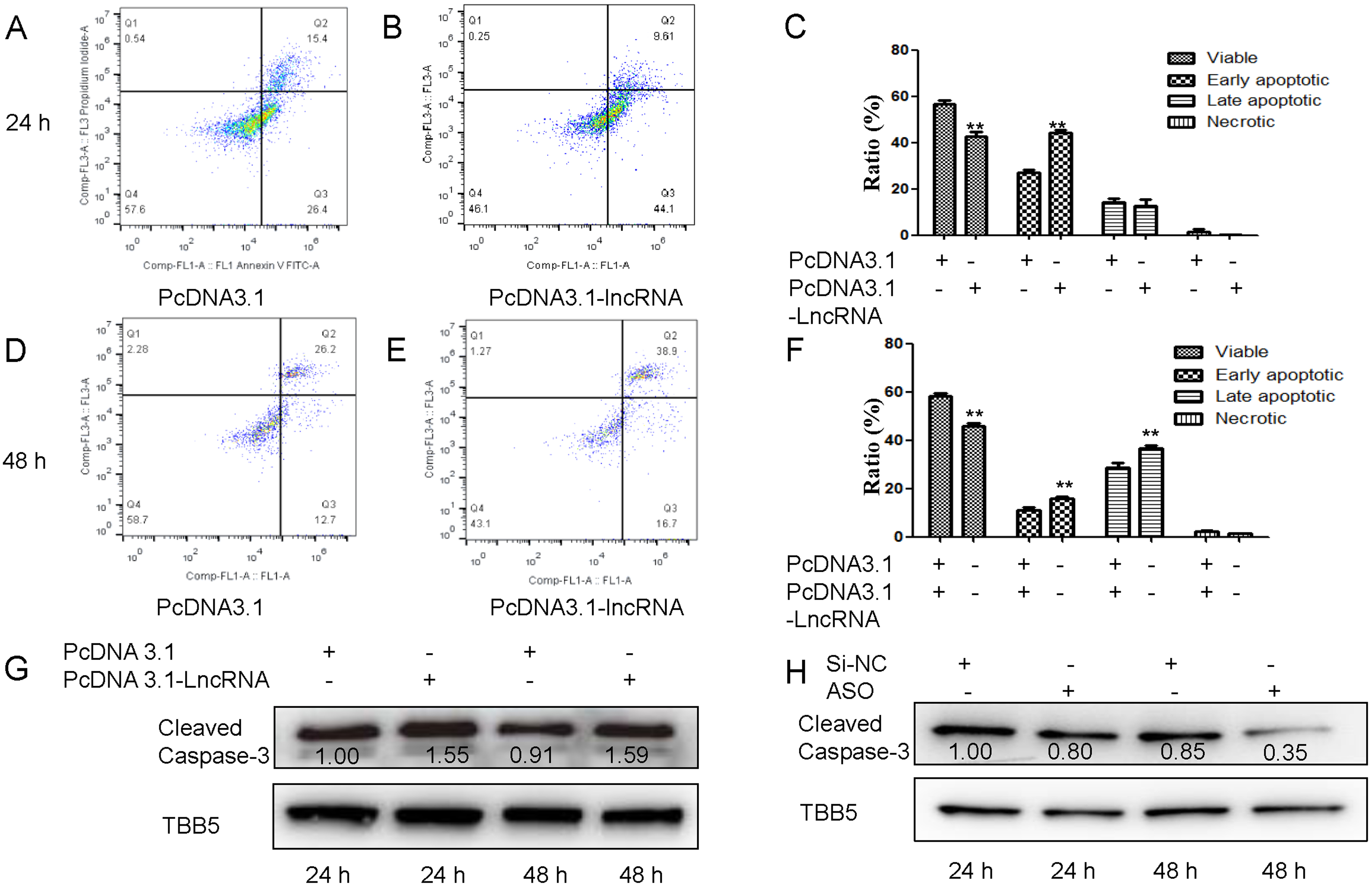 Fig. 6.
Effects of lncRNA-SUSAJ1 overexpression on cell Annexin V and
Caspase-3 levels. (A–B,D–E) PK15 cells were transfected with pcDNA3.1
lncRNA-SUSAJ1 and stained by the annexin V and PI reagent after JEV infected. The
annexin V and PI levels were determined by Flow cytometry. (C and F) The ratios
of viable, early apoptosis, late apoptosis, and necrotic cells detected by
Flow cytometry, C (24 h), D (48 h), **p
Caspase-3 is closely associated with apoptosis [39]; levels of cleaved caspase-3 protein were detected by Western blotting in this study. lncRNA-SUSAJ1 strongly affected cleaved caspase-3 protein expression. At 24 and 48 h after infection, lncRNA-SUSAJ1 overexpression promoted the expression of cleaved caspase-3 protein (Fig. 6G), whereas lncRNA-SUSAJ1 knockdown inhibited the expression of cleaved caspase-3 protein (Fig. 6H).
Although there is increasing evidence that lncRNAs are involved in regulating the host innate immune response and expression of virus-related genes and proteins, the specific mechanism underlying the effects of lncRNA-SUSAJ1 on JEV replication has not been elucidated.
You et al. [14] found that lncRNAs can serve as targets for new antiviral drugs. Zhang et al. [21] used a comprehensive and in-depth sequencing approach to confirm that lncRNAs participate in host antiviral responses during porcine reproductive and respiratory syndrome virus infection; in particular, lncRNAs regulate the apoptosis signalling pathway. Meng et al. [15] investigated the effects of lncRNAs on the immune response and viral replication; they identified key roles for lncRNAs in host–virus interactions. Torkzaban et al. [18] demonstrated that lncRNA-U1 directly upregulated the expression of NPBWR1 and induced apoptosis during infection with human immunodeficiency virus. Zou et al. [20] examined the role of lncRNA AX800134 in hepatitis B virus-related hepatocellular carcinoma. Viral proliferation and replication often require host cell components and energy sources. During apoptosis, infected cells die and the virus cannot use nutrients from these cells. This inhibits viral replication, transmission, and persistence. Here, we hypothesised that lncRNA-SUSAJ1 promotes apoptosis in JEV-infected PK15 cells, thereby inhibiting viral proliferation and replication.
To explore the effects of lncRNA-SUSAJ1 on JEV proliferation and replication, we
examined apoptosis-related genes via RT-qPCR. We found that lncRNA-SUSAJ1
overexpression promoted the expression of pro-apoptotic genes and inhibited that
of anti-apoptotic genes. We analysed apoptosis in PK15 cells that had been
transfected with pcDNA3.1 lncRNA-SUSAJ1 and NC plasmid using flow cytometry and
TUNEL staining. Flow cytometry showed that lncRNA-SUSAJ1 promoted apoptosis in
PK15 cells. TUNEL analysis showed that lncRNA-SUSAJ1 promoted late apoptosis
[40], and the proportion of late apoptotic PK15 cells increased with increasing
JEV infection duration (p
LncRNA-SUSAJ1 did not cause abnormal ROS accumulation in normal PK15 cells; however, in JEV-infected PK15 cells, lncRNA-SUSAJ1 promoted apoptosis, resulting in a significant increase in the proportion of cells experiencing late apoptosis, causing abnormal mitochondrial membrane potential and promoting the level of ROS produced by cell metabolism, perhaps due to lncRNA-SUSAJ1 interaction with novel proteins that may be present at very low levels in normal PK15 cells but are upregulated by JEV infection. This hypothesis requires further study.
Apoptosis is a type of programmed cell death; mitochondria-dependent apoptosis is the most common subtype. Mitochondria are sensitive to changes in ROS in cells. Some studies have shown that lncRNAs are involved in regulating cellular stress responses [41], such as oxidative stress and ER stress, which induce an antiviral response in the host. During oxidative stress, metabolism of ROS leads to mitochondrial damage [42]. The depolarisation of mitochondrial membrane potential displaces the pro-apoptotic protein Bax, releases cytochrome C from the inner membrane of mitochondria into the cytoplasm, activates the caspase-3 signalling pathway, and triggers a mitochondria-dependent apoptosis response. Therefore, caspase-3 is the most important enzyme in apoptosis. In this study, Western blotting showed that lncRNA-SUSAJ1 overexpression promoted the expression of cleaved caspase-3 protein in JEV-infected PK15 cells, while lncRNA-SUSAJ1 knockdown inhibited the expression of cleaved caspase-3 protein in JEV-infected PK15 cells. Furthermore, we demonstrated that lncRNA-SUSAJ1 overexpression promoted the release of ROS produced by PK15 cellular metabolism and reduced the mitochondrial membrane potential in JEV-infected PK15 cells.
HSP70 is a vital molecular chaperone that reduces UPR and helps proteins to fold correctly [36], resulting in the suppression of ER stress and promoting JEV proliferation. It remains unclear whether lncRNA-SUSAJ1 can inhibit JEV proliferation and replication by triggering a cellular stress response. To explore this, we performed RNA pull-down experiments, using biotin-labelled lncRNA-SUSAJ1 to analyse JEV-infected and uninfected PK15 cells after 36 h of incubation. Mass spectrometry analysis of the pull-down protein mixture indicated that lncRNA-SUSAJ1 has a regulatory role, in combination with the antioxidant protein Hsp70, which plays an important role in oxidative stress [43], when JEV infects PK15 cells. To verify the interaction between lncRNA-SUSAJ1 and Hsp70, we performed RIP and Western blotting experiments. The results revealed an interaction between lncRNA-SUSAJ1 and Hsp70, such that lncRNA-SUSAJ1 negatively regulated the Hsp70 expression.
ER stress and the UPR are involved in the regulation of viral replication [44, 45]. The ER is an important organelle for viral replication and maturation. After
viral infection of host cells, numerous viral proteins are synthesised in the ER
cavity; this increases the protein load in the ER, leads to the accumulation of
unfolded and misfolded proteins, induces the ER stress response, and activates
the UPR [46]. Three UPR signalling pathways have been identified: ATF6, PERK, and
IRE1 [47]. In this study, Western blotting was used to detect the marker protein
GRP78, which is activated by the ER stress response. The results showed that
lncRNA-SUSAJ1 antiviral activity was closely associated with the ER stress
response. lncRNA-SUSAJ1 overexpression significantly increased the expression of
GRP78 protein; it also inhibited JEV proliferation and replication by promoting
activation of the ER stress response. Subsequently, the specific pathway
underlying the promotion of UPR activation was examined. The expression levels of
PERK, IRE1 and ATF6 pathway-related proteins were detected by Western blotting.
Notably, lncRNA-SUSAJ1 positively regulated the ER stress response, promoted the
expression of IRE1, and promoted the phosphorylation of eIF2
NF-
We comprehensively explored the mechanism by which lncRNA-SUSAJ1 influences the
proliferation and replication of JEV in PK15 cells. When PK15 cells were infected
with JEV, lncRNA-SUSAJ1 promoted the ROS response and increased the production of
oxygen free radicals via cellular metabolism; lncRNA-SUSAJ1 also inhibited the
expression of Hsp70 and promoted the phosphorylation of eIF2
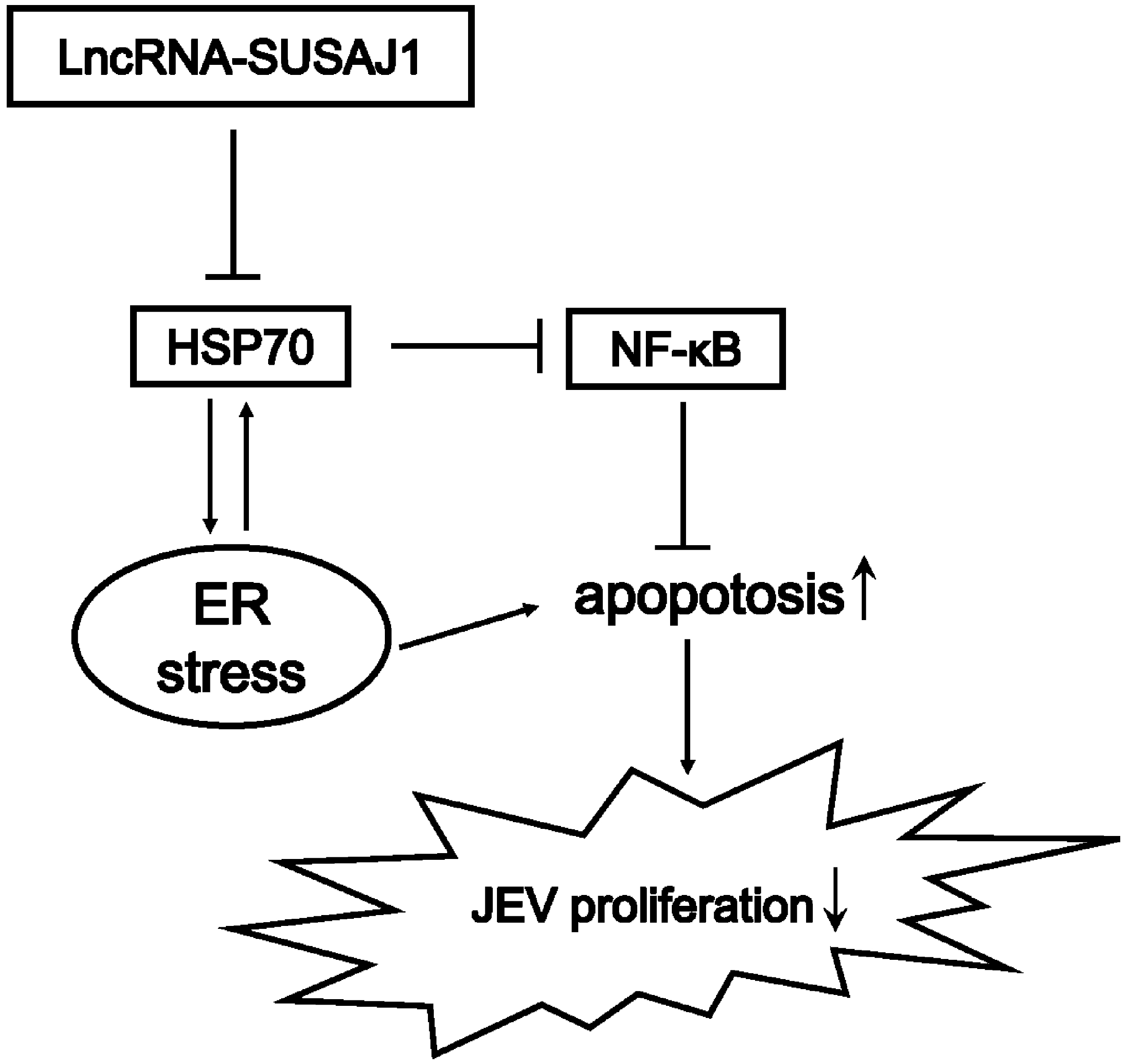 Fig. 7.
Model of lncRNA-SUSAJ1 activated the ER stress promoting apoptosis in PK15 cells.
JEV, Japanese encephalitis virus; lncRNAs, long non-coding RNAs; siRNAs, short interfering RNAs; ASO, antisense oligonucleotides; TBST, Tris-buffered saline-Tween-20; MMP, mitochondrial membrane potential; ROS, reactive oxygen species; ER, The endoplasmic reticulum; UPR, unfolded protein response; RIP, RNA immunoprecipitation.
XZ and AZ conceived and designed the study project. QY and JF performed the experiments and edited most of the manuscript. QY analyzed the data with support from HW, XL and SY revised the manuscript content. XZ, SY, and AZ contributed to result discussion and data interpretation. All authors read and approved the final manuscript.
Not applicable.
Not applicable.
This research was supported by grants from Natural Science Foundation of Zhejiang province (LY19C170003).
The authors declare no conflict of interest.
Publisher’s Note: IMR Press stays neutral with regard to jurisdictional claims in published maps and institutional affiliations.