

Frontiers in Bioscience-Elite (FBE) is published by IMR Press from Volume 13 Issue 2 (2021). Previous articles were published by another publisher on a subscription basis, and they are hosted by IMR Press on imrpress.com as a courtesy and upon agreement with Frontiers in Bioscience.
 , Diksha Sahai 3, Naveen Mangal 4, Jagadeesh Dhamodharan 5
1 College of Pharmacy, University of Iowa, Iowa City, IA, 52246
2 Pharmacology Unit, Faculty of Pharmacy, AIMST University, Semeling, 08100 Bedong, Kedah Darul Aman, Malaysia
3 College of Professional Studies, Northeastern University, Boston, MA, USA – 02115
4 Independent Clinical Pharmacologist, Billerica, MA 01821, USA
5 Department of Anatomy, Faculty of Medicine, AIMST University, Semeling, 08100 Bedong, Kedah Darul Aman, Malaysia
Abstract
Transdermal drug-delivery systems (TDDS) offer an attractive alternative to the oral route for delivery of biotherapeutics. Technological advancements in the past few decades have revolutionized the fabrication of micro-structured devices including creation of microneedles (MC). These devices are used for delivering peptides, macromolecules such as proteins and DNA, and other therapeutics through the skin. Here, we review the current use of MCs as a cost effective method for the self-administration of therapeutics. We will then review the current and common use of MCs as an effective treatment strategy for a broad range of diseases and their utility in the generation of effective vaccination delivery platforms. Finally, we will summarize the currently FDA approved MCs and their applications, along with the ongoing clinical trials that use such devices.
Keywords
- Alzheimer disease
- Diabetes mellitus
- Immunity
- Obesity
- Transdermal drug delivery
- vaccine therapy
- Review
Transdermal drug-delivery systems (TDDS) have generated considerable interest in the administration of drugs via the skin, as an alternative to oral route for delivery of biotherapeutics. However, the skin has proven to be a formidable barrier due to its unique underlying structure. Passive diffusion of drugs is mostly limited to small and lipophilic molecules. Current scientific research is heavily focused on the development of large molecules, such as DNA, RNA, vaccines, or other kinds of biomacromolecules, which are nearly impossible to deliver via skin in an unassisted manner (1, 2). Physical and chemical methods have been studied to show an increase in the skin permeability of large molecules. However, chemical methods usually cannot deliver biotherapeutics in a clinically meaningful concentration across the spectrum of diseases, and physical methods use bulky and expensive sophisticated devices, which requires trained personnel to operate (3). To overcome this problem, many biotherapeutics are delivered subcutaneously (s.c.) or intramuscularly (i.m.) using a hypodermic needle. However, the use of injections is often associated with the patient's discomfort, pain, generation of sharp waste, and potential of abuse (4). Oral route has its disadvantages with a primary hurdle of significant first-pass metabolism and poor absorption in the gastrointestinal tract (5).
As an alternative, MNs have emerged as an attractive option over the last decade which has demonstrated its usefulness and ability to overcome disadvantages associated with the use of oral and parenteral drug delivery. MNs are small enough that MNs can be self-administered while avoiding pain and discomfort, and still being able to deliver small and large molecules into and across the skin (6, 7). A plethora of scientific studies has demonstrated the successful application of MNs in the treatment, prevention, and management of a range of diseases, such as diabetes, obesity, and infectious diseases. This review article is an effort to highlight the impact of research studies focused on the application of MNs in disease management. This review article is not focused on the application of MNs in the treatment of cancer, since this body of work has already been accomplished, and readers are directed to a comprehensive review article (8). Duarah et al. have summarized the scientific literature for MNs applications in the pediatric population (9).
Skin (cutis) is the largest human organ, which covers the entire body and has a surface area of around 2 m2. Its thickness varies from 0.5 mm on eyelids to four mm or more on the palms of the hands and the soles of the feet. In total, skin accounts for around 16 percent of the body weight. Skin is closely integrated with blood-vessels (10), nerves (11), and lymphatic system (12). The skin is mainly comprised of three layers with varying degrees of specialization as follows.
The outermost layer of the epidermis is made of epithelial cells kertinocytes which are closely packed, making up the cornified skin layer or the stratum corneum (SC). ED is the primary barrier to drug delivery. Outer ED, is comprised of a a 5-layered assembly, composed of keratinocytes (95% of cells), which is generally 0.02–0.2 mm in thickness and typically is 50–150 μm in humans (13). The corneocytes provide suppleness and flexibility to the skin. ED represents the ‘bricks’ embedded in a ‘mortar’ which is composed of multiple lipid bilayers of ceramides, fatty acids, cholesterol, and cholesterol esters (14, 15). The most inner layer of ED is comprised of basal cells which reside on a basal lamina.
Dermis is the part of skin beneath the ED which provides an important barrier against pathogens (16). It provides mechanical strength to the skin (17). It is thicker than the ED (usually 2–4 mm) and contains collagen (mostly type I and III), immunologically active dermal dendritic cells and Langerhans cells, connective tissues, blood, and lymphatic vessels, hair follicles, eccrine glands and nerve endings (18–20).
Hypodermis is the layer underneath the dermis, and is comprised of fibroblasts, macrophages and adipose tissue made of adipocytes. The blood and lymphatic network in the dermis and hypodermis region re crucial for the systemic delivery of drugs.
Although skin appears to be fragile, it is a formidable barrier to breach and it acts as the body’s first natural defense mechanism against exogenous pathogens. In the context of drug delivery, a drug needs to have a unique physicochemical profile to get past the skin and into the systemic reservoir, unassisted (passive diffusion). This profile includes an optimum logP of around 1-3, the molecular weight of less than 500 Da, unionized form, and a melting point (MP) below 150-200°C (2, 21). Therefore, large molecules such as proteins often have a difficult time in crossing skin, and it becomes a challenge to attain the therapeutically meaningful plasma concentrations. Fortunately, over the years, many strategies have been developed to breach the skin and deliver not only significantly higher concentration of drugs if needed in case of small molecules, but also deliver proteins and vaccines, which used to be an elusive dream during early days of transdermal drug development. These strategies involve both chemical and physical approaches, including the addition of chemical penetration enhancers, electroporation, iontophoresis, sonophoresis, thermal ablation, or the synergistic combinations of two or more mechanisms, to deliver, small and large molecules as well as polymeric carriers. However, this review article will focus on the application of MN as an emerging tool for transdermal drug delivery and its application in prevention and disease management.
MNs are micron-sized needles, which are large enough to deliver macromolecules, proteins, and vaccines into the different layers of (or across) the skin but are short enough to avoid any pain experienced as traditionally seen with the parenteral drug delivery. It offers several unique advantages: localization of small or large molecules in the skin by overcoming the SC barrier, placement of drug in the proximity to the blood vessels near dermis for faster absorption and onset of action during systemic delivery, targeting dendritic or Langerhans cells of immune system present in the ED/dermis region which can be leveraged to develop vaccines; and eliminating pain (22, 23). The use of MN lowers the health hazard and potential abuse associated with hypodermic needles during parenteral administration and has the potential to reduce the cost of vaccines because of its dose sparing effect and elimination of cold storage. Most importantly MNs are designed to self-administer the vaccine/biotherapeutics with minimum training, consequently improving vaccination coverage in the pediatric population where parents can potentially vaccinate their kids without having to visit doctors, as well as in first world countries, where scarcity of well-trained nurses and doctors usually result in low vaccination coverage (24). The level of MNs entry in the skin layer is illustrated in Figure 1.
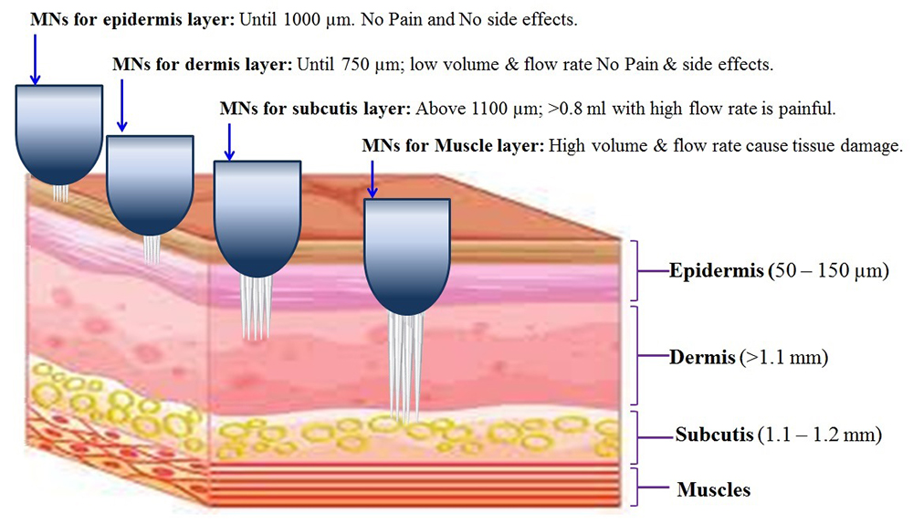 Figure 1
Effect of dermal application of MNs depth in drug delivery process. The lower level of MNs penetration does not produce any pain or adverse effects upto 1000 µm. Further, the volume is also one of the factors to cause the pain. Paradoxically, at higher volume i.e., >0.8 ml can induces the pain at 750 µm depth of MNs penetration with moderate constant flow rate. Even, the lower flow rate i.e., <0.1 ml/min is not shown any painful reactions.
Numerous studies have been conducted so far in the development of MNs. MNs have been fabricated in a variety of ways, each bringing its own set of advantages, and can be categorized into four different types:
These MNs are used for rapid bolus injection or slow infusion of the liquid formulation. Robust mechanical strength of micron-sized needles is necessary along with the adequate and constant flow rate. Dense dermal tissue might compress against the tip of the needle affecting the drug delivery rate and potentially targeting the wrong layer (24). Silicon is typically used for its high mechanical strength, but its high cost offsets its use in large-scale production (25). Hollow MN can directly place a drug in the ED-dermis region depending upon the length of the MN used making it possible to deliver macromolecules like proteins and vaccines. Insulin was delivered in the porcine skin using AdminPatch technology which uses hollow MNs (26). Hollow MNs can also be potentially useful in gene therapy as Luo et al. have demonstrated that localized delivery of oligonucleotides using hollow MN could be achieved in the 3D tissue model (27).
Solid MNs are used to breach the SC layer of the skin to create micro-channels followed by the application of a drug-loaded patch to deliver drugs via passive diffusion. Solid MNs can be mounted on a roller that can pierce the skin with a rolling application of the device. This approach was used by Kaur et al., where the 5-fold increase in verapamil flux and 11-fold increase in amlodipine flux was observed in the porcine skin (28). Verapamil regulates blood pressure whereas captopril is used for the management of hypertension and ischemia. Similarly, the transdermal flux of captopril and metoprolol was increased 75-fold and 4-fold respectively following the application of solid MN rollers (29). Conventionally, small molecules of up to 500 Da and molecules with logP of 1-3 are most suitable for topical administration. However, once micropores have been generated using solid MNs, large molecules, including proteins and more hydrophilic drugs can slowly diffuse across MN generated micro-conduits. In one proof-of-concept study, siRNA was delivered using this approach for localized effects in the skin (30). Localization of siRNA can have significant implications in the potential development of treatments for skin conditions such as alopecia, skin cancer, and hyperpigmentation.
Biodegradable, non-toxic polymers or water-soluble material is used to form MN tips encapsulating the drug in the matrix. Once inserted into the skin, tips are dissolved by the presence of water, releasing the encapsulated drug. This technique has a unique advantage of leaving no sharp waste behind, thus addressing needle abuse and proper disposal of bio-hazardous sharp waste (31). Ito et al. prepared insulin loaded in dextran/polypropylene dextran/polypropylene matrix. Insulin was found to be stable in the matrix for one month and when delivered into mice, all the loaded insulin was delivered within one hour, which correlated with the lowest plasma glucose level achieved (32). When the dissolving MN approach was combined with iontophoresis by Kumar et al., MNs demonstrated that it was possible to deliver significantly large quantities of small (calcein) and macromolecules (human growth hormone) in-vitro in the porcine skin than either of the methods alone (33). In a unique approach, Lee et al. fabricated dissolving MNs from carboxymethylcellulose or amylopectin as a base containing sulforhodamine B and bovine serum in the backing layer of MNs. This approach allowed the controlled release of drug from the reservoir after the formation of micro-conduits upon dissolving of MN tips (34). This approach can be pivotal in achieving the constant release of drug administration for prolonged pharmacological action, hallmarks for any medication intended to use in chronic disease management in the elderly population.
Drug containing matrix can be used to coat stainless steel or silicone MNs. Once inserted, a formulation containing the drug is dissolved off into the skin. In-vitro porcine skin studies showed that a higher amount of lidocaine delivered within three minutes compared to the one-hour topical application of its branded counterpart (35). The disadvantage of using coated MN is that only a limited amount of drug can be coated depending upon the number of MNs in the array and potential of leaving sharp waste behind. Also, the long-term stability of the drug in the matrix (e.g. PVP or PEG) while it is exposed to the environment, needs to be demonstrated to translate this approach to commercial success (36). Further, stainless steel coated MNs have potential ophthalmic drug delivery with safe and without irritation/edema in ocular regions. Experimentally, it is proved that, coated MNs possess the irritation free potential action for resealing of ophthalmic tissues with absence of allergic reactions, bleeding or infections (37, 38). Mainly corneal electrode used for the retinal drug delivery for the management of diabetic complication like retinopathy.
Hollow and solid MNs typically use metal e.g. silicon, which can have biocompatible problems and can result in the potential abuse of these needles. To overcome this problem and to enhance patient safety, hydrogel-forming MNs are also currently under investigation. This approach circumvents the problem of a limited amount of doses that can be delivered with other MNs (39). In one study, Donelly et al. created super swelling polymers that imbibed the water present in the skin creating micro-conduits between dermal circulations and attached lyophilized drug reservoir (40). Other advantages include easy sterilization and intact removal of hydrogel from skin post application (39).
The material used to form hydrogel should be sufficiently hard in the dry state to pierce the skin but should be capable of rapid swelling upon insertion. Once swollen, MNs should be able to maintain structural integrity. Sivaraman et al. delivered up to 11 µg/cm2 of methotrexate, an anti-cancer drug, from poloxamer hydrogels in human dermatomed skin (41). In another proof of concept study, this approach was used as a diagnostic tool to monitor lithium levels in rats (42).
A recent investigation into the use of MN for the delivery of poorly permeable proteins, e.g. insulin, has shown to be promising. Lee et al. demonstrated that insulin loaded into dissolving polymeric MNs rapidly decreased the glucose level in mice within two hours and to the same extent as s.c. the injection did, with a relatively lower amount of insulin delivered illustrating the dose sparing potential (43). Traditional s.c. injection for diabetes management has one key constraint, i.e. lack of glycemic control resulting in overtreatment with insulin, which can lead to brain damage (44). Ye et al. showed that MN patch integrated with exogenous pancreatic β cells coupled with glucose signal amplifiers released insulin in accordance with the glucose levels in mouse dorsum skin where the normal glucose level was maintained within the therapeutic window for six hours post treatment (45). Similar results were demonstrated in several other studies emphasizing the potential of MN in the management of diabetes (46–49).
MNs have been repeatedly shown in various studies to elicit a superior immune response, capable of providing greater protection against virus challenge, sustained immunity, and dose sparing effect. Due to the micron-size structure of needles, these devices have proven to be far more effective in targeting immunological rich layers of ED and dermis, demonstrating their usefulness in the development of next-generation vaccines. In the context of vaccine development, MNs provide the following key advantages over traditional vaccination routes:
MNs can place drugs closer to blood capillaries resulting in shorter lag time and faster therapeutic response. It has been demonstrated that ID vaccination enhances the immune response in equivalent doses as compared to s.c. or i.m. injections. Phase 2 study with BD Soluvia™ (FDA approved) microinjection system showed that 15 µg of trivalent inactivated influenza vaccine in elderly patients above 60, induced 1.7-fold higher GMT response compared with the same amount of i.m. dose. Furthermore, a 40% seroconversion rate (proportion of vaccinated individuals receiving a four-fold antibody titer increase) was achieved (50). These results were confirmed in Phase 3 study where ID delivery of an equivalent dose of 1µg HA/strain elicited superior GMT and seroprotection rates at 21-d post vaccination (51). Virus-like particles (VLPs), stabilized in the presence of trehalose and coated on MNs, produced 100% protection against the lethal viral challenge, a preventative immune response superior to i.m. administration (52).
MNs can be used for vaccination where their potential has been realized in several studies due to their ability to effectively target key immune cells in generating a robust immune response. An ED-dermal layer of skin has a rich population of antigen-presenting cells (APCs) called Langerhans cells, subclass of dendritic cells, which uptake foreign antigens, migrates to lymph node and initiates Th1 and Th2 dependent immune response. ID vaccination in this region has been shown to generate an equivalent antibody response at a much lower dose. This dose sparing, and increased potency, has been documented in several studies and is useful in not only reducing the cost of the vaccine but also in pandemics when a large population is at high risk of infections, and therefore, relatively lower mass production of vaccine is needed (53). In one study, ID vaccination with 1/5th of the dose of the conventional live attenuated yellow fever vaccine was able to achieve full protective immune response (54). Similar results were observed in another study where participants injected ID with 1/5th dose of inactivated polio vaccine achieved 100% seroconversion rate similar to the participants receiving the full dose of the same vaccine i.m. (55). Similarly, ID delivery of 20% dose of three different strains of influenza virus (H1N1, H3N2, and Malaysia B) achieved similar seroconversion rates, seroprotection rate, and GMT as compare to i.m. delivery (56). A similar dose sparing effect was observed for the trivalent inactivated influenza virus (Fluzone) (57), human diploid cell rabies vaccine (58, 59), and purified chick embryo cell rabies vaccine (PCECV) (60). However, it cannot be safely interpreted that the dose sparing effect will be seen in all vaccines as each vaccine has a different immunological benefit for ID delivery. Also, long-lasting antibody response for several years remains to be established for ID delivered vaccines. In one retrospective study, ID delivered rabies vaccine could elicit an effective antibody response for 10 years (61).
The minimally invasive approach of MN vaccination can help people to vaccinate children with minimum training. This also increases the chances of more children getting vaccinated as often it might be physically difficult for parents to schedule a clinic visit for vaccination. In one study, intent to vaccinate increased to 65% when participants were given a self-administered placebo MN patch with instructions (62). In another study, MN array with a leaflet containing instructions could be applied effectively and reproducibly by volunteers without the aid of a MN applicator device demonstrating the self-administering vaccination potential of MN (63).
There have been various studies involving the use of MNs for diabetes management in humans and animal models. Biodegradable polymers in conjunction with dissolving MNs were used in diabetic rats, where a hypoglycemic effect was seen in a dose-dependent manner. Subcutaneous injection dropped blood-glucose levels below the hypoglycemic threshold, whereas the same dose of insulin delivered via MNs kept blood-glucose levels above the hypoglycaemic threshold and maintained it at normal levels for longer periods, demonstrating the potential of MNs to deliver insulin in a controlled manner (64). This was further substantiated in a different study by Resnik et al., where sustained plasma insulin concentration was achieved after the application of hollow MNs as compared to the same dose delivered by s.c. infusion (65, 66). Thus, in diabetes management, where often multiple doses are required; steady-state concentration of insulin can mimic multiple dosing regimens. In an innovative approach by Yanqi et al., biodegradable MN arrays were integrated with pancreatic β cells and glucose signal amplifiers. The presence of amplifiers triggered the release of insulin in a hyperglycemic state in type-1 diabetic mice and stabilized blood glucose levels over 10 hours (67). Potential of MN was also demonstrated in the pediatric population, where 16 children and adolescents received insulin via hollow MNs. MN insertion pain was significantly lower and the onset of action was significantly faster as compared to s.c. treated arm indicating potentially greater patient compliance in children for the treatment of diabetes, as they are often afraid of needles (68). From the diagnostic point of view, MNs have been studied as the means of continuous glucose monitoring systems (CGMS). MNs are functionalized either to act as a sensing probe or a biological fluid collector. Both approaches have their unique challenges as it is quite difficult to develop functionalized MNs in a miniature form (66, 69).
MNs have found their application in the management of obesity, although it's still in the exploratory stage and relatively fewer studies have been conducted. In a study by Dangol et al., dissolving MN loaded with caffeine, resulted in significant weight loss as compared to obese mice. Triglyceride, total cholesterol, and lipoprotein-cholesterol levels also reduced within six weeks of treatment after dissolving MNs containing caffeine were applied in obese mice (70). Zhang et al. prepared rosiglitazone-loaded dextran nanoparticles embedded into polymeric MN-array patch for local delivery of browning agents, which facilitates browning of white adipose tissue resulting in dissipation of energy through the production of heat via non-shivering thermogenesis, thus, reducing obesity. In in-vivo mice studies, the application of MN loaded browning agents resulted in sustained release of rosiglitazone for three days and an increase in energy expenditure, fatty acid oxidation, and reduced-fat padding locally where MN was applied (71). An et al. fabricated gelatinized polymers where gelatin itself acted as a therapeutic agent. Gelatin was shown to reduce the suppression of lipogenesis-related genes in gene-expression studies. Furthermore, local application of gelatin MN in high-fat diet-induced obese rats decreased the amount of subcutaneous adipose tissue at the site of application (72). The same authors further extended the application of gelatin MNs in reducing the accumulation of adipose tissue where gelation derived from fish and swine were used in preparing MNs. Four-week treatment of either of the gelatin MNs to high-fat diet-induced obese rats resulted in smaller adipocytes in the region of application as well as a reduction in expression of fat metabolism-associated gene levels (73).
MN potential has also been realized in the management of chronic diseases, e.g. Alzheimer’s. Transcutaneous immunization (TCI) in mice with amyloid β-1 (Aβ-1) peptide seemed to recover cognitive function to some extent and higher anti-Aβ-1 IgG levels (74). However, this approach wasn’t effective as it didn’t produce epitopes or isotypes of anti-Aβ-1 IgG which are equally important in the complete restoration of cognitive function (75, 76). This suggests that future studies will require TCI to specifically elicit isotypes of anti-Aβ-1 IgG implicated in the treatment of Alzheimer’s disease. Similarly, donepezil HCL containing film integrated with hydrogel-forming MNs was used to achieve an optimum skin concentration level in-vivo in rats for 24 hours (77).
MNs can overcome disadvantages that are associated with the traditional vaccination method using hypodermic needles. Traditional vaccinations cause pain, generate sharp waste, require cold storage, and bypass the immune system of the skin. In contrast, several studies have demonstrated that MNs cause indiscernible pain (78–80), minimize the generation of sharp waste and associated hazard, stabilize proteins at room temperature eliminating the need of cold storage (81–85), and produce a superior robust immune response by targeting ED and dermis region of skin rich with immune cells (6, 86–90). In a study evaluating the cost-effectiveness of MN patches, Adhikari et al. demonstrated that the first dose of MN vaccination would cost US$0.95 compared to US$1.65 for the dose administered via SC (91). Rodgers et al. have provided a comprehensive review solely focused on combining nanoparticle delivery with MNs in vaccine development (92) as well as on the use of dissolving MN in the vaccine development (93).
Kolluru et al. developed dissolving MN containing inactivated polio vaccine (IPV) demonstrating stability in all three vaccine serotypes with > 70% activity maintained after 2 months and > 50% activity maintained after one-year storage at 5-25°C (94). In a study by Schipper et al., repeated fractional intradermal dosing of IPV serotype-1 using hollow MN in rats resulted in the 10-fold increase in IPV-specific IgG response as compared to i.m. bolus dosing (95). IPV was alternatively coated on MNs with N-trimethyl chitosan chloride (TMC) for efficient release during a pH-shift after in-vivo application in rats (96). In a human study involving 231 adults, ID vaccination with 40% of the standard dose, resulted in an increase of antibody titers by 64-fold (97).
Vescoco et al. conducted a study involving 66 healthy volunteers and injected rabies vaccine using DebioJect ID device containing 1/5th the dose delivered intramuscularly. Although, no difference in humoral response was observed between ID and i.m. routes, ID vaccination resulted in a significant decrease in pain associated with injections (96). Another study involving vaccination of dogs demonstrated that dissolving MN patch containing rabies vaccine can be at least as immunogenic as i.m. injection at the same dose (98). Laurent et al. delivered rabies vaccine using Beckton Dickinson's pre-filled ID delivery system. At 1/4th of the dose delivered i.m., protective seroconversion rate was achieved in healthy volunteers (99).
Edens et al. used coated MNs to immunize cotton rats with Edmonston-Zagreb measles vaccine strain. Full human dose or 20% of the dose was delivered, which generated virus-neutralizing antibodies at levels equivalent to the same dose delivered i.m. (100). Joyce et al. used dissolving MNs to immunize infant rhesus macaques. Higher, but not significant, neutralizing antibody titers were observed after MN application when compared to s.c. injection. On day 42 after vaccination, 100% of infant macaques achieved protective titers in MN treated group, whereas only 50% of infant macaques were able to achieve protective titers in s.c. group (101). The measles vaccine has also been demonstrated to be stable at elevated temperatures of up to 40°C when formulated with dissolving MN patch (102).
Nguyen et al. coated hepatitis B surface antigen, with Trehalose for stability, on MNs. In-vivo studies involving mice showed that antibody titers were significantly higher compared to vaccine delivered i.m., with alum as an adjuvant. In MN treated group, Th2 type immune response was predominant (103). In a study involving 16 rhesus macaques, dissolving MN patch, containing the hepatitis B vaccine, elicited seroprotective levels that could be correlated to seroprotection levels needed in humans. Induction of antigen-specific IFN-γ and IL-4 were higher, but not significant, in MN group than in i.m. group (87). A quadrivalent vaccine of hepatitis C, which is coded for four genotypes, was delivered with a MN device (Flugen) in Landrace pigs. At day 42 and 56 post-vaccination, antibody responses were significantly higher in the ID group (without adjuvant) compared to i.m. group, along with balanced Th-1 and Th-2 cytokines, strong T-cell, and granzyme B responses (104). Similar results were observed in several other studies involving different animal models and hepatitis B vaccines (7, 105–108). Coated MNs were coated with interferon-α, used in the treatment of hepatitis C, and achieved a similar reduction in tumor weight of mice when compared to s.c. administration (109). Wang et al. developed an oral mucosal vaccine of Hepatitis B antigen. When administered s.c. along with alum as an adjuvant, it produced Th2-biased immune response. However, dissolving MN arrays filled with mannose-PEG-cholesterol/lipid A-liposomes (MLLs) were applied to the oral mucosal region which produced Th-1 predominant, although balanced immune response. Moreover, IgA levels were significantly higher during oral mucosa immunization with MNs, and mucosal immunity in mice lasted nine months longer than the same vaccine delivered by vaccines. Levels of IFN-γ were significantly increased after MN treatment. Thus, robust cellular and humoral immunity were developed forming multiple lines of defense. MLLs delivered alone without MN resulted in poor immuno-protection (110).
Dissolving MN, fabricated from PVA and PVP, containing tetanus toxoids were applied on albino mice, which resulted in similar levels of IgG, IgG1, and IgG2α antibody titers when compared to i.m. group (88). Similar results were obtained when tetanus toxoid and diphtheria toxoid subunit vaccines were co-loaded in ceramic nano-porous dissolving MN arrays (111). Tetanus toxoid was loaded into chitosan nanoparticles and delivered via either solid or hollow MNs, which produced comparable IgG1 and IgG2α, compared with MN assisted traditional tetanus vaccine (without nanoparticles). However, Th1 cytokine levels i.e. IL-2, IL-6, were significantly higher in MN assisted tetanus-containing NP group, in comparison to IM administration of commercial tetanus vaccine. In contrast, Th2 cytokines, i.e. IFN-γ and IL-4 were comparable in i.m. and MN assisted groups. The higher cellular immune response observed after MN assisted vaccination of tetanus-containing NPs, might be due to the ability of MNs to deliver the vaccine in the skin layers where APCs mainly DCs, macrophages, and LCs are residing (112). Esser et al. prepared unadjuvanted tetanus toxoids containing dissolving MN fabricated from PVP and sucrose. MN treated pregnant mice conferred complete protection and 100% survival of all the mice born to vaccinated mothers when newborns were challenged with tetanus toxin at six weeks of age. Tetanus-specific antibodies were detectable up to 12-weeks of age. In contrast, mice born to i.m. vaccinated mothers could not survive the tetanus challenge (113). This illustrates the potential of vaccinating neonates while simultaneously vaccinating pregnant women (114).
Chitosan-based dissolving MNs elicited significantly higher influenza-specific antibody levels as compared to i.m. delivery. During the H1N1 viral challenge, MN immunized mice showed 100% survival as compared to 40% survival of i.m. treated mice (115). In a separate study, MN patch containing human influenza split vaccine and virus-like particles provided effective protection against heterosubtypic influenza (116). To explore the use of solid MNs, recombinant hemagglutinin (HA) was loaded into solid nanoporous MNs. After vaccination, mice showed complete protection when challenged with the mouse-adapted pH1N1 virus (117). Since the influenza virus has several subtypes, a universal vaccine against all flu subtypes is often desirable. Zhu et al. encapsulated inactivated influenza virus vaccines (H1N1 and H3N2) in dissolving MN patch. Mice immunized with this patch showed enhanced cross-protection against heterologous reassortant A/Shanghai/2013 H7N9 (rSH) influenza virus infection and antiviral efficacy against reassortant A/Vietnam/1203/2004 H5N1 (rVet) and A/Shanghai/2013 H7N9 (rSH) virus challenges (118). In a phase one study involving 370 healthy subjects, MicronJet600™ MN device was used to deliver 7.5, 15, and 45μg HA antigen/strain, and immunogenicity results were compared with Inflexal V™ (Janssen). MicronJet600™ MN demonstrated significantly higher immunogenicity than Inflexal at an equivalent dose (119).
Li et al. developed a long-lasting contraceptive MN biodegradable patch made out of PLGA polymer which maintained levonorgestrel concentration above the human contraceptive threshold for one month by slowly releasing the hormone over longer over a month (120). Prausnitz et al. discussed the application of “oral” MNs using the technology called luminal unfolding MN injector (LUMI) with the potential of delivering proteins and other biomolecules by bypassing harsh physiologic conditions of the gastrointestinal tract and releasing the drug in the intestinal wall for uptake into the bloodstream (121). Kolluru et al. developed a MN with plasmonic paper on the backing. Upon MN application, interstitial fluid (ISF) was collected on the plasmonic paper which can be later quantified for molecules in ISF using Raman scattering technique. This is an extremely useful approach for therapeutic monitoring and diagnostic purposes (122). ISF can be used as an alternative to blood, for the identification of biomarkers, as ISF do not clot and do not contains red blood cells. Indeed, anti-polio IgG levels were similar in ISF and serum when rats were immunized with polio vaccine (123). In an interesting study, snake-fang inspired MNs were constructed, with specialized design of multiple open groove architectures mimicking the grooved fangs of rear-fanged snakes. During hydrodynamic simulations of such specialized microstructures, MNs were shown to instantly (<15 s) deliver various liquids with a gentle thumb pressure circumventing the need of sophisticated and complex pumping system (124).
Over the years, several MN devices have been approved by the FDA. Furthermore, there have been various ongoing clinical trials listed in Table 1. Below is the summary of FDA approved MN devices as well as current clinical trials exploring various applications of MNs. The detail of MNs manufactures and their features are expressed in Table 2.
| MN | Condition | Study Type | Study Description | Primary Outcome | Sponsor |
|---|---|---|---|---|---|
| MN device | Psoriasis Vulgaris | Observational (n=11) | To collect skin biopsies and non-invasive MN device samples to use for transcriptomics profiling | Measurement of Expression by RNA-sequencing (RNAseq) of Extracted RNA Using Punch Biopsy Method Versus MN Device Sampling Method | Janssen R&D |
| MN patch | Healthy | Interventional (n=180) | To explore the rate of skin barrier recovery following MN treatment in healthy subjects of differing racial/ethnic backgrounds | Micropore closure kinetics | University of Iowa |
| Fractional MN Radiofrequency (FMR) | Skin Aging | Interventional (n=26) | Irradiate the high frequency by entering the dermis. The generated radiofrequency transfers heat energy and induce thermal denaturation in the surrounding tissue | Skin roughness and wrinkle | Yonsei University |
| Zolmitriptan-coated titanium MN array | Cluster Headache | Interventional (n=120) | Self-administration of patches for the treatment of cluster headache | The proportion of subjects who achieve sustained pain relief | Zosano Pharma Corporation |
| MN array-Doxorubicin | Cutaneous T-cell Lymphoma | Interventional (n=54); dose-finding study | In-situ MNA-directed chemo-immunotherapy using doxorubicin to kill tumor cells locally and alter the tumor microenvironment to induce durable systemic tumor-specific immunity | Evaluate the safety by confirming vital signs, hematology, comprehensive metabolic panel, assessment for skin toxicity, and adverse event evaluation | University of Pittsburgh |
| Dissolving MN containing doxorubicin (25-200 µg) | Basal cell carcinoma | Interventional (n=31); dose-finding study | To assess dose-limiting toxicity (DLT) and maximum tolerated dose (MTD), efficacy, safety, and tolerability | To evaluate DLT and MTD |
SkinJect, Inc |
| The solid microstructured transdermal system containing abaloparatide (300 µg) | Postmenopausal Osteoporosis | Interventional (n=474) | 12-month study to compare the efficacy and safety of abaloparatide-sMTS with abaloparatide-s.c. | Percent change from baseline in lumbar spine bone mineral density at 12 months | Radius Health, Inc. |
| Solid MN followed by application of penicillin (1200-2400 mg) | Healthy volunteers | Interventional (n=20) | In-house feasibility study of penicillin biosensor technology linked with closed-loop control for the automated delivery of penicillin antibiotics | Assessment of the biosensor's ability to track benzylpenicillin concentrations compared to observations made by microdialysis and blood sampling. |
Imperial College London |
| Radio-frequency MNs | Photoaged skin and actinic keratoses. | Interventional (n=24) | To investigate a non-ablative fractional thulium laser and a radio-frequency MN device as pre-treatment for combination photodynamic therapy | The 5-point categorical scale of photodamage severity, measuring changes in skin texture, pigmentation, telangiectasia, and wrinkles. | Merete Haedersdal, Bispebjerg Hospital |
| FMR | Periorbital Edema | Interventional (n=30) | To establish clinical efficacy, safety and patient satisfaction of reducing lower eyelid convexities or "bags" and/or malar crescents | Change in lower eyelid convexity scale |
InMode MD Ltd. |
| Dermaroller | Vitiligo | Interventional (n=36) | A comparative study between recipient site preparation using dermabrasion, liquid nitrogen induced blister and derma rolling system in autologous non-cultured epidermal cell suspension procedure | Comparison of extent of re-pigmentation in the vitiligo patches following dermabrasion, dermaroller system, and liquid nitrogen induced blister followed by autologous non-cultured epidermal cell suspension | Davinder Parsad, Postgraduate Institute of Medical Education and Research |
| Manufacturers | MNs Devices | Features |
|---|---|---|
| 3M | Hollow micro-structured transdermal system | Delivers the viscous formulations. |
| Corium Inc | MicroCor® MN system | Delivers the smaller peptides, proteins, monoclonal antibodies, and vaccines. |
| NanoPass Technologies Ltd | MicronJet MN system | Delivers the vaccine for the improvement of immunogenicity. |
| AdminMed | AdminPen MN | Delivers the vaccines. |
| AdminMed | AdminPatch® MN system | Delivery of vaccines and liquid medicines. |
| Tyndall National Institute (TNI) | Micro transdermal interface platform (MicroTIPs) | Delivers the vaccines, viscous & liquid medicines. |
3M’s hMTS technology is designed to improve ID delivery of biologics. It delivers a wide range of viscous formulations in quantities up to two ml within minutes depending upon the formulation and drug used. This self-administered patient-friendly system is designed for faster absorption and to achieve higher bioavailability of drugs. In a study, human growth hormone (hGH) showed higher Cmax when delivered using 3hMTS in Guinea pigs as compared to s.c. delivered hGH, although it was not statistically different. Peak concentration was achieved within 30-60 minutes for ID delivered dose as compared to 150 minutes for s.c delivery. This was attributed to faster absorption through highly vascularized ID tissue as compared to dense s.c. tissue. It comes with elderly patient-friendly features such as textured grip, audible click indicating actuation of drug and real-time status indicator (125).
Corium Inc. was issued a patent for dissolving MNs containing a matrix of biodegradable polymer containing drugs, vaccines, and biologics designed for rapid uptake, increase in skin concentration, and excellent skin tolerability. Aqueous nature of the skin rapidly dissolves the microstructures releasing the drug payload. Their technology includes the benefits of customizable release profiles, one-step administration, and negligible sharp wastage. Phase 2a clinical study in women aged 50-85, was completed for MicroCor® PTH containing teriparatide indicated in the treatment of osteoporosis, which is a common unmet medical need in the aging population. Traditionally, s.c. injection is required once daily, which needs refrigeration and produces sharp waste. However, MicroCor demonstrated comparable teriparatide exposure without leaving any sharp wastage behind and eliminating the need for cold storage, ultimately solving needle abuse and reducing cost. A recent preclinical proof-of-concept study with this technology was demonstrated in E-6 (GLP-1 agonist) which is used in diabetic patients, another common chronic disease in the elderly. E-6 is an engineering peptide manufactured to increase protein stability, potency, and serum half-life. In-vitro porcine skin study showed that MicroCor® technology allowed sustained blood concentration of E-6 for 96 hours after a 5-minute application, comparable to s.c. injection. This also opens up the possibility to extend this technology to other peptide hormones such as insulin, growth hormones, and oxytocin (126).
MicronJet (NanoPass Technologies Ltd) MN device has four 450 um needles, each 0.45 mm in length, compatible with any standard syringe to deliver liquid formulation of vaccines, drugs, and proteins. One-third equivalent dose of varicella-zoster virus administered in adults aged 50 years and older induced significantly higher GMT titers than s.c. dose, which persisted over 18 months (127). In another study, the dose sparing effect was demonstrated in healthy adults aged 18-40, where similar GMT levels were achieved using this device for ID delivery of 1/5th equivalent dose of H1N1 and B strain of influenza virus (56). The efficacy and safety of this device were also demonstrated to be comparable to traditional injection. However, pain experienced by subjects with Micronjet was much less than hypodermic needles, as expected (128, 129). Thus, MNs can be an effective tool in vaccinating the aging population, which is a critical unmet medical need. More than 90% of influenza-related deaths and hospitalizations have been seen in elderly people. Current vaccines are effective only in younger adults whereas their efficiency reduces considerably in the elderly due to weakened immune response (130).
AdminPen consists of a plastic syringe attached to the back of the MN array intended to deliver liquid formulation over 1cm2 area of skin. It can also be customized to attach to any standard syringe. In-vivo study in rats where vaccination containing whole-cell lysates of ID8 cell was delivered with AdminPen showed marked reduction in tumor growth and an increase in IgG1 and IgG2 titers (protective immunity) when delivered ID in addition to simultaneous being administered orally (131). Vismodegib, an anti-cancer small drug molecule, was delivered in superior concentration in excised porcine ear skin and there was a positive correlation between length of the needle and vismodegib concentration in the skin (132). Similarly, a 44-fold increase in flux was observed after six hours in rat skin in AdminPen delivered iron compared to its passive flux (133).
AdminPatch® MN arrays are 300-1500 um length that can be used either for diagnostic purposes or early detection of cancer biomarkers. It can also be used in conjunction with the drug-in-adhesive transdermal patch. About a two-fold increase in the skin concentration of insulin was achieved using 1500 µm length AdminPatch® array as compared to its passive flux (134). In-vitro study in porcine ear skin demonstrated a 9-fold increase in the flux of levodopa using AdminPatch® compared to passive permeation. Levodopa is used in the treatment of Parkinson’s, a chronic disease present in a large number of the aging population. However, therapeutically relevant flux was not achieved suggesting future use of larger arrays to deliver a higher dose resulting in clinically significant plasma concentration (135).
TNI’s wet etch technology allows fabricating silicon MNs of any height, density, and sharpness with a smooth surface and excellent structural robustness. These MNs are currently under investigation for their wide range of applications. Birchall et al. showed that pDNA could be localized in the ex-vivo human skin for cellular internalization and gene expression demonstrating the potential of MNs in cutaneous gene therapy (136). Genetic vaccination introduces DNA into cells, which is recognized as a foreign antigen resulting in the initiation of an immune response (137, 138). This approach takes advantage of excellent antigen-presenting cells located in the skin (139). TNIs wet etch MNs are currently being investigated for their use in monitoring ECG and treatment of skin tumors.
MNs are emerging as an important tool to revolutionize the field of transdermal drug delivery and broaden the applications of drugs for the treatment of different diseases. The major advantages of MNs are painless, non-invasive, and convenient to administer. Further, it doesn’t require cold storage and it is useful for developing and low-income countries. It will open Pandora’s Box for a newer dimension of the drug delivery process for vaccination, bio-peptides, and a large size of molecules. Since MNs are relatively easy to manufacture and it can significantly bring down the cost of medicine and therapy. Besides, it can be self-applicable compared to traditional hypodermic needles. Most important of MNs are the least wastage of medicine and easy to handle. Despite the dramatic progress of MNs manufacturing can differ depending upon the type of drug, dimensions of MNs array preparations, and purpose of MN device used. Hence, in the future; based on the guidance and support of technical expertise from the microelectronics industry; it can translate this MNs application from laboratory-scale to industrial-scale production. Furthermore, MNs can bring the hope to make the next generation of vaccination devices with the most convenient and efficient drug therapy by avoiding the first-pass metabolism, problems of conventional hypodermic needles, and drug delivery processes including multiple adverse drug reactions. Thus, MNs concepts are expected to make a potential impact on clinical therapy for a wide range of chronic disorders in near future.
This work did not receive any specific grant from funding agencies in the public, commercial, or not-for-profit sectors.
3D
3-dimension
3M-hollow Microstructured Transdermal System
antigen-presenting cells
amyloid β-1 peptide
becton dickinson
deoxyribonucleic acid
dose limited toxicity
epidermis
fractional microneedle radiofrequency
glucagon-like peptide-1
geometric mean titer
influenza A virus subtype
human growth hormone
intramuscularly
interferon gamma
immunoglobulin G
interleukin
inactivated polio vaccine
interstitial fluid
Langerhans cells
mannose-PEG-cholesterol/lipid A-liposomes
microneedles
maximum tolerated dose
sample size
purified chick embryo cell rabies vaccine
polyvinyl alcohol
polyvinyl pyrrolidone
ribonucleic acid
reassortant A/Shanghai/2013 H7N9
subcutaneously
stratum corneum
transcutaneous immunization
transdermal drug-delivery systems
N-trimethyl chitosan chloride
Tyndall National Institute