


 , Muhammad Reza Asgari Ghonche 2, Rodolfo Reda 5,*, Alessio Zanza 5, Luca Testarelli 5
1 Department of Endodontics, Dental Caries Prevention Research Center, Qazvin University of Medical Sciences, 4199-15315 Qazvin, Iran
2 Student Research Committee, Qazvin University of Medical Sciences, 4199-15315 Qazvin, Iran
3 Medical Microbiology Research Center, Qazvin University of Medical Sciences, 4199-15315 Qazvin, Iran
4 Department of Oral and Maxillofacial Radiology, Dental Caries Prevention Research Center, Qazvin University of Medical Sciences, 4199-15315 Qazvin, Iran
5 Department of Oral and Maxillofacial Sciences, Sapienza University of Rome, 00161 Rome, Italy
Abstract
Background: Mandibular first molars appear to be the most commonly
tooth subjected to a root canal treatment, therefore a better understanding of
the anatomy critical zones for resistance of this teeth may decrease the
treatment’s failure rate. So, this study was conducted to evaluate the dentin
thickness of the danger zone in mesial roots of mandibular first molars using
cone beam computed tomography in an Iranian population. Methods: In this
cross-sectional study, 210 Cone Beam Computed Tomography acquisition of the
mandibular first molars were collected from a radiology center in Qazvin. The
dentin thickness of the mesial roots (mesiobuccal and mesiolingual canals) was
measured from the furcation to 5 mm below. The relationship between the dentin
thickness in the danger zone and parameters, like age, gender, placement side,
root length, the curvature of the canal, canal type, presence of middle mesial
canal, and distance between the orifices of the mesial canals was investigated.
Frequency, mean and standard deviation for variables were calculated, and data
analysis was done by SPSS using simple and multiple linear regression and Pearson
correlation coefficient. Also, two-sample t-test was used to compare
mesiobuccal and mesiolingual on two sides. The significant level was also
considered at (p
Keywords
- cone beam computed tomography
- danger zone
- dentin thickness
Mandibular first molars are the foremost posterior teeth to erupt, and with an incidence of 17.0%, they are among the most common teeth that need root canal treatment (RCT) [1]. Successful RCT depends on chemical cleaning and mechanical shaping of the canals walls of the endodontic system. Commonly, inadequate cleaning and shaping of the endodontic system can occur for two reasons: insufficiency of understanding of root canal morphology, and inappropriate identification of canals [2]. Therefore, knowledge of the anatomical and morphological variations of the roots, of the intra-canal endodontic system and dentin thickness is necessary to avoid iatrogenic errors and treatment failures [3]. The thickness of the canal wall is an important factor because any mistake about it may lead to problems and compromise the treatment result [4]. Over-preparation of an area where the inner dentin wall is thin can lead to strip perforation, where a lateral, vertical, oblong perforation occurs and communicates between the internal canal system and the external surrounding periodontal ligament and bone [3, 5]. These perforations are related to the areas that “Abou-Rass” first introduced as the ‘danger zone’ concept in the 1980s [6]. In fact, he declared what clinicians had already reported: often, the mesial canals of lower molars don’t assume a central position in the root [7]. ‘Danger zones’ are described as areas where the dentin of the root wall is consistently thin and becomes more fragile and preferred locations for strip perforation during instrumentation [3, 8]. Danger zones are commonly seen in mandibular molars [9], maxillary premolars [10], and mesiobuccal roots of maxillary first molars [3, 11, 12]. On the other hand, ‘safe zones’ were described as thicker dentin layers, where there is more tooth structure compared to the danger zone of the root [7, 13]. Typically, the danger zone is 4–6 mm under the orifice of the pulp chamber [14] and the minimum distal dentin thickness is 1–2 mm under the furcation. The average dentin thickness in the danger zone in the mesial roots of mandibular molars is from 0.78–1.27 mm [1].
Several techniques have been used to evaluate the dentin thickness of root canals and studies have divided them into two types: clinical and laboratory techniques. Clinical techniques include observation during endodontic treatment and radiography such as micro-CT, Cone Beam Computed Tomography (CBCT), etc. Laboratory techniques include sectioning and observing the pulp chamber under a microscope [5]. Conventional radiographs were not a reliable method for measuring dentin thickness, as they showed greater thicknesses than those indicated above [1]. Micro-CT provides clear details about the root canal. Regardless, Micro-CT has a limited sample size and high radiation, so it cannot be used to scan living humans, which restricts its clinical application [1, 12]. The limitations of previous radiographic techniques can be overcome with 3D imaging techniques such as cone beam computed tomography (CBCT). CBCT technology has grown rapidly since it was first used in the 1980s because it provides a high-quality, precise, non-invasive 3D image for appropriate information about internal root canal anatomy and dentin thickness [1, 15, 16, 17, 18]. CBCT is a useful tool in clinical examination and morphological evaluation. It can provide three-dimensional information in axial, sagittal, and coronal sections and eliminates geometric distortion and anatomical superimposition in conventional radiographs [19, 20]. One of the limitations of CBCT is the spatial and contrast resolution lower than that of conventional or digital intraoral radiography. Radiographic artifacts are another problem of CBCT imaging. These artifacts may reduce the diagnostic accuracy of CBCT. Also, the movement of the patient during the scan can negatively affect the quality of the final image [21].
Therefore, this study aimed to evaluate the dentin thickness of the danger zone in mesial roots of mandibular first molars using CBCT.
In this cross-sectional study (analytical, descriptive), 210 CBCT acquisitions
of the mandibular first molar, prescribed for treatment plans including root
canal, orthodontics, implants, impacted teeth diagnosis, and trauma, were
collected using the sampling method available in the archive of a radiology
center in Qazvin. All CBCT images were collected by the ProMax 3D scanner device
(Romexis software version 3.8.3, Planmeca, Helsinki, Finland) with a voxel size of 0.15
1. CBCT with mandibular first molar teeth at least on one side.
2. Teeth with at least two distinct canals up to the middle third in the mesial root.
3. Mandibular first molar teeth with developed apex and without any root resorption and root fracture.
4. Teeth that don’t have any caries or resorption on the root surface, periapical lesions, or any odontogenic or non-odontogenic pathology.
5. Teeth that have not undergone root canal treatment and do not have any root canal fillings.
1. The presence of artifacts from nearby implants or metal crowns makes the measurement impossible.
2. Teeth with C-Shaped canals or calcification where the root canals cannot be identified.
3. Existence of complex root canal systems morphology.
4. Low quality of CBCT images due to movement of the patient during the scan.
First of all, the samples were evaluated by an endodontist and a radiologist
using Romexis software (R version 3.8.3, Planmeca, Helsinki, Finland) with 95%
inter-examiner agreement and border of roots was marked. If the two observers did
not agree on the border’s location of the root, an experienced oral and
maxillofacial radiologist would guide them to reach a single decision. Next, the
parameters were measured by a trained senior dental student. All the obtained
information was recorded in a pre-prepared form. After recording the demographic
information and other required information, the length of the mesial roots of
each sample, from the furcation to the apex, was measured in the sagittal axis of
the CBCT images. And based on the root length, the teeth were classified into 3
groups: long (
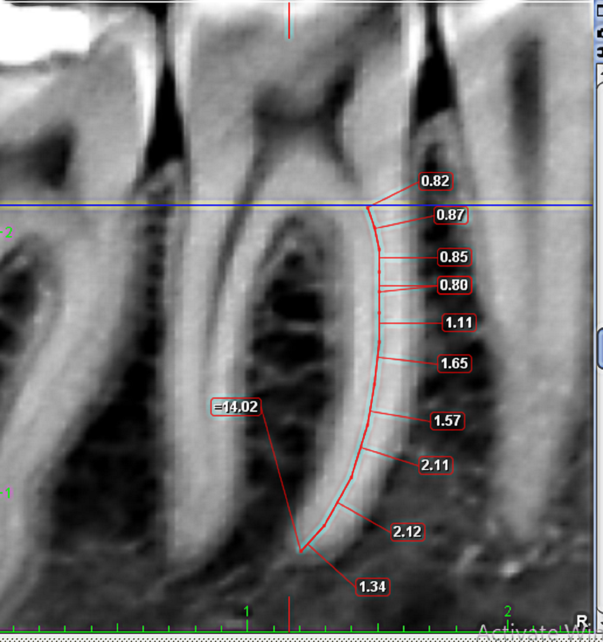 Fig. 1.
Measuring the root length in the coronal view of 1st molar by Romexis software.
Schneider’s classification [22] was used to check the curvature of the canal in the middle section of sagittal view. In this method, the angle resulting from the collision of two lines, that is, the line drawn parallel to the longitudinal axis of the coronal part of the canal and the line was drawn at the beginning point of the canal curvature to the apical foramen was used to calculate the curvature of the canal. Based on this, the teeth were divided into 3 groups: straight (10 degrees or less), moderately curved (10 to 20 degrees), and severely curved (20 to 70 degrees) [19, 23] (Fig. 2).
 Fig. 2.
Measuring the curvature of the canal in the coronal view of 1st molar by Romexis software.
To investigate the relationship between the mesial canals based on Vertucci’s classification [24], the axial view was used and by moving from the coronal to the apical side, the relationship between the two mesial canals was determined. During the examination, if there is a mid-mesial canal, its information was also recorded. The distance between the orifices of the mesial canals was also measured in the same view, at the cemento-enamel junction level of the tooth, and the distance between the buccal surface of the mesiobuccal (MB) canal and the lingual surface of the mesiolingual (ML) canal was also measured to estimate the distance between the entrances of the mesial canals [25] (Fig. 3).
 Fig. 3.
Measuring the distance between the orifices in the axial view of 1st molar by Romexis software.
Then, the minimum dentin thickness of the distal wall of the ML and MB canal was evaluated in the axial view of the CBCT images in sections 0–5 mm below the furcation. The measurement was done in such a way that first a hypothetical line was drawn which is tangent to the external walls of the distal side of the mesial root. Then, the depth of the concavity was measured by drawing a line perpendicular to this line in the deepest depression area on the distal side of the root. This work was done in every millimeter of the root surface in sections 0–5 mm below the furcation. The area with the greatest depth of concavity was used to calculate the minimum dentin thickness. This parameter was drawn and measured perpendicularly by drawing a line from the inner walls of the MB and ML canal to the outer surface of the root (Fig. 4).
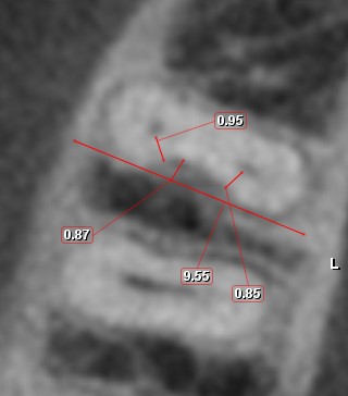 Fig. 4.
Measuring the minimum of dentin thickness in the danger zone in the axial view of 1st molar by Romexis software.
The collected data is entered into the SPSS v.23 (version 23, SPSS Inc.,
Chicago, IL, USA) and R software (version 4.1.1, R Foundation for Statistical
Computing, Vienna, Austria). Frequency, the average, and standard deviation for
variables were calculated and data analysis was done using simple and multiple
linear regression and Pearson correlation coefficient. Furthermore, for comparing
MB and ML on two sides used a two-sample t-test. The significant level
was also considered at p
This study was based on 210 CBCT to evaluate the relationship between the dentin thickness of the danger zone in the mesial roots of mandibular first molar teeth with the parameters of age, gender, placement side, root length, degree of canal curvature, canal type, presence of middle mesial canal, and the distance between the entrances of the mesial canals was done. The collected data was entered into the SPSS and subjected to statistical analysis (Tables 1,2).
| Minimum | Maximum | Average | Standard Deviation | |
|---|---|---|---|---|
| MB canal | 0.32 | 1.72 | 0.88 | 0.26 |
| ML canal | 0.42 | 1.59 | 0.90 | 0.20 |
| Distance to furcation | 0.14 | 4.50 | 1.58 | 0.94 |
| Age | 11 | 71 | 30.53 | 10.74 |
| Root length | 5.99 | 14.06 | 10.17 | 1.64 |
| Degree of curvature of the canal | 3.16 | 54.66 | 23.55 | 8.15 |
| Distance between the orifices of the mesial canals | 1.80 | 6.18 | 3.94 | 0.80 |
| Root type | Classification | Frequency | Percent |
|---|---|---|---|
| Root length | 0.00 | 41 | 19.5% |
| 1.00 | 43 | 20.5% | |
| 2.00 | 126 | 60.0% | |
| Degree of canal curvature | 0.00 | 10 | 4.8% |
| 1.00 | 55 | 26.2% | |
| 2.00 | 145 | 69.0% |
The mean of age of participants was 30.53
The minimum thickness of dentin in the danger zone in the MB canal varied from
0.32 to 1.72 mm and its average value was measured as 0.885
According to Fig. 5, the danger zone was usually seen in the range of 0 to 1 mm from the furcation more than other defined ranges, and it can be concluded that the location of the danger zone was seen in the range of 0 to 3 mm from the furcation with a probability of 93.4 % (196 samples).
 Fig. 5.
Distribution of Danger Zone distance from the furcation (mm).
There was no significant relationship between the dentin thickness in the danger
zone in the MB canal and the age of the patients (p-value = 0.178,
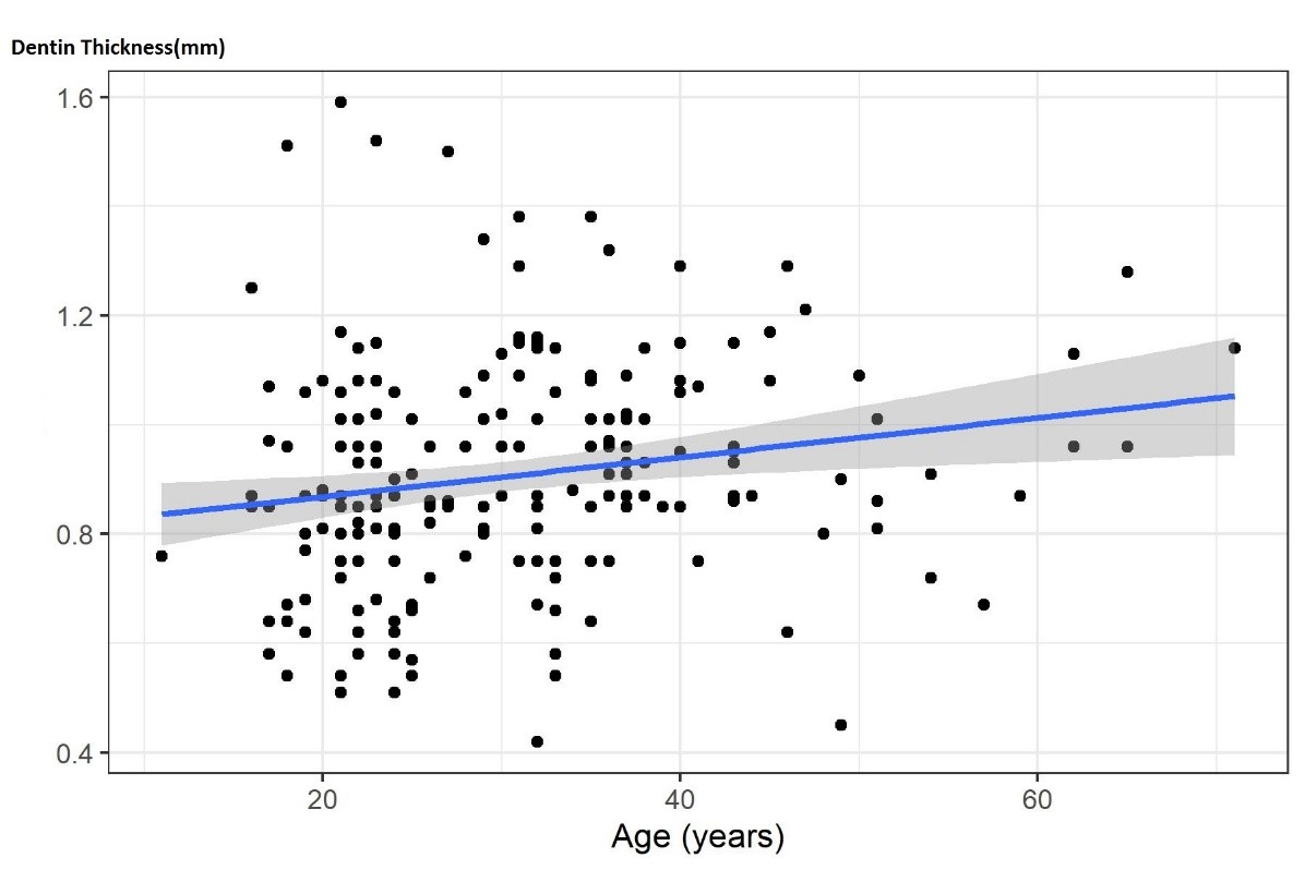 Fig. 6.
Association between the dentin thickness in danger zone of the mesiolingual canal and the patient’s age.
There was no significant relationship between the dentin thickness of the danger
zone in the MB canal (p-value = 0.59,
There was no significant difference between the dentin thickness in the danger zone with the right or left side of the tooth in the mesial canals (MB canal (p-value = 0.163) and ML canal (p-value = 0.671)).
The dentin thickness of the danger zone in the MB canal, medium root length
(p-value = 0.989,
A significant relationship was seen between the dentin thickness in the danger
zone in the MB canal and the degree of curvature of the canal. As the degree of
curvature of the canal increases, the dentin thickness in the danger zone
decreases moderate canal curvature (p-value = 0.008,
Also, there was no significant relationship between the dentin thickness of the
danger zone in the MB canal (p-value = 0.484,
Based on the multivariable model, only two variables, age, and degree of canal curvature were statistically significant. Based on the step-by-step algorithm and Akaike’s criterion (AIC), the best model was identified, and the results of this model showed that in addition to the two variables of age and degree of canal curvature, the presence of the middle mesial canal also had a predictive effect on the minimum thickness of dentin in the danger zone.
A significant relationship was seen between the dentin thickness of the danger
zone in the MB canal (p-value = 0.047,
Also, there was no significant relationship between the dentin thickness of the
danger zone in the MB canal (p-value = 0.925,
The mandibular first molar is the most expected tooth that undergoes endodontic treatment and is anatomically very challenging [26]. Comprehensive knowledge of tooth anatomy is a key factor influencing the outcome of the treatment, in addition to being aware of the most appropriate method for successful endodontic treatment [5]. On the other hand, awareness of the remaining dentin thickness in the roots, especially in the distal part of the mesial roots, which is described as a danger zone, minimizes but doesn’t eliminate the occurrence of strip perforation [26]. During canal shaping, dentin thickness is significantly reduced in the danger zone and is prone to excessive attenuation and subsequent complications [1]. According to the study by Berutty & Fedon, even differences of tenths or hundredths of millimeters can be very important to prevent strip perforation [9].
The present study evaluates the relationship between dentin thickness in the danger zone of the mesial roots of mandibular molars with age, gender, placement side, root length, degree of canal curvature, canal type, presence of middle mesial canal and distance between the orifices of the mesial canals based on CBCT images. Currently, the CBCT technique has become very popular in practices because it allows 3D visualization of anatomical structures and minimizes the overlap of surrounding structures.
Various studies have been conducted to measure the thickness of dentin in the danger zone and the factors affecting it [1, 26, 27]. But based on the searches, no study has comprehensively examined all the possible influential factors. For example, factors such as the curvature of the canal, the type of canal, and the presence of the middle mesial canal were not investigated in any of the available studies. On the other hand, despite the possibility of the effect of ethnicity on the dentin thickness of the furcation area, the available studies on the Iranian population are very limited. According to these limitations, we aimed to design this study and evaluate the mentioned factors.
Dentin thickness reduction is an important factor in canal instrumentation,
because widening the root canal too much can cause events such as perforation.
Lim and Stock have determined the ideal value of 200–300
Our results showed that the minimum dentin thickness of the danger zone in the
MB canal varied from 0.32 to 1.72 mm, and its average was measured as 0.885
Bryant et al. [29] reported that the average danger zone size for 200
canals was 0.79 mm. Keles et al. [30] also reported the thinnest wall of
the MB canal, 1.16
To prevent strip perforation in the danger zone, the selection of high taper NiTi rotary instruments should be cautious and also coronal flaring should be limited and instrumentation should be done along the walls that have thicker dentin and away from the walls where the danger zone is [1].
In the present study, the danger zone is observed in the range of 0 to 1 mm from the furcation more than in other areas, and in general, it can be concluded that the danger zone is located in the range of 0 to 3 mm from the furcation with the probability is 93.4%.
Despite our study, Zhou et al. [1] showed that the minimum dentin thickness of MB and ML canals was 3 to 4 mm below the furcation, and there was no difference between MB and ML canals. This difference between our results and Zhou’s results could be related to couple of reasons like the difference in the number of samples as well as the ethnical difference.
In the study of Kessler et al. [14], it was shown that the danger zone was located 4–6 mm below the orifice of the canal. This observed contrast could be related to ethnical dissimilarity, the different locations of the origin of the distance measurement, which in their study was the orifices of the canals.
Association between the minimum dentin thickness and age:
The present study declared that there was no significant relationship between the dentin thickness of the danger zone in the MB canal and the age of the patients (p-value = 0.178), but this relationship was significant in the ML canal (p-value = 0.008). In this way, older ages correlated to dentin thickness increment in the danger zone in the ML canal.
Zhou et al. [1] divided the patients into three groups: 18 to 30 years
old, 31 to 50 years old, and
So, it should be noted that age is an important factor that affects the thickness of the distal wall of the mesial roots of mandibular first molars. Mandibular first molars in younger people show larger canals and thinner root canal walls than older people [1].
Association between the minimum dentin thickness and gender:
The results of the present study showed that there was no significant relationship between the dentin thickness of the danger zone in the MB canal (p-value = 0.59) and the ML canal (p-value = 0.904) with the patients’ genders.
The results of Zhou’s study despite of our results showed that the dentin thickness of the danger zone in the MB and ML canals was greater in men than in women, except in mm 1 and 3 from the furcation in the ML canal [1]. The ethnical differences and the different number of samples can be accounted for as the main reasons for these observed differences.
Association between the minimum dentin thickness and the side:
There was no significant relationship between the dentin thickness of the danger zone in the MB canal (p-value = 0.59) and the ML canal (p-value = 0.904) with the tooth side in the jaw.
The results of Zhou et al.’s study [1] also showed that the dentin thickness of the danger zone in the MB and ML canals had no significant relationship with the right or left tooth side, which was in agreement with the present study.
Association between the minimum dentin thickness and the root lengths:
The results of the present study showed that there was no significant
relationship between the dentin thickness of the danger zone in the MB canal
(p-value
Furthermore, Zhou et al. [1] showed that the dentin thickness of the danger zone was significantly different between long and short teeth. So short teeth had less dentin thickness. In addition to the difference in the number of samples, the reason for these inconsistent results with our data can be related to the ethnical difference, which is one of the important factors affecting the thickness of the distal wall of the mesial roots of the mandibular first molars.
In the study by Dwivedi et al. [26] and Sauáia et al. [27], it was shown that the distal thickness of the mesial roots of mandibular first molars in longer teeth was thinner compared to shorter teeth. Similarly, the reason for this observed difference can be related to the ethnical difference, the different study method, the smaller number of samples, and the different origin of the length measurement, which was the tip of the cusps in the two mentioned studies, and the furcation in the present study.
Association between the minimum dentin thickness and the degree of curvature:
The accumulation of stress in the root should be considered during the treatment. Since it is closely related to the vertical fracture of the root. Concerning the accumulation of stress, the curvature of the canal seems to be more important than the external morphology of the root [1].
In this study as the degree of curvature of the canal increases, the dentin thickness in the danger zone decreases. We determined a significant correlation between the dentin thickness of the danger zone in the MB canal and the moderately curved (p-value = 0.008) and severely curved (p-value = 0.046) degree of curvature of the canal according to Schneider’s classification, but no significant relationship was found between the dentin thickness in the danger zone in the ML canal and the degree of curvature of the canal.
Based on the searches, no similar study was found on the relationship between the minimum dentin thickness in the danger zone and the degree of curvature of the canal.
Association between the minimum dentin thickness and the canal type:
In the present study, it was also found that there was no significant relationship between the dentin thickness in the danger zone in the MB canal (p-value = 0.484) and the ML canal (p-value = 0.691) with the canal type. Based on the author’s information, no similar study was found on the relationship between the minimum dentin thickness of the danger zone and canal type.
Association between the minimum dentin thickness and middle mesial canal:
We also found a significant relationship between the dentin thickness in the danger zone in the MB canal and the ML canal with the presence of the middle mesial canal (p-value = 0.047, 0.044). In the samples where the middle mesial canal was seen, the dentin thickness was less.
Likewise, no similar study was found regarding the relationship between the minimum thickness of dentin in the danger zone and the presence of the middle mesial canal.
Association between the minimum dentin thickness and distance between the orifices of the mesial canals:
Our study demonstrated it was no significant relationship between the dentin thickness in the danger zone in the MB canal and the ML canal with the distance between the orifices of the mesial canals.
In contrast to our results, De-deus et al. [31] showed that there was a positive correlation between the dentin thickness of the danger zone and the distance between the orifices of the mesial canals. This means that the more distance between the orifices of the mesial canals, the greater the dentin thickness in the danger zone. The possible reason for this difference can be expressed in the smaller sample size, the different methods of measuring the distance between the orifices, and ethnical differences [31, 32].
In the present study, the investigations were limited to the first molar teeth in the furcation area, and not the whole root, so it’s suggested to design a study in which the second molars with the whole root are also examined.
Other limitations of this study include the sensitivity of the measurement method in determining the exact size and location of the studied indicators, the impossibility of separating the cement thickness from the dentin, the impossibility of using methods such as micro-CBCT as a result of conducting this study in vivo.
In this study, individually the CBCT was used; therefore, it’s better to design a study in this direction by using the Micro-CT method.
Less dentin thickness in the danger zone in the mesial roots of mandibular first molars was seen in younger patients in ML canal, with a greater degree of canal curvature in the MB canal and teeth with a mid-mesial canal. Therefore, it is suggested that high taper instruments should be used should be used sparingly to prevent root canal perforation and other complications in younger patients, also teeth with a high degree of canal curvature, and teeth with middle mesial canal, especially in the range of 0 to 1 mm from the furcation.
The data that support the findings of this study are available from the corresponding authors.
Conceptualization—MB and RR; methodology—AZ; software—MR; validation—MR, MV and MT; formal analysis—AA; investigation—AZ and MRAG; resources—AA; data curation—MRAG; writing—original draft preparation—MT; writing—review and editing—RR; visualization—AZ; supervision—LT; project administration—LT. All authors contributed to editorial changes in the manuscript. All authors read and approved the final manuscript.
The study protocol was approved by the ethics committee of Qazvin University of Medical Sciences (IR.QUMS.REC.1400.307).
Not applicable.
This research received no external funding.
The authors declare no conflict of interest. Luca Testarelli is serving as one of the Editorial Board Members and Guest Editors of this journal. We declare that Luca Testarelli had no involvement in the peer review of this article and has no access to information regarding its peer review. Full responsibility for the editorial process for this article was delegated to Marco Tatullo.
References
Publisher’s Note: IMR Press stays neutral with regard to jurisdictional claims in published maps and institutional affiliations.