IMR Press / FBL / Volume 28 / Issue 9 / DOI: 10.31083/j.fbl2809225
Open Access Review
Impact of Immunosenescence on Viral Infections with an Emphasis on COVID-19
Show Less
1 Department of Internal Medicine, University of Genoa, 16132 Genoa, Italy
2 IRCCS Ospedale Policlinico San Martino, 16132 Genoa, Italy
3 Department of Clinical and Experimental Medicine, University of Messina, 98100 Messina, Italy
4 Allergy and Clinical Immunology Unit, Department of Clinical and Experimental Medicine, University of Messina, 98125 Messina, Italy
*Correspondence: giuseppe.murdaca@unige.it (Giuseppe Murdaca)
These authors contributed equally.
§These authors contributed equally.
Front. Biosci. (Landmark Ed) 2023, 28(9), 225; https://doi.org/10.31083/j.fbl2809225
Submitted: 5 July 2023 | Revised: 28 August 2023 | Accepted: 8 September 2023 | Published: 26 September 2023
Copyright: © 2023 The Author(s). Published by IMR Press.
This is an open access article under the CC BY 4.0 license.
Abstract

During aging, the immune system (IS) undergoes remarkable changes known as immunosenescence, a multifactorial and dynamic phenomenon that affects both natural and acquired immunity and plays an important role in most chronic diseases in older people. Among the determinants of immunosenescence, we find a low-grade sterile chronic inflammation, known as “inflamm-aging”. This condition of chronic inflammation causes a progressive reduction in the ability to trigger antibody and cellular responses effective against infections and vaccinations. In this review, we wanted to explore the role of immunosenescence and inflamm-aging as determinants of the immunological aging process and predisposing viral infections phenomena, with a particular reference to cytomegalovirus (CMV), varicella zoster virus (VZV), influenza virus (IFV) diseases and SARS-CoV2. IS aging is also reflected in a reduction in the antibody response to vaccinations, hence there is a need to expand trials to elderly patients, in order to identify the most appropriate methods for developing effective and safe vaccination and preventive strategies.

Keywords
immunosenescence
inflamm-aging
CMV
VZV
IFV
COVID-19
vaccine immunogenicity
1. Introduction

Aging is one of the most difficult biological phenomena to understand, with the ability to influence the functions of many organs and systems, and represents the main risk factor for geriatric diseases [1]. The aging process could be considered a result of the dysregulation of different body systems leading to a decline from the normal (homeostatic) regulation level to an altered state of dyshomeostasis, a reflection of the adaptation of the organism to intrinsic and extrinsic stimuli to which it is subjected during aging [2, 3].

This phenomenon that we could define as “bioaging” is dominated by two distinct processes: immunosenescence and chronic inflammation, known as “inflamm-aging”, which places the human organism in the continuum of an altered state of bioreactivity [4]. The immune system (IS) is characterized, in older age, by a series of pathophysiological variations that go under the term “immunosenescence”, i.e., a multifactorial process that affects both natural and acquired immunity and which plays a fundamental pathogenetic in most of the chronic diseases of the elderly [5].

Inflamm-aging, a systemic state of chronic low-grade inflammation, is one of the hallmarks of immunosenescence; it is characterized by an overproduction of upregulated circulating inflammatory markers and is considered the central pillar of aging [6]. Accumulation of damaged macromolecules is responsible for inflammation and endogenous host cellular debris is the source of chronic tissue damage [7]. This condition of chronic inflammation causes a progressive reduction in the ability to trigger antibody and cellular responses, which are effective against infections and vaccinations [8].

2. Age-Associated Changes in Adaptive and Innate Immunity

Aging also affects the human hematopoietic system, resulting in a decrease in bone marrow cellularity, a decline in the adaptive immune response, and an increase in hematological disorders and malignancies. These changes observed in hematopoietic stem cells play an important role in the generation of the T and B cell repertoire. In the case of T cells, this is exacerbated by the involution of the thymus and the decline of its function [9]. Specifically, in over 65 patients, a progressive decrease in the absolute values of CD4+ and CD8+ T cells was observed; on the contrary, the activated peripheral T cells (HLA-DR1) underwent a progressive increase in their absolute number. In contrast, the percentage and the absolute number of “virgin/unprimed” (CD45RA1) and “memory/activated” (CD45R01) CD4+ and CD8+ T cells did not change significantly during old age, so much that centenarians still retained a substantial reservoir of “virgin” CD4+ T (about 20%) and “virgin” CD8+ (about 50%) cells [10].

Virgin T cells from older people also show several functional defects, including significantly shorter telomeres, a restricted receptor repertoire, and some decreased interleukin (IL)-2 production. As a result, their ability to mediate effective immune responses against new antigens diminishes [11].

This progressive loss of naïve T cell function is compensated in about 30% of the elderly, with the expansion of CD8+, CD45RO+, and CD25+ T clones that can produce IL-2, and with a protective humoral capacity towards vaccinations with the expansion of memory effector cells. Furthermore, in the elderly, there is a progressive increase in CD28 cells, a particular subpopulation of cells subjected to replicative senescence, due to the shortening of telomeres and a reduction in proliferative capacity [12].

Downregulation of CD28 expression due to chronic immune activation of human T cells is one of the signatures of replicative senescence and has been associated with impaired vaccine responses [13].

Changes in the bone marrow microenvironment that occur with aging (e.g., reduced cytokine IL-7) result in decreased pro-B cell survival and a tendency for hematopoietic stem cells (HSCs) to generate myeloid lineage cells rather than lymphoid [14].

Although there is a decrease in the genesis of B cells in older patients, the ability to synthesize and secrete antibodies is unaffected. The downregulation of the transcription factors XBP-1 and Blimp-1, and the upregulation of the transcription factor PAX-5 lead to a decrease in the number of IgM B-1 cells in this category of patients [15].

Frasca D et al. [16] showed that B cells stimulated with anti-CD40/IL-4 from old mice proliferate 2 times less than young controls, while B cells from old mice produce 6 times less IgG1 and 12 times less IgE in culture. Therefore, this evidence suggests that humoral immune responses are dramatically impaired in older subjects, which translates into a reduced response to anti-viral vaccinations.

Aging also has significant effects on all cells of the innate immune system. In elderly subjects, there is an impairment of multiple functions of neutrophils, for example, such as phagocytic capacity, the synthesis of reactive oxygen intermediates, and the efficiency of intracellular killing. Macrophages are also affected by the aging of the individual, causing a progressive loss of effectiveness of their immunological defense functions (e.g., phagocytosis, secretion of cytokines and chemokines, and presentation of the antigen) [17].

Natural Killer (NK) cells also demonstrate a significant susceptibility to aging, which results in a defect of the cellular cytotoxic capacity as a consequence of a reduced generation of inositol triphosphate (IP3). IL-2-induced IFN-γ production and IL-2 or IL-12-induced chemokine production such as MIP-1α, RANTES, and IL-8 were found to decrease in NK cells of aged individuals. During aging, there is also a reduction in the lytic efficiency of NK cells, partly compensated by an increase in their number. The aging process affecting the NK compartment would therefore favor the risk of infection and mortality in the elderly [18].

“Inflamm-aging” represents one of the main features of the aging process; it is understood as a progressive chronic increase in the low-grade proinflammatory state. Inflamm-aging is a determinant of the speed of the aging process and of the duration of life, and it is highly correlated to some chronic pathologies typical of advanced age such as Alzheimer’s disease, Parkinson’s disease, atherosclerosis, heart disease, type II diabetes, osteoporosis and insulin resistance, cancer and other diseases. Inflammatory aging also increases morbidity and mortality, significantly harming patients’ health and causing a decline in patients’ quality of life [19, 20].

Several possible pathogenetic mechanisms can lead to a condition of chronic inflammation, including the process of accumulation of macromolecules and damaged cells (auto-debris) auto-debris that occurs with aging due to increased production and/or inadequate elimination. Self-debris can mimic bacterial products and function as molecular patterns associated with endogenous “damage”, which activate innate immunity by activating a network of sensors (including the Nlrp3 inflammasome) that recognize them as “danger” signals and initiate immune reactions necessary for physiological repair [21].

Studies about oldest/old subjects have highlighted significant differences between their microbiota and that of younger patients, in particular a rearrangement in the Firmicutes population and an enrichment of facultative anaerobes, specifically pathogens, have been highlighted. The impairment of the microbiota in elderly patients is related to an increase in inflamm-aging, determined by markers such as IL-6 and IL-8. The microbiota of centenarians showed a marked decrease in Faecalibacterium prauznitzii and symbiotic species endowed with anti-inflammatory properties and an increase in Eubacterium limosum and its relatives [22].

In the immunological aging process, there is a mitochondria-mediated over-production of anti-inflammatory mediators such as mitokines (HN, FGF21, GDF15), thus allowing to preserve the balance between inflammatory and specific immune responses. Unsuccessful aging is characterized, however, by an imbalance between the inflammatory and anti-inflammatory immune responses, with a prevalence of the former and a consequent increase in the production of specific inflammation mediators such as reactive oxygen species (ROS) and associated molecular models to danger (DAMP), leading to an increase in procytokines [23, 24].

An increased number of senescent cells during the aging process contributes to chronic low-grade inflammation via the senescence-associated secretory phenotype, or SASP, which includes pro-inflammatory cytokines (e.g., IL-6, IL-1, HMGB1, S100), chemokines (e.g., IL-8, MCP-1), soluble receptors (for example, sTNFR), metalloproteases (e.g., collagenase), some protease inhibitors including SERPIN, and growth factors. Chronic inflammation can induce in turn telomere dysfunction, promoting cellular senescence. This highlights how inflammation and senescence reinforce each other in a sort of vicious circle [25, 26].

3. The Role of Immunosenescence on Susceptibility to Viral Infections

Immunosenescence contributes to increased susceptibility to viral infectious diseases, resulting in increased incidence of cytomegalovirus (CMV), varicella zoster virus (VZV), and influenza virus (IFV) diseases [27].

A prospective Canadian study of patients over 65 in 32 nursing homes found that a high representation of CMV-reactive CD4+ Regulatory T cells (Treg) and T cells were predictive of the risk of respiratory viral infections. Thus CMV seropositivity (with high IgG levels) and related changes in T cell populations appear to be related to the risk of viral infectious diseases among the elderly [28]. Treg cells play an indispensable role in the homeostasis of the immune system, particularly for their role as immunosuppressants. Among several cellular markers that have been associated with Treg cell fate and function, expression of the transcriptional regulator FoxP3 is the most specific feature that distinguishes them from other T helper cell lines [29, 30].

Persistent CMV infection has been hypothesized to trigger a pro-inflammatory environment that enhances T cell differentiation. Indeed, CMV-induced IFN-α was found to result in the loss of CD27 and CD28 receptors on CD4+ T cells on CD8+ T cells in vitro. While total CD8+ T cell levels appear to be higher in CMV seropositive elderly, uncertainty remains regarding total CD4+ T cell levels, which may be similar in CMV seropositive and seronegative elderly [31]. In elderly patients, a significant relationship is observed between high latent viral load and greater amplitude and magnitude of CMV-specific CD8 T cell functional responses. However, Jackson SE et al. [32] demonstrated that latent viral load does not correlate with an increase in the number of differentiated memory T cell populations.

With aging, there is a defect in aspects of the adaptive cellular and humoral response to CMV, resulting in an increased rate of viral replication in older individuals. CMV viral reactivation results in a systemic inflammatory condition, which may be responsible for increased morbidity and mortality in elderly patients with co-existing infections [33].

CMV seropositivity is not only associated with modified CD8+ T cell immunity, but also with increased serum antibody titers against VZV. CMV reactivation in older individuals is linked to decreased immunity against VZV and therefore a higher incidence of VZV reactivation [34].

Elevated CMV antibody levels have been associated with an increase in the cytokines IL-6 and TNF-α in the elderly, as well as an increase in C-reactive protein (CRP) levels. CMV, and more recently EBV, have also been implicated in the development of other diseases such as coronary heart disease and cognitive impairment in older adults with cardiovascular disease [35].

VZV reactivation increases during aging, predominantly manifesting in the skin (shingles). In elderly patients, the number of circulating VZV-specific CD4+ T cells secreting IFN-γ is significantly decreased, while an increase in VZV-specific cells is observed in the skin. In contrast, the number of FoxP3+ Tregs and expression of the inhibitory PD-1 receptor on CD4+ T cells are significantly increased in the skin of older humans. The increase in the number of FoxP3+ Treg cells and their suppressive activity could contribute to the decrease of specific VZV responses in older subjects, and therefore to its more frequent reactivation [36, 37].

Burke BL et al. [38] conducted an in vivo study of the immune function of elderly patients in response to VZ antigen and phytohaemagglutinin (PHA) skin testing, mitogen (PHA)- and antigen (VZ)-induced lymphocyte stimulation, and antibodies to VZ. The study demonstrated that the stimulation of lymphocytes with PHA in vitro supported the hypothesis of a progressive decline in reactivity with advancing age. In contrast to this decline in cellular immune responses to VZ with aging, humoral immunity remains relatively intact. In cases where the viral infection becomes chronic, i.e., a persistence of viral antigens, there is typically a failure in the development of memory CD8+ T cells and impairment of the effector functions of CD8+ T cells, thus limiting the potential of T cells to effectively eliminate viral infection. The name of the CD8 T cell state is “exhaustion”, which is a progressive and hierarchical impairment of effector functions (IL-2 production and proliferative capacity are impaired early, followed by some defects in TNF production and cytotoxicity) [39, 40, 41]. Depleted T cells (Tex) are epitope-specific and are characterized by the expression of specific markers, including CD279 (marker of programmed cell death-1, PD-1) and CD366 (T cell immunoglobulin and mucin domain-3, Tim-3), differentiating from anergic T cells. Tim-3-expressing CD8+T cells that accumulate with aging show progressive functional loss in proliferative capacity, inhibitory profile of surface molecules, and cytokine production, resulting in immune system dysfunction typical of aging [42, 43, 44].

Shahbazi M et al. [42] highlighted that in patients with severe forms of COVID-19, there is an increasing number of CD8+ T cells characterized by the expression of the markers CD39, PD-1, and TIM-3, which suggests that SARS-CoV2 can lead to early overactivation and high cytotoxicity of lymphocytes, contributing to the severity of the viral infection.

Age-related changes in adaptive immunity are also seen during influenza virus infections. In fact, during aging, there is a progressive impairment of the signaling capacity of the T cell receptor (TCR). CD4+ T cells also demonstrate a reduced ability to synthesize IL-2 upon antigenic stimulation with age, as well as a reduced ability to stimulate B cells, thus influencing humoral immunity in a deficient sense [45, 46].

The decline of B-cell precursors in old age is associated with the preferential loss of lymphoid-polarized hematopoietic stem cells. With aging, there is a decrease in germinal center formation during influenza infection and B cells produce lower quality antibodies. Furthermore, the increase in senescent B cells with aging is negatively associated with a protective response after influenza vaccination [47].

In addition, memory CD8+ T cells in the spleen progressively change from a more activated to a less activated phenotype during aging, probably as a consequence of low levels of CD43 expression [48, 49]. Indeed, after influenza A virus (IAV) infection, older lungs show prolonged inflammation and excessive numbers of CD8+ resident memory Ts (TRMs), which are intrinsically hyporesponsive to antigenic challenge and ineffective at protecting against respiratory infections. Aged CD8+ TRM cells are also immunologically active in the lungs, promoting chronic lung inflammation and fibrotic damage [50].

Interestingly, although the total number of CD4+ T lymphocytes in the lung after influenza infection is not different between young and old mice, the distribution ratio of the T-helper (TH) subgroup is affected by aging. Indeed, older adults have been shown to have a CD4+ T cell compartment biased towards primarily regulatory, memory, and type 2 (TH2) helper T cells. Although these subsets are critical in the resolution of influenza infection in its acute phase, reduced initial effector response could lead to poor viral clearance resulting in much longer recovery time, increased lung injury, and potentially increased risk of bacterial infections secondary [51].

Fig. 1 shows the main functional alterations that occur in some of the immune cells (B and T cells, alveolar macrophages - AM, and neutrophils). These immunological changes predispose the elderly patient to viral infections, resulting in increased morbidity and mortality.
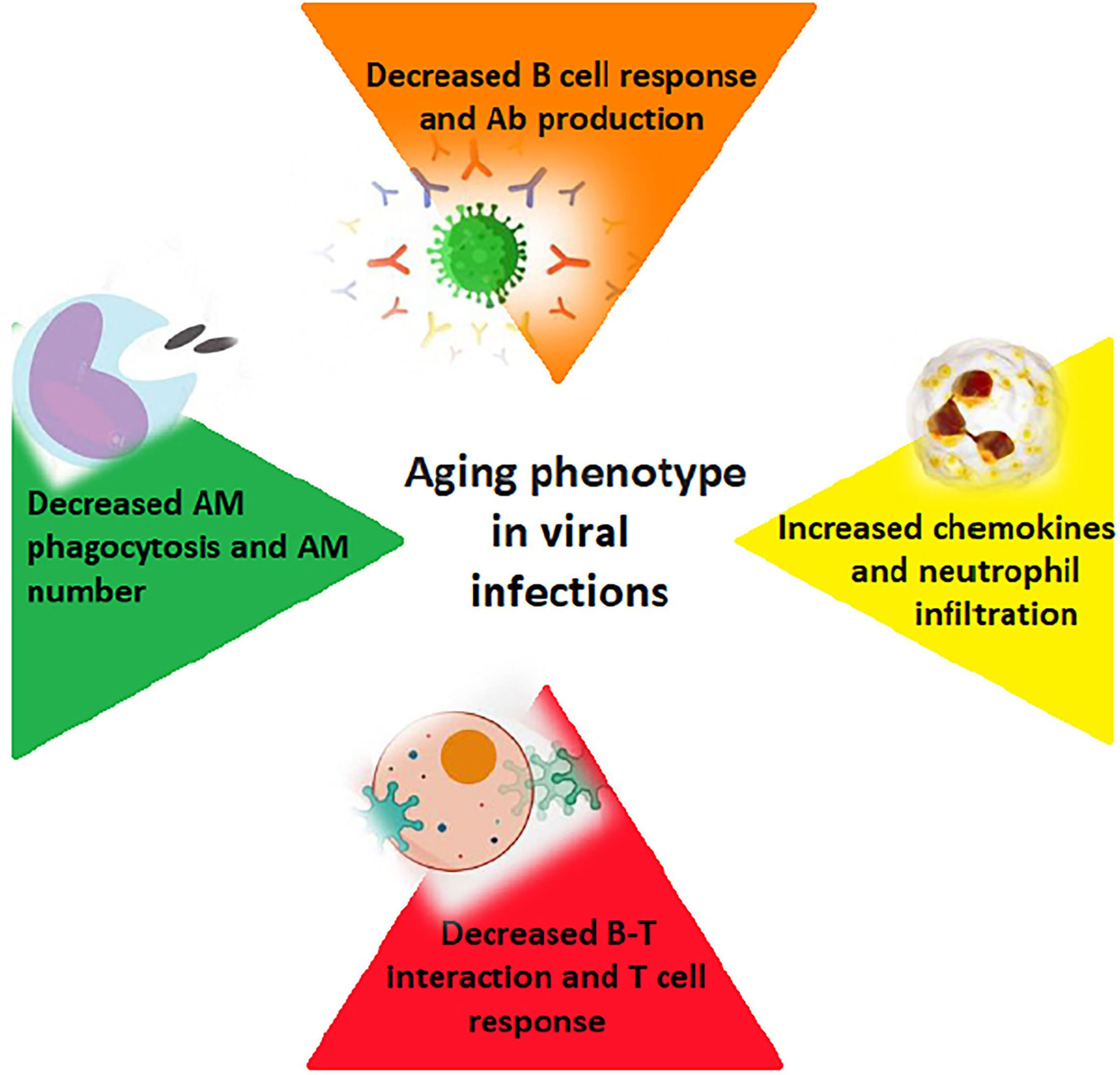
Fig. 1.

How aging affects the immune response to viral infections (B and T cells; alveolar macrophages – AM; Ab - antibodies).

4. Aging and COVID-19: Immune Aging as a Risk Factor for Severe Disease Outcomes

Coronavirus disease 2019 (COVID-19) is a viral disease affecting the upper and lower airways caused by the infectious agent severe acute respiratory syndrome coronavirus 2 (SARS-CoV2), which can cause a large variety of clinical forms ranging from almost asymptomatic cases to critical and potentially lethal forms [52, 53]. The aging of the immune system and the state of systemic inflammation contribute significantly to the development of clinically severe forms of COVID-19 disease, predisposing over 65 subjects to a higher rate of complications and mortality than younger subjects [54].

The increased susceptibility to SARS-CoV2 infection in elderly subjects is primarily related to age-related alterations in the cellular receptor of angiotensin-converting enzyme 2 (ACE2), through which the virus penetrates inside the target cells [55]. For example, elderly patients with severe aortic stenosis had a four-fold higher circulating level of ACE2 than in hypertensive patients. This increase could be justified by a combined effect of reduced left ventricular systolic function, increased pulmonary pressure, and age in this patient population, who are therefore more at risk for SARS-CoV2 infections [56].

Hematopoietic stem cells (HSC cells) also undergo qualitative alterations during aging (e.g., insufficient DNA repair capacity, increase in HSCs with a simultaneous decrease in their homing efficiency, and myeloid bias of their differentiation potential), resulting in a downregulation of lymphoid differentiation genes in favor of an upregulation of myeloid differentiation genes. This proneness to myeloid differentiation would seem to contribute to the deterioration of immune system competence in elderly individuals, thus influencing the increased susceptibility to SARS-CoV2 infection in this particular category of patients [57, 58]. COVID-19 patients display a typical picture of lymphopenia, with marked reductions in the absolute number of circulating CD4+ CD8+ T cells against a prevalence of mononuclear cells (monocytes and macrophages) in the target tissues of the lesion. In the aging immune system, this picture of progressive lymphopenia with CD4+ T lymphocyte wasting and reduced regulatory T lymphocyte function is physiological and determines a propensity for excessive autoimmune and inflammatory responses, also associates with the reduced ability of senescent macrophages to phagocytose cells, apoptotic cells. In the elderly patient, SARS-CoV2 infection would induce further CD4+ CD8+ T cell depletion and macrophage migration, thus increasing the risk of developing secondary bacterial infection and viral sepsis [59, 60, 61].

Macrophages localized in the lungs of elderly patients have also been observed to have a more pronounced production of IL-6 and other pro-inflammatory cytokines in response to stimuli [62]. IL-6 inhibits the production of Interferon-γ, necessary for the activation of CD8+ cells, already deficient in elderly patients. In patients suffering from clinically severe forms of COVID-19 disease, a higher IL-6/Interferon-γ ratio is typically observed, probably as an expression of the cytokine storm and therefore of tissue damage in the lung [63, 64].

In addition to macrophages, neutrophils in patients with severe forms of COVID-19 also show changes in number, phenotype, and function, in particular, an increase in the number of immature neutrophils characterized by the surface marker CD10LowCD101 has been demonstrated to associate with clinically severe forms of respiratory disease. Furthermore, COVID-19 patients who developed ARDS had significantly higher neutrophil counts and this factor may be implicated in the development of cytokine storm syndrome [65, 66].

In older patients, there is typically a preponderance of CD56dim NK cells, endowed with distinctly cytotoxic properties and secretion of IFN-γ, to the detriment of CD56bright NK cells, which instead exhibit cytokine and chemokine synthesis properties. Furthermore, during cellular aging, there is a reduction in the NK cells’ ability to synthesize some mediators capable of counteracting viral infections, with a consequent increased susceptibility to them. In elderly patients, impaired NK function has been observed in patients with severe COVID-19, who express higher levels of the cell depletion marker receptor NKG2A, indicating impaired antiviral immunity [67, 68].

Taken together, these results indicate that senescence is associated with a progressive loss of functional NK cells, partially compensated by an increase in the number of mature cells NK cells. Longitudinal studies have shown that the reduction in terms of number and/or NK cell function in the elderly is associated with serious infections and increased risk of mortality; they support the significance of these cells in the control of infectious diseases in the elderly [69].

5. Vaccine Immunogenicity in the Elderly

Changes in the immune system that occur with aging can affect the antibody response to vaccination. Among the various changes, the most significant is the picture of inflamm-aging with consequent macrophage activation, capable of creating a harmful environment for the generation of an adequate immune response to a vaccine. This picture of low-grade inflammation is associated with impaired antigen presentation by dendritic cells (DCs), which are unable to efficiently process and present antigens to T cells with aging. Changes seen in lymph nodes with aging contribute to impaired vaccine response [70].

Antigen-presenting cells (APCs) also show changes in older individuals, particularly, this category of patients displays a decrease in the number of naïve cells mainly in the subpopulation of CD8 T cells, thus precluding priming by new antigens [71].

Thymic involution involved in impaired immune response to vaccines manifests with a decrease in naïve T cell production by 3% per year. Despite the thymus undergoing a progressive involution starting from puberty, there is a well-preserved number of T cells in elderly subjects; thus its role is assumed by thymic residues or substitute lymphoid organs, which are able to produce and select a large number of T cells every day, up to the very limit of human life. The increase in the number of mature T cells in the periphery and the increase in the ratio of memory T cells to naïve T cells contributes to the maintenance of homeostatic equilibrium in peripheral lymphocyte cell clusters [72, 73].

Age-related thymic involution causes a collapse of the TCR repertoire, which then becomes a monitoring system of thymic function. In particular, sjTREC - signal junction T cell receptor excision circles, i.e., a particular TREC that arises from an intermediate rearrangement at the TCRD/A locus in developing TCRαβ+ T lymphocytes, has been successfully used as a marker for the prediction of survival in old age [74].

Furthermore, there is a progressive loss of lymphocyte proliferative capacity in the elderly as a consequence of the shortening of telomeres, which, once the critical limit is reached, block the ability of the latter to divide, resulting in a cluster of “old” T cells which are no longer able to proliferate, but have limited receptor diversity. This type of aged lymphocytes is characterized by a loss of receptor expressive capacity, as happens, for example, for the costimulatory molecule CD28, which is essential to allow the differentiation of B cells into plasma cells and therefore the production of specific antibodies [75, 76].

In the management of the COVID-19 pandemic emergency, the new messenger RNA (mRNA) vaccines have already played a fundamental role. Once the mRNA for the spike protein is encoded, SARS-CoV2 is injected directly into the host, where the mRNA is translated by ribosomes, resulting in the production of a vital protein responsible for the immune response. The lack of involvement of the infectious agent in their production makes this vaccine technology safer, with a low potential for mutations and, thus a lower risk of degradation of the antigen in vivo [77].

Phase III studies investigating vaccine efficacy in elderly subjects demonstrated that two doses of the Pfizer-BioNTech vaccine produce immunogenicity, regardless of the elderly’s health condition, and provide strong humoral immunity in elderly people who are 80 and 96 years old [78].

Demonstration of post-vaccine immunogenicity in the elderly is an important aspect of the fight against COVID-19, although disease presentation, severity, and mortality are the main outcomes that need to be evaluated in this type of future observational study.

In this perspective, the enrollment of older participants in COVID-19 vaccine trials is essential to understand the vaccine response of this vulnerable population [79].

6. Discussion and Conclusions

Immunosenescence and inflammation predispose frail and elderly people to viral infections. As far as COVID-19 is concerned, immunological aging processes favor the development of more serious forms of the disease and complications.

In this area, recent studies have particularly highlighted that skin immunosenescence has a role not only in the onset of dermatological disorders, but also has fascinating system-level implications, whereby respiratory and neurological diseases have emerged as potentially related to skin immunosenescence [80, 81].

Chronic inflammation predisposes the body to acute inflammatory reactions that can be devastating, such as in the case of most elderly patients with COVID-19.

Variations in the efficiency of the immune system and an individual’s inflammatory state can contribute to the severity of the infection by both affecting viral replication and increasing the production of pro-inflammatory cytokines.

Inflammation usually has the ability to suspend itself when it is no longer needed, but if the immune system is confronted with repeated attacks, the inflammatory process can become chronic, becoming latent (symptom-free and hardly detectable) and damaging all tissues.

Since the vaccine response in elderly subjects is altered, with reduced production of neutralizing antibodies, the implementation of vaccine enrollment and trials in patients over 65 is of fundamental importance, in order to identify adequate vaccine formulations able to effectively stimulate the immune system.

This paper has some limitations related mainly to the variability of the type of works that have been analyzed and to the still partial knowledge regarding the immunopathological mechanisms of SARS-CoV2 concerning immunosenescence.

Author Contributions

Conceptualization, SG and GMu; methodology FP and GMa; investigation, FP; resources, GMu; data curation, GMu and GMa; writing—original draft preparation, GMa, FP, GMu; writing—review and editing, FP, GMu, GMa, SG; visualization, FP; supervision, SG and GMa; project administration, SG, GMu and GMa. All authors have read and agreed to the published version of the manuscript.

Ethics Approval and Consent to Participate

Not applicable.

Acknowledgment

Not applicable.

Funding

This research received no external funding.

Conflict of Interest

The authors declare no conflict of interest. Given their role as Guest Editor or Editorial Board Member, Dr. Giuseppe Murdaca, Dr. Francesca Paladin and Dr. Sebastiano Gangemi had no involvement in the peer-review of this article and has no access to information regarding its peer review. Full responsibility for the editorial process for this article was delegated to Vijay Kumar.

References
[1]
Aronson L. Healthy Aging across the Stages of Old Age. Clinics in Geriatric Medicine. 2020; 36: 549–558.
[2]
Santoro A, Bientinesi E, Monti D. Immunosenescence and inflammaging in the aging process: age-related diseases or longevity? Ageing Research Reviews. 2021; 71: 101422.
[3]
Campisi J, Kapahi P, Lithgow GJ, Melov S, Newman JC, Verdin E. From discoveries in ageing research to therapeutics for healthy ageing. Nature. 2019; 571: 183–192.
[4]
Witkowski JM, Fulop T, Bryl E. Immunosenescence and COVID-19. Mechanisms of Ageing and Development. 2022; 204: 111672.
[5]
Rodriguez JE, Naigeon M, Goldschmidt V, Roulleaux Dugage M, Seknazi L, Danlos FX, et al. Immunosenescence, inflammaging, and cancer immunotherapy efficacy. Expert Review of Anticancer Therapy. 2022; 22: 915–926.
[6]
Soma T, Nagata M. Immunosenescence, Inflammaging, and Lung Senescence in Asthma in the Elderly. Biomolecules. 2022; 12: 1456.
[7]
Banić M, Pleško S, Urek M, et al. Immunosenescence, Inflamm-aging and Resilience: An Evolutionary Perspective of Adaptation in the Light of COVID-19 Pandemic. Psychiatr Danub. 2021; 33: 427–431.
[8]
Liu Z, Liang Q, Ren Y, et al. Immunosenescence: molecular mechanisms and diseases. Signal Transduction and Targeted Therapy. 2023; 8: 200.
[9]
Cossarizza A, Ortolani C, Monti D, Franceschi C. Cytometric analysis of immunosenescence. Cytometry. 1997; 27: 297–313.
[10]
Sadighi Akha AA. Aging and the immune system: an overview. Journal of Immunological Methods. 2018; 463: 21–26.
[11]
Weiskopf D, Weinberger B, Grubeck-Loebenstein B. The aging of the immune system. Transplant International. 2009; 22: 1041–1050.
[12]
Ventura MT, Casciaro M, Gangemi S, Buquicchio R. Immunosenescence in aging: between immune cells depletion and cytokines up-regulation. Clinical and Molecular Allergy. 2017; 15: 21.
[13]
Frasca D, Diaz A, Romero M, Garcia D, Blomberg BB. B Cell Immunosenescence. Annual Review of Cell and Developmental Biology. 2020; 36: 551–574.
[14]
Labi V, Derudder E. Cell signaling and the aging of B cells. Experimental Gerontology. 2020; 138: 110985.
[15]
de Mol J, Kuiper J, Tsiantoulas D, Foks AC. The Dynamics of B Cell Aging in Health and Disease. Frontiers in Immunology. 2021; 12: 733566.
[16]
Frasca D, Van der Put E, Riley RL, Blomberg BB. Reduced Ig Class Switch in Aged Mice Correlates with Decreased E47 and Activation-Induced Cytidine Deaminase. Journal of Immunology. 2004; 172: 2155–2162.
[17]
Gomez CR, Nomellini V, Faunce DE, Kovacs EJ. Innate immunity and aging. Experimental Gerontology. 2008; 43: 718–728.
[18]
Panda A, Arjona A, Sapey E, Bai F, Fikrig E, Montgomery RR, et al. Human innate immunosenescence: causes and consequences for immunity in old age. Trends in Immunology. 2009; 30: 325–333.
[19]
Xia S, Zhang X, Zheng S, Khanabdali R, Kalionis B, Wu J, et al. An Update on Inflamm-Aging: Mechanisms, Prevention, and Treatment. Journal of Immunology Research. 2016; 2016: 8426874.
[20]
Franceschi C, Bonafè M, Valensin S, Olivieri F, De Luca M, Ottaviani E, et al. Inflamm-aging. An evolutionary perspective on immunosenescence. Annals of the New York Academy of Sciences. 2000; 908: 244–254.
[21]
Franceschi C, Campisi J. Chronic Inflammation (Inflammaging) and its Potential Contribution to Age-Associated Diseases. the Journals of Gerontology Series a: Biological Sciences and Medical Sciences. 2014; 69: S4–S9.
[22]
Bischoff SC. Microbiota and aging. Current Opinion in Clinical Nutrition and Metabolic Care. 2016; 19: 26–30.
[23]
Conte M, Martucci M, Chiariello A, Franceschi C, Salvioli S. Mitochondria, immunosenescence and inflammaging: a role for mitokines? Seminars in Immunopathology. 2020; 42: 607–617.
[24]
Fulop T, Larbi A, Pawelec G, Khalil A, Cohen AA, Hirokawa K, et al. Immunology of Aging: the Birth of Inflamm-aging. Clinical Reviews in Allergy & Immunology. 2023; 64: 109–122.
[25]
Teissier T, Boulanger E, Cox LS. Interconnections between Inflammageing and Immunosenescence during Ageing. Cells. 2022; 11: 359.
[26]
Loeser RF, Collins JA, Diekman BO. Ageing and the pathogenesis of osteoarthritis. Nature Reviews Rheumatology. 2016; 12: 412–420.
[27]
Oh SJ, Lee JK, Shin OS. Aging and the Immune System: the Impact of Immunosenescence on Viral Infection, Immunity and Vaccine Immunogenicity. Immune Network. 2019; 19: e37.
[28]
Li X, Zheng Y. Regulatory T cell identity: formation and maintenance. Trends in Immunology. 2015; 36: 344–353.
[29]
Sawant DV, Vignali DAA. Once a Treg, always a Treg? Immunological Reviews. 2014; 259: 173–191.
[30]
Kadambari S, Klenerman P, Pollard AJ. Why the elderly appear to be more severely affected by COVID-19: The potential role of immunosenescence and CMV. Reviews in Medical Virology. 2020; 30: e2144.
[31]
Weltevrede M, Eilers R, de Melker HE, van Baarle D. Cytomegalovirus persistence and T-cell immunosenescence in people aged fifty and older: a systematic review. Experimental Gerontology. 2016; 77: 87–95.
[32]
Jackson SE, Sedikides GX, Okecha G, Poole EL, Sinclair JH, Wills MR. Latent Cytomegalovirus (CMV) Infection Does Not Detrimentally Alter T Cell Responses in the Healthy Old, But Increased Latent CMV Carriage Is Related to Expanded CMV-Specific T Cells. Frontiers in Immunology. 2017; 8: 733.
[33]
Davies EL, Noor M, Lim EY, Houldcroft CJ, Okecha G, Atkinson C, et al. HCMV carriage in the elderly diminishes anti-viral functionality of the adaptive immune response resulting in virus replication at peripheral sites. Frontiers in Immunology. 2022; 13: 1083230.
[34]
Ogunjimi B, Hens N, Pebody R, Jansens H, Seale H, Quinlivan M, et al. Cytomegalovirus seropositivity is associated with herpes zoster. Human Vaccines & Immunotherapeutics. 2015; 11: 1394–1399.
[35]
Stowe RP, Peek MK, Cutchin MP, Goodwin JS. Reactivation of herpes simplex virus type 1 is associated with cytomegalovirus and age. Journal of Medical Virology. 2012; 84: 1797–1802.
[36]
Vukmanovic-Stejic M, Sandhu D, Seidel JA, Patel N, Sobande TO, Agius E, et al. The Characterization of Varicella Zoster Virus–Specific T Cells in Skin and Blood during Aging. Journal of Investigative Dermatology. 2015; 135: 1752–1762.
[37]
Weinberg A, Pang L, Johnson MJ, Caldas Y, Cho A, Tovar-Salazar A, et al. The Effect of Age on the Immunogenicity of the Live Attenuated Zoster Vaccine is Predicted by Baseline Regulatory T Cells and Varicella-Zoster Virus-Specific T Cell Immunity. Journal of Virology. 2019; 93: e00305–e00319.
[38]
Burke BL, Steele RW, Beard OW, Wood JS, Cain TD, Marmer DJ. Immune Responses to Varicella-Zoster in the Aged. Archives of Internal Medicine. 1982; 142: 291–293.
[39]
Rha M, Shin E. Activation or exhaustion of CD8+ T cells in patients with COVID-19. Cellular & Molecular Immunology. 2021; 18: 2325–2333.
[40]
De Biasi S, Meschiari M, Gibellini L, Bellinazzi C, Borella R, Fidanza L, et al. Marked T cell activation, senescence, exhaustion and skewing towards TH17 in patients with COVID-19 pneumonia. Nature Communications. 2020; 11: 3434.
[41]
Weinberg A, Canniff J, Rouphael N, Mehta A, Mulligan M, Whitaker JA, et al. Varicella-Zoster Virus–Specific Cellular Immune Responses to the Live Attenuated Zoster Vaccine in Young and Older Adults. Journal of Immunology. 2017; 199: 604–612.
[42]
Shahbazi M, Moulana Z, Sepidarkish M, Bagherzadeh M, Rezanejad M, Mirzakhani M, et al. Pronounce expression of Tim-3 and CD39 but not PD1 defines CD8 T cells in critical Covid-19 patients. Microbial Pathogenesis. 2021; 153: 104779.
[43]
Lee KA, Shin KS, Kim GY, Song YC, Bae EA, Kim IK, et al. Characterization of age-associated exhausted CD8+ T cells defined by increased expression of Tim-3 and PD-1. Aging Cell. 2016; 15: 291–300.
[44]
Chiappelli F, Khakshooy A, Greenberg G. COVID-19 Immunopathology and Immunotherapy. Bioinformation. 2020; 16: 219–222.
[45]
Lorenzo EC, Bartley JM, Haynes L. The impact of aging on CD4+ T cell responses to influenza infection. Biogerontology. 2018; 19: 437–446.
[46]
Lanzer KG, Johnson LL, Woodland DL, Blackman MA. Impact of ageing on the response and repertoire of influenza virus-specific CD4 T cells. Immunity & Ageing. 2014; 11: 9.
[47]
Keilich SR, Bartley JM, Haynes L. Diminished immune responses with aging predispose older adults to common and uncommon influenza complications. Cellular Immunology. 2019; 345: 103992.
[48]
Ely K, Roberts A, Kohlmeier J, Blackman M, Woodland D. Aging and CD8+ T cell immunity to respiratory virus infections. Experimental Gerontology. 2007; 42: 427–431.
[49]
Goplen NP, Wu Y, Son YM, Li C, Wang Z, Cheon IS, et al. Tissue-resident CD8+ T cells drive age-associated chronic lung sequelae after viral pneumonia. Science Immunology. 2020; 5: eabc4557.
[50]
Shenoy AT, Mizgerd JP. Seedy CD8+ TRM cells in aging lungs drive susceptibility to pneumonia and sequelae. Cellular & Molecular Immunology. 2021; 18: 787–789.
[51]
McElhaney JE, Kuchel GA, Zhou X, Swain SL, Haynes L. T-Cell Immunity to Influenza in Older Adults: A Pathophysiological Framework for Development of More Effective Vaccines. Frontiers in Immunology. 2016; 7: 41.
[52]
Witkowski JM, Fulop T, Bryl E. Immunosenescence and COVID-19. Mechanisms of Ageing and Development. 2022; 204: 111672.
[53]
Solana R, Mariani E. NK and NK/T cells in human senescence. Vaccine. 2000; 18: 1613–1620.
[54]
Chen Y, Klein SL, Garibaldi BT, Li H, Wu C, Osevala NM, et al. Aging in COVID-19: Vulnerability, immunity and intervention. Ageing Research Reviews. 2021; 65: 101205.
[55]
Fagyas M, Kertész A, Siket IM, Bánhegyi V, Kracskó B, Szegedi A, et al. Level of the SARS-CoV-2 receptor ACE2 activity is highly elevated in old-aged patients with aortic stenosis: implications for ACE2 as a biomarker for the severity of COVID-19. GeroScience. 2021; 43: 19–29.
[56]
Chen J, Jiang Q, Xia X, Liu K, Yu Z, Tao W, et al. Individual variation of the SARS‐CoV‐2 receptor ACE2 gene expression and regulation. Aging Cell. 2020; 19: e13168
[57]
Müller L, Di Benedetto S. How Immunosenescence and Inflamm-aging May Contribute to Hyperinflammatory Syndrome in COVID-19. International Journal of Molecular Sciences. 2021; 22: 12539.
[58]
Weinberger B. Vaccination of older adults: Influenza, pneumococcal disease, herpes zoster, COVID-19 and beyond. Immunity & Ageing. 2021; 18: 38.
[59]
Napoli C, Tritto I, Mansueto G, Coscioni E, Ambrosio G. Immunosenescence exacerbates the COVID-19. Archives of Gerontology and Geriatrics. 2020; 90: 104174.
[60]
Bencivenga L, Rengo G, Varricchi G. Elderly at time of COronaVIrus disease 2019 (COVID-19): possible role of immunosenescence and malnutrition. GeroScience. 2020; 42: 1089–1092.
[61]
Wang Y, Pang SC, Yang Y. A potential association between immunosenescence and high COVID-19 related mortality among elderly patients with cardiovascular diseases. Immunity & Ageing. 2021; 18: 25.
[62]
Cunha LL, Perazzio SF, Azzi J, Cravedi P, Riella LV. Remodeling of the Immune Response With Aging: Immunosenescence and Its Potential Impact on COVID-19 Immune Response. Frontiers in Immunology. 2020; 11: 1748.
[63]
Lynch SM, Guo G, Gibson DS, Bjourson AJ, Rai TS. Role of Senescence and Aging in SARS-CoV-2 Infection and COVID-19 Disease. Cells. 2021; 10: 3367.
[64]
Mirbeyk M, Saghazadeh A, Rezaei N. Geriatrics and COVID-19. Advances in Experimental Medicine and Biology. 2021; 22: 209–222.
[65]
Tizazu AM, Mengist HM, Demeke G. Aging, inflamm-aging and immunosenescence as risk factors of severe COVID-19. Immunity & Ageing. 2022; 19: 53.
[66]
Müller L, Di Benedetto S. How Immunosenescence and Inflamm-aging May Contribute to Hyperinflammatory Syndrome in COVID-19. International Journal of Molecular Sciences. 2021; 22: 12539.
[67]
Ligotti ME, Pojero F, Accardi G, Aiello A, Caruso C, Duro G, et al. Immunopathology and Immunosenescence, the Immunological Key Words of Severe COVID-19. Is There a Role for Stem Cell Transplantation? Frontiers in Cell and Developmental Biology. 2021; 9: 725606.
[68]
Pietrobon AJ, Teixeira FME, Sato MN. I mmunosenescence and Inflamm-aging: Risk Factors of Severe COVID-19 in Older People. Frontiers in Immunology. 2020; 11: 579220.
[69]
Banić M, Pleško S, Urek M, Babić Ž, Kardum D. Immunosenescence, Inflamm-aging and Resilience: An Evolutionary Perspective of Adaptation in the Light of COVID-19 Pandemic. Psychiatr Danub. 2021; 33: 427–431.
[70]
Crooke SN, Ovsyannikova IG, Poland GA, Kennedy RB. Immunosenescence and human vaccine immune responses. Immunity & Ageing. 2019; 16: 25.
[71]
Fulop T, Larbi A, Pawelec G, Cohen AA, Provost G, Khalil A, Lacombe G, Rodrigues S, Desroches M, Hirokawa K, Franceschi C, Witkowski JM. Immunosenescence and Altered Vaccine Efficiency in Older Subjects: A Myth Difficult to Change. Vaccines. 2022; 10: 607.
[72]
Franceschi C, Monti D, Barbier D, Salvioli S, Grassilli E, Capri M, et al. Successful immunosenescence and the remodelling of immune responses with ageing. Nephrology Dialysis Transplantation. 1996; 11: 18–25.
[73]
Soegiarto G, Purnomosari D. Challenges in the Vaccination of the Elderly and Strategies for Improvement. Pathophysiology. 2023; 30: 155–173.
[74]
Paparazzo E, Geracitano S, Lagani V, Citrigno L, Bartolomeo D, Aceto MA, Bruno F, Maletta R, Passarino G, Montesanto A. Thymic function and survival at advance ages in nursing home residents from Southern Italy. Immunity & Ageing. 2023; 20: 16.
[75]
Fulop T, Larbi A, Pawelec G, Cohen AA, Provost G, Khalil A, et al. Immunosenescence and Altered Vaccine Efficiency in Older Subjects: A Myth Difficult to Change. Vaccines. 2022; 10: 607.
[76]
O’Driscoll M, Ribeiro Dos Santos G, Wang L, Cummings DAT, Azman AS, Paireau J, et al. Age-specific mortality and immunity patterns of SARS-CoV-2. Nature. 2021; 590: 140–145.
[77]
Soiza RL, Scicluna C, Thomson EC. Efficacy and safety of COVID-19 vaccines in older people. Age and Ageing. 2021; 50: 279–283.
[78]
Gao J, Lun P, Ding YY, George PP. COVID-19 Vaccination for Frail Older Adults in Singapore - Rapid Evidence Summary and Delphi Consensus Statements. The Journal of Frailty & Aging. 2022; 11: 236–241.
[79]
Salmerón Ríos S, Mas Romero M, Cortés Zamora EB, Tabernero Sahuquillo MT, Romero Rizos L, Sánchez-Jurado PM, et al. Immunogenicity of the BNT162b2 vaccine in frail or disabled nursing home residents: COVID-A study. Journal of the American Geriatrics Society. 2021; 69: 1441–1447.
[80]
Papa V, Li Pomi F, Borgia F, Vaccaro M, Pioggia G, Gangemi S. Immunosenescence and Skin: A State of Art of Its Etiopathogenetic Role and Crucial Watershed for Systemic Implications. International Journal of Molecular Sciences. 2023; 24: 7956.
[81]
Chen B, Yang J, Song Y, Zhang D, Hao F. Skin Immunosenescence and Type 2 Inflammation: A Mini-Review With an Inflamm-aging Perspective. Frontiers in Cell and Developmental Biology. 2022; 10: 835675.

Publisher’s Note: IMR Press stays neutral with regard to jurisdictional claims in published maps and institutional affiliations.

Share
Back to top