IMR Press / FBL / Volume 28 / Issue 9 / DOI: 10.31083/j.fbl2809210
Open Access Review
The Role of NLRP3 Inflammasomes in Trained Immunity
Show Less
1 College of Veterinary Medicine and Institute of Veterinary Science, Kangwon National University, 24341 Chuncheon, Republic of Korea
*Correspondence: leegeun@kangwon.ac.kr (Geun-Shik Lee)
Front. Biosci. (Landmark Ed) 2023, 28(9), 210; https://doi.org/10.31083/j.fbl2809210
Submitted: 4 May 2023 | Revised: 14 August 2023 | Accepted: 31 August 2023 | Published: 22 September 2023
Copyright: © 2023 The Author(s). Published by IMR Press.
This is an open access article under the CC BY 4.0 license.
Abstract

Inflammasomes are cytosolic multi-protein complexes that play an important role in the innate immune system, inducing cytokine maturation and pyroptosis. Trained immunity is the induction of memory in innate immune cells by epigenetic reprogramming due to repeated inflammatory stimuli that alter the inflammatory response and increase resistance to infection or disease. Although it is speculated that nucleotide-binding oligomerization domain (NOD), leucine-rich repeat (LRR), and the NLR family pyrin domain containing 3 (NLRP3) inflammasomes respond to various inflammatory stimuli and are associated with trained immunity, the exact relationship is still unclear. This paper aims to introduce data from recent research on the role of inflammasomes in trained immunity through cellular immunometabolic and epigenetic reprogramming. It also suggests a new therapeutic strategy for inflammatory diseases through the complementary regulation of inflammasomes and trained immunity.

Keywords
trained immunity
NLRP3
inflammasome
inflammatory diseases
1. Introduction

The immune system is categorized into innate and adaptive immunity. While innate immunity is the first line of defense against pathogens and is present at birth, adaptive immunity develops over time, and has a ‘memory’ [1]. The immunological memory cells represented by T and B lymphocytes, provide a rapid and effective response against subsequent encounters with the same antigen, thereby offering a long-term defense against re-infection [1]. Interestingly, this classification of the immune system based on memory has been challenged by the discovery of ‘memory-like’ phenomena in innate immunity [2]. The most prominent example is the Bacillus Calmette-Guérin (BCG) vaccine against tuberculosis, which has shown non-specific protective effects in infants by reducing susceptibility to other respiratory infections, a response proposed to be mediated by the long-term boosting of innate immunity [3]. Also, lymphocytes and immunological memory associated with them have been believed to be present only in vertebrates, and invertebrates were believed to have no immune memory due to the absence of T and B cells. However, current evidence is to the contrary [2]. For example, although the immune response of copepods (small crustaceans) is dependent only on innate immunity, it is this immunity that has been shown to interfere with re-infection by parasitic tapeworms, indicating that even lower animals have immune memory [4]. Another such example is seen in honeybees. When exposed to bacteria, bees increase their hemocyte count and the expression of antimicrobial peptides [5, 6]. These changes are maintained long-term, allowing bees to defend themselves better against other pathogens [5, 6]. Overall, memory associated with the innate immune system is observed in invertebrates, as well as in vertebrates, including humans, indicating a clear evolutionary conservation that could be used for therapeutic benefits [7]. In general, innate immune cells respond to external stimuli and then return to an inactive state when the stimulus disappears. However, under certain circumstances, the innate immune cells undergo epigenetic and metabolic changes that result in a rapid and robust response to similar stimuli [8]. This modified innate immune response against similar stimuli is called ‘trained immunity’, a concept first introduced by Netea et al. in 2011 [2]. It is defined as ‘the immunological memory of innate immune cells to past insults’ [2] and is achieved through the epigenetic and metabolic modifications induced by proinflammatory cytokines and microbial cellular components [2].

Inflammasomes are multi-protein complexes present in the cytoplasm of innate immune cells and some epithelial cells that initiate and amplify inflammatory responses [9]. They are assembled by recognizing intracellular danger signals such as pathogen-, danger-, and lifestyle-associated molecular patterns (PAMPs, DAMPs, and LAMPs) via the induction of cytosolic homeostatic changes (e.g., K+ efflux and mitochondrial reactive oxygen species [mROS]) [9, 10]. The inflammasome assembly promotes the maturation and secretion of proinflammatory cytokines (e.g., interleukin [IL]-1β and IL-18), as well as inflammatory cell death (i.e., pyroptosis) [9]. Inflammasomes are distinguished based on the sensor proteins, such as the nucleotide-binding oligomerization domain (NOD), leucine-rich repeat (LRR), and the NLR family pyrin domain containing 3 (NLRP3), and upon activation, they form inflammasome complexes by binding with apoptosis-associated speck-like protein containing a caspase recruitment domain (ASC) and caspase-1 [9]. The activation of the NLRP3 inflammasomes is a two-step process involving “priming” and “activation”. The process of expressing NLRP3 and the proforms of IL-1β is known as “priming”, and the assembly of inflammasome components, inducing the maturation of IL-1β and IL-18 through caspase-1 activation is known as “activation” [9]. The activated caspase-1 cleaves gasdermin D (GSDMD), a pore-forming protein that causes pyroptosis, by inducing the formation of pores in the cell membrane through which alarmins, DAMPs, and cytokines are released [9, 11]. Although inflammasome activation promotes pathogen clearance and tissue repair, excessive or dysregulated inflammasome activation exacerbates inflammation leading to tissue damage, worsening various inflammatory diseases and even making the host more vulnerable to infections [9, 11]. Therefore, the precise regulation of inflammasome activation is crucial [11]. The structure, mechanisms, and regulation of inflammasomes are being actively studied, and they have emerged as important therapeutic targets for various inflammatory and autoimmune disease therapies.

The epigenetic and metabolic changes which lead to the trained immunity of the innate immune system are primarily driven by the proinflammatory cytokines (i.e., IL-1β), which are regulated by inflammasomes [9]. Therefore, a close relationship between inflammasomes and trained immunity is inevitable. This review aims to explain the emerging role of inflammasomes in trained immunity, thus providing directions for future research on the control of trained immunity.

2. Trained Immunity

Trained immunity refers to the increase in the non-specific immune response through metabolic changes and the epigenetic reprogramming of immune cells [8, 12]. An example of trained immunity is the non-specific protective effects of vaccines as mentioned above (BCG vaccines administered to infants) which provide increased protection against secondary infections through acquired immunity and individual responsiveness [13]. The stimuli that induce trained immunity are diverse and include infections such as bacterial (e.g., Mycobacterium bovis in the BCG vaccine), fungal (e.g., Candida albicans), parasitic (e.g., Nippostrongylus brasiliensis), viral (e.g., cytomegalovirus), as well as inflammatory signals (e.g., interferon [IFN]-γ) [12]. These processes involved in trained immunity occur through the activation of the pattern recognition receptors (PRRs), which recognize the PAMPs and generate greater responses in cells with abundant PRRs [13]. Therefore, trained immunity is mainly observed in monocytes and macrophages, but has also been observed in natural killer (NK) cells, neutrophils, group 2 innate lymphoid cells, and stem cells [13, 14, 15]. While trained immunity plays a protective role against infectious diseases, it has negative effects on chronic inflammatory diseases or immune-mediated disorders [12]. Due to the protective role and negative effects of trained immunity, more detailed research is needed on its mechanisms.

2.1 Trained Immunity is Induced by Epigenetic Reprogramming

Innate immune cells are reprogrammed through epigenetic mechanisms, and the epigenetic changes train subsequent immune responses [12]. Monocytes/macrophages rich in PRRs are rapidly activated by danger signals (PAMPs and DAMPs) and initiate a transcriptional cascade regulated at the chromosomal level [16]. The changes in transcription patterns such as nuclear factor (NF)-κB induce epigenetic changes [16]. Even after the stimulus disappears, epigenetic changes in the gene expression persist, leading to long-term alterations in the immune response to a second stimulus [16]. Overall, epigenetic changes are classified into three categories as follows: DNA methylation, histone modification after transcription, and non-coding RNAs [16]. Most epigenetic mechanisms involve the recruitment of protein complexes associated with nucleosome modification and remodeling or protein-DNA interactions that affect gene expression through chemical modifications of DNA bases such as 5-methylcytosine [16]. Example of histone modification could be given here. There are also other epigenetic mechanisms, such as the production of non-coding RNAs that regulate mRNA during transcription, leading to mRNA degradation or translational inhibition [16].

A comparative study of BCG-vaccinated individuals who have the ability to limit the growth of Mycobacterium tuberculosis (Mtb) and those who do not, suggested that the differences in response to the pathogen are probably associated with the ability of macrophages to suppress Mtb growth more significantly in some individuals [17]. This difference was found to be associated with increased IL-1β production [17]. Subsequent research identified the methylation of 43 genes as a predictor of trained immunity [18], highlighting the influence of DNA methylation on trained immunity.

The epigenetic markers involved in regulating trained immunity are histone 3 with monomethylation at 4th lysine residue (H3K4me1), histone 3 with trimethylation at 4th lysine residue (H3K4me3), histone 3 with acetylation at 27th lysine residue of the N-terminus of histone H3 (H3K27ac), histone 3 with trimethylation at 9th lysine residue (H3K9me3), and histone 3 with trimethylation at 27th lysine residue (H3K27me3) [7]. H3K4me1 is an epigenetic modification that is associated with enhancers. H3K4me3 is involved in regulation of gene expression [7]. H3K27ac is defined as an active enhancer marker due to its association with higher transcriptional activation [7]. The preceding three markers increase during trained immunity [7]. H3K9me3 is associated with heterochromatin [7]. H3K27me3 is associated with the formation of heterochromatic regions and the downregulation of neighboring genes [7]. H3K9me3 and H3K27me3 decrease during the training period [7]. These markers are important indicators of trained immunity because they induce changes in the transcription levels upon re-stimulation.

The role of non-coding RNA in trained immunity is not yet fully understood, but some studies suggest that these RNA may be involved in regulating gene expression related to immune response [13]. For example, long non-coding RNA-cyclooxygenase 2 (Cox2) is co-expressed with the Cox-2 gene in lipopolysaccharide (LPS)-stimulated mouse macrophages and regulates the expression of inflammatory genes by interacting with the NF-κB and signal transducers and activators of transcription 3 (STAT3) transcription factors [19]. Additionally, long non-coding RNA-nuclear paraspeckle assembly transcript 1 (NEAT1) has been reported to enhance the effects of the BCG vaccine in inducing trained immunity [13].

2.2 Metabolic Changes Induce Epigenetic Reprogramming

Metabolic rewiring within cells induces trained immunity [20]. Several sub-signaling pathways involved in trained immunity have been studied, and some metabolites from these pathways, namely, peptides such as muramyl dipeptide (MDP) and BCG through the NOD2 receptor, oxidized low-density lipoprotein (oxLDL) through direct action on the protein kinase B (PKB or Akt) [21], and a western diet through NLRP3 leading to the Akt-mammalian target of rapamycin (mTOR)-hypoxia-inducible factor-1α (HIF-1α) pathway, have been reported to mediate trained immunity [20]. Additionally, rapamycin inhibits trained immunity through mTOR inhibition [20].

An increase in Akt phosphorylation leads to an increase in aerobic glycolysis in macrophages [22]. Specific metabolites generated from this process, such as acetyl coenzyme A (acetyl-CoA) and fumarate, epigenetically restructure histones [23, 24]. These results indicate that the Akt-mTOR-HIF-1α-mediated increase in aerobic glycolysis is a major mechanism for supplying energy and essential nutrients for innate immune activation and regulating trained immunity [25]. Moreover, acetyl-CoA increases the methylation and acetylation of the histones of the genes involved in innate immune response, resulting in epigenetic markers such as H3K4mel, H3K4me3, and H3K27ac [23, 24]. Acetyl-CoA also modifies the cholesterol synthesis pathway through mevalonate, inducing trained immunity through epigenetic changes [26]. Fumarate also increases the methylation and acetylation of the histones of genes involved in the innate immune response through the inhibition of lysine-specific demethylase 5 (KDM5) [23, 24].

In transcriptomic and epigenetic studies of β-glucan-trained macrophages, unique metabolic-epigenetic characteristics have been discovered that suggest a connection between metabolic pathways and epigenetic reprogramming [27]. This is mediated through dectin-1 which activates p38, one of the mitogen-activated protein kinases (MAPKs) and enhances the trimethylation profiles at the lysine residue on the DNA packaging protein histone H3 (H3K4) levels [28, 29]. These findings suggest that metabolic changes from oxidative phosphorylation to glycolysis are important for the induction of β-glucan-mediated trained immunity.

Citrate is generated during glycolysis and derived from other metabolites such as glutamine and transformed into α-ketoglutarate (α-KG) to enter the tricarboxylic acid (TCA) cycle [30]. The supply of glutamine results in the accumulation of fumarate through citrate and α-KG and integrates the immune and metabolic circuits inducing the epigenetic reprogramming of monocytes by inhibiting KDM5 histone demethylases [30]. The generation of α-KG through glutaminolysis regulates the activation of macrophages via metabolic changes mediated by demethylase Jumonji domain-containing protein 3 (JMJD3) fatty acid oxidation, and epigenetic reprogramming [31]. Conversely, itaconate induces immune tolerance by the alkylation of cysteine residues in the Kelch-like ECH-associated protein 1 (KEAP1) protein, activating NF-E2–related factor 2 (NRF2) anti-inflammatory transcription factors that increase the expression of anti-inflammatory genes [32]. β-glucan responds to the induction of immune tolerance by inhibiting the expression of immune-responsive gene 1, an enzyme that regulates itaconate synthesis [33]. Mevalonate is an essential intermediate in the cholesterol synthesis pathway and induces trained immunity via the insulin-like growth factor 1 (IGF1) receptor and mTOR signaling [26]. In addition, statins, which inhibit 3-hydroxy-3-methylglutaryl coenzyme A reductase, hinder the induction of trained immunity [34]. Therefore, patients with hyper immunoglobulin D syndrome (HIDS), who have a mevalonate kinase deficiency and accumulate mevalonate, have a trained immunity phenotype and experience periodic attacks of sterile inflammation [35]. Increased cholesterol synthesis is observed in trained hematopoietic stem and progenitor cells (HSPCs) stimulated by β-glucan [36], and is associated with the accumulation of fats with cholesterol esters and more powerful saturated acyl chains [37].

Overall, the reprogramming of cellular metabolic pathways is considered an important mediator of trained immunity regulation. Among these, changes in the Akt-mTOR-HIF-1α and TCA pathway have been proposed as major mechanisms [12, 38]. Immune metabolism (immunometabolism) induces trained immunity by regulating the histone acetylation and methylation of inflammatory cytokine gene promoters and enhancers.

2.3 Reprogramming of Hematopoietic Stem and Progenitor Cells (HSPCs)

Trained immunity was first described based on the characteristics of myeloid cells such as monocytes and macrophages [2]. However, since monocytes and macrophages have relatively short lifespans [39], the reason for the presence of an effective defense against pathogens due to immune memory even after at least one year of BCG vaccination in infants who have not yet developed adaptive immunity could not be explained [3]. It is speculated that this long-term trained immunity is due to epigenetic changes in the precursors of the myeloid cells. In 2018, two research groups showed that trained immunity occurs in hematopoietic stem and progenitor cells (HSPCs) [40, 41]. Kaufmann et al. [41] demonstrated that the intravenous administration of BCG trains HSPCs to produce functionally reprogrammed macrophages that provide subsequent non-specific protection. Mitroulis et al. [40] showed the expansion of HSPCs utilizing glucose metabolism and cholesterol biosynthetic pathways induced by β-glucan in mice, and these cells had a protective effect against secondary LPS challenge and chemotherapy-induced myelosuppression. Subsequent studies have demonstrated that the reprogramming of HSPCs is induced by β-glucan in a murine HSPC transplantation model [42, 43], a western-style diet in mice [44], LPS in mice [45], BCG vaccination in humans [46], and extracellular unstable heme in mice [47], thereby contributing to trained immunity.

3. Role of Trained Immunity in Inflammasome Activation
3.1 Role of Immunometabolism on the Priming and Activation of Inflammasomes

The induction of trained immunity is achieved through immunometabolic and epigenetic changes in immune cells [48]. Immunometabolic changes that induce trained immunity also affect the activation of inflammasomes [49]. Although research on the effects of trained immunity on inflammasomes is limited, the impact of immunometabolic and epigenetic modifications on the priming and activation steps of inflammasome activation, as well as the reciprocal effect of these two steps on immunometabolism and epigenetics are discussed.

The priming step induces the upregulation of inflammasome components. The lack of glucose transporter (GLUT) 1 suppresses the expression of NLRP3 and the proform of IL-1β (pro-IL-1β) affects immunometabolism [49]. HIF-1α, which regulates the expression of genes involved in glucose metabolism and promotes this process [49], upregulates the expression of NLRP3, pro-IL-1β, and caspase-1, promoting the priming of the NLRP3 inflammasomes [49]. Succinate increase induced by LPS stabilizes HIF-1α and promotes the transcription of pro-IL-1β [49].

There is evidence to suggest that immunometabolism regulates the activation step. The inhibition of GLUT1-dependent glycolysis suppresses NLRP3 inflammasome activation [50]. In addition, hexokinase (HK), through its interaction with the mitochondrial outer membrane, activates the NLRP3 inflammasomes [49], while the inhibitors of HK attenuate inflammasome activation [51]. In mouse macrophages stimulated by Mtb, the decreased expression of phosphofructokinase, muscle type (PFK-m) inhibits NLRP3 inflammasome activation [52]. Pyruvate kinase (PK; PKM2, an abundant type in macrophages) induces the assembly of NLRP3 and Absent in melanoma 2 (AIM2) inflammasomes and stimulates the release of inflammatory factors through the phosphorylation of protein kinase R [49]. HIF-1α also leads to IL-1β maturation through inflammasome activation when it is stabilized by LPS treatment [49]. As such, metabolic enzymes tightly regulate the activation of inflammasomes.

Metabolites are the other regulators of inflammasome activation. Accumulation of succinate through succinate dehydrogenase (SDH) promotes the production of mROS by increasing mitochondrial membrane potential and succinate oxidation [49, 53]. Inhibition of glyceraldehyde-3-phosphate dehydrogenase (GAPDH) and α-enolase suppresses nicotinamide adenine dinucleotide (NAD) + hydrogen (H) (NADH) generation, induces mROS generation and activates NLRP3 inflammasomes [54]. Oxidative phosphorylation, an energy production process that occurs in mitochondria, consists of the electron transport chain and ATP synthase. Inhibition of oxidative phosphorylation in trained immunity activates NLRP3 inflammasomes through mitochondrial destabilization and ROS production [55]. Overall, metabolic changes during trained immunity regulate inflammasome activation at both the priming and activation steps (Table 1, Ref. [49, 50, 51, 52, 54]).

Table 1.Role of trained immunity in inflammasome activation.
Target Mechanism Effect on the NLRP3 inflammasome Ref.
GLUT 1 Deficiency of GLUT 1 suppresses glycolysis Inhibition of priming step [49, 50]
Inhibition of GLUT1 suppresses glycolysis Inhibition of activation step [50]
HIF-1α HIF-1α promotes glycolysis Induction of priming step [49]
Succinate Succinate stabilized HIF-1α Induction of priming step [49]
HK HK disrupts the mitochondrial outer membrane Induction of activation step [49]
Inhibitors of HK Inhibition of activation step [51]
PFK-m Suppression glycolysis Induction of activation step [52]
PK (PKM2) Phosphorylation of protein kinase R Induction of activation step [49]
SDH SDH induces mitochondrial ROS Induction of activation step [49]
GAPDH Inhibition of GAPDH suppresses NADH generation Induction of activation step [54]
α-enolase Inhibition of α-enolase suppresses NADH generation Induction of activation step [54]

Abbreviations: NLRP3, NLR family pyrin domain containing 3; GLUT 1, glucose transporter 1; HIF-1α, hypoxia-inducible factor-1α; HK, hexokinase 2; PFK-m, phosphofructokinase-m; PK (PKM2), pyruvate kinase muscle isoenzyme 2; SDH, succinate dehydrogenase; GAPDH, glyceraldehyde-3-phosphate dehydrogenase; ROS, reactive oxygen species; NADH, nicotinamide adenine dinucleotide (NAD) + hydrogen (H).

3.2 Role of Epigenetic Rewiring on the Priming of Inflammasomes

Components of inflammasomes are regulated at the transcriptional level by factors such as H3K4me1, H3K4me3, and H3K27ac, which facilitate the binding of transcription factors like NF-κB by weakening the electrostatic interaction between histones and DNA and opening up the chromatin structure [56, 57, 58]. Signaling of inflammasome priming is induced by PAMPs or DAMPs, which provide priming signals that activate NF-κB, resulting in the upregulation of components such as NLRP3 and pro-IL-1β [9, 11]. Increased levels of H3K4me1, H3K4me3, and H3K27ac during trained immunity result in the regulation of gene expression through NF-κB [56]. Furthermore, long non-coding RNAs, such as enhancer RNAs, upregulated by H3K27ac promote the binding and activation of NF-κB [59]. These epigenetic modifications contribute to the priming of inflammasome. On the other hand, H3K9me3 and H3K27me3 interrupt the accessibility of NF-κB on the chromatin [60]. Additionally, H3K9me3 and H3K27me3 interact with heterochromatin protein 1 (HP1) and polycomb repressive complex 2 (PRC2) to form and maintain heterochromatin [61, 62]. This inhibits the gene expression by NF-κB and suppresses inflammasome priming [61, 62]. Taken together, epigenetic changes regulate the priming step (Table 2, Ref. [56, 57, 58, 59, 61, 62, 63, 64, 65, 66, 67, 68, 69]).

Table 2.Regulation of NLRP3 inflammasome by epigenetic reprogramming.
Epigenetic modification Mechanism Effect on NLRP3 inflammasome Ref.
H3K4me1, H3K4me3, H3K27ac ↑ Changes in chromatin structure and increased binding of NF-κB Induction of priming step [56, 57, 58]
H3K27ac ↑ Activation of NF-κB through enhancer RNAs [59]
H3K9me3 ↓ Heterochromatin formation by binding with HP1 [61]
H3K27me3 ↓ Heterochromatin formation by binding with PRC2 [62]
H3K4me1, H3K4me3, H3K27ac ↑ NEAT1 promotor Inhibition of priming step [69]
H3K4me1 ↑ GPR17-PRC1 increasing Polycomb repressive complex 1-mediated Induction of activation step via ROS [63]
H3K4me3 ↑ The transcriptional regulation of genes related to ROS generation [64]
H3K4me1, H3K4me3, H3K27ac ↑ Transcriptional activation of p53 [67, 68]
H3K9me3, H3K27me3 ↓ Glucose-6-phosphate dehydrogenase [65, 66]
H3K4me1, H3K4me3, H3K27ac ↑ TLR-MYD88/TRIF-IRF1 pathway Induction of activation step via mtDNA [56]

, increase; , decrease.

Abbreviations: H3K4me1, histone 3 with methylation at the 4th lysine residue; H3K4me3, histone 3 with trimethylation at the 4th lysine residue; H3K27ac, histone 3 with acetylation at the 27th lysine residue; NF-κB, Nuclear factor kappa B; H3K9me3, histone 3 with trimethylation at the 9th lysine residue; HP1, heterochromatin protein 1; PRC2, Polycomb repressive complex 2; NEAT1, nuclear paraspeckle assembly transcript 1; GPR17, G Protein-coupled receptor 17; TLR, toll-like receptor; MYD88, myeloid differentiation primary response 88; TRIF, TIR domain-containing adaptor protein; IRF1, interferon regulatory factor 1; mtDNA, oxidized mitochondrial DNA; ROS, reactive oxygen species.

3.3 Role of Epigenetic Rewiring on Inflammasome Activation

Although there may be opposing views, ROS is generally considered a common trigger that induces NLRP3 inflammasome assembly [70]. H3K4me1 was found in the promoter of the G-protein-coupled receptor (GPR) 17, which increases ROS generation through Polycomb repressive complex 1 (PRC1) [63]. Also, a demethylase (Jar1) and a methyltransferase (SETD7) of H3K4me3 regulate the transcription of genes involved in ROS generation [64]. H3K27ac was found in the promoter of a non-coding RNA called NORAD, which increases ROS generation [71]. H3K27ac was also found in the promoters of autophagy-related genes ATG5 and ATG12, which are involved in ROS generation [72]. H3K9me3 and H3K27me3 were found in the promoter of glucose-6-phosphate dehydrogenase (G6PD), a pentose phosphate pathway-related gene that suppresses ROS generation [65, 66]. In summary, in trained immunity, the increase in H3K4me1, H3K4me3, and H3K27ac and the decrease in H3K9me3 and H3K27me3 regulate the expression of genes involved in ROS generation and response, leading to an increase in ROS generation.

Some NLRP3 inflammasome activators are known to activate the inflammasomes through lysosomal destabilization [70]. H3K4me1, H3K4me3, and H3K27ac induce lysosomal destabilization through the transcriptional activation of proteins such as p53 [67, 68, 73, 74] which can induce lysosomal destabilization through various mechanisms [67, 75]. These include binding with other proteins to decrease membrane stability within the lysosome [76], or decreasing the expression of proteins involved in cholesterol transport, leading to increased cholesterol levels within the lysosome and decreased stability [76]. However, p53 also inhibits inflammasome priming by competing with the Enhancer of zeste homolog 2 (Ezh2) for binding to the NEAT1 promoter region [69].

It is known that mitochondrial damage leads to the production of oxidized mitochondrial DNA (mtDNA), which is a trigger to assemble NLRP3 inflammasomes [77]. The two adapters, myeloid differentiation primary response 88 (MYD88) and toll/interleukin-1 receptor (TIR) domain-containing adaptor protein (TRIF), in toll-like receptor (TLR) activation induce an increase in oxidized mtDNA synthesis in mitochondria, which depends on the downstream interferon regulatory factor 1 (IRF1) [77, 78]. H3K4me1, H3K4me3, and H3K27ac are enhancer regions associated with genes involved in the TLR-MYD88/TRIF-IRF1 pathway, promoting gene expression and activating the pathway [56, 79], leading to increased oxidized mtDNA production and contributing to NLRP3 activation. Thus, epigenetic changes in trained immunity regulate NLRP3 activation at both the priming and activation levels (Table 2).

4. Regulation of Trained Immunity by Inflammasomes
4.1 Effect of Inflammasomes on Cellular Metabolism

Various metabolic pathways within cells are altered by immune-inducing factors through epigenetic reprogramming, thereby regulating trained immunity [12]. Thus, inflammasome activation could regulate trained immunity by influencing cellular metabolism [43].

IL-1β regulates the expression of proteins that control the rate of glucose transport [80] and the conversion of pyruvate to lactate [81]. Thus, IL-1β upregulates glycolysis by increasing the expression of glycolysis-related proteins (i.e., GLUT1, GLUT3, phosphofructokinase, liver type [PFKL], hexokinase 2 [HK2], PKM2, monocarboxylate transporter [MCT] 1, MCT4, and lactate dehydrogenase A [LDHA]) [81, 82]. In addition, NLRP3 inflammasomes induced by LPS with amyloid β lead to trained immunity by increasing glycolysis through IL-1β-dependent 6-phosphofructo-2-kinase/fructose-2,6-biphosphatase 3 (PFKB3) expression in macrophages [83]. The increased glucose transport and expression of related enzymes cause changes in cellular metabolism, and the accumulation of its end product, lactate, induces inflammatory responses, leading to trained immunity [81, 84, 85].

Inflammasomes can be involved in trained immunity through changes in mitochondrial function. OxLDL is known to induce trained immunity through the Akt-mTOR-HIF1α axis and mitochondrial metabolic reprogramming, and also activates the NLRP3 inflammasome through a common mechanism that generates ROS [86]. The mechanism by which inflammasomes regulate trained immunity through mitochondria includes the inhibition of oxidative metabolism by IL-1β, which moves the Myddosome, a complex of Myd88 and IL-1 receptor-associated kinase (IRAK), to mitochondria and is associated with inflammation related to obesity [87]. Additionally, the IL-1 receptor antagonist, anakinra, reduces the effects of amyloid-β oligomers (AβO) by mediating changes in the expression levels of mitochondrial membrane potential and the fusion/fission proteins associated with AβO, and is involved in memory loss caused by neuroinflammation in mice [88]. Thus, inflammasomes might induce trained immunity through mechanisms that alter mitochondrial metabolism.

4.2 Effect of Inflammasomes on Epigenetics

While inflammasomes indirectly induce the development of trained immunity by inducing changes in cellular metabolism, IL-1β directly regulates gene expression through histone modification, an example of epigenetic reprogramming [89]. IL-1β activates the NF-κB pathway [90], which disrupts the balance of histone acetyltransferase (HAT) and histone deacetylase (HDAC) [91]. When exposed to IL-1β, there is a decrease in active histone modifications such as H3K9ac and H3K4me3 and an increase in repressive histone modification such as H3K27me3 [89]. Furthermore, IL-1β increases DNA methylation to promote activation of the promoter, which also increases proinflammatory gene expression [92]. Therefore, inflammasomes through IL-1β affect epigenetic reprogramming which influences the development of trained immunity.

4.3 Effect of Inflammasomes on the Expansion of HSPCs

IL-1β alters the metabolism in hematopoietic progenitors and interacts with IL-1 receptors on HPSCs to activate transcription factors such as NF-κB, increasing the expression of myeloid lineage-specific genes [40]. This induces myelopoiesis, which suggests that inflammasomes are necessary factors for the training of HSPCs, inducing mature myeloid cells to produce a stronger inflammatory response [40]. Myelopoiesis induces changes in the function (e.g., cytokine secretions and phagocytosis) of peripheral innate immune cells over a long period of time [40]. Therefore, inflammasomes regulate trained immunity through the myelopoiesis of HSPCs. In summary, inflammasomes regulate trained immunity by affecting immune metabolism, epigenetics, and HSPC expansion (Table 3, Ref. [40, 80, 81, 82, 87, 88, 89, 92, 93]).

Table 3.Regulation of trained immunity by inflammasomes.
Target Mechanism Ref.
Immunometabolism Glycolysis Increased expression of GLUT1 and GLUT3 [80]
Upregulation of glycolysis by increasing the expression of glycolysis-related proteins [82, 93]
Increased expression of LDH [81]
Oxidative metabolism Movement of the Myddosome [87]
Changes in the expression levels of mitochondrial membrane potential and fusion/fission proteins [88]
Epigenetics Histone modifications H3K9ac and H3K4me3 and an increase in repressive histone modification H3K27me3 [89]
DNA methylation Promotion promoter activation, which increases pro-inflammatory gene expression [92]
HSPCs expansion Myelopoiesis Increased expression of myeloid lineage-specific genes [40]

Abbreviations: GLUT 1, glucose transporter 1; GLUT3, glucose transporter 3; LDH, lactate dehydrogenase; H3K9ac, histone 3 with acetylation at the 27th lysine residue; H3K4me3, histone 3 with trimethylation at the 4th lysine residue; HSPCs, hematopoietic stem and progenitor cells.

5. The Potential Role of NLRP3 Inflammasomes in Trained Immunity
5.1 Trained Immunity and Inflammasomes in Diseases

It has been suggested that trained immunity helps defend against various bacterial, fungal, and viral infections [12]. In the same context, trained immunity has been proposed to potentially assist in the treatment of coronavirus disease-19 (COVID-19) caused by severe acute respiratory syndrome coronavirus 2 (SARS-CoV-2) [12]. However, attempts to find a correlation between the BCG vaccination status and COVID-19 severity have yielded inconclusive results [12, 94]. Conversely, the presence of pre-existing inflammatory conditions and excessive inflammatory response induced by SARS-CoV-2 infection have been identified as poor prognostic factors for COVID-19 progression [95]. Various metabolic pathways within cells are altered by immune-inducing factors through epigenetic reprogramming, thereby regulating trained immunity [94]. This highlights the ambivalence of trained immunity, which might be either beneficial or harmful to our health depending on how it is regulated.

5.2 NLRP3 Inflammasomes in Disease

Among inflammasomes, the NLRP3 inflammasome has been well studied due to its involvement in both age-related and metabolic diseases, such as type 2 diabetes, obesity, atherosclerosis, and Alzheimer’s disease [96, 97]. Further, inflammasomes have been reported to promote host defense responses and aid in pathogen clearance in infectious diseases [9, 11, 70]. Thus, NLRP3 inflammasomes respond to various pathogens such as bacteria, viruses, and fungi by secreting IL-1β and IL-18, which activate the adaptive immune system and increase the production of antimicrobial peptides [9, 11, 70]. Additionally, NLRP3 inflammasomes induce pyroptosis to prevent pathogen spread and recruit other immune cells through the release of alarmins and DAMPs into the extracellular space [9, 11, 70]. Thus, inflammasomes promote host defense responses and aid in pathogen clearance in infectious diseases.

5.3 Utilization of Trained Immunity through the Regulation of Inflammasomes

As mentioned above, trained immunity promotes the host’s defense response and aids in pathogen clearance in infectious diseases [13]. Additionally, well-regulated inflammasome activation in early immune response enhances the development of acquired/adaptive immunity, greatly increasing the host’s defense capability against infectious diseases [98]. However, excessive inflammasome activation has detrimental effects on the host [99], and the impact on the host defense capabilities varies depending on the type of pathogen and inflammasome sensor protein [98, 100]. Therefore, utilizing trained immunity through inflammasome regulation requires a meticulous approach. Further research on enhancing the defense against infectious diseases through the regulation of inflammasome activation and trained immunity is necessary.

Due to changes in modern lifestyle [10], the prevalence of inflammatory and autoimmune diseases has been steadily increasing, and consequently, the demand for treatment of these diseases is also on the rise [101]. Inflammasomes and trained immunity could be good targets for inflammatory and autoimmune diseases because they regulate the host’s response in inflammatory diseases and affect the development and progression of diseases such as atherosclerosis, rheumatoid arthritis, and neurodegenerative diseases (i.e., Alzheimer’s and Parkinson’s diseases) [70, 102, 103]. Therefore, research on the interaction between trained immunity and inflammasome regulation could provide new insights into the development of effective treatments for inflammatory and autoimmune diseases.

6. Conclusion

This review provides a comprehensive overview of the current research trends and knowledge on the role of NLRP3 inflammasomes in trained immunity (Fig. 1). However, many unresolved issues remain, and more research is needed to elucidate the relationship between trained immunity and inflammasomes. Specifically, the role of the potassium ion efflux as a major activation pathway for NLRP3 inflammasomes in trained immunity, the impact of inflammasomes other than NLRP3 on trained immunity, how trained immunity is induced and regulated in humans, how the danger signal (i.e., LAMPs) resulting from altered lifestyles affect inflammasomes and trained immunity, and how the interaction between trained immunity and inflammasomes can be clinically utilized are still questions that remain unanswered.
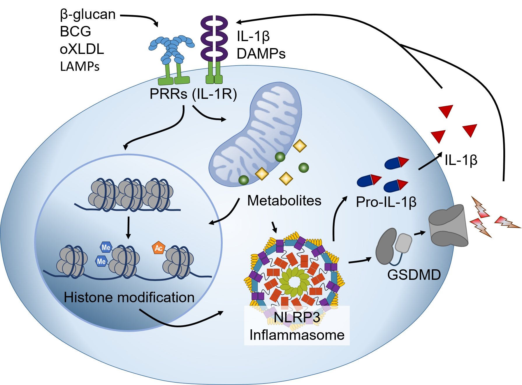
Fig. 1.

Interactions of trained immunity and the NLRP3 inflammasome. The stimuli that trigger trained immunity (e.g., β-glucan, BCG, oXLDL, and LAMPs) activate PRRs, inducing DNA methylation, histone modification, or metabolic changes in innate immune cells. These changes persist for a long time and regulate the transcription of genes related to the inflammatory response. The changes also impact NLRP3 inflammasome activation, ultimately influencing the secretion of proinflammatory cytokines, DAMPs, and LAMPs via gasdermin D. These inflammatory responses, in turn, affect the trained immunity of immune cells through IL-1R and PRRs. Abbreviations: BCG, Bacillus Calmette-Guérin; DAMPs, danger-associated molecular pattern; IL-1R, Interleukin-1 receptor; LAMPs, lifestyle-associated molecular pattern; GSDMD, gasdermin D; oXLDL, oxidized low-density lipoprotein; PRRs, pattern recognition receptors.

Studies on these aspects will help to deepen our understanding of the function and regulation of trained immunity and inflammasomes and to develop new strategies for the prevention and treatment of inflammatory and infectious diseases. Trained immunity and inflammasomes are important components of the innate immune system at the intersection of immunology and metabolism and are worth exploring further.

Author Contributions

GL: Conceptualization, Writing - Original Draft. HA: Conceptualization, Review & Editing. EL: Conceptualization, Review & Editing. GSL: Conceptualization, Writing - Original Draft, Review & Editing, Supervision. All authors have read and approved the final manuscript. All authors have participated sufficiently in the work and agreed to be accountable for all aspects of the work.

Ethics Approval and Consent to Participate

Not applicable.

Acknowledgment

Not applicable.

Funding

This study was supported by a National Research Foundation of Korea (NRF) grant funded by the Korean government (No. RS-2023-00208354 and RS-2023-00244078).

Conflict of Interest

The authors declare no conflict of interest. The Guest Editor, Geun-Shik Lee has not been involved in the peer-review of this article and has no access to information regarding its peer review. Full responsibility for the editorial process for this article was delegated to Amedeo Amedei.

References
[1]
Bonilla FA, Oettgen HC. Adaptive immunity. The Journal of Allergy and Clinical Immunology. 2010; 125: S33–S40.
[2]
Netea MG, Quintin J, van der Meer JWM. Trained immunity: a memory for innate host defense. Cell Host & Microbe. 2011; 9: 355–361.
[3]
Calmette A. Preventive Vaccination Against Tuberculosis with BCG. Proceedings of the Royal Society of Medicine. 1931; 24: 1481–1490.
[4]
Kurtz J, Franz K. Innate defence: evidence for memory in invertebrate immunity. Nature. 2003; 425: 37–38.
[5]
Corona M, Robinson GE. Genes of the antioxidant system of the honey bee: annotation and phylogeny. Insect Molecular Biology. 2006; 15: 687–701.
[6]
Moreno MA, Whitehill JM, Quach V, Midamba N, Manskopf I. Marijuana experiences, voting behaviors, and early perspectives regarding marijuana legalization among college students from 2 states. Journal of American College Health. 2016; 64: 9–18.
[7]
Hu Z, Lu SH, Lowrie DB, Fan XY. Trained immunity: A Yin-Yang balance. MedComm. 2022; 3: e121.
[8]
Divangahi M, Aaby P, Khader SA, Barreiro LB, Bekkering S, Chavakis T, et al. Trained immunity, tolerance, priming and differentiation: distinct immunological processes. Nature Immunology. 2021; 22: 2–6.
[9]
Ahn H, Kwon HM, Lee E, Kim PH, Jeung EB, Lee GS. Role of inflammasome regulation on immune modulators. Journal of Biomedical Research. 2018; 32: 401–410.
[10]
Zindel J, Kubes P. DAMPs, PAMPs, and LAMPs in Immunity and Sterile Inflammation. Annual Review of Pathology. 2020; 15: 493–518.
[11]
Strowig T, Henao-Mejia J, Elinav E, Flavell R. Inflammasomes in health and disease. Nature. 2012; 481: 278–286.
[12]
Netea MG, Domínguez-Andrés J, Barreiro LB, Chavakis T, Divangahi M, Fuchs E, et al. Defining trained immunity and its role in health and disease. Nature Reviews. Immunology. 2020; 20: 375–388.
[13]
Bekkering S, Domínguez-Andrés J, Joosten LAB, Riksen NP, Netea MG. Trained Immunity: Reprogramming Innate Immunity in Health and Disease. Annual Review of Immunology. 2021; 39: 667–693.
[14]
Martinez-Gonzalez I, Mathä L, Steer CA, Ghaedi M, Poon GFT, Takei F. Allergen-Experienced Group 2 Innate Lymphoid Cells Acquire Memory-like Properties and Enhance Allergic Lung Inflammation. Immunity. 2016; 45: 198–208.
[15]
Moorlag SJCFM, Rodriguez-Rosales YA, Gillard J, Fanucchi S, Theunissen K, Novakovic B, et al. BCG Vaccination Induces Long-Term Functional Reprogramming of Human Neutrophils. Cell Reports. 2020; 33: 108387.
[16]
Chen S, Yang J, Wei Y, Wei X. Epigenetic regulation of macrophages: from homeostasis maintenance to host defense. Cellular & Molecular Immunology. 2020; 17: 36–49.
[17]
Verma D, Parasa VR, Raffetseder J, Martis M, Mehta RB, Netea M, et al. Anti-mycobacterial activity correlates with altered DNA methylation pattern in immune cells from BCG-vaccinated subjects. Scientific Reports. 2017; 7: 12305.
[18]
Das J, Verma D, Gustafsson M, Lerm M. Identification of DNA methylation patterns predisposing for an efficient response to BCG vaccination in healthy BCG-naïve subjects. Epigenetics. 2019; 14: 589–601.
[19]
Flores-Concha M, Oñate ÁA. Long Non-coding RNAs in the Regulation of the Immune Response and Trained Immunity. Frontiers in Genetics. 2020; 11: 718.
[20]
Riksen NP, Netea MG. Immunometabolic control of trained immunity. Molecular Aspects of Medicine. 2021; 77: 100897.
[21]
Keating ST, Groh L, Thiem K, Bekkering S, Li Y, Matzaraki V, et al. Rewiring of glucose metabolism defines trained immunity induced by oxidized low-density lipoprotein. Journal of Molecular Medicine. 2020; 98: 819–831.
[22]
Saz-Leal P, Del Fresno C, Brandi P, Martínez-Cano S, Dungan OM, Chisholm JD, et al. Targeting SHIP-1 in Myeloid Cells Enhances Trained Immunity and Boosts Response to Infection. Cell Reports. 2018; 25: 1118–1126.
[23]
Wong CC, Qian Y, Yu J. Interplay between epigenetics and metabolism in oncogenesis: mechanisms and therapeutic approaches. Oncogene. 2017; 36: 3359–3374.
[24]
Dai X, Lv X, Thompson EW, Ostrikov KK. Histone lactylation: epigenetic mark of glycolytic switch. Trends in Genetics. 2022; 38: 124–127.
[25]
Cheng SC, Quintin J, Cramer RA, Shepardson KM, Saeed S, Kumar V, et al. mTOR- and HIF-1α-mediated aerobic glycolysis as metabolic basis for trained immunity. Science. 2014; 345: 1250684.
[26]
Kones R. Rosuvastatin, inflammation, C-reactive protein, JUPITER, and primary prevention of cardiovascular disease–a perspective. Drug Design, Development and Therapy. 2010; 4: 383–413.
[27]
Saeed S, Quintin J, Kerstens HHD, Rao NA, Aghajanirefah A, Matarese F, et al. Epigenetic programming of monocyte-to-macrophage differentiation and trained innate immunity. Science. 2014; 345: 1251086.
[28]
Zarubin T, Han J. Activation and signaling of the p38 MAP kinase pathway. Cell Research. 2005; 15: 11–18.
[29]
Quintin J, Saeed S, Martens JHA, Giamarellos-Bourboulis EJ, Ifrim DC, Logie C, et al. Candida albicans infection affords protection against reinfection via functional reprogramming of monocytes. Cell Host & Microbe. 2012; 12: 223–232.
[30]
Liu Y, Xu R, Gu H, Zhang E, Qu J, Cao W, et al. Metabolic reprogramming in macrophage responses. Biomarker Research. 2021; 9: 1.
[31]
Arts RJW, Novakovic B, Ter Horst R, Carvalho A, Bekkering S, Lachmandas E, et al. Glutaminolysis and Fumarate Accumulation Integrate Immunometabolic and Epigenetic Programs in Trained Immunity. Cell Metabolism. 2016; 24: 807–819.
[32]
Mills EL, Ryan DG, Prag HA, Dikovskaya D, Menon D, Zaslona Z, et al. Itaconate is an anti-inflammatory metabolite that activates Nrf2 via alkylation of KEAP1. Nature. 2018; 556: 113–117.
[33]
Ferreira AV, Netea MG, Domínguez-Andrés J. Itaconate as an immune modulator. Aging. 2019; 11: 3898–3899.
[34]
Mulder WJM, Ochando J, Joosten LAB, Fayad ZA, Netea MG. Therapeutic targeting of trained immunity. Nature Reviews. Drug Discovery. 2019; 18: 553–566.
[35]
Bekkering S, Arts RJW, Novakovic B, Kourtzelis I, van der Heijden CDCC, Li Y, et al. Metabolic Induction of Trained Immunity through the Mevalonate Pathway. Cell. 2018; 172: 135–146.e9.
[36]
Chavakis T, Mitroulis I, Hajishengallis G. Hematopoietic progenitor cells as integrative hubs for adaptation to and fine-tuning of inflammation. Nature Immunology. 2019; 20: 802–811.
[37]
Oguro H. The Roles of Cholesterol and Its Metabolites in Normal and Malignant Hematopoiesis. Frontiers in Endocrinology. 2019; 10: 204.
[38]
Netea MG, Joosten LAB, Latz E, Mills KHG, Natoli G, Stunnenberg HG, et al. Trained immunity: A program of innate immune memory in health and disease. Science. 2016; 352: aaf1098.
[39]
Patel AA, Zhang Y, Fullerton JN, Boelen L, Rongvaux A, Maini AA, et al. The fate and lifespan of human monocyte subsets in steady state and systemic inflammation. The Journal of Experimental Medicine. 2017; 214: 1913–1923.
[40]
Mitroulis I, Ruppova K, Wang B, Chen LS, Grzybek M, Grinenko T, et al. Modulation of Myelopoiesis Progenitors Is an Integral Component of Trained Immunity. Cell. 2018; 172: 147–161.e12.
[41]
Kaufmann E, Sanz J, Dunn JL, Khan N, Mendonça LE, Pacis A, et al. BCG Educates Hematopoietic Stem Cells to Generate Protective Innate Immunity against Tuberculosis. Cell. 2018; 172: 176–190.e19.
[42]
Bono C, Martínez A, Megías J, Gozalbo D, Yáñez A, Gil ML. Dectin-1 Stimulation of Hematopoietic Stem and Progenitor Cells Occurs In Vivo and Promotes Differentiation Toward Trained Macrophages via an Indirect Cell-Autonomous Mechanism. mBio. 2020; 11: e00781-20.
[43]
Dos Santos JC, Barroso de Figueiredo AM, Teodoro Silva MV, Cirovic B, de Bree LCJ, Damen MSMA, et al. β-Glucan-Induced Trained Immunity Protects against Leishmania braziliensis Infection: a Crucial Role for IL-32. Cell Reports. 2019; 28: 2659–2672.e6.
[44]
Christ A, Günther P, Lauterbach MAR, Duewell P, Biswas D, Pelka K, et al. Western Diet Triggers NLRP3-Dependent Innate Immune Reprogramming. Cell. 2018; 172: 162–175.e14.
[45]
de Laval B, Maurizio J, Kandalla PK, Brisou G, Simonnet L, Huber C, et al. C/EBPβ-Dependent Epigenetic Memory Induces Trained Immunity in Hematopoietic Stem Cells. Cell Stem Cell. 2023; 30: 112.
[46]
Cirovic B, de Bree LCJ, Groh L, Blok BA, Chan J, van der Velden WJFM, et al. BCG Vaccination in Humans Elicits Trained Immunity via the Hematopoietic Progenitor Compartment. Cell Host & Microbe. 2020; 28: 322–334.e5.
[47]
Jentho E, Ruiz-Moreno C, Novakovic B, Kourtzelis I, Megchelenbrink WL, Martins R, et al. Trained innate immunity, long-lasting epigenetic modulation, and skewed myelopoiesis by heme. Proceedings of the National Academy of Sciences of the United States of America. 2021; 118: e2102698118.
[48]
Hajishengallis G, Li X, Mitroulis I, Chavakis T. Trained Innate Immunity and Its Implications for Mucosal Immunity and Inflammation. Advances in Experimental Medicine and Biology. 2019; 1197: 11–26.
[49]
Yu Q, Guo M, Zeng W, Zeng M, Zhang X, Zhang Y, et al. Interactions between NLRP3 inflammasome and glycolysis in macrophages: New insights into chronic inflammation pathogenesis. Immunity, Inflammation and Disease. 2022; 10: e581.
[50]
Renaudin F, Orliaguet L, Castelli F, Fenaille F, Prignon A, Alzaid F, et al. Gout and pseudo-gout-related crystals promote GLUT1-mediated glycolysis that governs NLRP3 and interleukin-1β activation on macrophages. Annals of the Rheumatic Diseases. 2020; 79: 1506–1514.
[51]
Alatshan A, Kovács GE, Aladdin A, Czimmerer Z, Tar K, Benkő S. All-Trans Retinoic Acid Enhances both the Signaling for Priming and the Glycolysis for Activation of NLRP3 Inflammasome in Human Macrophage. Cells. 2020; 9: 1591.
[52]
Hackett EE, Charles-Messance H, O’Leary SM, Gleeson LE, Muñoz-Wolf N, Case S, et al. Mycobacterium tuberculosis Limits Host Glycolysis and IL-1β by Restriction of PFK-M via MicroRNA-21. Cell Reports. 2020; 30: 124–136.e4.
[53]
Mills EL, Kelly B, Logan A, Costa ASH, Varma M, Bryant CE, et al. Succinate Dehydrogenase Supports Metabolic Repurposing of Mitochondria to Drive Inflammatory Macrophages. Cell. 2016; 167: 457–470.e13.
[54]
Sanman LE, Qian Y, Eisele NA, Ng TM, van der Linden WA, Monack DM, et al. Disruption of glycolytic flux is a signal for inflammasome signaling and pyroptotic cell death. eLife. 2016; 5: e13663.
[55]
Chung IC, Chen LC, Tsang NM, Chuang WY, Liao TC, Yuan SN, et al. Mitochondrial Oxidative Phosphorylation Complex Regulates NLRP3 Inflammasome Activation and Predicts Patient Survival in Nasopharyngeal Carcinoma. Molecular & Cellular Proteomics. 2020; 19: 142–154.
[56]
Bae S, Lesch BJ. H3K4me1 Distribution Predicts Transcription State and Poising at Promoters. Frontiers in Cell and Developmental Biology. 2020; 8: 289.
[57]
Piatek P, Tarkowski M, Namiecinska M, Riva A, Wieczorek M, Michlewska S, et al. H3K4me3 Histone ChIP-Seq Analysis Reveals Molecular Mechanisms Responsible for Neutrophil Dysfunction in HIV-Infected Individuals. Frontiers in Immunology. 2021; 12: 682094.
[58]
Peterson JM, Wang DJ, Shettigar V, Roof SR, Canan BD, Bakkar N, et al. NF-κB inhibition rescues cardiac function by remodeling calcium genes in a Duchenne muscular dystrophy model. Nature Communications. 2018; 9: 3431.
[59]
Brown JD, Lin CY, Duan Q, Griffin G, Federation A, Paranal RM, et al. NF-κB directs dynamic super enhancer formation in inflammation and atherogenesis. Molecular Cell. 2014; 56: 219–231.
[60]
Pelinski Y, Hidaoui D, Stolz A, Hermetet F, Chelbi R, Diop MK, et al. NF-κB signaling controls H3K9me3 levels at intronic LINE-1 and hematopoietic stem cell genes in cis. The Journal of Experimental Medicine. 2022; 219: e20211356.
[61]
Methot SP, Padeken J, Brancati G, Zeller P, Delaney CE, Gaidatzis D, et al. H3K9me selectively blocks transcription factor activity and ensures differentiated tissue integrity. Nature Cell Biology. 2021; 23: 1163–1175.
[62]
Nakamura M, Batista RA, Köhler C, Hennig L. Polycomb Repressive Complex 2-mediated histone modification H3K27me3 is associated with embryogenic potential in Norway spruce. Journal of Experimental Botany. 2020; 71: 6366–6378.
[63]
Liu H, Xing R, Ou Z, Zhao J, Hong G, Zhao TJ, et al. G-protein-coupled receptor GPR17 inhibits glioma development by increasing polycomb repressive complex 1-mediated ROS production. Cell Death & Disease. 2021; 12: 610.
[64]
Hou J, Feng HQ, Chang HW, Liu Y, Li GH, Yang S, et al. The H3K4 demethylase Jar1 orchestrates ROS production and expression of pathogenesis-related genes to facilitate Botrytis cinerea virulence. The New Phytologist. 2020; 225: 930–947.
[65]
Lu C, Yang D, Klement JD, Colson YL, Oberlies NH, Pearce CJ, et al. H3K9me3 represses G6PD expression to suppress the pentose phosphate pathway and ROS production to promote human mesothelioma growth. Oncogene. 2022; 41: 2651–2662.
[66]
Cai Y, Zhang Y, Loh YP, Tng JQ, Lim MC, Cao Z, et al. H3K27me3-rich genomic regions can function as silencers to repress gene expression via chromatin interactions. Nature Communications. 2021; 12: 719.
[67]
Zhang Y, Qian M, Tang F, Huang Q, Wang W, Li Y, et al. Identification and Analysis of p53-Regulated Enhancers in Hepatic Carcinoma. Frontiers in Bioengineering and Biotechnology. 2020; 8: 668.
[68]
Xu A, Liu M, Huang MF, Zhang Y, Hu R, Gingold JA, et al. Rewired m6A epitranscriptomic networks link mutant p53 to neoplastic transformation. Nature Communications. 2023; 14: 1694.
[69]
Yuan J, Zhu Q, Zhang X, Wen Z, Zhang G, Li N, et al. Ezh2 competes with p53 to license lncRNA Neat1 transcription for inflammasome activation. Cell Death and Differentiation. 2022; 29: 2009–2023.
[70]
Yang Y, Wang H, Kouadir M, Song H, Shi F. Recent advances in the mechanisms of NLRP3 inflammasome activation and its inhibitors. Cell Death & Disease. 2019; 10: 128.
[71]
Wang J, Sun Y, Zhang X, Cai H, Zhang C, Qu H, et al. Oxidative stress activates NORAD expression by H3K27ac and promotes oxaliplatin resistance in gastric cancer by enhancing autophagy flux via targeting the miR-433-3p. Cell Death & Disease. 2021; 12: 90.
[72]
Cui C, Li T, Xie Y, Yang J, Fu C, Qiu Y, et al. Enhancing Acsl4 in absence of mTORC2/Rictor drove β-cell dedifferentiation via inhibiting FoxO1 and promoting ROS production. Biochimica et Biophysica Acta. Molecular Basis of Disease. 2021; 1867: 166261.
[73]
Wang H, Fan Z, Shliaha PV, Miele M, Hendrickson RC, Jiang X, et al. H3K4me3 regulates RNA polymerase II promoter-proximal pause-release. Nature. 2023; 615: 339–348.
[74]
Sungalee S, Liu Y, Lambuta RA, Katanayeva N, Donaldson Collier M, Tavernari D, et al. Histone acetylation dynamics modulates chromatin conformation and allele-specific interactions at oncogenic loci. Nature Genetics. 2021; 53: 650–662.
[75]
Rahnamoun H, Hong J, Sun Z, Lee J, Lu H, Lauberth SM. Mutant p53 regulates enhancer-associated H3K4 monomethylation through interactions with the methyltransferase MLL4. The Journal of Biological Chemistry. 2018; 293: 13234–13246.
[76]
Yamashita G, Takano N, Kazama H, Tsukahara K, Miyazawa K. p53 regulates lysosomal membrane permeabilization as well as cytoprotective autophagy in response to DNA-damaging drugs. Cell Death Discovery. 2022; 8: 502.
[77]
Zhong Z, Liang S, Sanchez-Lopez E, He F, Shalapour S, Lin XJ, et al. New mitochondrial DNA synthesis enables NLRP3 inflammasome activation. Nature. 2018; 560: 198–203.
[78]
Bordon Y. mtDNA synthesis ignites the inflammasome. Nature Reviews. Immunology. 2018; 18: 539.
[79]
Beacon TH, Delcuve GP, López C, Nardocci G, Kovalchuk I, van Wijnen AJ, et al. The dynamic broad epigenetic (H3K4me3, H3K27ac) domain as a mark of essential genes. Clinical Epigenetics. 2021; 13: 138.
[80]
Kol S, Ben-Shlomo I, Ruutiainen K, Ando M, Davies-Hill TM, Rohan RM, et al. The midcycle increase in ovarian glucose uptake is associated with enhanced expression of glucose transporter 3. Possible role for interleukin-1, a putative intermediary in the ovulatory process. The Journal of Clinical Investigation. 1997; 99: 2274–2283.
[81]
Tan Q, Huang Q, Ma YL, Mao K, Yang G, Luo P, et al. Potential roles of IL-1 subfamily members in glycolysis in disease. Cytokine & Growth Factor Reviews. 2018; 44: 18–27.
[82]
Tan Q, Duan L, Huang Q, Chen W, Yang Z, Chen J, et al. Interleukin -1β Promotes Lung Adenocarcinoma Growth and Invasion Through Promoting Glycolysis via p38 Pathway. Journal of Inflammation Research. 2021; 14: 6491–6509.
[83]
Finucane OM, Sugrue J, Rubio-Araiz A, Guillot-Sestier MV, Lynch MA. The NLRP3 inflammasome modulates glycolysis by increasing PFKFB3 in an IL-1β-dependent manner in macrophages. Scientific Reports. 2019; 9: 4034.
[84]
Xie M, Yu Y, Kang R, Zhu S, Yang L, Zeng L, et al. PKM2-dependent glycolysis promotes NLRP3 and AIM2 inflammasome activation. Nature Communications. 2016; 7: 13280.
[85]
Lin HC, Chen YJ, Wei YH, Lin HA, Chen CC, Liu TF, et al. Lactic Acid Fermentation Is Required for NLRP3 Inflammasome Activation. Frontiers in Immunology. 2021; 12: 630380.
[86]
Groh LA, Ferreira AV, Helder L, van der Heijden CDCC, Novakovic B, van de Westerlo E, et al. oxLDL-Induced Trained Immunity Is Dependent on Mitochondrial Metabolic Reprogramming. Immunometabolism. 2021; 3: e210025.
[87]
Zhou H, Wang H, Yu M, Schugar RC, Qian W, Tang F, et al. IL-1 induces mitochondrial translocation of IRAK2 to suppress oxidative metabolism in adipocytes. Nature Immunology. 2020; 21: 1219–1231.
[88]
Batista AF, Rody T, Forny-Germano L, Cerdeiro S, Bellio M, Ferreira ST, et al. Interleukin-1β mediates alterations in mitochondrial fusion/fission proteins and memory impairment induced by amyloid-β oligomers. Journal of Neuroinflammation. 2021; 18: 54.
[89]
Li R, Ong SL, Tran LM, Jing Z, Liu B, Park SJ, et al. Chronic IL-1β-induced inflammation regulates epithelial-to-mesenchymal transition memory phenotypes via epigenetic modifications in non-small cell lung cancer. Scientific Reports. 2020; 10: 377.
[90]
Liu T, Zhang L, Joo D, Sun SC. NF-κB signaling in inflammation. Signal Transduction and Targeted Therapy. 2017; 2: 17023.
[91]
Bhatt D, Ghosh S. Regulation of the NF-κB-Mediated Transcription of Inflammatory Genes. Frontiers in Immunology. 2014; 5: 71.
[92]
Bellavia D, Costa V, De Luca A, Cordaro A, Fini M, Giavaresi G, et al. The Binomial “Inflammation-Epigenetics” in Breast Cancer Progression and Bone Metastasis: IL-1β Actions Are Influenced by TET Inhibitor in MCF-7 Cell Line. International Journal of Molecular Sciences. 2022; 23: 15422.
[93]
Riera MF, Galardo MN, Pellizzari EH, Meroni SB, Cigorraga SB. Participation of phosphatidyl inositol 3-kinase/protein kinase B and ERK1/2 pathways in interleukin-1beta stimulation of lactate production in Sertoli cells. Reproduction. 2007; 133: 763–773.
[94]
O’Neill LAJ, Netea MG. BCG-induced trained immunity: can it offer protection against COVID-19? Nature Reviews. Immunology. 2020; 20: 335–337.
[95]
Zhou F, Yu T, Du R, Fan G, Liu Y, Liu Z, et al. Clinical course and risk factors for mortality of adult inpatients with COVID-19 in Wuhan, China: a retrospective cohort study. Lancet. 2020; 395: 1054–1062.
[96]
Mangan MSJ, Olhava EJ, Roush WR, Seidel HM, Glick GD, Latz E. Targeting the NLRP3 inflammasome in inflammatory diseases. Nature Reviews. Drug Discovery. 2018; 17: 588–606.
[97]
Sharma BR, Kanneganti TD. NLRP3 inflammasome in cancer and metabolic diseases. Nature Immunology. 2021; 22: 550–559.
[98]
Anand PK, Malireddi RKS, Kanneganti TD. Role of the nlrp3 inflammasome in microbial infection. Frontiers in Microbiology. 2011; 2: 12.
[99]
Vora SM, Lieberman J, Wu H. Inflammasome activation at the crux of severe COVID-19. Nature Reviews. Immunology. 2021; 21: 694–703.
[100]
Zhao C, Zhao W. NLRP3 Inflammasome-A Key Player in Antiviral Responses. Frontiers in Immunology. 2020; 11: 211.
[101]
Frediani B, Falsetti P, Storri L, Bisogno S, Baldi F, Campanella V, et al. Evidence for synovitis in active polymyalgia rheumatica: sonographic study in a large series of patients. The Journal of Rheumatology. 2002; 29: 123–130.
[102]
Mangan MSJ, Olhava EJ, Roush WR, Seidel HM, Glick GD, Latz E. Targeting the NLRP3 inflammasome in inflammatory diseases. Nature Reviews. Drug Discovery. 2018; 17: 688.
[103]
Municio C, Criado G. Therapies Targeting Trained Immune Cells in Inflammatory and Autoimmune Diseases. Frontiers in Immunology. 2021; 11: 631743.

Publisher’s Note: IMR Press stays neutral with regard to jurisdictional claims in published maps and institutional affiliations.

Share
Back to top