IMR Press / FBL / Volume 28 / Issue 9 / DOI: 10.31083/j.fbl2809207
Open Access Review
The Roles and Targeting of Tumor-Associated Macrophages
Show Less
1 Shenzhen Bay Laboratory, 518132 Shenzhen, Guangdong, China
*Correspondence: gaoqq@szbl.ac.cn (Qianqian Gao)
Front. Biosci. (Landmark Ed) 2023, 28(9), 207; https://doi.org/10.31083/j.fbl2809207
Submitted: 27 February 2023 | Revised: 28 April 2023 | Accepted: 9 May 2023 | Published: 15 September 2023
Copyright: © 2023 The Author(s). Published by IMR Press.
This is an open access article under the CC BY 4.0 license.
Abstract

Tumor-associated macrophages (TAMs) are the most abundant infiltrating immune cells in the tumor microenvironment (TME) and play an important role in tumor progression. Clinically, the increase of TAMs infiltration is linked to poor prognosis of patients with various cancer types. Multiple studies have demonstrated that reducing or reprogramming TAMs can inhibit the occurrence or development of tumors. Therefore, TAMs have been identified as novel targets for the treatment of cancer therapy. In this review, the origin, polarization, roles, and targeting of TAMs in malignancies, are discussed.

Keywords
tumor-associated macrophages
tumor microenvironment
macrophage roles
macrophage targeting
cancer therapy
1. Introduction

The morbidity and mortality of malignant tumors are rising worldwide, threatening human life and health and becoming a leading cause of death [1]. Growing evidence suggests that the tumor microenvironment (TME) has a major role in determining tumor progress, in addition to the aggressive biological behavior of tumor cells [2]. The TME, which is made up of blood vessels, immune cells, fibroblasts, inflammatory cells generated from bone marrow, different signaling chemicals and extracellular matrix, is a complex microenvironment that surrounds the tumor site [3]. Immunosuppressive TME promotes the proliferation and metastasis of tumor cells [4]. Macrophages are the most abundant immune cells in the TME and play a central regulatory role. Macrophages infiltrating TME are defined as tumor-associated macrophages (TAMs), accounting for approximately 30–50% of total immune cell counts [5]. TAMs have been proven to be related to the occurrence, development, angiogenesis, and metastasis of tumors [6], suggesting that TAMs could be a potential therapeutic target and prognostic biomarker for tumors.

Current antitumor strategies targeting TAMs include inhibiting the recruitment of macrophages, promoting TAMs depletion, regulating its polarization, and enhancing TAM phagocytosis. Targeting TAMs has become one of the main anti-tumor therapeutic strategies. In this review, we attempt to discuss the origins, polarization, roles, and reprogramming of TAMs, as well as the therapeutic implications of targeting TAMs in malignancies.

2. The Origins of TAMs

There has been controversy over the exact origins of TAMs. However, with the development of modern lineage-tracing techniques, our understanding of the origins of TAMs has been improved significantly. It is currently believed that TAMs are mainly composed of two parts, namely, tissue-resident macrophages (TRMs) and bone marrow-derived macrophages (BMDMs) (Fig. 1). TRMs originate from the embryonic precursors of yolk sac or fetal liver and persist in specific organs, such as Langerhans cells in the skin, Kupffer cells in the liver, alveolar macrophages, and microglia in the brain [7]. In the early stage of tumorigenesis, resident macrophages from surrounding tissues constitute the initial TAMs inside the tumor, forming a microenvironment conducive to tumor growth. As the tumor grows, various chemokines secreted by stromal and tumor cells in the inflammatory state of the TME induce the recruitment of a large number of BMDMs to form the typical TAMs. Although various types of macrophages coexist in tumors, BMDMs make up the majority of TAMs and its role in different types and stages of tumors needs further study [8].
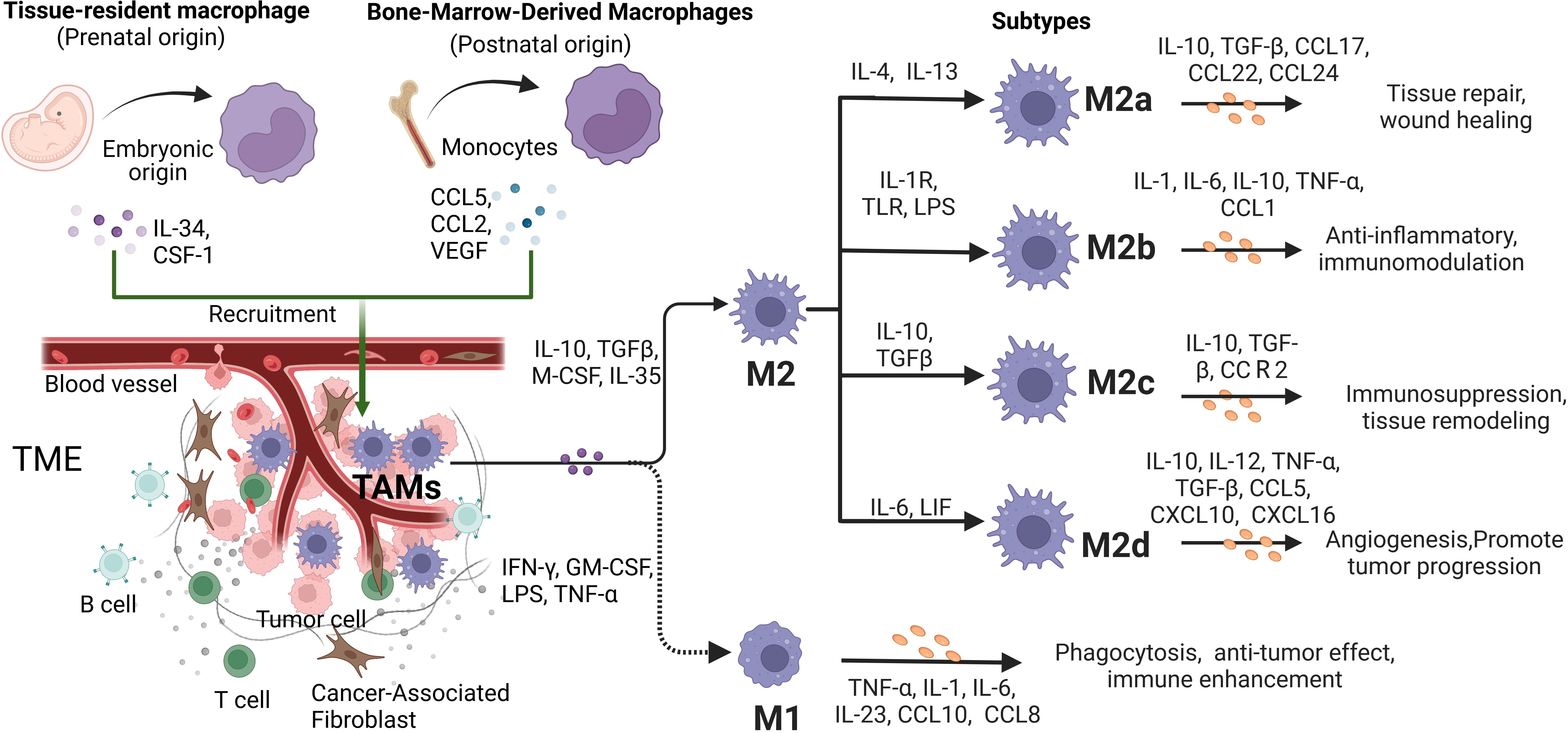
Fig. 1.

The origins, differentiation and function characteristics of Tumor-associated macrophages (TAMs). TAMs can be developed from embryonic-derived tissue-specific resident macrophages and bone marrow-derived macrophages (BMDMs). As the ratio of cytokine types and concentrations in tumor microenvironment (TME) changes, the activation phenotype of TAMs changes dynamically to play different functional roles. In conventional binary model, macrophages polarize into two subtypes, M1-type and M2-type. In response to stimulation by different factors, M2-type TAMs further polarize to four phenotypes, including M2a, M2b, M2c and M2d. They secrete various factors that have significant impacts on the tumor progression.

3. The Categories of TAMs

TAMs are usually divided into the classically activated type 1 (M1-type) macrophages, and the alternatively activated type 2 (M2-type) macrophages according to their secreted products and functions. M1-type TAMs are primarily responsible for tumor killing and inhibition, while M2-type TAMs are involved in tumor incidence, development and metastasis [9]. Moreover, M1-type TAMs can be induced and activated by interferon-γ (IFN-γ), granulocyte-macrophage colony-stimulating factor (GM-CSF), lipopolysaccharide (LPS), tumor necrosis factor-α (TNF-α) and other proinflammatory factors, and release anti-tumor cytokines such as interleukin (IL)-1, IL-6 and IL-23, which can promote the inflammatory response and antigen presentation ability. M2-type TAMs, on the other hand, are primarily activated by cytokines such as IL-4, IL-13, and transforming growth factor β (TGF-β), which promote the expression of arginase-1 (Arg-1), mannose receptor 1 (CD206) and scavenger receptor cysteine-rich type 1 protein M130 (CD163). M2 TAMs secret anti-inflammatory cytokines, including IL-10, chemokine (C-C motif) ligand (CCL) 17, CCL18, CCL22 and promote Th2 cell differentiation and angiogenesis, thus facilitating tumor progress [10] (Fig. 1).

M2-type TAMs can be further divided into types M2a, M2b, M2c, and M2d. Among them, M2a-type TAMs are related to type II inflammation, that is, the Th2 reaction accompanied by the release of IL-4 and IL-13 and an allergic reaction. M2b-type TAMs are involved in Th2 activation and immunomodulation through immune complexes and toll-like receptor ligands. M2c-type TAMs induce immunosuppressive tissue repair through IL-10. M2d-type TAMs are activated by IL-6, which promotes tumor growth through angiogenesis [11] (Fig. 1).

M1-type TAMs exhibit a pro-inflammatory and anti-tumor phenotype, which is characterized by the expression and production of high levels of pro-inflammatory cytokines. M2-type TAMs take parts in promoting tumor progression and suppressing immunity by expressing mannose receptors, CD206, CD163, and arginase, while also producing IL-10 and TGF-β [12]. The metabolism of TAMs encompasses lipid metabolism, purine metabolism, amino acid metabolism, oxidative phosphorylation metabolism and glycolysis. TAMs with purine metabolism, amino acid metabolism and glycolysis expressed high levels of pro-angiogenesis genes [13]. Glycolysis was up-regulated in M1-type TAMs, while fatty acid oxidation and oxidative phosphorylation metabolism were enhanced in M2-like TAMs [14].

TAMs have remarkable heterogeneity and plasticity, and they change dynamically under the stimulation of different signals in a specific microenvironment. TAMs can be polarized from M1-type to M2-type [15]. At the initial stage of tumor progression, M1-type is the dominant phenotype. Tumor cells or CD4+ T cells, however, release cytokines such as IL-4, CSF-1, TGF-β, and Arg-1, which induce the transformation of M1-type TAMs into M2-type phenotype gradually and enhance tumor growth and angiogenesis.

Human and mouse TAMs can be identified by cell surface markers, including CD11b, CD86, CD11c and chemokine (C-C motif) receptor (CCR) 5. As for human M1-type TAMs, CD14, CD80 and CD68 are used as markers, while M2-type TAMs express CD11b, CD206, IL-4Ra and CD14. In mice, CD11b, CD86, F4/80, CCR5 and major histocompatibility complex class II (MHC-II) are used to isolate M1-type TAMs, while M2-type TAMs can be distinguished by CD11b, CD206, F4/80, IL-4Rα, CCR-5 and MHC-Ⅱ [16].

4. The Antigen-Presentation Role of TAMs

TAMs are the largest number of antigen-presenting cells in the TME. M1-type TAMs enhance antigen presentation by increasing MHC-II and costimulatory molecules CD80 and CD86 on the cell surface, while M2-type TAMs decrease T-cell antigen presentation. M1 TAMs phagocytize tumor antigen, and the most characteristic antigen peptides are retained after lysosomal decomposition. The peptides are then combined with MHC-II and presented to T lymphocytes in the form of antigen peptide-MCH-II molecular complex on the surface of M1-type TAMs. When the complex is recognized by T cell receptor (TCR), T lymphocytes are activated [17]. Exhaustion of T cells produced a large number of myeloid-related factors, which recruited monocytes and induced them to differentiate into antigen-presenting TAMs. TAMs continued to stimulate TCR through antigen presentation, thus aggravating the exhaustion of T cells [18].

5. The Roles of TAMs in Tumor Progression

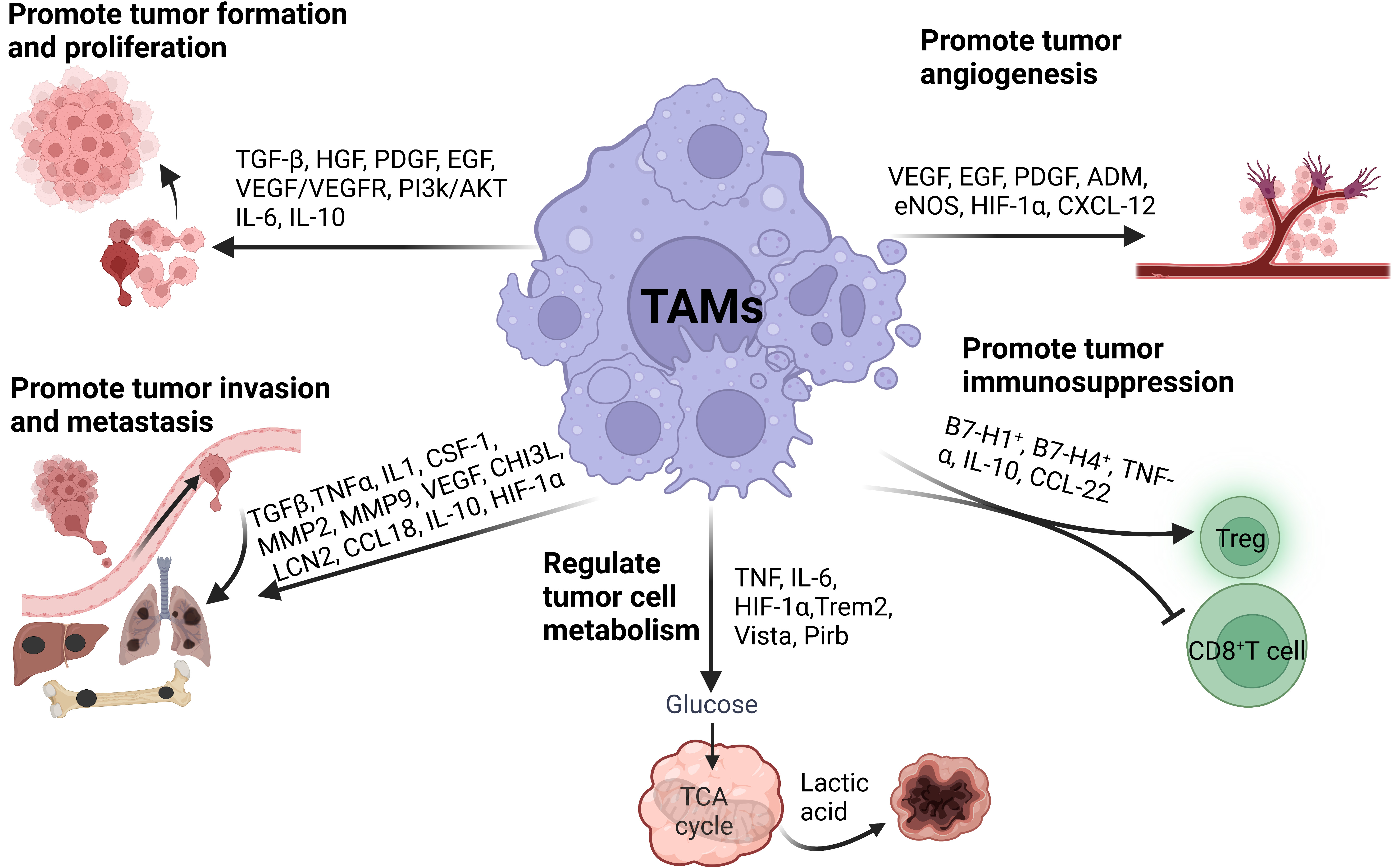
After infiltrating into tumor tissue, macrophages develop a new phenotype, aiding in tumor occurrence and development. TAMs promote tumor cell proliferation, invasion and migration, angiogenesis and immunosuppression and regulate tumor cell metabolism through complex autocrine and paracrine pathways (Fig. 2).

Fig. 2.

The roles of TAMs in tumor progression. TAMs facilitate tumor progression by promoting tumor cell proliferation, invasion, migration, angiogenesis, immunosuppression, and metabolism.

5.1 Promote Tumor Formation and Proliferation

A large number of animal experiments and clinical studies have proved that all stages of tumorigenesis are affected by tumor-related inflammation. TAMs release a large number of reactive oxygen species and reactive nitrogen mediators, which lead to DNA damage and genome instability and increase the frequency of gene mutation. Accumulatively, the DNA repair function becomes abnormal, thus forming cells with the potential to develop into tumors. If the damage continues, it will further expand the cloning of cells with tumor potential, increase genomic instability and cell atypia, and finally form tumor cells, thus starting tumor occurrence. It was reported that the pro-inflammatory effect of M1-type TAMs increased the genomic instability of malignant tumor cells and became the driving force of tumorigenesis [19].

TAMs are closely related to tumor growth. TAMs stimulate tumor cell proliferation by interacting with them and secreting cytokines such as epidermal growth factor (EGF), platelet-derived growth factor (PDGF), TGF-β, hepatocyte growth factor (HGF), epidermal growth factor receptor (EGFR) and basic fibroblast growth factor (bFGF) (Fig. 2). In the mouse model of ovarian cancer, TAMs secreted a large amount of EGF to activate EGFR in surrounding tumor cells, up-regulate the VEGF/vascular endothelial growth factor (VEGFR) signaling pathway, and promote the proliferation and migration of tumor cells [20]. In hepatocellular carcinoma, TAMs promoted the stem cell-like characteristics of tumor cells through high TGF-β expression [21]. In the mouse model of bladder cancer, type I collagen protein produced by TAMs activated the PI3K/AKT signaling pathway through integrin 21, which promoted tumor proliferation. In addition, co-culture of TAMs and esophageal squamous cell carcinoma promoted the high expression of annexin A10 (ANXA) 10 in tumor cells and then phosphorylated the AKT/ERK pathway to promote tumor cell proliferation [22].

5.2 Promote Tumor Invasion and Metastasis

Tumor cells usually leave the primary site and migrate to other sites. The ability of tumor cells to spread distantly is dependent on the TME. As the main component, TAMs play an important role in tumor metastasis. TAMs promote the invasion and migration of tumor cells by secreting TNF-α, TGF-β, IL1, CSF-1, and metalloproteinases (MMPs) (e.g., MMP-2 and MMP-9) to change the connection between cells and destroy the basement membrane (Fig. 2). For instance, TAMs triggered local and systemic expression of MMP-9, VEGF, chitinase-3-like protein 1 (CHI3L1), and Lipocalin-2 (LCN2) to promote tumor metastasis in a mouse model of triple-negative breast cancer [23]. In another study, TAMs secreted large amounts of CCL18 and activated the NF-κB/miR-21/PTEN/AKT axis in myofibroblasts through its receptor PIPTNM3, further promoting the invasion and metastasis of malignant breast tumors [24]. In one mouse lung cancer model, hypoxia increased the generation of M2 macrophages and simultaneously the expression of IL-10, VEGF, and hypoxia-inducible factor-1α (HIF-1α), which further promoted cancer cell metastasis and recruited more macrophages to infiltrate the primary tumor tissue [25]. Moreover, MMP-2 and MMP-9 secreted by TAMs degraded the matrix and promoted gastric and colon cancer cell invasion [26].

TAMs induce epithelial-mesenchymal transition (EMT) in the TME. Breast cancer cells activated macrophages to transform into TAMs through GM-CSF, and TAMs secreted CCL18 to induce EMT and promote tumor metastasis [27]. Experiments in mice in vitro and in vivo showed that TAMs induced EMT in colon cancers by regulating STAT3/miR-506-3p/FoxQ1 axis, and then promoted the production of CCL2 to recruit more TAMs, which significantly increased the rate of tumor invasion and metastasis [28]. Ovarian cancer cells induced macrophages to polarize into M2-type TAMs by secreting macrophage colony-stimulating factor (M-CSF). M2 TAMs induced EMT by releasing CCL18, which further increased M-CSF transcription in ovarian cancer cells through ZEB1 protein. Thus, the CCL18-ZEB1-M-CSF interaction loop between ovarian cancer cells and TAMs increased tumor metastasis [29].

TRMs are also involved in the progression of different tumors and have organ specificity. At the early stages of NSCLCs, TRMs gathered nearby tumor cells and promoted tumor invasion and Treg cell reaction. TRMs promoted Treg cell differentiation by up-regulating the expression of CD73 and cytotoxic T-lymphocyte-associated protein 4 (CTLA-4) in Treg cells, and protected tumor cells from being killed by CD8+ T cells. These results show that TRMs provide a perfect microenvironment for tumor progression [8]. In one developmental model of rat liver cancer induced by diethylmitrosamine, TNF-α secreted by Kupffer cells (KCs) in the liver triggered the chromosomal instability of liver prognosticator cells (LPCs) through the disorder of ubiquitin D and checkpoint kinase 2. The self-renewal of LPCs was enhanced, which promoted the transformation of LPCs into liver cancer stem cells, thus facilitating the development of liver cancer [30]. The anti-inflammatory factors macrophage galactose-type C-type lectin 2 (Mgl2), IL-10, Mgl1 and Arg1 in KCs were found increased significantly in the liver cancer model constructed by injecting AKT/Ras plasmid. KCs cells were polarized to M2-type and led to the occurrence of liver cancer. Interestingly, miR-206 in KCs cells reversed the polarization of KCs from M2-type to M1-type, and activated CCL2/CCR2 signal to recruit CD8+ T cells for an anti-tumor effect [31].

Microglia, the resident macrophages in the central nervous system, played a similar role in promoting the progress of glioblastoma. When microglia and glioma cells were co-cultured in vitro, microglia lost phagocytosis activity and simultaneously secreted factors such as MMP-9, EGF, IL-6 and VEGF to enhance the invasion of glioblastoma [32].

In one mouse pancreatic cancer model, TRMs were amplified in the tumor tissue. Depletion of pancreatic resident macrophages led to a significant reduction in the tumor load and showed a transcription profile of cancer fibrosis [33].

5.3 Promote Tumor Angiogenesis

The foundation of tumor angiogenesis is endothelial cells in the TME, where new blood vessels serve as the primary route for tumor invasion and metastasis as well as supplying nutrients and oxygen for tumor growth. The density of TAMs and tumor blood vascularity are highly correlated in malignancies. TAMs promote the growth of tumor microvessels and lymphatic vessels by secreting VEGF and EGF to accelerate the proliferation of tumor cells (Fig. 2). IL-17 expressed by TAMs promoted angiogenesis in laryngeal squamous cell carcinoma [34]. In the co-culture system of TAMs and melanoma cells, TAMs promoted angiogenesis and melanoma growth by secreting adrenomedullin (ADM), which stimulated the endothelial nitric oxide synthase (eNOS) signal pathway [35]. Under hypoxic conditions, TAMs expressed the transcription factor HIF-1, which induced the transcription of VEGF, PDGF, and EGF and promoted angiogenesis. Furthermore, TAMs secreted MAPK-activated protein kinase-2 in a mice colon cancer model to increase the expression of chemokine (C-X-C motif) ligand 12(CXCL-12) in the tumor, thus promoting tumor progression and angiogenesis [36].

5.4 Promote Immunosuppression

TAMs promote tumor progression through immunosuppression. TAMs are the main immunoregulatory cells in tumors and participate in the suppression of cytotoxic T lymphocytes (CTLs) responses in the TME (Fig. 2). Effective antitumor immunity mainly depends on the activation of CD8+ T cells. However, TAMs employ various mechanisms to directly or indirectly inactivate CTLs, contributing to tumor immune evasion and development [37]. TAMs with high expression of B7-H1 and B7-H4 inhibited the proliferation of CD4+ T cells and the secretion of IFN-γ, and promoted the immune escape and metastasis of gastric cancer cells [38]. The increased TGF-β secreted by TAMs in lung cancer caused CD4+ and CD8+ T cell dysfunction, allowing the tumor to evade immune surveillance [39]. Moreover, TNF-α and IL-10 secreted by TAMs increased PD-L1 expression in monocytes. The interaction of PD-L1 expressed by monocytes and PD-1 receptor expressed on activated T cells led to tumor immune escape. Monocytes expressing PD-L1 significantly inhibited tumor-specific T cell proliferation, cytokine production, and cytotoxic potential in vitro. Moreover, PD-L1+ monocytes inhibited tumor-specific immunity in vivo and fostered tumor growth in NOD/SCID mice bearing human tumors [40].

5.5 Regulate Tumor Cell Metabolism

TAMs promote tumor growth by regulating tumor cell metabolism and increasing the aerobic glycolysis (Fig. 2). During tumor growth, the TME presents different degrees of hypoxia. Hypoxia inhibited the uptake of glucose by TAMs, leading to an increase in the content of glucose in the TME, which further increased the utilization of glucose by tumor cells, and ultimately promoted tumor angiogenesis and metastasis [41]. TAMs with tumor-inducing metabolic features, including purine metabolism, amino acid (AA) metabolism, and glycolysis, expressed high levels of pro-angiogenic genes. During the transition from lipid metabolism to purine metabolism, macrophages gradually lost the expression of genes related to antigen presentation, while gained the expression of genes related to angiogenesis and immunosuppression, including Trem2, v-domain Ig suppressor of T cell activation (Vista) and paired immunoglobulin like receptor b (Pirb) [13]. In patients with medullary thyroid carcinoma, lactic acid produced by tumor cells caused the glucose metabolism mode of TAMs to change from oxidative phosphorylation to glycolysis, which led TAMs to secrete more lactic acid, TNF, and IL-6 to further promote tumor development [42].

In TME, the metabolism of TAMs is always in a dynamic process, which plays an important role in tumor development and anti-tumor immune response. Nutrients such as glucose, lipids and amino acids are absorbed by TAMs and catabolized or converted into biosynthetic intermediates or signal metabolites to regulate TAMs function. M1-type TAMs had a high level of aerobic glycolysis activity, which produced reactive oxygen species to kill pathogens. M2-type TAMs relied on oxidative phosphorylation and produced IL-10 and VEGF to promote the growth of malignant tumor cells [43]. In another study, M2-type TAMs expressed high-level glucose transporter and had stronger glucose-uptake capacity than M1-like TAMs. Glucose uptake promoted O-GlcN protein acylation in M2-type TAMs, which further promoted tumor metastasis [44].

6. TAMs Targeting Strategies in Cancer Therapies

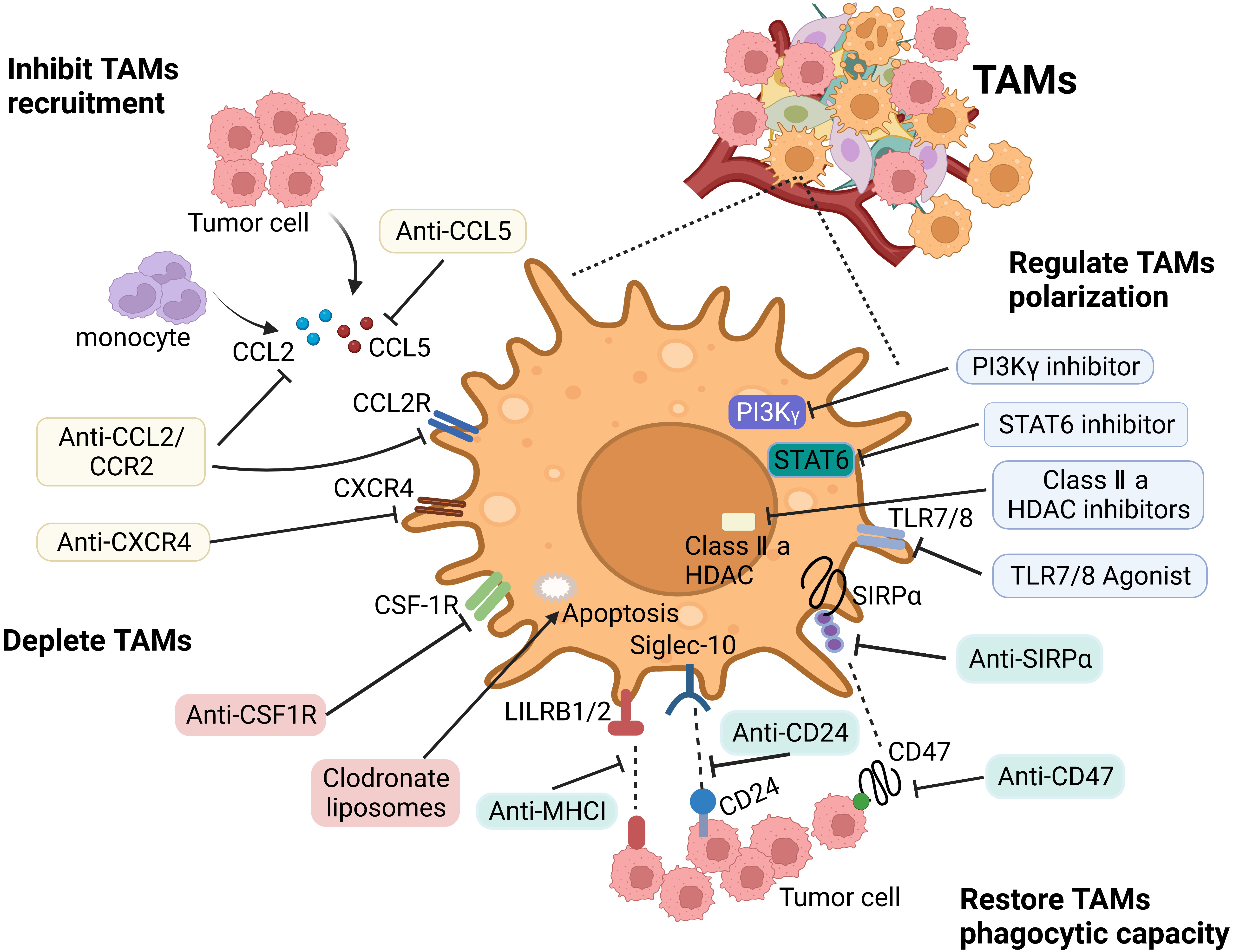
TAMs play an important role in the occurrence and development of tumors. Due to their remarkable heterogeneity and plasticity, the current treatment strategies for TAMs mainly include inhibiting their recruitment, depleting or reprogramming TAMs, and improving their phagocytic ability (Fig. 3).

Fig. 3.

Main therapeutic strategies to target TAMs. The therapeutic strategies targeting TAMs include: (1) Inhibiting macrophages recruitment via targeting CCL2/CCR2 axis and CXCL12/CXCR4 pathway. (2) Depleting TAMs by blocking CSF-1/CSF-1R axis, or inducing their apoptosis by drugs, such as trabectedin and bisphosphonates. (3) Repolarizing macrophages by anti-CD47 antibodies, CD40 agonists, PI3Kγ inhibitors and HDACs inhibitors. (4) Restoring macrophage phagocytic activity by blocking the signaling pathways CD47/SIRPa, CD24/Siglec-10, and LILRB/MHCI.

6.1 Inhibit TAMs Recruitment

In TME, various chemokines such as CCL2, CCL5, and CSF-1 secreted by tumor cells and stromal cells recruit monocytes circulating in the blood to the tumor area to differentiate into TAMs. Blocking the relevant signaling pathways with small molecule inhibitors or specific antibodies can inhibit the recruitment of TAMs.

In the liver cancer xenotransplantation mice model, the CCR2 antagonist RDC018 blocked the CCL2/CCR2 axis, and inhibited the recruitment of inflammatory monocytes and the infiltration and M2-type polarization of TAMs. The blockade resulted in the reversal of the immunosuppressive status of the TME and the activation of an antitumor CD8+ T cell response [45]. A phase II clinical trial of pancreatic ductal adenocarcinoma (PDAC) showed that the CCR2 inhibitor PF-04136309 effectively blocked the CCL2-CCR2 pathway, and its combined treatment with FOLFIRINOX (leucovorin + fluorouracil + irinotecan + oxaliplatin) reduced the number of CCR2+ monocytes in the tumor [46]. The monoclonal antibody Leronlimab, and the small-molecule inhibitors maraviroc and vicriviroc, blocked the CCL5/CCR5 signal axis and inhibited the recruitment of TAMs and tumor growth [47, 48]. A single-arm IIa trial in patients with metastatic PDAC was conducted with the CXCR4 inhibitor BL-8040, the PD-1 antagonist pembrolizumab, and NAPOLI-1. It was found that the disease control rate was 77% and the average effective duration was 7.8 months, which brought hope for PDAC treatment [49].

6.2 Deplete TAMs

TAMs depletion can reduce the density of TAMs in the tumor tissue and the immunosuppressive TME. CSF-1 is necessary for the maturation, differentiation, and survival of the mononuclear phagocytes, which express the CSF-1 receptor (CSF-1R) exclusively. TAMs were massively apoptotic when the CSF-1/CSF-1R axis was blocked [50]. Given its importance in macrophages, many clinical drugs targeting CSF-1R have been developed, such as BLZ945, PLX3397, PLX7486 and PLX7486. In a mouse model of PDAC, CSF-1 neutralizing antibodies not only reduced the number of T cells but also reprogramed the remaining T cells to enhance antigen presentation and anti-tumor responses. The combination of CSF-1 neutralizing antibodies and CTLA-4 antagonists resulted in tumor regression [51]. The monoclonal antibody (RG-7155), which inhibits CSF-1R activation, induced macrophage apoptosis in vitro, and significantly reduced TAMs density and increased the ratio of CD8+/CD4+ T cells in the mouse model to activate anti-tumor immunity [52].

Another strategy to promote the depletion of TAMs is the application of bisphosphonates, including zoledronic acid and clodronate. Both in vitro and in vivo studies have shown that bisphosphonate treatment induced apoptosis in macrophages. In mouse PDAC model, clodronate liposomes significantly reduced the density of TAMs and enhanced the anti-tumor effect of CD8+ T lymphocytes to inhibit tumor growth [53]. In addition, trabectedin exerted an anti-tumor immune effect by mediating the depletion of TAMs in an ovarian cancer model [54].

6.3 Regulate TAMs Polarization

Given the plasticity of macrophages, reprogramming TAMs to an antitumor phenotype is a highly desirable cancer treatment strategy. In addition to inducing TAMs to differentiate into M1-type, M2-type TAMs also have the potential to be repolarized into M1-type. Recent research has shown that the cyclodextrin nanoparticles (CDNP-R848) loaded with R848, an agonist of Toll-like receptors TLR7 and TLR8, delivered drugs to TAMs efficiently in vivo. Application of CDNP-R848 in multiple mouse tumor models polarized TAMs towards the M1-phenotype and inhibited tumor growth. Furthermore, the combination of CDNP-R848 and anti-PD-1 antibodies had a synergistic effect and led to tumor shrinkage [55]. In a xenograft mice model of breast cancer, the nanomedicine of STAT6 inhibitor AS1517499 and inhibitor of kappa B kinase (IKK) siRNA effectively induced the polarization of TAMs from M2-type to M1-type, reshaped the TME and significantly inhibited tumor growth [56]. Inhibition of PI3k-γ pathway and targeting CSF-1R in M2-type TAMs upregulated the secretion of pro-inflammatory cytokines IFN-γ and IL-12, while reduced the inhibitory cytokines IL-10 and IL-6, thereby promoting TAMs transformation from M2-type to M1-type and inhibiting tumor growth [57]. Another strategy is to induce the polarization of TAMs at the epigenetic level. The histone deacetylase inhibitor TMP195 changed the epigenetic characteristics of TAMs and made them differentiate into M1-type, which induced TAMs accumulation in breast cancer TME, enhanced their phagocytic activity, and reduced tumor burden and lung metastases [58]. Agonists for stimulator of interferon genes (STING) reprogrammed M2-type TAMs into M1-type, and overcame the immunosuppression and resistance to PARP inhibition in BRCA1 deficient breast cancer [59]. Knock-down of STING and STING activator promoted both human PBMC-derived macrophages and mouse BMDMs into M1-like subtype, and induced apoptosis of gastric cancer cells through activation of IL-6R-JAK-STAT pathway and its downstream target IL24 [60].

6.4 Block Checkpoint Molecules in TAMs

Checkpoint molecules, including PD-1, PD-L1, Tim-3 and Tim-4 are expressed in TAMs, which inhibit TAMs phagocytosis and promote tumor immune escape. Recent studies showed that PD-L1 antibody therapy increased the proliferation, survival and size of TAMs from mouse bone marrow. Moreover, PD-L1 antibody therapy increased the phosphorylation level of Akt and mTOR, thus inducing TAMs activation by up-regulating the expression of MHC-II and CD86. TAMs produced more TNFα and IL-12, which induced the polarization of TAMs to M1-type. The in vivo study results showed that the tumor growth was significantly inhibited, and the number and activation of TAMs, as well as the tumor-infiltrating T cells, were increased. Moreover, the proliferation of TAMs and its MHC-II expression were increased in PD-L1-/- mice compared with wild-type mice [61].

Another study showed that almost all PD-1+ TAMs were M2-type. Upregulation of PD-1 expression in M2-type TAMs decreased its phagocytic ability. After transplantation of PD-L1-overexpressed and PD-L1-knockedout colon cancer CT26 cells in mice, it was found that PD-L1 knockout significantly increased the phagocytosis of PD-1+ TAMs and reduced the tumor growth. Human colon cancer DLD-1 cells expressing PD-L1 but unable to express mouse PD-1 were xenografted into NSG mice. The treatment with anti-mouse PD-1 or PD-L1 antibody significantly reduced mouse tumor growth, while it had no effect on tumor growth after TAMs depletion. The results suggested that the anti-mouse PD-1 antibody triggered the anti-tumor effect by binding with PD-1 on macrophages rather than on tumors [62].

Tim-3 knocked-down macrophages and hepatocellular carcinoma H22 cells were injected subcutaneously into mice. Compared with control macrophages, Tim-3 knocked-down macrophages significantly inhibited H22 tumor growth. Mechanism study found that the inhibition of Tim-3 significantly reduced the phosphorylation level of STAT-6 in macrophages, and impaired the polarization of macrophages to M2-type, thus hindering tumor growth [63]. Tim-4+ macrophages in pleural cavity or abdominal cavity damaged the proliferation of CD8+ T cells. Antibody blockade and gene elimination of Tim-4 improved the therapeutic effect of PD-1 antibody on mice bearing MC38 colon cancer [64].

6.5 Restore TAMs Phagocytic Capacity

Increasing the phagocytic ability of TAMs is another important anti-tumor strategy, which can directly kill tumor cells. At present, three main pathways inhibit the phagocytosis of TAMs: the signal regulatory protein α (SIRPα)/CD47 pathway, the major histocompatibility complex class I/leukocyte immunoglobulin (Ig)-like receptor subfamily B member 1 (MHC-1/LILRB1) pathway, and the CD24/sialic acid-binding Ig-like lectin 10 (Siglec-10) pathways. CD47 is a membrane protein that suppresses anti-tumor immunity by inhibiting phagocytosis and participates in cell proliferation, migration, apoptosis, and immune homeostasis. The CD47 major ligand SIRP, is a membrane protein mainly expressed on macrophages and myeloid cells. The N-terminus of its extracellular domain binds to CD47, resulting in tyrosine phosphorylation on immune receptor tyrosine inhibitory motifs (ITIMs) and the release of the “don’t eat me” signal, thereby inhibiting the phagocytosis of macrophages and protecting normal cells from the destruction by the immune system [65]. Anti-CD47 antibodies (e.g., Hu5F9-G4, SRF231, and IBI188) and anti-SIRP antibodies blocked the CD47/SIRP signaling pathway and enhanced the phagocytosis of TAMs to tumor cells. A phase 1b study in patients with Non-Hodgkin Lymphoma showed that Hu5F9-G4 and rituximab synergistically enhanced the phagocytosis of TAMs to inhibit Non-Hodgkin Lymphoma [66]. Anti-SIRP monoclonal antibodies blocked the recognition of the ligand CD47 by SIRP, thus enhancing the phagocytosis of TAMs [67].

The high expression of MHC-I in tumor cells inhibited the phagocytosis of TAMs. MHC-I is coupled with two members of the leukocyte immunoglobulin-like receptor (LILR) family, LILRB1 and LILRB2, and contains ITIMs to help inhibit intracellular signal transduction. Blockade of the MHC-1/LILRB1 pathway in the tumor that exhibited drug resistance after blocking the SIRP/CD47 axis enhanced the phagocytosis of MHC-I+ tumor cells by TAMs in vitro and in vivo [68].

CD24 is a highly glycosylated surface protein that interacts with Siglec-10. Tumor cells highly express CD24, while TAMs highly express Siglec-10. After binding to CD24, the ITIM of Siglec-10 recruited and activated the SH2-domain-containing tyrosine phosphatase SHP-1 or SHP-2, thereby inhibiting the phagocytosis of TAMs [69]. Knocking out the CD24/Siglec-10 gene or blocking the CD24/Siglec-10 axis with monoclonal antibodies increased the ability of TAMs to phagocytize tumor cells and reduced tumor growth [70]. Recent studies showed that both mouse and human TAMs expressed PD-1, which was related to the decrease of phagocytosis. The expression level of PD-1 in M2-type TAMs was significantly higher than that in M1-type TAMs. In vivo blocking of PD-1/PD-L1 signaling increased macrophage phagocytosis, reduced tumor growth, and prolonged mouse survival time in a macrophage-dependent manner [63] (Fig. 3).

7. Conclusions

The interaction between tumor cells and TAMs is very complicated. TAMs in different activation states in the TME have different functions. They are a two-edged sword in that they can act as M1-type TAMs, recognize tumor antigens, and phagocytize or kill tumor cells, while they can also be domesticated by TME into M2-type to stimulate tumor development. Which aspect plays the dominant role depends on the activation state of macrophages, the stage of the tumor, and the influences of the TME. Targeting M2-type TAMs is a promising strategy for cancer therapy. Given many targets of TAMs still have not been discovered, or suitable targeted drugs have not yet been developed, it will be necessary to further study the molecular mechanism of the interaction between tumor cells and TAMs to find more effective new targets and drugs.

Abbreviations

M-CSF, Macrophage colony-stimulating factor; TGF-β, Transforming growth factor β; CSF-1, Colony stimulating factor 1; VEGF, Vascular endothelial growth factor; IL-1R, interleukin-1 receptors; TLR, Toll-like receptors; LPS, Lipopolysaccharides; LIF, leukemia inhibitory factor isoform 1 precursor; IL, interleukin-34; TNF-α, Tumor necrosis factor-α; CCL, Chemokine (C-C motif) ligand; CCR2, chemokine (C-C motif) receptor 2; CXCL, Chemokine (C-X-C motif) ligand; HGF, Hepatocyte growth factor; PDGF, Platelet-derived growth factor; EGF, epidermal growth factor; VEGF, Vascular endothelial growth factor; VEGFR, Vascular endothelial growth factor receptors; PI3K, Phosphoinositide 3 kinase; AKT, protein kinase B; MMP, Metalloproteinases; CHI3L, Chitinase-3-like protein; LCN2, Lipocalin-2; HIF-1α, hypoxia inducible factor-1α; ADM, Adrenomedullin; eNOS, Enos Endothelial nitric oxide synthase; CXCL-12, Chemokine (C-X-C motif) ligand 12; B7-H1+, Recombinant Human Programmed Cell Death 1 Ligand 1; B7-H4+, V-set domain-containing T-cell activation inhibitor 1; Trem2, triggering receptor expressed on myeloid cells 2; Vista, V-type immunoglobulin domain-containing suppressor of T cell activation; Pirb, paired immunoglobulin-like receptor b; LILRB1/2, Leukocyte immunoglobulin-like receptor subfamily B member 1/2; SIRPa, signal-regulatory protein alpha; Siglec-10, sialic acid-binding Ig-like lectin 10; MHCI, Major histocompatibility complex I; TLR7/8, Toll-like receptors 7/8; STAT6, Signal transducer and activator of transcription 6; PI3Kγ, Phosphoinositide 3 kinase γ.

Author Contributions

QG, YL and RW designed the study. YL wrote the manuscript. RW provided help with the writing. QG reviewed and revised the manuscript. All authors contributed to editorial changes in the manuscript. All authors read and approved the final manuscript. All authors have participated sufficiently in the work and agreed to be accountable for all aspects of the work.

Ethics Approval and Consent to Participate

Not applicable.

Acknowledgment

Not applicable.

Funding

This research received no external funding.

Conflict of Interest

The authors declare no conflict of interest.

References
[1]
Sung H, Ferlay J, Siegel RL, Laversanne M, Soerjomataram I, Jemal A, et al. Global Cancer Statistics 2020: GLOBOCAN Estimates of Incidence and Mortality Worldwide for 36 Cancers in 185 Countries. CA: A Cancer Journal for Clinicians. 2021; 71: 209–249.
[2]
Bejarano L, Jordāo MJC, Joyce JA. Therapeutic Targeting of the Tumor Microenvironment. Cancer Discovery. 2021; 11: 933–959.
[3]
Hui L, Chen Y. Tumor microenvironment: Sanctuary of the devil. Cancer Letters. 2015; 368: 7–13.
[4]
Liu Q, Zhang H, Jiang X, Qian C, Liu Z, Luo D. Factors involved in cancer metastasis: a better understanding to “seed and soil” hypothesis. Molecular Cancer. 2017; 16: 176.
[5]
Mantovani A, Allavena P, Marchesi F, Garlanda C. Macrophages as tools and targets in cancer therapy. Nature Reviews. Drug Discovery. 2022; 21: 799–820.
[6]
Gao J, Liang Y, Wang L. Shaping Polarization Of Tumor-Associated Macrophages In Cancer Immunotherapy. Frontiers in Immunology. 2022; 13: 888713.
[7]
Christofides A, Strauss L, Yeo A, Cao C, Charest A, Boussiotis VA. The complex role of tumor-infiltrating macrophages. Nature Immunology. 2022; 23: 1148–1156.
[8]
Casanova-Acebes M, Dalla E, Leader AM, LeBerichel J, Nikolic J, Morales BM, et al. Tissue-resident macrophages provide a pro-tumorigenic niche to early NSCLC cells. Nature. 2021; 595: 578–584.
[9]
Mills CD, Kincaid K, Alt JM, Heilman MJ, Hill AM. M-1/M-2 macrophages and the Th1/Th2 paradigm. Journal of Immunology (Baltimore, Md.: 1950). 2000; 164: 6166–6173.
[10]
Fleetwood AJ, Lawrence T, Hamilton JA, Cook AD. Granulocyte-macrophage colony-stimulating factor (CSF) and macrophage CSF-dependent macrophage phenotypes display differences in cytokine profiles and transcription factor activities: implications for CSF blockade in inflammation. Journal of Immunology (Baltimore, Md.: 1950). 2007; 178: 5245–5252.
[11]
Murray PJ, Allen JE, Biswas SK, Fisher EA, Gilroy DW, Goerdt S, et al. Macrophage activation and polarization: nomenclature and experimental guidelines. Immunity. 2014; 41: 14–20.
[12]
Anders CB, Lawton TMW, Smith HL, Garret J, Doucette MM, Ammons MCB. Use of integrated metabolomics, transcriptomics, and signal protein profile to characterize the effector function and associated metabotype of polarized macrophage phenotypes. Journal of Leukocyte Biology. 2022; 111: 667–693.
[13]
Li S, Yu J, Huber A, Kryczek I, Wang Z, Jiang L, et al. Metabolism drives macrophage heterogeneity in the tumor microenvironment. Cell Reports. 2022; 39: 110609.
[14]
Wang F, Zhang S, Vuckovic I, Jeon R, Lerman A, Folmes CD, et al. Glycolytic Stimulation Is Not a Requirement for M2 Macrophage Differentiation. Cell Metabolism. 2018; 28: 463–475.e4.
[15]
Yunna C, Mengru H, Lei W, Weidong C. Macrophage M1/M2 polarization. European Journal of Pharmacology. 2020; 877: 173090.
[16]
Wculek SK, Dunphy G, Heras-Murillo I, Mastrangelo A, Sancho D. Metabolism of tissue macrophages in homeostasis and pathology. Cellular & Molecular Immunology. 2022; 19: 384–408.
[17]
Farooque A, Afrin F, Adhikari JS, Dwarakanath BSR. Polarization of macrophages towards M1 phenotype by a combination of 2-deoxy-d-glucose and radiation: Implications for tumor therapy. Immunobiology. 2016; 221: 269–281.
[18]
Kersten K, Hu KH, Combes AJ, Samad B, Harwin T, Ray A, et al. Spatiotemporal co-dependency between macrophages and exhausted CD8+ T cells in cancer. Cancer Cell. 2022; 40: 624–638.e9.
[19]
Elinav E, Nowarski R, Thaiss CA, Hu B, Jin C, Flavell RA. Inflammation-induced cancer: crosstalk between tumours, immune cells and microorganisms. Nature Reviews. Cancer. 2013; 13: 759–771.
[20]
Yin M, Li X, Tan S, Zhou HJ, Ji W, Bellone S, et al. Tumor-associated macrophages drive spheroid formation during early transcoelomic metastasis of ovarian cancer. The Journal of Clinical Investigation. 2016; 126: 4157–4173.
[21]
Fan QM, Jing YY, Yu GF, Kou XR, Ye F, Gao L, et al. Tumor-associated macrophages promote cancer stem cell-like properties via transforming growth factor-beta1-induced epithelial-mesenchymal transition in hepatocellular carcinoma. Cancer Letters. 2014; 352: 160–168.
[22]
Kodaira H, Koma YI, Hosono M, Higashino N, Suemune K, Nishio M, et al. ANXA10 induction by interaction with tumor-associated macrophages promotes the growth of esophageal squamous cell carcinoma. Pathology International. 2019; 69: 135–147.
[23]
Steenbrugge J, Breyne K, Demeyere K, De Wever O, Sanders NN, Van Den Broeck W, et al. Anti-inflammatory signaling by mammary tumor cells mediates prometastatic macrophage polarization in an innovative intraductal mouse model for triple-negative breast cancer. Journal of Experimental & Clinical Cancer Research: CR. 2018; 37: 191.
[24]
Nie Y, Chen J, Huang D, Yao Y, Chen J, Ding L, et al. Tumor-Associated Macrophages Promote Malignant Progression of Breast Phyllodes Tumors by Inducing Myofibroblast Differentiation. Cancer Research. 2017; 77: 3605–3618.
[25]
Zhang J, Cao J, Ma S, Dong R, Meng W, Ying M, et al. Tumor hypoxia enhances Non-Small Cell Lung Cancer metastasis by selectively promoting macrophage M2 polarization through the activation of ERK signaling. Oncotarget. 2014; 5: 9664–9677.
[26]
Cardoso AP, Pinto ML, Pinto AT, Pinto MT, Monteiro C, Oliveira MI, et al. Matrix metalloproteases as maestros for the dual role of LPS- and IL-10-stimulated macrophages in cancer cell behaviour. BMC Cancer. 2015; 15: 456.
[27]
Su S, Liu Q, Chen J, Chen J, Chen F, He C, et al. A positive feedback loop between mesenchymal-like cancer cells and macrophages is essential to breast cancer metastasis. Cancer Cell. 2014; 25: 605–620.
[28]
Wei C, Yang C, Wang S, Shi D, Zhang C, Lin X, et al. Crosstalk between cancer cells and tumor associated macrophages is required for mesenchymal circulating tumor cell-mediated colorectal cancer metastasis. Molecular Cancer. 2019; 18: 64.
[29]
Long L, Hu Y, Long T, Lu X, Tuo Y, Li Y, et al. Tumor-associated macrophages induced spheroid formation by CCL18-ZEB1-M-CSF feedback loop to promote transcoelomic metastasis of ovarian cancer. Journal for Immunotherapy of Cancer. 2021; 9: e003973.
[30]
Li XF, Chen C, Xiang DM, Qu L, Sun W, Lu XY, et al. Chronic inflammation-elicited liver progenitor cell conversion to liver cancer stem cell with clinical significance. Hepatology (Baltimore, Md.). 2017; 66: 1934–1951.
[31]
Liu N, Wang X, Steer CJ, Song G. MicroRNA-206 promotes the recruitment of CD8+ T cells by driving M1 polarisation of Kupffer cells. Gut. 2022; 71: 1642–1655.
[32]
Coniglio SJ, Eugenin E, Dobrenis K, Stanley ER, West BL, Symons MH, et al. Microglial stimulation of glioblastoma invasion involves epidermal growth factor receptor (EGFR) and colony stimulating factor 1 receptor (CSF-1R) signaling. Molecular Medicine (Cambridge, Mass.). 2012; 18: 519–527.
[33]
Zhu Y, Herndon JM, Sojka DK, Kim KW, Knolhoff BL, Zuo C, et al. Tissue-Resident Macrophages in Pancreatic Ductal Adenocarcinoma Originate from Embryonic Hematopoiesis and Promote Tumor Progression. Immunity. 2017; 47: 323–338.e6.
[34]
Meng CD, Zhu DD, Jiang XD, Li L, Sha JC, Dong Z, et al. Overexpression of interleukin-l7 in tumor-associated macrophages is correlated with the differentiation and angiogenesis of laryngeal squamous cell carcinoma. Chinese Medical Journal. 2012; 125: 1603–1607.
[35]
Chen P, Huang Y, Bong R, Ding Y, Song N, Wang X, et al. Tumor-associated macrophages promote angiogenesis and melanoma growth via adrenomedullin in a paracrine and autocrine manner. Clinical Cancer Research: An Official Journal of the American Association for Cancer Research. 2011; 17: 7230–7239.
[36]
Suarez-Lopez L, Kong YW, Sriram G, Patterson JC, Rosenberg S, Morandell S, et al. MAPKAP Kinase-2 Drives Expression of Angiogenic Factors by Tumor-Associated Macrophages in a Model of Inflammation-Induced Colon Cancer. Frontiers in Immunology. 2021; 11: 607891.
[37]
Cheng N, Bai X, Shu Y, Ahmad O, Shen P. Targeting tumor-associated macrophages as an antitumor strategy. Biochemical Pharmacology. 2021; 183: 114354.
[38]
Matsunaga T, Saito H, Ikeguchi M. Increased B7-H1 and B7-H4 Expressions on Circulating Monocytes and Tumor-Associated Macrophages are Involved in Immune Evasion in Patients with Gastric Cancer. Yonago Acta Medica. 2011; 54: 1–10.
[39]
Li L, Yang L, Wang L, Wang F, Zhang Z, Li J, et al. Impaired T cell function in malignant pleural effusion is caused by TGF-β derived predominantly from macrophages. International Journal of Cancer. 2016; 139: 2261–2269.
[40]
Kuang DM, Zhao Q, Peng C, Xu J, Zhang JP, Wu C, et al. Activated monocytes in peritumoral stroma of hepatocellular carcinoma foster immune privilege and disease progression through PD-L1. The Journal of Experimental Medicine. 2009; 206: 1327–1337.
[41]
Wenes M, Shang M, Di Matteo M, Goveia J, Martín-Pérez R, Serneels J, et al. Macrophage Metabolism Controls Tumor Blood Vessel Morphogenesis and Metastasis. Cell Metabolism. 2016; 24: 701–715.
[42]
Arts RJW, Plantinga TS, Tuit S, Ulas T, Heinhuis B, Tesselaar M, et al. Transcriptional and metabolic reprogramming induce an inflammatory phenotype in non-medullary thyroid carcinoma-induced macrophages. Oncoimmunology. 2016; 5: e1229725.
[43]
Bulle A, Dekervel J, Deschuttere L, Nittner D, Libbrecht L, Janky R, et al. Gemcitabine Recruits M2-Type Tumor-Associated Macrophages into the Stroma of Pancreatic Cancer. Translational Oncology. 2020; 13: 100743.
[44]
Shi Q, Shen Q, Liu Y, Shi Y, Huang W, Wang X, et al. Increased glucose metabolism in TAMs fuels O-GlcNAcylation of lysosomal Cathepsin B to promote cancer metastasis and chemoresistance. Cancer Cell. 2022; 40: 1207–1222.e10.
[45]
Li X, Yao W, Yuan Y, Chen P, Li B, Li J, et al. Targeting of tumour-infiltrating macrophages via CCL2/CCR2 signalling as a therapeutic strategy against hepatocellular carcinoma. Gut. 2017; 66: 157–167.
[46]
Nywening TM, Wang-Gillam A, Sanford DE, Belt BA, Panni RZ, Cusworth BM, et al. Targeting tumour-associated macrophages with CCR2 inhibition in combination with FOLFIRINOX in patients with borderline resectable and locally advanced pancreatic cancer: a single-centre, open-label, dose-finding, non-randomised, phase 1b trial. The Lancet. Oncology. 2016; 17: 651–662.
[47]
Jiao X, Nawab O, Patel T, Kossenkov AV, Halama N, Jaeger D, et al. Recent Advances Targeting CCR5 for Cancer and Its Role in Immuno-Oncology. Cancer Research. 2019; 79: 4801–4807.
[48]
Pervaiz A, Zepp M, Mahmood S, Ali DM, Berger MR, Adwan H. CCR5 blockage by maraviroc: a potential therapeutic option for metastatic breast cancer. Cellular Oncology (Dordrecht). 2019; 42: 93–106.
[49]
Gong R, Ren H. Targeting chemokines/chemokine receptors: a promising strategy for enhancing the immunotherapy of pancreatic ductal adenocarcinoma. Signal Transduction and Targeted Therapy. 2020; 5: 149.
[50]
Hamilton JA. Colony-stimulating factors in inflammation and autoimmunity. Nature Reviews. Immunology. 2008; 8: 533–544.
[51]
Zhu Y, Knolhoff BL, Meyer MA, Nywening TM, West BL, Luo J, et al. CSF1/CSF1R blockade reprograms tumor-infiltrating macrophages and improves response to T-cell checkpoint immunotherapy in pancreatic cancer models. Cancer Research. 2014; 74: 5057–5069.
[52]
Ries CH, Cannarile MA, Hoves S, Benz J, Wartha K, Runza V, et al. Targeting tumor-associated macrophages with anti-CSF-1R antibody reveals a strategy for cancer therapy. Cancer Cell. 2014; 25: 846–859.
[53]
Yang X, Lin J, Wang G, Xu D. Targeting Proliferating Tumor-Infiltrating Macrophages Facilitates Spatial Redistribution of CD8+ T Cells in Pancreatic Cancer. Cancers. 2022; 14: 1474.
[54]
Germano G, Frapolli R, Belgiovine C, Anselmo A, Pesce S, Liguori M, et al. Role of macrophage targeting in the antitumor activity of trabectedin. Cancer Cell. 2013; 23: 249–262.
[55]
Rodell CB, Arlauckas SP, Cuccarese MF, Garris CS, Li R, Ahmed MS, et al. TLR7/8-agonist-loaded nanoparticles promote the polarization of tumour-associated macrophages to enhance cancer immunotherapy. Nature Biomedical Engineering. 2018; 2: 578–588.
[56]
Xiao H, Guo Y, Li B, Li X, Wang Y, Han S, et al. M2-Like Tumor-Associated Macrophage-Targeted Codelivery of STAT6 Inhibitor and IKKβ siRNA Induces M2-to-M1 Repolarization for Cancer Immunotherapy with Low Immune Side Effects. ACS Central Science. 2020; 6: 1208–1222.
[57]
Li M, Li M, Yang Y, Liu Y, Xie H, Yu Q, et al. Remodeling tumor immune microenvironment via targeted blockade of PI3K-γ and CSF-1/CSF-1R pathways in tumor associated macrophages for pancreatic cancer therapy. Journal of Controlled Release: Official Journal of the Controlled Release Society. 2020; 321: 23–35.
[58]
Guerriero JL, Sotayo A, Ponichtera HE, Castrillon JA, Pourzia AL, Schad S, et al. Class IIa HDAC inhibition reduces breast tumours and metastases through anti-tumour macrophages. Nature. 2017; 543: 428–432.
[59]
Wang Q, Bergholz JS, Ding L, Lin Z, Kabraji SK, Hughes ME, et al. STING agonism reprograms tumor-associated macrophages and overcomes resistance to PARP inhibition in BRCA1-deficient models of breast cancer. Nature Communications. 2022; 13: 3022.
[60]
Miao L, Qi J, Zhao Q, Wu QN, Wei DL, Wei XL, et al. Targeting the STING pathway in tumor-associated macrophages regulates innate immune sensing of gastric cancer cells. Theranostics. 2020; 10: 498–515.
[61]
Hartley GP, Chow L, Ammons DT, Wheat WH, Dow SW. Programmed Cell Death Ligand 1 (PD-L1) Signaling Regulates Macrophage Proliferation and Activation. Cancer Immunology Research. 2018; 6: 1260–1273.
[62]
Gordon SR, Maute RL, Dulken BW, Hutter G, George BM, McCracken MN, et al. PD-1 expression by tumour-associated macrophages inhibits phagocytosis and tumour immunity. Nature. 2017; 545: 495–499.
[63]
Yan W, Liu X, Ma H, Zhang H, Song X, Gao L, et al. Tim-3 fosters HCC development by enhancing TGF-β-mediated alternative activation of macrophages. Gut. 2015; 64: 1593–1604.
[64]
Chow A, Schad S, Green MD, Hellmann MD, Allaj V, Ceglia N, et al. Tim-4+ cavity-resident macrophages impair anti-tumor CD8+ T cell immunity. Cancer Cell. 2021; 39: 973–988.e9.
[65]
Chao MP, Weissman IL, Majeti R. The CD47-SIRPα pathway in cancer immune evasion and potential therapeutic implications. Current Opinion in Immunology. 2012; 24: 225–232.
[66]
Advani R, Flinn I, Popplewell L, Forero A, Bartlett NL, Ghosh N, et al. CD47 Blockade by Hu5F9-G4 and Rituximab in Non-Hodgkin’s Lymphoma. The New England Journal of Medicine. 2018; 379: 1711–1721.
[67]
Alvey CM, Spinler KR, Irianto J, Pfeifer CR, Hayes B, Xia Y, et al. SIRPA-Inhibited, Marrow-Derived Macrophages Engorge, Accumulate, and Differentiate in Antibody-Targeted Regression of Solid Tumors. Current Biology: CB. 2017; 27: 2065–2077.e6.
[68]
Barkal AA, Weiskopf K, Kao KS, Gordon SR, Rosental B, Yiu YY, et al. Engagement of MHC class I by the inhibitory receptor LILRB1 suppresses macrophages and is a target of cancer immunotherapy. Nature Immunology. 2018; 19: 76–84.
[69]
Panagiotou E, Syrigos NK, Charpidou A, Kotteas E, Vathiotis IA. CD24: A Novel Target for Cancer Immunotherapy. Journal of Personalized Medicine. 2022; 12: 1235.
[70]
Barkal AA, Brewer RE, Markovic M, Kowarsky M, Barkal SA, Zaro BW, et al. CD24 signalling through macrophage Siglec-10 is a target for cancer immunotherapy. Nature. 2019; 572: 392–396.

Publisher’s Note: IMR Press stays neutral with regard to jurisdictional claims in published maps and institutional affiliations.

Share
Back to top