


 , Lisa Barnes 1, Melanie Riggs 1, Helen Knaggs 1, Zoe Diana Draelos 2
1 Center for Anti-Aging Research, Global Product Research and Development, Nu Skin Enterprises, Provo, UT 84601-4432, USA
2 Dermatology Consulting Services, PLLC, High Point, NC 27265-8501, USA
Abstract
Background: Humans are exposed to physical, biological, chemical, and
psychological stressor throughout their life span. In recent years many medicinal
plants have been shown to induce stress adapting and protective functions.
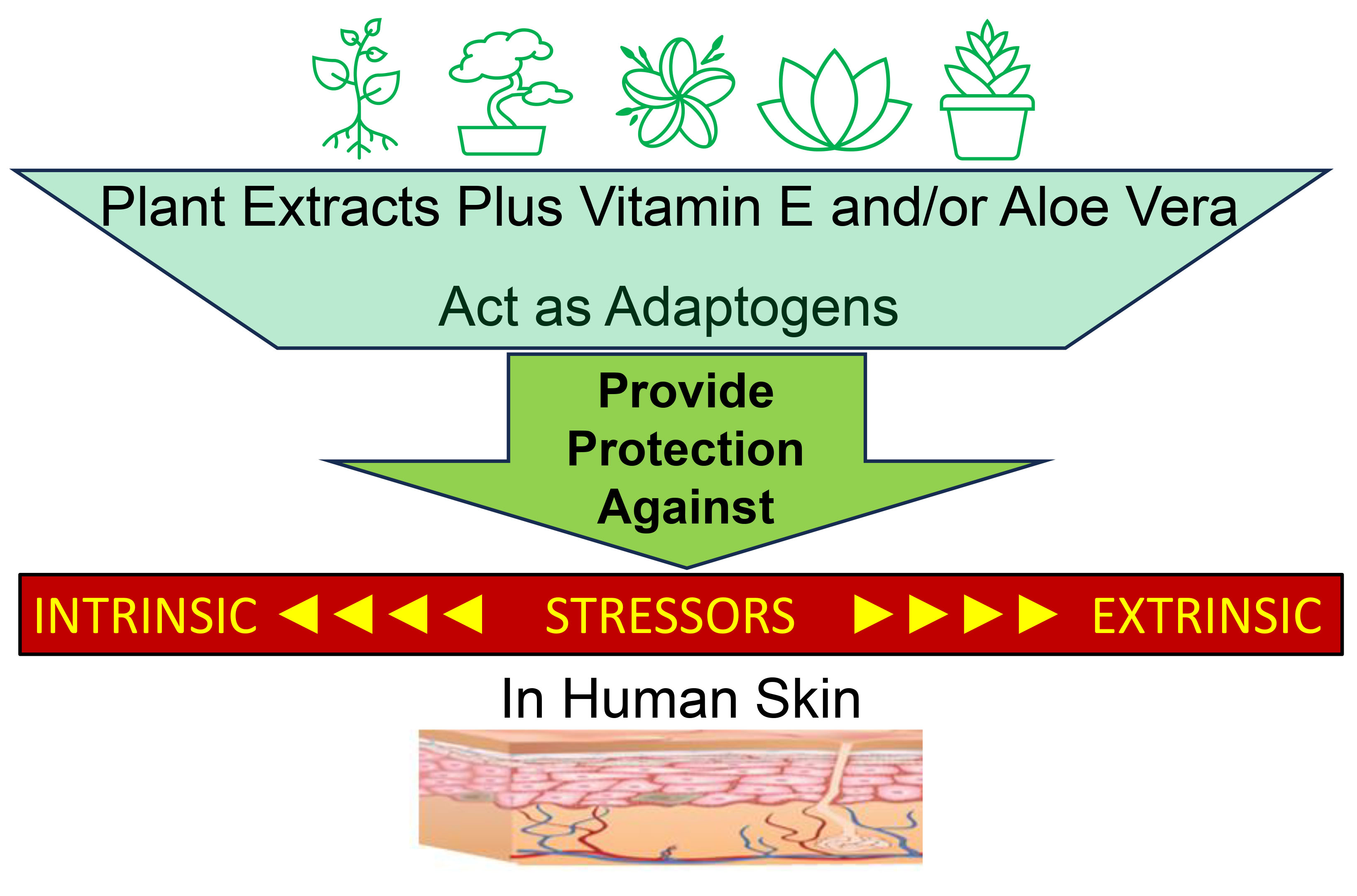
Plant-derived extracts and vitamin E exhibit stress protection or resistance by
normalizing cellular homeostasis and enhancing resistance to toxic stimuli to
overcome cellular damage. Here we report the evaluation of a topical preparation
(product test materials; PTM) containing an ingredient blend of Rhodiola Rosea,
Eleutherococcus Senticosus (Siberian Ginseng), Rhaponticum Carthamoides, Inonotus
Obliqus, and Slegainella Lepidophylla as the base formula and tested the addition
of Lespedeza Capitata (leaf/stem) extract plus vitamin E and/or Aloe Vera to
determine the induced protective functions in human skin when challenged with
intrinsic and extrinsic stressors. Methods: The base topical preparation
plus Lespedeza Capitata extract plus vitamin E or the base topical preparation
plus vitamin E and Aloe Vera were assayed in vitro on (a) intrinsically
stressed excised abdominoplasty skin, (b) full thickness (FT) skin equivalent
models post-treated with a combination of ultra-violet (UV) B light (250
mJ/cm
Graphical Abstract
Keywords
- product test materials
- adaptogen ingredients
- diesel particulate matter
- UVB
- full thickness skin equivalent models
Skin is the largest organ of human body and is exposed to a variety of intrinsic stressors such as hormonal changes, fatigue [1], insomnia, and extrinsic stressors such as UV radiation and pollution [2]. Acute, prolonged or chronic exposure to intrinsic or extrinsic stressors perturbs the state of cellular homeostasis in skin. For example, a variety of disorders such as atopic dermatitis, dyspigmentation, hyperpigmentation, increased fragmentation and disorganization of extracellular matrix proteins collagen and elastin, and thinning of the basement membrane results in significant aesthetic deterioration on the face and body leading to an aged appearance [3, 4]. Most treatments for improving stress are focused on prevention and protection, and some claim to treat some characteristics of stress induced disorders such as hyperpigmentation and dermal matrix fragmentation by increasing extracellular matrix protein levels [5]. Less attention is given to the induction of stress response, which could be the first line of treatment before application of treatment regimen that involves prevention and treatment. Several plants extracts have been found to be beneficial in maintaining and restoring cellular homeostasis, promoting immunomodulatory activity, improving endurance against fatigue, and resisting cellular damage from stressors [6, 7]. Rhodiola Rosea [8], Eleutherococcus Senticosus [9] Rhaponticum Carthamoides [10], Inonotus Obliquus [11] and Selaginella Lepidophylla [12] have been found to exhibit adaptgenic functions by boosting antioxidant and anti-inflammatory functions [6, 8, 9, 10, 11, 12, 13]. Phytochemicals such as stilbenes, anthocyanins, procyanidins, epicatechin, gallocatechingallate, acetogenins, isorhamnetin, sulforaphane, allyl isothiocyanate, lycopene, tomatine, lectin, curcumin, 6-shogaol, and 6-gingerol have been found to activate stress response signaling associated with oxidative stress, inflammation, and heat shock proteins [14]. The mechanism of stress protective response by adaptogenic plants in enhancing stress resistance and adaptation in humans have been shown to involve regulation of cortisol, nitric oxide, stress-activated protein kinase JNK, the forkhead box O transcription factor, and upregulation of heat shock protein pathway [15, 16].
Also, the biological activities of the legume crop Lespedeza Capitata have been shown to have skin care and pharmaceutical applications [17]. For example, some evidence from the topical use of Lespedeza Capitata have reported skin moisturizing properties along with protecting against photoaging [17]. Also, the known skin benefits of vitamin E [18] and Aloe Vera [19] are well established. For example, Vitamin E’s antioxidant and anti-inflammatory properties have been reviewed [18]. Moreover, Aloe Vera contains multiple vitamins, enzymes, minerals, sugars, and fatty acids that contribute to it healing activities [19]. In our review of the literature, we found that all studies showing stress protective function with adaptogenic plant extracts involved oral administration of botanical extracts. Notably, to date, no studies have shown topical application of botanical actives showing adaptogenic response to intrinsic or extrinsic stressors have been reported.
Plants with adaptogenic function have been described in the literature to
provide resilience, resistance, and adaptogenicity against intrinsic and
extrinsic stress by enhancing stress protective function including antioxidant,
anti-inflammation, anti-fatigue, antidepressive, neuroprotection, and CNS
stimulating activity [14]. Oral administration of the tincture extract from
Rhodiola rosea roots inhibited pro-inflammatory enzymes cyclooxygenase-1 (COX-1),
cyclooxygenase-2 (COX-2), and phospholipase A2 (PLA2) by preventing the release
of arachidonic acid from cell membranes, thereby, rendering membrane
stabilization and enhancing anti-fatigue functions [20]. In a clinical study,
repeated oral administration of Rhodeola rosea for 4 weeks reduced symptoms of
fatigue and improved attention span and cognitive function [20, 21]. Further, oral
administration of ADAPT-232 capsules 199 containing fixed combination
of standardized extracts of adaptogens, Rhodiola Rosea, Eleutherococcus
senticosis, and Schisandra Chinesis extract characterized for the content of
active markers eleutherosides, schisandrins, salidroside, tyrosol, and rosavin to
BALB/c mice prior to stress exposure increased alertness and endurance leading to
reduced fatigue. This study found that the adaptogen blends stimulated levels of
stress response proteins Heat Shock Protein 70 (HSP70) and Heat Shock Protein 72
(HSP72) in the serum of mice suggesting increased protection and tolerance to
stress [22]. In recent years, activation of autophagy mechanisms as an adaptive
response to stressors by natural substances, which includes resveratrol (vitis
vinifera) and curcumin (curcuma longa). Resveratrol activated autophagy
mechanisms by inhibiting mammalian Target of Rapamycin (mTOR) signaling [23], and
curcumin stimulated autophagy by inducing caspase-3 signaling [24]. Both
resveratrol and curcumin were shown to exert anti-tumor activity by stimulating
autophagic death of tumor cells. In addition, many other polyphenols were also
shown to exhibit adaptogenic effects. Quercetin glycosides stimulated glucose
update, and thus, overcoming mitochondrial dysfunction to improve response to
treatment of type 1 diabetes [25]. Hydroxytyrosol (HT), found in olives,
significantly upregulated mitochondrial biogenesis pathway genes PPARG
Coactivator 1 Alpha (PGC1-
To the best of our knowlege, there are no studies in the literature that have shown stress adapting and stress protective functions of adaptogen ingredients alone or in combination, when applied topically to intrinsically or extrinsically stressed skin. Since many of these ingredients have been associated with adaptogenic response when administered orally or systemically, we hypothesized that the adaptogen ingrediens may provide stress response activity, when applied topically against intrinsic and extrinsic stressors. Therefore, the purpose of this study was to determine whether or not different adaptogen plant-derived extracts as active ingredients provide protection against against intrinsic and extrinsic stressor via in vitro and clinical testing.
The product test materials (PTMs) with adaptogen ingredients (AI) are shown in Fig. 1.
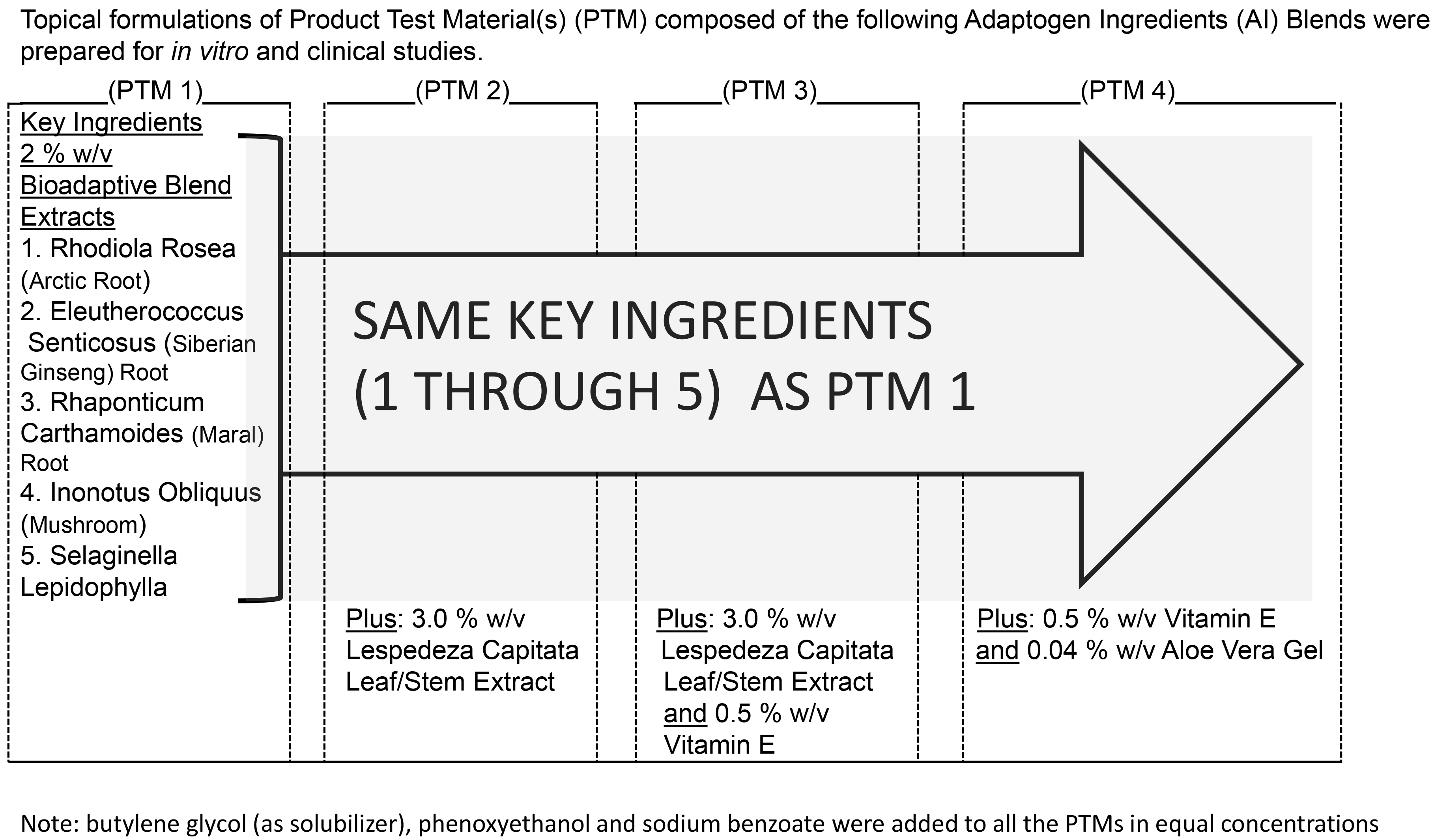 Fig. 1.
Schematic of the five Product Test Materials (PTM), PTM1, PTM2, PTM3, PTM4, PTM5 and their ingredient composition.
Human skin explant used in the assay was a surgical waste from an abdominal
procedure performed on a Caucasian female of 50 years of age in a plastic surgery
clinic in Beverly Hills, CA, USA. The explant skin was partitioned into
~2 cm
The study was conducted to understand how stress gene expression in the skin is
influenced by specific Product Test Materials (PTMs) under the influences of
stressors such as UVB exposure and Diesel Particulate Matter (DPM). The study was
performed using a full-thickness in vitro skin culture model containing
epidermal and dermal cell layers (EFT-400, MatTek Corp, Ashland, MA, USA). Four
separate groups of tissues had PTMs applied to the surface four hours prior to
DPM treatment. One-hour post-DPM treatment, the tissues were then exposed to 250
mJ/cm
The following groups were included in the study (N = 3/group):
PTM + UVB + DPM group: In this group, skin tissues were pre-treated with each of the test materials, PTM1, PTM2, PTM3, PTM4 followed by UVB + DPM; Stress Alone Group: Treated with UVB + DPM only and Naïve Group: Not treated with UVB, DPM and topicals. Gene expression was assessed on a custom Open Array (OA) panel format 112 (Supplementary Table 2). The panel consisted of 103 target genes belonging to stress response pathway along with 9 endogenous control genes (Ubiquitin C (UBC), Transferrin receptor (TFRC), Peptidylprolyl isomerase A (PPIA), hypoxanthine phosphoribosyltransferase 1 (HPRT1), glucuronidase beta (GUSB), Glyceraldehyde-3-phosphate dehydrogenase (GAPDH), Actin beta (ACTB), Eukaryotic 18S rRNA (18S), and beta-2-microglobulin (B2M). The UV light source and Diesel Particulate Matter (DPM) preparation used for extrinsic stress protection assessment is described below.
SOL500 Sun Simulator (Dr Hönle AG, Munich, Germany) with an H2 lamp filter
was used for generating UVB light for extrinsic stress. The full thickness (FT)
skin tissues were placed on an irradiation platform 30 cm from the H2 lamp filter
and irradiated with 250 mJ/cm
Diesel particulate matter NIST 1650b (Sigma-Aldrich, Inc. St. Louis, MO, USA) was used to prepare stock solution of 25 mg/mL in dimethyl sulfoxide (DMSO) and sonicated for 30 min to avoid agglomeration of the suspended Particulate Matter 2.5 (PM2.5) particles. DPM was diluted to 100 µg/mL in DMSO within 1 h of stock preparation to avoid variability in the NIST1650 composition for testing on skin models.
To demonstrate the bioadaptive capacity of test materials, a clinical study design to investigate the functional activity of test formulations to provide resilience and protection against UV induced erythema was developed. The study endeavored to demonstrate that pre-treatment of test materials suppresses development of erythema following UV exposure in healthy volunteers. IRB approval was obtained for the study protocol prior to study initiation at the clinical site. The study was performed on 20 female healthy volunteers at 18 to 78 years of age (average age 51.1 years) median BMI of 26.5 at Dermatology Consulting Services, PPLC, NC under the supervision of Zoe Draelos, MD (see Table 1). Subjects who signed informed consent and met all of inclusion criteria and none of the exclusion criteria were enrolled in the study. Subjects were dispensed a bar of Dove soap for cleansing of the entire buttock area and body. The subjects were instructed to apply one skin care product/regimen to the randomized right buttock and a second skin care product/regimen to the randomized left buttock and nothing to the central buttock which was defined as the untreated control site. The total number of sites for product application was 40 with each topical applied to 10 sites on either left or right side of buttock in a randomized design. A compliance check was conducted at 4 weeks.
| N = 20 |
| AGE range 18 to 78 (mean 51.5 |
| Body Mass Index (BMI) 26.5 |
| All subjects completed the study without any adverse events. |
| N = number of subjects |
Subjects returned to the research center at week 8 for irradiation. The left, right, and central buttock (3 sites) was irradiated with 2MED of UVB from a solar simulator (150W xenon arc bulb, Solar Light, PA, Philadelphia, USA). Subjects returned to the clinic 24 hours after irradiation. At the clinic, dermaspectrophotometer readings and photographs of erythema were obtained from the two-product application treated sites on the right and left buttocks and the one untreated control site on the central buttocks. The photographs of erythema intensity in UVB treated alone group, combination of UVB plus product treatment group were assessed using Image J open-source software (version 1.48; National Institute of Health, Bethesda, MD, USA). Safety parameters which included potential signs of irritation, blistering, and edema were assessed throughout the duration of the study. Subjects who were found to be noncompliant were dropped from the study and excluded from the data analysis. Statistical assessment of the variance of the mean of dermaspectrophotmeteric assessment from control group and topical PTM treated group were analysed using analysis of variance (ANOVA) test. A p value of 0.05 or less between the control and PTMs treated group were considered statistically significant.
For the gene expression data, genes were considered statistically significant if
the level of expression was 2-fold or greater compared to control levels as
determined by two-tailed t-test (p
We developed a set of 4 topical formulations containing adaptogenic ingredients
(AI) blends (Fig. 1) for testing on intrinsic as well as extrinsically stressed
human skin models. We used surgically excised ex vivo abdominoplasty skin as a
representative model for simulating intrinsic stress since ex vivo skin secrete
stress inducers including Adrenocorticotropic Hormone (ACTH), cortisol and
pro-inflammatory cytokines TNF-
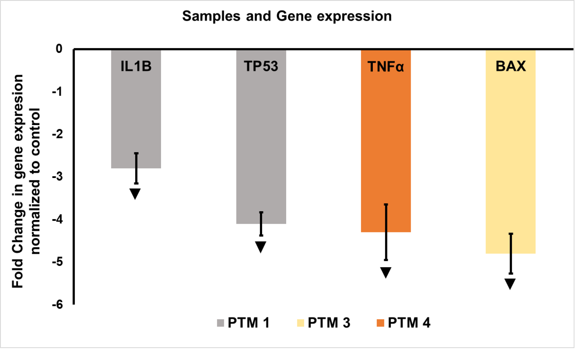 Fig. 2.
Effect of adaptogen ingredients (AI) topical materials (PTM1,
PTM3, PTM4) on intrinsically stressed skin: Assessment of stress gene markers
normalized to control. ▼ = Significant change in gene expression in
product test materials (PTM) treated samples compared to control. Topical PTM
samples were treated to excised abdominoplasty skin tissues for 24 hours
following which RNA was extracted and cDNA synthesized. The RNA samples were
assayed for gene expression on a panel of genes that affect stress response.
Results showed downregulation of pro-inflammatory cytokine Interleukin
-1
Based on the capacity of the AI topical formulations to reduce expression of key
markers of stress induction on intrinsically stressed skin, we hypothesized that
topical application of AI may also provide stress protective response in skin
exposed to extrinsic stressors UVB and DPM. UVB is a major environmental stressor
associated with DNA damage through induction of Cyclibutane Pyrimidine Dimers
(CPD) and perturbation of cellular apoptosis in human skin specifically, when
exposed to 250 mJ/cm
Pre-treatment with 4 topicals PTM1, PTM2, PTM3 and PTM4 significantly suppressed
pro-inflammatory cytokine IL-1
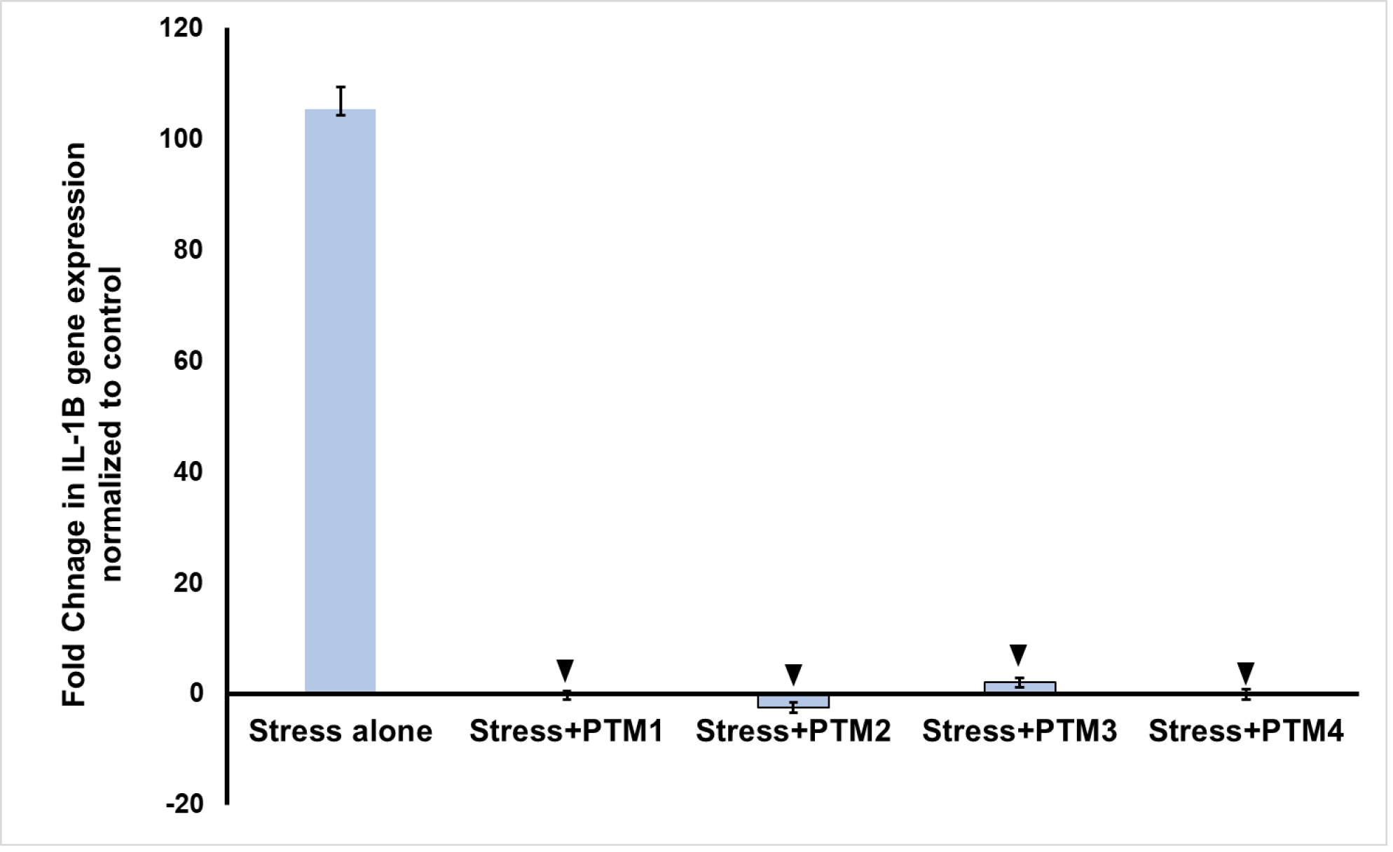 Fig. 3.
Effect of AI topical materials PTM1, PTM2, PTM3, PTM4 on
extrinsically stressed skin: Assessment of stress gene markers normalized to
control. ▼ = Significant reduction in IL-1
Pre-treatment with PTM1 and PTM2 upregulated 2-fold and higher antioxidant gene Heme Oxygenase 1 (HMOX1) expression, anti-apoptotic B-cell lymphoma 2 gene (BCL2) and 3 Heat Shock Protein (HSP) family genes heat shock transcription factor 4 (HSF4), heat shock protein family B (small) member 1 (HSPB1) and Crystallin Alpha B (CRYAB) (Part of HSP20 gene family) (Fig. 4). PTM3 and, however, did not induce significant differential (2-fold change) in expression of HMOX1, BCL2 and HSP family genes HSF4, HSPB1 and CRYAB other antioxidant, apoptotic and HSP family genes in the open array panel.
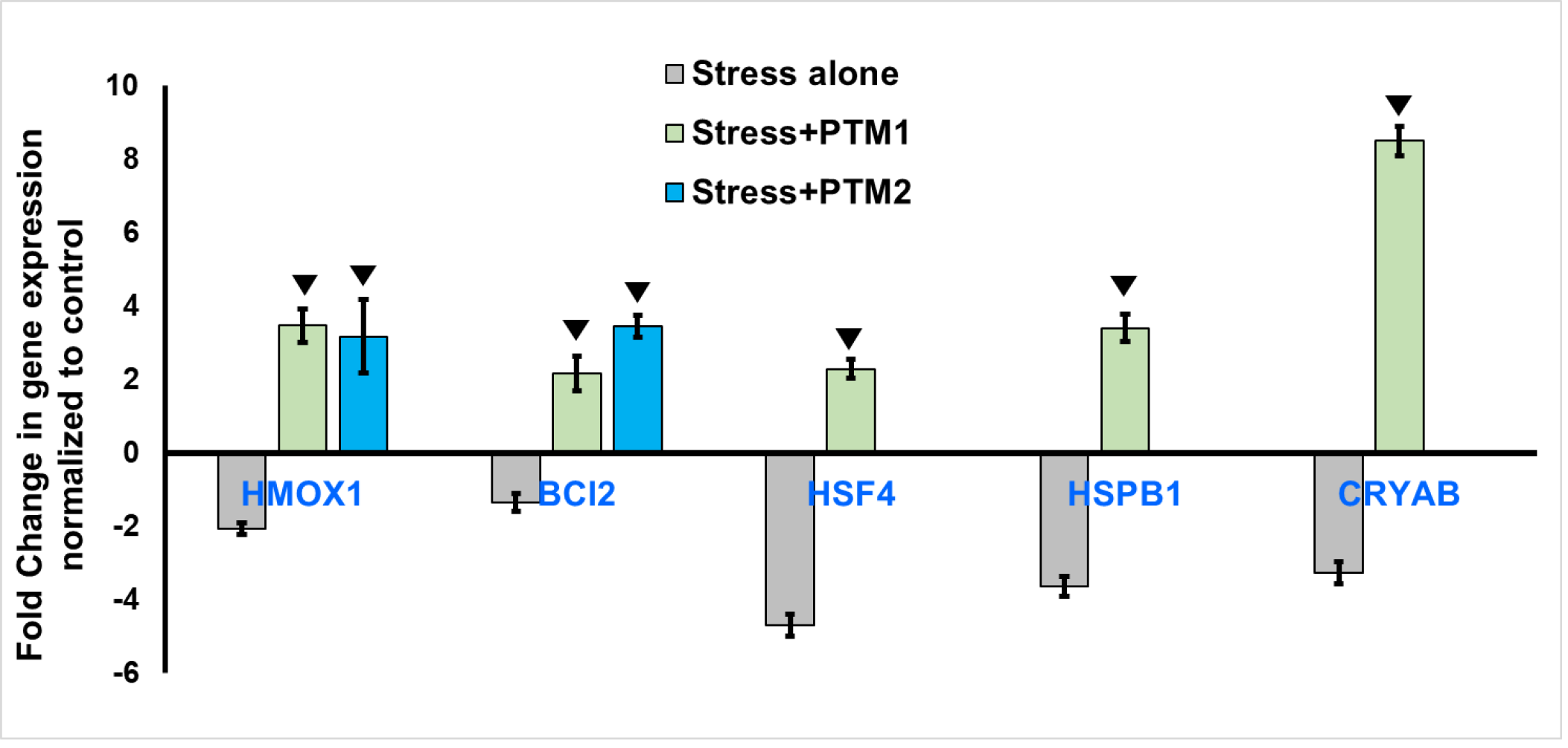 Fig. 4.
Effect of AI topical materials PTM1, PTM2, PTM3, PTM4 on extrinsically stressed skin: Assessment of stress gene markers normalized to control. ▼ = Significant change in gene expression in PTM treated samples compared to control. Methods detailed in Fig. 3. PTM1 and PTM2 significantly stimulated antioxidant gene Heme Oxygenase 1 (HMOX1), anti-apoptotic B-cell lymphoma 2 gene (BCL2), and HSP family genes heat shock transcription factor 4 (HSF4), heat shock protein family B (small) member 1 (HSPB1) and Crystallin Alpha- B (CRYAB). This figure is a representation of two independent experiments.
Our data is consistent with the induction of stress response genes such as HSP70
and HSP72 in studies involving oral administration of adaptogenic ingredients
[23]. In summary, our data suggests that IL-1
Based on positive input from mechanistic studies that demonstrated capacity of
the adaptogen topical compositions to resist development of intrinsic and
extrinsic stressors by downregulation of pro-inflammatory cytokines primarily
IL-1
Assessment of erythema intensity by dermaspectrophotometric analysis at 24 hours
following UVB exposure showed that all 4 formulations reduced the intensity of
erythema. However, the reduction in spectrophotometric measurement was slightly
more pronounced in the areas treated with PTM 3 and PTM 4 (Fig. 5). The
improvement of erythema intensity by PTM1, PTM2, PTM3 and PTM4 AI topical
materials shown with dermaspectrophotometric analysis was further examined by
photographic images taken at 24 hours following UVB exposure. Since PTM3 and PTM4
compositions are almost identical with the exception of Lespedeza Capitata
Leaf/Stem Extract, only PTM3 treated location was photographed. The images showed
that compared to control site treated with 2 MED of UVB, the sites treated with
PTM1 regimen (Fig. 6), PTM2 (Fig. 7) and PTM3 (Fig. 8) showed suppression of
erythema intensity. The area of the control UVB treated area assessed by Image J
software was 3.1 cm
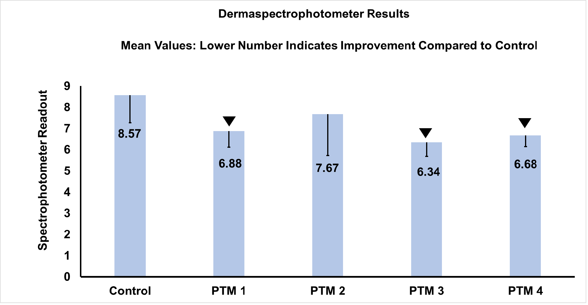 Fig. 5.
Assessment of erythema intensity by dermaspectrophotometer measurement (n = 10 site per sample). ▼ = Significant erythema suppression in PTM treated subjects compared to control (subjects treated with UV alone). Twenty Subjects who signed informed consent and met all of inclusion criteria and none of the exclusion criteria were enrolled in the study. Subjects were dispensed a bar of Dove soap for cleansing of the entire buttock area and body. The subjects were instructed to apply one skin care product/regimen to the randomized right buttock and a second skin care product/regimen to the randomized left buttock and nothing to the central buttock which was defined as the untreated control site. The total number of sites for product application was 40 with each topical applied to 10 sites on either left or right side of buttock in a randomized design. Subjects returned to the research center at week 8 for irradiation. The left, right, and central buttock (3 sites) was irradiated with 2MED of UVB from a solar simulator (150W xenon arc bulb, Solar Light, Philadelphia). Subjects returned to the clinic 24 hours after irradiation. At the clinic, dermaspectrophotometer readings and photographs of erythema were obtained from the two-product application treated sites on the right and left buttocks and the one untreated control site on the central buttocks. The photographs of erythema intensity in UVB treated alone group, combination of UVB plus product treatment group were assessed using Image J open-source software.
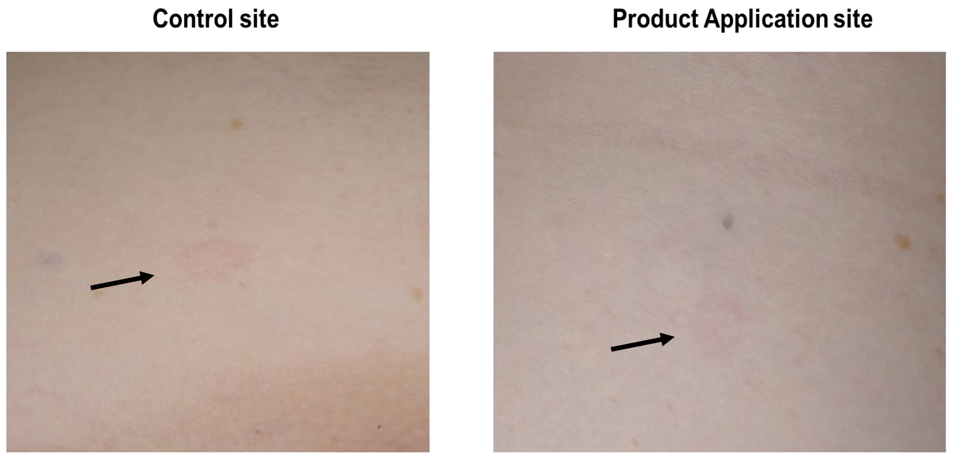 Fig. 6.
Photographic assessment of intensity of erythema. Methods detailed in Fig. 4. Pre-treatment with PTM1 for 8 weeks prior to 2 minimal erythema dose (MED) UVB exposure significantly suppressed UV induced erythema. UV induced erythema in control site and product application site are indicated by black arrows.
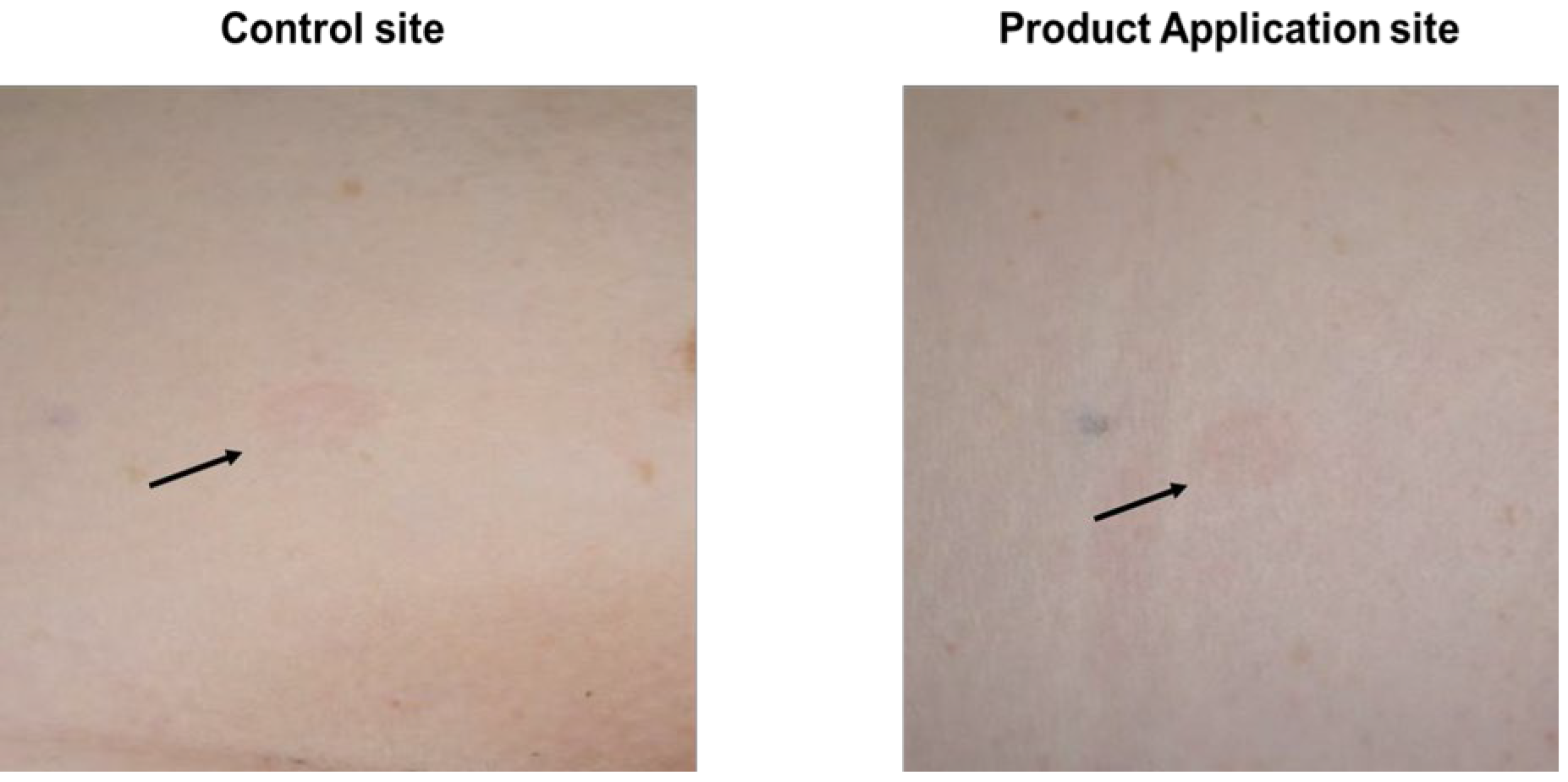 Fig. 7.
Photographic assessment of intensity of erythema. Methods detailed in Fig. 4. Pre-treatment with PTM2 for 8 weeks prior to 2 MED UVB exposure significantly suppressed UVB induced erythema. UV induced erythema in control site and product application site are indicated by black arrows.
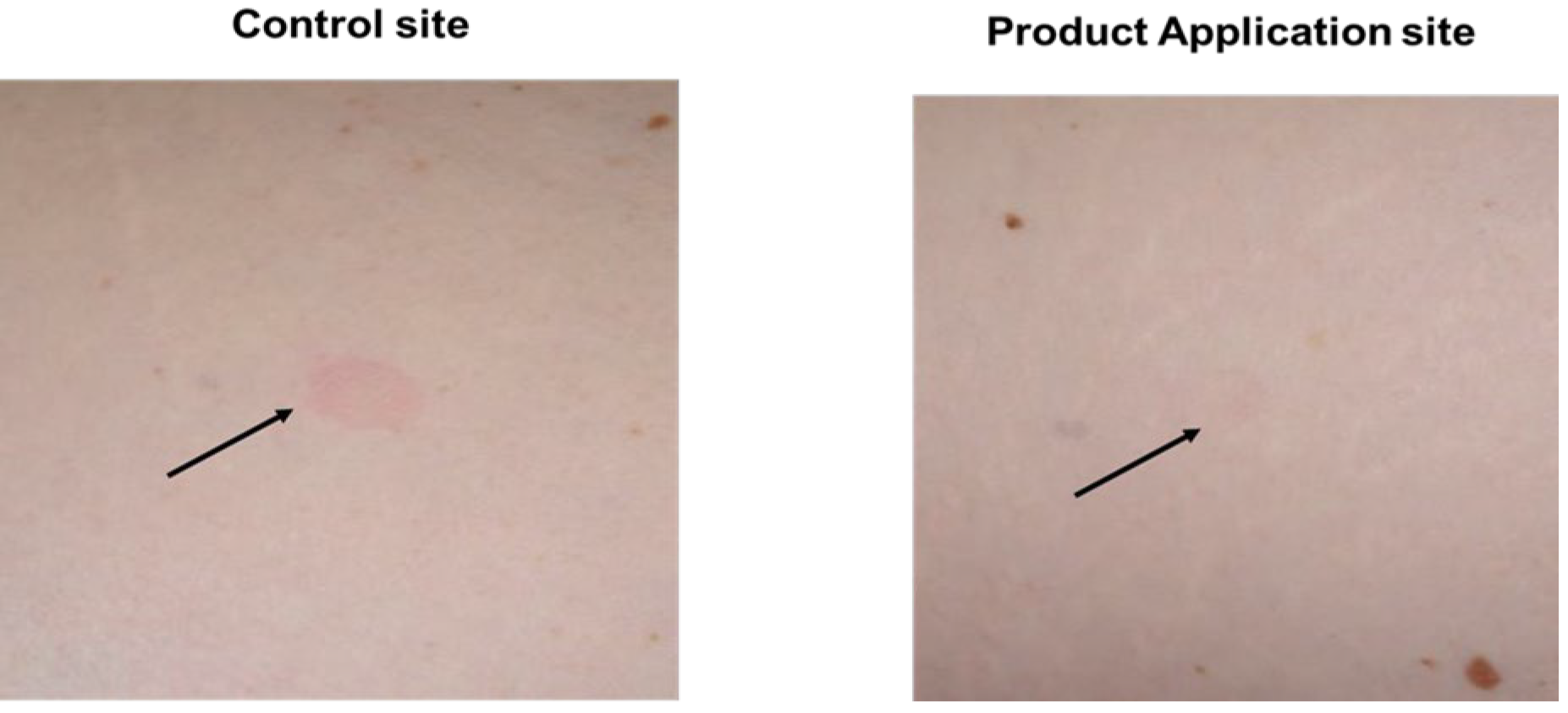 Fig. 8.
Photographic assessment of intensity of erythema. Methods detailed in Fig. 4. Pre-treatment with PTM3 product regimen for 8 weeks prior to 2 MED of UVB exposure significantly suppressed UVB induced erythema. UV induced erythema in control site and product application site are indicated by black arrows.
The present results demonstrate that topical application of adaptogen ingredients (AI) activates stress protective response in human skin against intrinsic stressors as well as major extrinsic stressors UVB and DPM by suppressing proinflammatory cytokines, activating antioxidant gene expression and inducing stress response by upregulating HSP gene family. Our data is consistent with the induction of stress response genes such as HSP70 and HSP72 in studies involving oral administration of adaptogenic ingredients. The gene expression results corroborated with our clinical data, which showed the capacity of AI topical compositions to suppress intensity of erythema induced by UVB. In summary, the results provide an indication of potential benefits of including adaptogenic ingredients in topical skin products. More research including mechanistic and clinical studies with long term administration of AI compositions are needed to further characterize the usefulness of topical application of adaptogens on human skin and its clinical benefits in protection against intrinsic and extrinsic stressors.
PTM, Product Test Materials; AI, Adaptogen Ingredients; DPM, Diesel Particulate Matter; UVB, Ultra-violet B; FT, Full Thickness skin equivalent models.
All datasets including table and figures, developed and analyzed during this study are included in this published article. The corresponding author will make available all data published in this manuscript upon reasonable request.
GD, LB and HK designed and executed the in vitro studies with Sunnybio discovery Inc. MR and ZDD designed the clinical study in consultation with LB and executed the study. All authors contributed to editorial changes in the manuscript. All authors read and approved the final manuscript. All authors have participated sufficiently in the work and agreed to be accountable for all aspects of the work
The study was carried out in compliance with 21CFR Part 50 Protection of Human Subjects. The primary investigator verbally consented all subjects. Subjects were allowed to take as much time as necessary to read the consent form. The consent form was approved by the Allendale Institutional Review Board (AIRB), Old Lyme, CT. The investigator answered all subject questions. Following answering of all questions, the subjects and the investigator signed the consent form. The subject were provided with a copy of the consent form. The ethics approval number is: DCS-09-20, ethics committee approval date: March 01, 2020.
The authors would like to thank Krys Bojanowski Sunny Biodiscovery Inc., Santa Paula, CA for performing the experiments and collecting data on intrinsic stress evaluation. The authors would also like to thank Genemarkers Inc., Kalamazoo MI for performing experiments and collecting data on extrinsic stress evaluation. Further, we express our sincere gratitude to Edwin Lephart, Brigham Young University, for his critical review and comments and support for development of this manuscript.
This work was supported by funding from Nu Skin Enterprises.
Ganesh Diwakar, Lisa Barnes, Melanie Riggs, Helen Knaggs are employees of Nu Skin enterprises, Provo UT USA. Zoe Diana Draelos is an owner and Principal Investigator at Dermatology Consulting Services, PLLC, NC, USA. The authors declare no conflict of interest.
References
Publisher’s Note: IMR Press stays neutral with regard to jurisdictional claims in published maps and institutional affiliations.