IMR Press / FBL / Volume 28 / Issue 11 / DOI: 10.31083/j.fbl2811317
Open Access Review
MicroRNA in Fibrotic Disorders: A Potential Target for Future Therapeutics
Show Less
1 Science and Math Program, Asian University for Women, 4000 Chattogram, Bangladesh
2 Department of Clinical and Molecular Sciences, Università Politecnica Delle Marche, 60126 Ancona, Italy
3 Department of Gynecology and Obstetrics, Division of Reproductive Sciences & Women’s Health Research, Johns Hopkins Medicine, Baltimore, MD 21205, USA
*Correspondence: soriful84@gmail.com; mislam18@jhmi.edu (Md Soriful Islam)
These authors contributed equally.
Front. Biosci. (Landmark Ed) 2023, 28(11), 317; https://doi.org/10.31083/j.fbl2811317
Submitted: 16 June 2023 | Revised: 17 September 2023 | Accepted: 10 October 2023 | Published: 29 November 2023
Copyright: © 2023 The Author(s). Published by IMR Press.
This is an open access article under the CC BY 4.0 license.
Abstract

Fibrotic disorders are defined by accumulating excessive extracellular matrix (ECM) components, especially collagens, in various organs, leading to tissue scarring and organ dysfunction. These conditions are associated with significant challenges in the healthcare system because of their progressive nature and limited treatment options. MicroRNAs (miRNAs) are small non-coding RNA molecules (approximately 22 nucleotides) that modulate gene expression by selectively targeting mRNAs for degradation or translational repression. MiRNAs have recently been identified as potential targets for therapeutic developments in fibrotic disorders. They play vital roles in inducing fibrotic phenotype by regulating fibroblast activation and ECM remodeling. Multiple strategies for targeting specific miRNAs in fibrotic disorders have been explored, including antisense oligonucleotides, small molecule modulators, and natural compounds. This review discussed the role of miRNAs in different fibrotic disorders, including cardiac fibrosis, liver fibrosis, kidney fibrosis, lung fibrosis, dermal fibrosis, and primary myelofibrosis, with recent advances in developing miRNA-based therapeutics.

Keywords
microRNA
fibrotic disorders
cardiac fibrosis
liver fibrosis
kidney fibrosis
lung fibrosis
dermal fibrosis
primary myelofibrosis
miRNA therapeutic
natural compounds
1. Introduction

Fibrosis is defined by excessive accumulation of extracellular matrix (ECM) components, especially collagens, and loss of cellular homeostasis. The highly progressive nature of fibrosis is associated with the loss of tissue architecture and organ function [1, 2]. Current data suggests the major role that fibrosis plays in a wide range of diseases, including scleroderma, Crohn’s disease, ulcerative colitis, myelofibrosis, systemic lupus erythematosus (SLE), systemic sclerosis, liver cirrhosis, progressive kidney disease, cystic fibrosis, cardiac fibrosis and idiopathic pulmonary fibrosis (IPF), and cancer [2, 3]. There are around 4968 fibrosis cases per 100000 individuals annually [4]. Fibrosis has been reported to be associated with up to 45% of all deaths in industrialized nations [5]. These conditions are associated with a significant burden in global healthcare systems because of their progressive nature and limited treatment options.

Fibrosis is a complex process influenced by a combination of various molecular factors, including transforming growth factor-β (TGF-β), platelet-derived growth factor (PDGF), connective tissue growth factor (CTGF) [3], interleukin-13 (IL-13), interleukin-4 (IL-4) [6] and nuclear factor kappa-light-chain-enhancer of activated B cells (NF-κB) [7]. Myofibroblast is a specialized cell type that plays a vital role in wound healing and tissue homeostasis [8] and the pathogenesis of fibrotic disorders [8]. In recent years, microRNAs (miRNAs) have been recognized as critical players in the pathogenesis of fibrotic diseases [9, 10, 11]. MiRNAs are small, non-coding RNA molecules (approximately 22 nucleotides) that modulate gene expression by binding to the 3 untranslated region (UTR) of target mRNAs. This interaction leads to the degradation of the mRNA or translational repression [12]. MiRNAs play vital roles in the progression of fibrotic diseases by regulating the activation of fibroblasts, ECM remodeling, and the advancement of tissue scarring [2, 3]. Thus, targeting miRNAs holds great potential as a therapeutic strategy to modulate fibrosis and restore tissue homeostasis. Indeed, multiple approaches, including antisense oligonucleotides, small molecule modulators, and natural compounds, for targeting specific miRNAs in fibrotic disorders have been studied.

This review provides a comprehensive overview of the role of miRNAs in fibrotic diseases, with a particular focus on their potential as therapeutic targets for future therapeutics. Particularly, we discuss: (1) the functional roles of miRNAs in the fibrosis process, (2) the role of miRNAs in various fibrotic diseases, including cardiac, liver, kidney, lung, dermal, and primary myelofibrosis, and (3) the recent advances in developing miRNA-based therapeutics for fibrotic diseases.

2. Methods

This review provides a comprehensive overview of the role of miRNAs in fibrotic diseases and explores potential therapeutic approaches. An extensive literature search has been performed until May 2023 using electronic databases such as PubMed and Google Scholar. Review articles were used as a source to identify additional articles relevant to miRNA and fibrosis. Only articles published in English have been included in this review. The search strategy involved using the following keywords: “fibrosis”, “fibrotic disorders”, “fibrotic diseases”, “extracellular matrix”, “wound healing”, “microRNA or miRNA”, “cardiac fibrosis”, “liver fibrosis”, “kidney fibrosis”, “lung fibrosis”, “dermal fibrosis”, “primary myelofibrosis,”, “miRNA therapeutic”, “miRNA inhibitor”, “natural compounds”, “antisense oligonucleotides”, “antagomirs”, “carvedilol”, “ivabradine”, “nebivolol”, “astragaloside IV”, “baicalin”, “berberine”, “celastrol”, “corilagin”, “EGCG”, “quercetin”, “resveratrol”, “tanshinone IIA”, and “ursolic acid”.

3. MiRNA Biogenesis

MiRNAs are small, single-stranded, non-coding RNA molecules that are evolutionarily conserved [12]. They are transcribed from specific DNA regions and exhibit a broad spectrum of functions. The primary role of miRNA is to pair with mRNA molecules that contain a complementary sequence, resulting in the downregulation of their expression. A single miRNA molecule can target multiple mRNA molecules, thereby regulating numerous cellular pathways [13]. MiRNA biogenesis is complex and regulated by several mechanisms facilitated by transportation, RISC (RNA-induced silencing complex) biding, miRNA decay, and processing performed by Drosha and Dicer enzymes (Fig. 1).

Fig. 1.

The biogenesis steps of microRNAs (miRNAs). The miRNA biogenesis involves several steps, including transcription, pri-miRNA processing, export to the cytoplasm, dicer processing, strand selection, and target recognition and regulation. After several sequential process, the pre-miRNA is recognized in the cytoplasm, and cleaved by Dicer, and miRNA duplex is generated. In the final step, the guide strand of the miRNA duplex is preferentially selected and loaded onto an argonaute (AGO) protein within the RNA-induced silencing complex (RISC). The miRNA-loaded RISC complex recognizes target mRNA molecules through base pairing, usually resulting in mRNA degradation or translational repression. Figure created with biorender.com. GTP, Guanosine triphosphate; mRNA, messenger RNA.

The process of miRNA biogenesis begins with specific miRNA genes in the DNA, which can be transcribed as individual miRNAs or as part of polycistronic transcripts [14]. In addition to being transcribed as individual miRNAs or polycistronic transcripts, miRNA genes can also be found within introns or UTRs of protein-encoding genes [15].

Typically, the transcription of a miRNA gene occurs through RNA polymerase III, which produces primary miRNA (pri-miRNA) transcripts. These pri-miRNA transcripts then undergo capping and polyadenylation. They undergo a processing step mediated by the microprocessor complex, which converts them into pre-miRNAs (precursor miRNAs) [16, 17]. The microprocessor complex consists of at least two essential proteins: Drosha, a ribonuclease III enzyme, and DGCR8 (DiGeorge syndrome critical region 8), a dsRNA binding protein, along with some less studied components such as DDX5 (DEAD-box RNA helicases p68) and DDX17 (p72) [18, 19]. These two proteins work together to process pri-miRNA into pre-miRNA during the miRNA biogenesis pathway. The microprocessor identifies distinct motifs in the primary miRNA and cuts it at the stem of the classic hairpin loop formation, creating a 5-monophosphate and a 2-nt 3 overhang on the precursor miRNA [20]. After being processed into pre-miRNAs, they are exported from the nucleus to the cytoplasm via an active interplay between the exportin 5 receptor and the pre-miRNA transcripts [21, 22]. This transport mechanism ensures that the pre-miRNAs reach the cytoplasm, undergoing further processing to become mature miRNAs. Once in the cytoplasm, the pre-miRNA is recognized and cleaved by an enzyme called Dicer, which is an RNase III enzyme. Dicer processes the pre-miRNA, producing a double-stranded RNA (dsRNA) molecule that is approximately 20-25 base pairs in length [23, 24]. This dsRNA molecule is the precursor for the mature miRNA strand. In humans, the functionality of Dicer is supported by its collaboration with TARBP2 (trans-activation-responsive RNA binding protein of dsRBP) [25]. TARBP2 binds with Dicer and increases its activity in processing pre-miRNAs into mature miRNAs [26]. This collaboration between Dicer and TARBP2 is vital for the accurate and efficient production of functional miRNAs in the cells. Finally, during the RISC loading process, the double-stranded miRNA (dsmiRNA) produced by Dicer is transferred to one of the proteins from the AGO (Argonaute) family of proteins [27]. AGO proteins are essential components of the RISC complex. Once the dsmiRNA is loaded onto an AGO protein, it undergoes strand selection, where one of the strands is selected as the mature miRNA strand. In contrast, the other strand is considered the passenger strand, possibly degraded. The mature miRNA loaded onto the AGO protein is then ready to exert its regulatory function by binding to target mRNA molecules and guiding the RISC complex to induce gene silencing or translational repression (Fig. 1). It should be noted, however, that not all miRNAs follow canonical processing pathways and a number of non-canonical miRNA biogenesis routes, such as those independent of either Drosha or Dicer, have recently been identified [17, 25]. As an example, a small RNA precursor could be generated starting from the products of mRNA splicing. Behind splicing from host mRNAs, the trimmed precursor refolds into a short stem-loop structure that functions as a pre-miRNA. Other non-canonical pathways do not require Dicer action but involve AGO2 catalytic activity [17].

4. MiRNAs and Extracellular Matrix

MiRNAs have been studied in various biological processes, including fibrosis. Understanding the interaction between miRNAs and ECM accumulation is crucial for tissue remodeling, wound healing, and fibrosis. The ECM microenvironment is a complex network of macromolecules that provides structural support to cells and tissues, serving as a physical barrier between cells and their surroundings [28, 29]. ECM consists of diverse proteins, including collagens, fibronectin, laminins, and proteoglycans, maintaining tissue integrity and function [30]. Dysregulation of ECM remodeling and excessive deposition can result in the development of fibrotic conditions [31].

Each specific tissue cell type possesses a particular combination of ECM components that influence mechanical properties and organization of cells. Specialized proteins, such as the integrin families of trans-membrane receptors, regulate cell adhesion to the ECM. Cell adhesion is essential for cell proliferation and survival and, more generally, for developing the typical architecture constitutive of healthy tissues. Structural changes in the ECM are strongly associated with disease development and progression [29]. Along with its structural role, ECM steers cell-cell and cell-matrix interactions. When triggered by specific signals, growth factors and cytokines stored in the ECM are released, stimulating proper cellular responses toward cell growth, differentiation, adhesion, and migration. It is the substrate for fibroblast activation and differentiation into myofibroblasts, the primary effector cells in fibrotic tissue formation [32].

The ECM maintains tissue integrity by undergoing quantitative and qualitative remodeling mediated by specific enzymes in particular glycosidases and MMPs [32, 33]. Altered ECM composition encompasses either excessive matrix degradation, as in osteoarthritis, or its abnormal production and accumulation, leading to tissue scarring and impaired organ function, both hallmarks of fibrosis [32]. This unbalanced process disrupts the regular cell-matrix interactions, leading to stiffening, decreased perfusion, and hypoxia, further promoting cell injury and fibroblast-to-myofibroblast differentiation [34]. Surprisingly, the ECM translationally contributes to pathological gene expression, thus promoting the establishment of a positive feedback loop of self-amplifying fibroblast activation and augmented tissue fibrosis, culminating in disease progression [35].

Some of the most investigated pro-fibrotic regulators are the TGF-β family, members of the PDGF family [36, 37, 38], and Wnt/β-catenin signaling pathways [39, 40]. MiRNAs have been implicated in ECM-related signaling pathways. For instance, miR-29 family members target mRNAs that encode proteins involved in fibrosis, including collagens, elastin, and fibrillin and inhibit their expression [41]. Experimental evidence suggests a fibrotic ECM environment downregulates miR-29c expression in fibroblasts, supporting a causal link between this miRNA level and fibrosis progression [35]. Notably, miR-29 acts as a downstream regulator of TGF-β/Smad3-mediated fibrosis, exerting its effects by specifically targeting collagen synthesis [42]. Specific miRNAs can regulate the production and accumulation of collagens. In murine models of lung fibrosis, miR-21 increases the production of a specific type of collagen (Col-1), disrupting the typical organization of cells, known as epithelial polarity [43].

Similarly, miR-21 influences the upregulation of other types of ECM proteins [44, 45, 46], while miR-30 influences the downregulation of genes involved in ECM degradation [47]. MiR-155 is upregulated in fibrotic disorders, promoting ECM production by targeting the TGF-β1 signaling pathway [48]. Additionally, miR-145 [49], miR-192 [50], and miR-200a [51], among other miRNAs, have been associated with the regulation of collagen synthesis. Furthermore, evidence suggests that miRNAs can be produced by cells and packaged into extracellular vesicles, including exosomes. These miRNAs can be transferred or transported to recipient cells, to regulate gene expression and contribute to ECM remodeling [52, 53]. According to current knowledge, modulating miRNA expression to target abnormal ECM signaling pathways in fibrotic diseases is considered a promising approach for developing new therapeutics [32, 54].

5. Role of miRNAs in Fibrotic Disorders

MiRNA dysregulation can significantly impact fibrosis-related key cellular processes, including fibroblast activation and ECM remodeling [55, 56]. Studies have shown different patterns of miRNA expression in multiple fibrotic disorders, including cardiac fibrosis, liver fibrosis, kidney fibrosis, lung fibrosis, and dermal fibrosis, as well as primary myelofibrosis (Fig. 2).
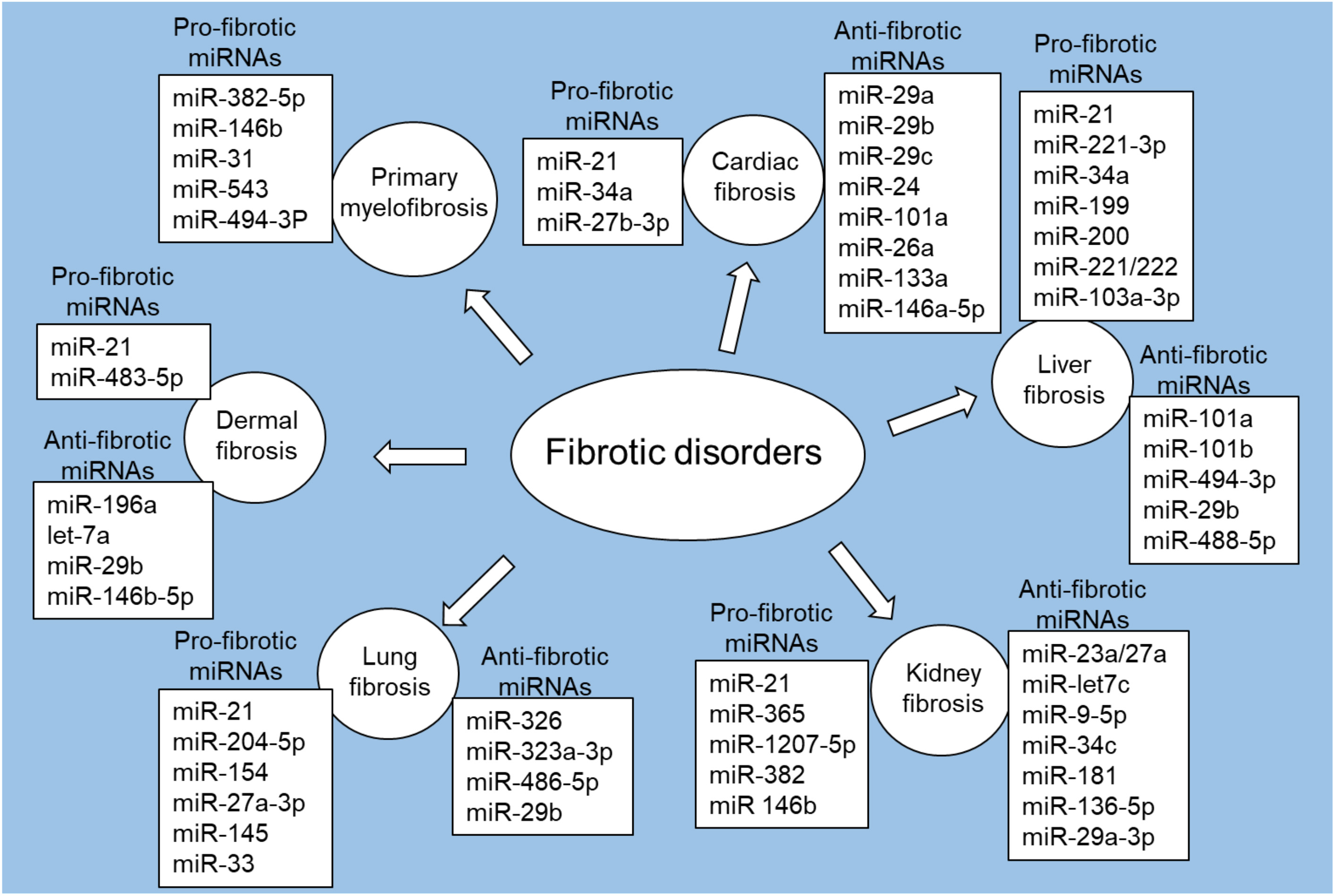
Fig. 2.

Pro-fibrotic and anti-fibrotic miRNAs in fibrotic disorders. Figure created with biorender.com.

5.1 Cardiac Fibrosis

Cardiac fibrosis is defined by the excessive buildup of ECM components in the heart that leads to the rigidity and impaired function of the cardiac tissue [57]. The development of cardiac fibrosis can be influenced by various risk factors, including diabetes, aging, genetic factors, alcohol abuse, hypertension (high blood pressure), coronary artery disease, myocardial infarction (heart attack), heart failure, and SLE and rheumatoid arthritis [58, 59]. Emerging data suggest that miRNAs play an important role in developing and progressing cardiac fibrosis [9].

Among the miRNAs associated with pro-fibrotic effects, miR-21 and miR-34 have received significant attention. MiR-21 is upregulated in cardiac fibrosis and is associated with fibroblast activation, collagen synthesis, and ECM remodeling [60, 61]. In a mouse model of pressure-overload-induced cardiac disease, specific inhibition of miR-21 resulted in reduced interstitial fibrosis and cardiac dysfunction mediated by the ERK/MAPK signaling cascade [62]. Similarly, in chronic Chagas disease cardiomyopathy (CCC), where intense cardiac fibrosis and inflammation occur, silencing miR-21 through locked nucleic acid (LNA)-anti-miR-21 inhibitor led to a significant reduction in cardiac fibrosis in Trypanosoma cruzi-infected mice [61]. The upregulation of miR-21 was associated with elevated content of collagens in cardiac fibroblasts, and its increase in collagen content was partially mediated by the downregulation of TGF-βRIII [63]. Notably, overexpression of TGF-βRIII effectively inhibited miR-21 expression and attenuated collagen production in fibroblasts, suggesting a reciprocal loop between miR-21 and TGF-βRIII involved in cardiac fibrosis. Additionally, TGF-β1 was identified as a positive regulator of miR-21 [63]. MiR-34a has also been implicated in myocardial fibrosis, particularly following myocardial infarction [64]. Inhibition of miR-34a was associated with a reduction in experimental cardiac fibrosis severity in mice [64]. TGF-β1, a key player in fibrosis, was found to increase miR-34a expression in cardiac fibroblasts [64]. Data suggest that overexpression of miR-34a enhanced the profibrogenic activity of TGF-β1 in cardiac fibroblasts, while inhibition of miR-34a weakened this activity [64]. The study further found that Smad4, an important mediator of TGF-β1 signaling, can act as a target of miR-34a during cardiac fibrosis [64].

In a mouse model of transverse aortic constriction (TAC)-induced cardiac hypertrophy, miR-27b-3p showed elevated expression, and miR-27b-knockout mice exhibited remarkably attenuated cardiac hypertrophy, inflammation, and fibrosis. Analysis of the transcriptome revealed that miR-27b deletion significantly reduced TAC-induced cardiac hypertrophy, fibrosis, and inflammatory genes. Interestingly, miR-27b-3p directly targets the fibroblast growth factor 1 (FGF1), and the administration of rFGF1 decreased fibrosis and cardiac hypertrophy in TAC-models [65].

Several miRNAs, including miR-29, miR-24, miR-101a, miR-26a, and miR-133a, have exhibited antifibrotic effects in various studies [41, 66, 67, 68, 69]. The antifibrotic properties of the miR-29 family (such as miR-29a, miR-29b, and miR-29c) have been extensively investigated in cardiac fibrosis [41, 70, 71]. The lower expression levels of miRNAs were detected in the myocardial region adjacent to the infarct, and their down-regulation was linked to increased expression of collagens [41]. In vitro and in vivo studies have shown that down-regulation of miR-29 was associated with increased collagen expression levels, while overexpression of miR-29 decreased collagen expression levels in fibroblasts [41]. Additionally, the down-regulation of miR-29a was accompanied by up-regulation of vascular endothelial growth factor A (VEGF-A) in cardiac fibrosis tissues, and overexpression of miR-29a reduced cardiac fibrosis and fibroblast proliferation by targeting the VEGF-A/MAPK signaling pathway [70]. Furthermore, overexpression of miR-29b decreased angiotensin II-induced cardiac fibrosis, partly by inhibiting the TGF-β signaling pathway [71].

MiR-24 was downregulated in heart tissue from subjects with myocardial infarction [66]. The overexpression of miR-24 improved heart function, decreased fibrosis, and reduced the differentiation and migration of cardiac fibroblasts [66]. The overexpression of miR-24 also reduced TGF-β secretion and Smad2/3 phosphorylation in cardiac fibroblasts [66]. Furthermore, it has been shown that miR-24 can target furin, which controls the processing of latent TGF-β activation [72], indicating its role in inhibiting fibrosis [73].

The antifibrotic properties of miR-101a have been reported in cardiac fibrosis [67]. The downregulation of miR-101a/b was observed in the peri-infarct area of rats four weeks after coronary artery ligation [67]. Overexpression of miR-101a in rats with chronic myocardial infarction was associated with improved cardiac performance and reduced interstitial fibrosis [67]. Moreover, overexpression of miR-101a/b decreased the proliferation and collagen secretion in rat neonatal cardiac fibroblasts by targeting c-Fos and TGF-β1 [67]. MiR-26a expression levels were downregulated in hypertension-induced heart fibrosis in rats and mice [68]. The deficiency of miR-26a was associated with heart fibrosis, while upregulation of miR-26a levels prevented hypertension and myocardial fibrosis by targeting CTGF and Smad4 [68]. Additionally, miR-26a decreased the proliferation of cardiac fibroblasts by the enhancer of the zeste homolog 2/p21 pathway [68].

A systematic review of miRNAs in cardiac fibrosis and aortic stenosis showed that miR-133a is associated with increased left ventricular mass regression post-surgery, whereas in the aortic valve, the downregulation of miR-665, miR-602, and miR-939, but the upregulation of miR-193b and miR-214 [74]. Several researchers have recently uncovered an extensive crosstalk between the TGF-β and Wnt signaling pathways. TGF-β induces Wnt expression via TGF-β-activated kinase 1 (TAK) and triggers the canonical Wnt signaling pathway, which plays a crucial role in normal development, although it is also implicated in various pathological processes. The next step of this process activates NF-κB [9]. The miR-25-3p, for example, regulates Wnt signaling by targeting Dickkopf (DKK), which in turn activates Smad3 and fibrosis-related genes. In cardiac fibrosis, the NF-κB signaling pathway could upregulate miR-25-3p expression levels, reinforcing that the crosstalk between signaling pathways in cardiac fibrosis is associated with miRNAs [9]. Noteworthy, NF-κB is a master regulator of the inflammatory process that modulates the expression of a range of pro-inflammatory genes. It has been hypothesized that the persistent activation of NF-κB might trigger the transcription of specific inflammamiRNAs whose identification could be of therapeutic relevance for the most common inflammatory-related and age-related diseases [75].

Isoproterenol (ISO) is recognized for its propensity to elevate levels of proinflammatory cytokines, thereby inducing cardiac fibrosis through the upregulation of MMPs expression in cardiac fibroblasts. A study exploring ISO-induced cardiac fibrosis observed a notable decreased levels of miR-146a-5p in both ISO-treated rat cardiac tissue and ISO-induced cardiac fibroblasts [76]. On the other hand, the expression levels of FGF2, Col-1, and α-SMA were increased in ISO-treated rat cardiac tissue and cardiac fibroblasts [76]. The overexpression of miR-146a-5p was associated with downregulation of FGF2, while knockdown of miR-146a-5p enhanced the expression levels of FGF2 [76]. Furthermore, the direct inhibition of FGF2 induced suppression of FGF2, Col-1, and α-SMA expression levels within cardiac fibroblasts. These findings suggest that the miR-146a-5p/FGF2 pathway may represent a promising avenue for novel therapeutic interventions in cardiac fibrosis [76].

In a study investigating myocardial infarction in rats, miR-133a levels were found to be lower in cardiac fibrosis [69]. Upregulation of miR-133a in rats with myocardial infarction significantly increased cardiac function and decreased collagen content, accompanied by reduced expression levels of fibrotic factors, such as TGF-β1, CTGF, Col-1, collagen type 3 (Col-3), and α-SMA [69]. Another study found that transgenic expression of miR-133a improved TAC-induced myocardial fibrosis [77]. It has been shown that both mouse and human models of cardiac hypertrophy demonstrated lower levels of miR-133 [78]. Overexpression of miR-133 was associated with reduced cardiac hypertrophy and targeted RhoA, Cdc42, WHSC2, and CTGF [78, 79]. Other miRNAs such as miR-155 [80], miR-208a [81], miR-205-5p [82], mR-548c-3p [83], miR-23b-3p and miR-27b-3p [83], miR-181a [84], miR-378 [85, 86], miR‑370 [87], miR-433 [87], miR-7a/b [88], and miR-122 [89] have also been studied in cardiac fibrosis, either as pro-fibrotic or anti-fibrotic regulators.

5.2 Liver Fibrosis

Liver fibrosis may develop due to liver damage, resulting in excessive deposition of ECM proteins in the liver tissue [90]. The major causes of liver fibrosis include chronic injury, local inflammation, viral infection, and oxidative stress [91].

A number of miRNAs have been reported in liver fibrosis, with distinct roles in either promoting or inhibiting the fibrotic process [92]. Among them, miR-21 has been extensively studied in liver fibrosis and is known to be upregulated in fibrotic liver tissues [93]. Notably, several studies have shown that a major feature of hepatic fibrosis is the extensive activation of hepatic stellate cells (HSCs) [94], and the upregulation of miR-21 contributed to fibrogenesis by activating HSCs and increasing ECM secretion [95]. A miRNA profiling study identified a significant dysregulation of miR-451a, miR-142-5p, let-7f-5p, and miR-378a-3p upon in vitro activation of HSCs [96]. These miRNAs were upregulated in extracellular vesicles, indicating their potential as promising biomarkers for liver fibrosis. Another study by Tsay et al. [97] focused on miR-221-3p in hepatocytes as a therapeutic target for liver fibrosis. They found that blocking miR-221-3p in hepatocytes promoted the recovery of liver function and accelerated the resolution of ECM deposition [97]. This effect was attributed to the post-transcriptional regulation of GNAI2 by miR-221-3p, leading to reduced CCL2 secretion [97]. The reduction in CCL2 secretion may be associated with the mitigating effect of miRNA-221-3p on liver fibrosis.

In a study by Tu et al. [92], the impact of miR-101a and miR-101b on the TGF-β signaling pathway in liver fibrosis was examined. They found a significant reduction in miR-101 expression in the fibrotic liver, activated HSCs, and compromised hepatocytes induced by CCL4 [92]. Mechanistically, miR-101 showed an inhibitory effect on the TGF-β signaling pathway in both HSCs and hepatocytes, thus reversing activated HSCs to an inactive state [92]. Likewise, miR-488-5p has been recognized as a potential therapeutic target because it inhibits the activation of HSCs and hepatic fibrosis by regulating the expression of TET methylcytosine dioxygenase 3 (TET3) involved in the TGF-β/Smad2/3 signaling pathway [94]. On the other hand, miR-494-3p exhibited an antifibrotic effect in liver fibrosis, as demonstrated in a study by Li et al. [98] in the context of alcoholic hepatitis (AH). The lower expression levels of miR-494-3p were observed in AH livers. They found that miR-494-3p mimic effectively decreased the expression of α-SMA and prevented the development of liver fibrosis in a mouse model [98]. They further revealed that miR-494-3p can target TNF receptor-associated factor 3 (TRAF3), and overexpressing TRAF3 could rescue the effects of miR-494-3p [98]. Overall, the study suggests that miR-494-3p has an antifibrotic effect and holds the potential as a target for treating alcoholic hepatitis. Notably, alcohol-fed mice have shown a significant increase in circulating miRNAs, such as miR-122, miR-130a, miR-153, miR-182, miR-192, miR-30a, miR-30b, and miR-744, which are predominantly inflammation-associated [10]. MiR-103a-3p has been found to be upregulated in the serum of patients with nonalcoholic fatty liver disease (NAFLD), a metabolic-associated disease characterized by lipid accumulation, inflammation, and fibrosis. Inhibition of miR-103a-3p in a NAFLD mouse model improved liver function by decreasing lipid accumulation, suppressing inflammatory response, and reducing liver fibrosis [99].

The antifibrotic effect of miR-29b in liver fibrosis has been reported by Wang and colleagues [100]. Human and mouse fibrotic liver tissues and primary HSCs showed significant downregulation of miRNA-29b expression [100]. The overexpression of miR-29b prevented CCL4-induced liver fibrosis in mice [100]. This protective effect was mediated by the downregulation of α-SMA, Col-1, and TIMP-1 (tissue inhibitor of metalloproteinase-1) [100]. Mechanistically, miR-29b prevented liver fibrosis, at least in part, by downregulating HSCs activation and inducing HSCs apoptosis by inhibiting the PI3K/AKT pathway [100]. These findings support the therapeutic potential of miR-29b in combating liver fibrosis. Other miRNAs, including miR-122 [101], miR-34a [102], miR-199 and miR-200 [103], and miR-221/222 [104] have also been implicated in liver fibrosis, either as pro-fibrotic or antifibrotic regulators.

Hepatitis C virus (HCV) is a leading global cause of chronic liver diseases. Many HCV-infected individuals progress from mild symptoms of acute infection to chronic infection, which can advance to a late-stage condition denoted cirrhosis and characterized by the replacement of healthy liver tissue with nodules and scar tissue resulting from long-term liver damage. In some cases, irreversible cirrhosis can ultimately lead to developing hepatocellular carcinoma (HCC) [105]. It has been shown that plasma levels of several miRNAs are deregulated in the various stages of liver fibrosis. Some of them were able to differentiate patients among early/late cirrhotic stage and HCC, such as circulatory miR-221 and miR-542 [105], miR-484, miR-524, miR-615 and miR-628 expression profiling in HCV mediated HCC [106], and lastly miR-650, 552-3p, 676-3p, 512-5p and 147b [107].

5.3 Kidney Fibrosis

Kidney fibrosis, also known as renal fibrosis, is defined by the excessive deposition of ECM components, such as collagens, in the kidney tissue. It is a common feature of various chronic kidney diseases (CKD). Diabetes and high blood pressure stand as the predominant causes of CKD. Recent studies have suggested the substantial role of miRNAs in the pathogenesis of kidney fibrosis [89].

Zarjou et al. [108] identified a dynamic miRNA signature associated with renal fibrosis induced by unilateral ureteral obstruction (UUO). Among the altered miRNAs, miR-21 showed the maximum increase in UUO kidneys, and its upregulation in renal tubular epithelial cells was stimulated by TGF-β1 or TNF-α [108]. Blocking miR-21 was associated with attenuated UUO-induced renal fibrosis [108]. Chau et al. [109] explored the role of miR-21 in kidney fibrosis. They found that miR-21 is upregulated in the kidney after injury and is also increased in human kidneys with kidney problems [109]. Mice lacking miR-21 showed reduced fibrosis compared to mice expressing miR-21, suggesting its role in promoting kidney fibrosis [109]. Further analysis found that miR-21 can regulate a set of genes involved in metabolic pathways, particularly fatty acid (FA) and lipid oxidation. Peroxisome proliferator-activated receptor α (PPARα), a regulator of lipid metabolism, was identified as a direct target of miR-21 [109].

In a diabetic nephropathy rat model induced by streptozotocin, upregulation of miR-365 was observed in renal tissues, and similar elevated expression of miR-365 was reported in high glucose-treated HK-2 cells [110]. Silencing miR-365 was associated with the accumulation of ECM components and inflammatory cytokines, suggesting its inhibitory effect on renal fibrosis [110]. Alvarez et al. [111] reported that miR-1207-5p, a PVT1 (plasmacytoma variant translocation 1)-derived miRNA, is highly expressed in kidney cells and is increased by glucose and TGF-β1. They also found that miR-1207-5p independently promoted the expression of TGF-β1, PAI-1, and FN1 involved in ECM deposition in the glomeruli under hyperglycemic conditions [111].

Other miRNAs, such as miR-23a/27a [112], miR-let7c [113], miR-9-5p [114], miR-34c [115], miR‑181 [116], miR-136-5p [117], miR-29a-3p [118], miR-382 [119] and miR‑146b [120] have also been reported in kidney fibrosis.

MiR-23a and miR-27a, located together in a gene cluster, regulate proteins involved in muscle atrophy. Zhang et al. [112] explored the role of miR-23a/27a in reducing muscle atrophy and renal fibrosis. They demonstrated that miR-23a/27a could reduce muscle loss and kidney fibrosis. Injection of miR-23a/27a into the muscle of diabetic mice was associated with reduced fibrosis in the kidneys and downregulated fibrotic proteins [112]. In experimental studies, the administration of miR-let7c-modified mesenchymal stem cells improved kidney injury associated with UUO by decreasing Col-4α1, MMP-9, TGF-β1, and TGF-βR1 levels [113].

MiR-9-5p exhibited a protective effect against renal fibrosis in a mouse model of UUO [114]. This protective effect was partially mediated through the downregulation of profibrotic proteins, reduction in the number of infiltrating monocytes/macrophages, mitigation of tubular epithelial cell injury, and the inhibition of TGF-β1-dependent de-differentiation in human kidney proximal tubular cells [114]. In mice with UUO, the administration of miR-34c was associated with a notable reduction in the kidney fibrosis area [115]. Moreover, the expression levels of critical fibrotic markers, including CTGF, α-SMA, Col-1, Col-3, and fibronectin, were significantly downregulated [115]. Furthermore, in in vitro experiments, it has been demonstrated that miR-34c can attenuate epithelial-mesenchymal transition (EMT) induced by TGF-β in mouse tubular cells [115], indicating that miR-34c can inhibit the process of EMT and reduce kidney fibrosis in UUO mice. Reduced expression of miR181 has been reported to be associated with renal fibrosis, and miR181 agonist improved kidney impairment induced by UUO [116]. Mechanistically, miR‑181 mimics effectively reduced the expression of α‑SMA, CTGF, COL1A1, and COL3A1 in renal cells, with Egr1 identified as a direct of miR-181 [116].

It has been shown that noncoding RNA activated by DNA damage (NORAD) can activate the TGF-β1/Smad3 pathway through miR-136-5p, promoting tacrolimus-induced renal fibrosis [117]. The in vitro and in vivo experiments demonstrated that NORAD was upregulated, miR-136-5p was downregulated, and the expression of the TGF-β1/Smad3 pathway was altered in response to tacrolimus induction [117], suggesting their potential involvement in the development of renal fibrosis. Castoldi et al. [118] explored the impact of angiotensin II on miRNA expression and its association with renal fibrosis in an experimental model of angiotensin II-dependent hypertension. Results showed that angiotensin II treatment increased systolic blood pressure, albuminuria, and renal fibrosis [118]. MiR-29a-3p was found to be lower in renal tubules and higher in glomeruli, and its target gene MMP-2, was identified [118]. These findings indicate a potential role of miR-29a-3p in the development of angiotensin II-induced renal fibrosis. In summary, the above studies provide evidence for the involvement of miRNAs, including miR-21, miR-365, miR-1207-5p, miR-23a/27a, miR-let7c, miR-9-5p, miR-34c, miR‑181, miR-136-5p, miR-29a-3p, miR-382 and miR‑146b in the regulation of renal fibrosis.

5.4 Lung Fibrosis

Lung fibrosis, also known as pulmonary fibrosis (PF), is a pathological condition characterized by the excessive and abnormal deposition of fibrous tissue in the lungs [121]. A number of studies demonstrated the differential expressions of miRNAs and their role in lung fibrosis [122].

Zhou et al. [123] investigated the molecular mechanism underlying the role of miR-21 in PF. They found that inhibition of miR-21 was associated with reduced levels of ECM proteins in a mouse model of PF induced by bleomycin and in human pulmonary fibroblasts treated with TGF-β1 [123]. The study further demonstrated that TGF-β1 activated TGF-β1RI/SMAD2/3/SMAD4 signaling cascade and regulated miR-21 expression, thereby promoting BLM-induced PF and TGF-β1-induced ECM production in human pulmonary fibroblasts [123]. In a recent study, Zhu et al. [124] found that the expression levels of miR-204-5p were upregulated in PF-derived exosomes (PF-Exo) and that injection of PF-Exo potentiated PF progression and fibroblast proliferation in both in vivo and in vitro models. Treatment with PF-Exo and miR-204-5p antagomir improved lung tissue characteristics, including reduced expression of fibrotic markers and collagen content [124]. Furthermore, AP1S2 was identified as a direct target of miR-204-5p [124]. Inhibition of miR-204-5p counteracted the fibroblast proliferation induced by PF-Exo, and this effect was reversed by silencing AP1S2 [124], suggesting the regulatory role of miR-204-5p in pulmonary fibrosis.

A study by Das et al. [125] utilized a combination of computational analysis, laboratory experiments, and animal models to explore miRNAs involved in TGF-β1 regulation and the role of miR-326 in lung fibrosis. They found that miR-326 can regulate TGF-β1 expression, and its levels were inversely correlated with TGF-β1 protein levels in human cell lines and during the progression of lung fibrosis in mice [125]. Furthermore, restoration of miR-326 levels was associated with the inhibition of TGF-β1 expression and attenuation of the fibrotic response [125], suggesting its potential as a therapeutic agent for lung fibrosis. A separate study identified miR-323a-3p as a downregulated miRNA in lung epithelium affected by bronchiolitis obliterans syndrome (BOS) after lung transplantation, IPF, and murine bleomycin-induced fibrosis [126]. Pharmacomanipulation of miR-323a-3p levels demonstrated its role in regulating lung fibrosis, with antagomirs enhancing fibrosis and mimics suppressing it [126]. It was also reported that miR-323a-3p exhibits regulatory actions by attenuating TGF-α and TGF-β signaling pathways, reducing caspase-3 expression, and inducing apoptosis [126]. In 2015, genome-wide profiling of miRNA expression in a silica-induced mouse model of PF was conducted [127]. A number of dysregulated miRNAs, including miR-21, miR-455, miR-151-3p, miR-486-5p, and miR-3107, were identified and confirmed in both silica-induced and bleomycin-induced mouse models of lung fibrosis [127]. Noteworthy, miR-486-5p levels were found to be downregulated in the serum samples of patients with silicosis and in the lung tissues of patients with silicosis and IPF [127]. Further observations demonstrated that miR-486-5p targets SMAD2, an important mediator of pulmonary fibrosis. It has been shown that overexpression of miR-486-5p in animal models exhibited potential therapeutic significance by reducing the severity of lung lesions [127].

The miR-33 family, encoded within the introns of sterol regulatory element binding protein (SREBP) genes, serves as pivotal regulators of sterol and FA metabolism. miR-33 plays a central role in modulating the immunometabolic response of macrophages, facilitating mitochondrial biogenesis, enhancing FA oxidation, and promoting cholesterol efflux [128]. Elevated levels of miR-33 have been found in bronchoalveolar lavage cells obtained from individuals with IPF when compared to healthy controls. Interestingly, the pharmacological inhibition of miR-33 in macrophages through the administration of anti-miR-33 peptide nucleic acids (PNA-33) effectively mitigates fibrotic processes in various in vivo and ex vivo models of PF in both mice and humans [128].

Decreased levels of miR-29b in human peripheral blood from IPF patients were associated with increased mortality [129]. Notably, in animal models, miR-29 mimic MRG-201 systemic delivery reduced fibrosis, and the next-generation miR-29 mimic, with improved chemical stability, downregulated profibrotic genes with lower doses than the original compound and was well tolerated without adverse effects observed [129]. MiR-29, renamed as miR-29a-3p according to the new nomenclature, appears to be involved in lung inflammation and PF induced by neodymium oxide (Nd2O3) by a mechanism triggered by the lncRNA H19 [130].

Exosomes, nanovesicles released by almost all cell types, represent fundamental mediators of cell-cell communication and are emerging as essential elements in regulating several diseases through carrying condition-specific miRNAs. This feature represents a potential mechanism to be investigated for developing novel therapeutics [131]. In PF exosomes carry fibrotic mediators and multiple differentially expressed miRNAs were detected in exosomes from plasma and lung tissue of patients with PF compared with controls, such as let-7d-5p, miR-103-3p, miR-27b-3p, miR-30a-5p, miR-125b-5p, miR-128-3p, miR-21-5p, miR-100-5p, miR-140-3p, and miR-374b-5p [131]. Finally, alterations in the composition of the lung microbiota have a apparent impact on the development of lung fibrosis. Recent investigations have indentified disparities in lung microbiota composition among healthy individuals, those afflicted with IPF, and individuals experiencing acute exacerbation-IPF [132]. Several studies have started investigating the relationship between immune response modulation, lung microbiota dynamics, exosome-mediated miRNA regulation, and preserving pulmonary homeostasis. These inquiries may explore the relationships among these elements and their collective influence on the development of PF [132].

To summarize, the above studies highlighted the role of specific miRNAs, including miR-21, miR-204-5p, miR-154, miR-27a-3p, miR-145, miR-326, miR-323a-3p, miR-486-5p, miR-33 and miR-29b in lung fibrosis.

5.5 Dermal Fibrosis

Dermal fibrosis is defined by the excessive accumulation of fibrous connective tissue in the skin, leading to the affected area’s thickening, hardening, and stiffening. Dermal fibrosis may occur when the normal balance of ECM production and remodeling in the skin is disturbed. Several conditions, including keloids, hypertrophic scars, scleroderma or systemic sclerosis (SSc), and graft versus host disease, are associated with the presence of skin fibrosis. Many studies have demonstrated the differential expression of several miRNAs in skin fibrosis.

In a recent study by Jafarinejad-Farsangi et al. [133], the expression patterns of miR-29a and miR-21, two miRNAs with opposing effects on collagen production in fibroblasts, were examined in SSc patients and healthy controls. The study found that miR-21 was upregulated, while miR-29a was downregulated in SSc and TGF-β-treated fibroblasts [133]. The restoration of miR-29a expression and blockade of miR-21 function was associated with negative regulation of collagen production, suggesting their opposing roles in fibrosis regulation [133]. Another study demonstrated that overexpression of miR-21 in SSc fibroblasts increased the expression of Bcl-2, an inhibitor of apoptosis. At the same time, antisense inhibition of miR-21 was associated with a higher rate of apoptosis [134]. These findings suggest that miR-21 could regulate apoptosis in SSc fibroblasts, further linking its function to fibrotic processes in systemic sclerosis. MiR-483-5p has been identified in driving fibrosis in SSc by targeting crucial mediators involved in ECM production and remodeling [135]. These findings collectively suggest the role of miR-21 and miR-483-5p in the fibrotic mechanisms of systemic sclerosis.

Honda et al. [136] found that miR-196a was found to be lower in SSc fibroblasts, both in vitro and in vivo, and its expression was restored by inhibition of TGF-β. Manipulation of miR-196a levels affected the expression of Col-1 [136], suggesting its association with the regulation of fibrotic processes. Indeed, lower expression levels of serum miR-196a were found to be associated with more severe SSc phenotypes [136]. Another study demonstrated that let-7a exhibited decreased expression in systemic and localized forms of scleroderma, leading to elevated production of Col-1 [137]. This suggests that let-7a may contribute to the fibrotic processes of scleroderma by regulating collagen synthesis.

In the study by Gallant-Behm et al. [138], the pharmacodynamic activity of a miR-29b mimic (remlarsen) was examined for its therapeutic potential in fibrotic conditions. The miR-29 mimic effectively regulated the expression levels of collagens. In a clinical trial, miR-29 mimic demonstrated the ability to suppress collagen expression and prevent fibroplasia in skin wounds [138]. This result suggests that miR-29b mimic could be a promising therapeutic approach for preventing fibrotic scarring and cutaneous fibrosis.

In the SSc primary skin fibroblasts, Zhou et al. [139] reported the overexpression of miR-146b levels. In a recent study by Fujisawa et al. [140], the antifibrotic properties of miR-146b-5p were investigated in skin wounds. They found that basic fibroblast growth factor (bFGF) upregulated miR-146b-5p, and its target gene PDGFRα was identified in fibroblasts [140]. Transfection of fibroblasts with a miR-146b-5p mimic was associated with the downregulation of PDGFRα and Col-1 expression [140]. In wound models, treatment with the miR-146b-5p mimic significantly reduced cutaneous fibrosis, while the miR-146b-5p inhibitor enhanced fibrosis and increased levels of PDGFRα and Col-1 [140]. Overall, miR-196a, let-7a, miR-483-5p, miR-29a and miR-21, miR-29b, and miR-146b-5p have shown potential roles in regulating fibrosis and offer potential therapeutic strategies for dermal fibrosis.

5.6 Primary Myelofibrosis

Primary myelofibrosis, also called chronic idiopathic myelofibrosis, is a relatively uncommon and chronic hematological disorder belonging to the group of myeloproliferative neoplasms. Primary myelofibrosis is a type of blood cancer defined by the formation of fibrous tissue in the bone marrow. The progressive fibrosis in the bone marrow disturbs the normal production of blood cells and is associated with anemia, thrombocytopenia (low platelet count), and abnormal levels of white blood cells. Numerous studies have identified distinct miRNA expression patterns and their functional roles in dermal fibrosis.

Rossi et al. [141] identified that the upregulation of miR-382-5p in CD34+ cells was associated with the downregulation of superoxide dismutase 2 (SOD2), resulting in the accumulation of reactive oxygen species (ROS) and oxidative DNA damage. Additionally, TGF-β1 increased miR-382-5p expression and decreased SOD2 activity, further contributing to ROS accumulation [141]. These findings suggest the involvement of the TGF-β1/miR-382-5p/SOD2 axis in primary myelofibrosis pathogenesis, linking it to oxidative stress and inflammation. In another study by Ha and Jung, the expression levels of miRNAs miR-150, miR-146b, miR-31, and miR-95 were examined in patients with primary myelofibrosis and other myeloproliferative neoplasms [142]. The expression of miR-146b was significantly higher in primary myelofibrosis patients compared to normal controls and exhibited a positive correlation with the severity of fibrosis [142]. MiR-31 was also associated with fibrosis, while miR-150 showed upregulated expression in essential thrombocythemia patients [142]. However, miR-95 expression negatively correlated with platelet count [142].

Aberrant regulation of several miRNAs, including miR-10b-5p, miR-19b-3p, miR-29a-3p, miR-379-5p, and miR-543 was observed in both primary myelofibrosis granulocytes and CD34+ cells, along with consistent downregulation of a microRNA-offset RNAs (moRNAs) called 3-moR-128-2, that is a microRNA-like small RNA generated by the precursor sequence of miR-128-3p [143]. Additionally, miR-543 has been suggested as a potential biomarker for identifying myelofibrosis patients [144]. Furthermore, miR-494-3P was found to be higher in hematopoietic stem and progenitor cells of primary myelofibrosis patients, with the protein suppressor of cytokine signaling 6 (SOCS6) as its target [145]. SOCS6 plays a role in regulating the JAK/STAT signaling pathway, which is involved in the development of primary myelofibrosis [146].

Collectively, it is apparent that multiple miRNAs, including miR-382-5p, miR-146b, miR-31, miR-128-2, miR-543, and miR-494-3p could potentially contribute to the development and progression of primary myelofibrosis. Consequently, targeting and modulating the expression levels and functions of these specific miRNAs could present promising therapeutic approaches for primary myelofibrosis.

6. miRNA as a Therapeutic Target for Fibrotic Disorders

MiRNAs exert their regulatory influence upon an extensive array of target genes. Table 1 (Ref. [41, 63, 64, 65, 66, 67, 68, 69, 70, 71, 72, 76, 78, 79, 92, 93, 94, 95, 97, 98, 100, 108, 110, 111, 113, 114, 115, 116, 117, 118, 123, 124, 125, 126, 127, 133, 134, 135, 136, 137, 138, 139, 140, 145]) provides a comprehensive view of major miRNA targets involved in fibrotic diseases. Several compounds have been assessed for their potential to target and modulate miRNA expression in fibrotic diseases (Table 2, Ref. [62, 77, 109, 147, 148, 149, 150, 151, 152, 153, 154, 155, 156, 157, 158, 159, 160, 161, 162, 163, 164, 165, 166, 167, 168, 169, 170, 171, 172]). These include antisense oligonucleotides, small molecule modulators, and natural compounds.

Table 1.The targets of miRNAs in different fibrotic disorders.
MiRNA species MiRNA expression in fibrotic disorders MiRNA targets
let-7a ↓let-7a (dermal fibrosis) [137]. Col 1 [137].
miR-let7c ↓miR-let7c (kidney fibrosis) [113]. Collagen Ivα1, MMP-9, TGF-β1, and TGF-βR1 [113].
miR-9-5p miR-9-5p (kidney fibrosis) [114]. Profibrotic factors (i.e., ACTA2, COL1A1, FN1) [114].
miR-21 ↑miR-21 (cardiac fibrosis, liver fibrosis, kidney fibrosis, lung fibrosis, dermal fibrosis) [63, 93, 108, 123, 133]. Collagen [63, 133], ECM secretion [95], PPARα) [109], collagen I and α‐SMA [123], Bcl-2 [134].
miR-24 ↓miR-24 (cardiac fibrosis) [66]. TGF-β secretion [66], Smad2/3 phosphorylation [66], furin [72].
miR-26a ↓miR-26a (cardiac fibrosis) [68]. CTGF, Smad4, EZH2/p21 pathway [68].
miR-27b-3p ↑miR-27b-3p (cardiac fibrosis) [65]. FGF1 [65].
miR-29a ↓miR-29a (dermal fibrosis, cardiac fibrosis, kidney fibrosis) [41, 118, 133]. Collagen [41, 133], MMP-2 [118]. VEGF-A/MAPK [70].
miR-29b ↓miR-29b (dermal fibrosis, cardiac fibrosis, liver fibrosis) [41, 100, 138]. Collagens [41, 138], TGF-β signaling pathway [71], α-SMA, ColI, and TIMP-1, PI3K/AKT pathway [100].
miR-29c ↓miR-29c (cardiac fibrosis) [41]. Collagen [41].
miR-34a ↑miR-34a (cardiac fibrosis) [64]. TGF-β1 [64].
miR-34c ↑miR-34c (kidney fibrosis) [115]. CTGF, α-SMA, collagen type 1, collagen type 3, and fibronectin [115].
miR-101a/b ↓miR-101a/b (cardiac fibrosis, liver fibrosis) [67, 92]. Collagen secretion, c-Fos and TGF-β1 [67], TGF-β signaling [92].
miR-133 ↓miR-133a (cardiac fibrosis) [69], ↓miR-133 (cardiac fibrosis) [78]. TGF-β1, CTGF, col1, col3, and α-SMA [69], RhoA, Cdc42, WHSC2, and CTGF [78, 79].
miR-136-5p ↓miR-136-5p (kidney fibrosis) [117]. TGF-β1/Smad3 pathway [117].
miR-146a-5p ↓miR-146a-5p [76]. FGF2, Col-1, and α-SMA [76].
miR-146b-5p ↑miR-146b-5p (dermal fibrosis) [139]. PDGFRα and Col-1 [140]
miR181 ↓miR181 (kidney fibrosis) [116]. α‑SMA, CTGF, COL1A1, and COL3A1 [116].
miR-196a ↓miR-196a (dermal fibrosis) [136]. Col 1 [136].
miR-204-5p ↑miR-204-5p (lung fibrosis) [124]. AP1S2 [124].
miR-221-3p ↑miR-221-3p (liver fibrosis) [97]. GNAI2, CCL2 [97].
miR-323a-3p ↓miR-323a-3p (lung fibrosis) [126]. TGF-α and TGF-β signaling pathways, caspase-3 [126].
miR-326 ↓miR-326 (lung fibrosis) [125]. TGF-β1 [125].
miR-365 ↑miR-365 (kidney fibrosis) [110]. ECM components and inflammatory cytokines [110].
miR-483-5p ↑miR-483-5p (dermal fibrosis) [135]. Col4A1 and Col4A2, Col1A2, αSMA and SM22A [135].
miR-486-5p ↓miR-486-5p (lung fibrosis) [127]. SMAD2 [127].
miR-488-5p ↓miR-488-5p (liver fibrosis) [94]. TET3 [94].
miR-494-3p ↓miR-494-3p (liver fibrosis) [98], ↑miR-494-3p (primary myelofibrosis) [145]. α-SMA, TRAF3 [98], SOCS6 [145].
miR-1207-5p ↑miR-1207-5p (kidney fibrosis) [111]. TGF-β1, PAI-1, and FN1 [111].

, downregulation; , upregulation.

Table 2.Compounds that target miRNAs in fibrotic disorders.
Therapeutic agent Fibrotic disorders Targeted miRNAs Function
Antisense oligonucleotides Antagomirs Cardiac fibrosis ↓miR-34 LNA-antimiR-34 (8-mer) decreased cardiac fibrosis, by targeting miR-34 [147].
↓miR-21 The inhibition of miR-21 was associated with the attenuation of cardiac dysfunction and interstitial fibrosis [62].
Kidney fibrosis ↓miR-21 Anti-miR-21 was associated with reduction of kidney fibrosis after injury [109].
MiRNA mimics Liver fibrosis ↑miRNA-200a MiR-200a-mimic can regulate the Keap1/Nrf2 pathway in hepatic stellate cells (HSCs) and fibrosis [148].
Carvedilol Cardiac fibrosis ↑miR-133 Carvedilol treatment upregulated miR-133a expression, which was associated with reduced fibrosis and improved cardiac function of rat model [77].
↑miR-29b Carvedilol showed cardioprotective effects against myocardial fibrosis by upregulating miR-29b expression [149].
Carvedilol showed cytoprotective effects against cardiomyocyte apoptosis by upregulating miR-133 expression [150].
Liver fibrosis ↑miR-200a Carvedilol decreased liver fibrosis and improved liver function in rats treated with CCl4 by upregulating circulating miR-200a [151].
Ivabradine Cardiac fibrosis ↑miRNA-133a Ivabradine reduced cardiac fibroblast activation and myocardial fibrosis by upregulation of miRNA-133a [152].
Nebivolol Cardiac fibrosis ↑miR-27a In a rodent model of hypertension, nebivolol attenuated left ventricular hypertrophy, fibrosis, and functional deterioration, at least in part, by regulating miR-27a and miR-29a [153].
↑miR-29a
Astragaloside IV Kidney fibrosis ↓miR-21 Astragaloside IV improved renal function and fibrosis in diabetic kidney disease by inhibiting miR-21 [154].
Baicalin Kidney fibrosis ↑miRNA-124 Baicalin attenuated renal fibrosis in human kidney cells by upregulation of miRNA-124 [155].
↑miR-3595 Baicalin showed anti-fibrotic effects in HSCs involved in liver fibrosis by induction of miR-3595 expression [156].
Lung fibrosis ↓miR-21 Baicalein showed antifibrotic effects on bleomycin-induced PF and decreased miR-21 [157].
Baicalein reduced differentiation of human lung fibroblasts by inhibiting the expression of miR-21 [158].
Berberine Liver fibrosis ↑miR-30a-5p Berberine inhibited autophagy in activated HSCs through upregulation of miR-30a-5p, leading to HSCs apoptosis and improvement of liver fibrosis in mice [159].
Celastrol Cardiac fibrosis ↓miR-21 Celastrol attenuated cardiac fibrosis and improved cardiac function in a mouse model of myocardial fibrosis by inhibition of the miR-21 [160].
Corilagin Liver fibrosis ↓miR-21 Corilagin reduced miR-21 expression and CCl4-induced liver fibrosis of rats [161].
EGCG Liver fibrosis ↑miR-221 In in vitro and in vivo models of hepatic fibrosis, EGCG effectively inhibited osteopontin (OPN)-dependent injury and fibrosis by upregulation of miR-221 [162].
Quercetin Cardiac fibrosis ↓miR-223-3p Quercetin inhibited the expression of miR-223-3p, and activated the autophagy pathway, which was associated with reduction in myocardial fibrosis and improvement in atrial remodeling [163].
Kidney fibrosis ↓miR-21 Quercetin showed anti-fibrotic effects against renal fibrosis, at least in part, by inhibition of miR-21 [164].
Resveratrol Cardiac fibrosis ↓miR-17 Resveratrol inhibited the proliferation of cardiac fibroblasts and collagen secretion induced by TGF-β1, by downregulating miR-17 [165].
Liver fibrosis ↑miR-20a Resveratrol alleviated liver fibrosis by inducing autophagy and activating the miR-20a-mediated PTEN/PI3K/AKT signaling pathway [166].
↓miR-21 Resveratrol showed protective effects against bile duct ligation-induced liver fibrosis and injury, inhibiting the TGFβ1-Smad3-miR21 axis [167].
↓miR-190a-5p Resveratrol alleviated liver fibrosis by inhibiting miR-190a-5p [168]–, and protected against thioacetamide-induced liver fibrosis by attenuating miR-155 expression [169].
↓miR-155
Lung fibrosis Resveratrol attenuated bleomycin-induced PF by downregulating miR-21 [170].
Tanshinone IIA Cardiac fibrosis ↑miR-133 Tanshinone IIA protected neonatal cardiomyocytes from hypoxia-induced apoptosis, at least in part, by upregulation of miR-133 expression [171].
Ursolic acid Cardiac fibrosis ↓miR-21 Ursolic acid attenuated myocardial fibrosis and pathological cardiac hypertrophy in mice, by inhibition of the miR-21/ERK signaling pathway [172].

EGCG, Epigallocatechin-3-gallate; PF, Pulmonary fibrosis.

6.1 Antisense Oligonucleotides

Antisense oligonucleotides are synthetic molecules formulated to inhibit or mimic the activity of specific miRNA. Indeed, based on the canonical processes of miRNA biogenesis and RNA interference, two distinct categories of miRNA-based antisense oligonucleotides have been formulated: antagomiRs and miRNA mimics.

AntagomiRs, also called antimiRs or blockmiRs, constitutively inhibit the activity of a specific miRNA target with high specificity and are mainly used for loss-of-function investigations [173]. In particular, antagomiRs are synthesized in the form of single-stranded oligonucleotides that exhibit complementarity to the guide strands of endogenous miRNAs or pre-miRNA sequences. Chemical modifications enhance antagomiR stability and prevent degradation. When delivered in the cytoplasm, they are engaged in homologous binding with the target miRNAs, thus blocking their interaction with the AGO2 protein and loading into the RISC complex. Consequently, antagomiRs impede mRNA degradation or the repression of translation induced by miRNAs [173].

Cardiac fibrosis: Bernardo et al. [147] examined the therapeutic capacity of inhibiting the miR-34 family in mice with preexisting pathological cardiac remodeling and dysfunction triggered by myocardial infarction or pressure overload. They utilized an LNA-modified antimiR called LNA-antimiR-34 to inhibit the miR-34 family. The results showed that LNA-antimiR-34 effectively silenced all three miR-34 family members in both cardiac stress models and contributed to the attenuation of cardiac remodeling, atrial enlargement, and improved outcomes. Interestingly, no benefit was observed in the myocardial infarction model when miR-34a alone was hindered through LNA-antimiR-34a (15-mer) [147]. However, inhibition of the whole miR-34 family with LNA-antimiR-34 (8-mer) offered substantial therapeutic effects in terms of systolic function improvement, decreased lung congestion, minimized cardiac fibrosis, enhanced angiogenesis, increased Akt activity, decreased atrial natriuretic peptide gene expression, and maintained sarcoplasmic reticulum Ca2+ ATPase gene expression in mice subjected to pressure overload [147]. Additionally, the enhanced outcomes in the LNA-antimiR-34 (8-mer)-treated mice were linked to the upregulation of several direct miR-34 targets, including VEGF, vinculin, protein O-fucosyltranferase 1, Notch1, and semaphorin 4b [147]. These data suggest the potential of silencing the whole miR-34 family as an intervention approach to protect against pathological cardiac remodeling and improve cardiac function. In a study by Thum et al. [62], antagomirs were utilized to silence miR-21 in a mouse model of pressure-overload-induced heart disease. The researchers were able to attenuate cardiac dysfunction and decrease interstitial fibrosis by blocking miR-21 [62], showing that antagomirs can be used to treat cardiovascular disease by altering miRNA activity.

Kidney fibrosis: Studies utilizing anti-miR-21 oligonucleotides have offered proof of their efficacy and specificity in animal models of kidney diseases [109, 174]. Chau et al. [109] reported that inhibition of miR-21 through complementary oligonucleotides in mice was associated with reduced kidney fibrosis after injury. These data highlight that targeting miR-21 through anti-miR-21 would be an effective therapeutic approach in preventing kidney fibrosis.

MiRNA mimics consist of dsRNA oligonucleotides spanning 20–22 nucleotides in length. These mimics of endogenous miRNAs emulate the duplex structures generated by DICER, making them suited for interaction with AGO2 and incorporation into RISCs and ready for translational repression activities. An essential attribute of miRNA mimics lies in their capacity to concurrently target multiple mRNA molecules, thus potentially downregulating and upregulating a range of proteins.

Liver fibrosis: In hepatic cells, the nuclear factor-erythroid-2-related factor 2 (Nrf2) is a vital transcription factor responsible for inducing a protective effect from oxidative stress through the expression of numerous detoxifying enzymes, including glutathione S-transferases (GSTs), NQO1 (NAD(P)H-quinone oxidoreductase 1), and glucuronosyl transferases. Nrf2 activation depends on the Keap1 (Kelch-like ECH-associated protein 1) adaptor protein, a direct target of miRNA-200a [148]. Functional studies based on miR-200a-mimic highlighted that this miRNA regulates the Keap1/Nrf2 pathway in HSCs and fibrosis, and interestingly, the epigenetic therapy can restore Keap1 expression triggered by miRNA-200a, therefore restoring the Nrf2-dependent antioxidant pathway in liver fibrosis [148]. MiR-122 is a conserved liver-specific miRNA essential for maintaining liver homeostasis through regulating various functions such as glucose, iron homeostasis, lipid and cholesterol metabolism, and infection of HCV [175]. It has been shown that miR-122 expression is downregulated in nonalcoholic steatohepatitis (NASH) patients and in a subset of HCC patients. MiR-122 is a miRNA showing therapeutic potential both as an anti-mir against HCV infection and as a mimic in combating various liver diseases, including fibrosis, cirrhosis, and HCC. MiR-122 represents the first miRNA to have undergone efficacious clinical trials among HCV-infected patients [175].

6.2 Carvedilol

Carvedilol is a medication classified as a member of the drugs known as β-blockers, mainly used for treating hypertension, heart failure, and a few different kinds of cardiac problems.

Cardiac fibrosis: Carvedilol therapy upregulated miR-133a expression in a study on rats with myocardial infarction-induced cardiac fibrosis, which was connected to decreased fibrosis and enhanced cardiac function [77]. Another study found that carvedilol prevented myocardial fibrosis by increasing miR-29b expression and inhibiting Smad3 activation, which resulted in the downregulation of fibrotic markers [149]. Furthermore, carvedilol showed cytoprotective effects against cardiomyocyte apoptosis by enhancing the expression of miR-133 and suppressing caspase-9-mediated apoptotic pathways, ameliorating impaired cardiac function and protecting against oxidative stress-induced damage [150]. These reports propose that carvedilol is helpful in heart fibrosis via modulating miRNA expression and inhibiting fibrotic pathways.

Liver fibrosis: Carvedilol was reported to enhance circulating miR-200a expression, decreasing liver fibrosis and improving liver function in rats treated with CCl4 [151]. This effect was linked to the upregulation of SMAD7 gene expression and protein content, resulting in the minimization of the pro-fibrogenic marker TGF-β1 and the inflammatory markers p-38 MAPK and p-S536-NF-κB p65 [151].

6.3 Ivabradine

Ivabradine is a therapeutic compound that belongs to a class of drugs called hyperpolarization-activated cyclic nucleotide-gated (HCN) channel blockers. Ivabradine targets and blocks the HCN channels in the sinoatrial node, lowering the heart rate without remarkably affecting other cardiovascular parameters.

Cardiac fibrosis: In mouse models of heart failure with preserved ejection fraction (HFpEF) and heart failure with reduced ejection fraction (HFrEF), high-dose ivabradine ameliorated diastolic cardiac function in HFpEF and enhanced both diastolic and systolic function and ventricular tachycardia incidence in HFrEF, while decreasing cardiac fibroblast activation and myocardial fibrosis [152]. The overexpression of miRNA-133a, which targets CTGF and Col-1, may contribute to ivabradine’s protective effects [152].

6.4 Nebivolol

Nebivolol is a medication belonging to the class of drugs known as beta-blockers, essentially used for treating hypertension and heart failure. Nebivolol acts by inhibiting the effects of certain hormones, such as adrenaline, on the heart and blood vessels, reducing heart rate and blood pressure.

Cardiac fibrosis: In a rodent model of hypertension, nebivolol, a selective β1-blocker with nitric oxide-enhancing effects, exhibited better effects compared to atenolol in attenuating left ventricular hypertrophy, fibrosis, and functional deterioration [153]. These favorable results of nebivolol were associated with its ability to prevent the downregulation of miR-27a and miR-29a expression, which target Sp1, and the decrease in miR-133a expression, which targets Cdc42 [153], propose a potential role of miRNAs in regulating the cardioprotective effects of nebivolol.

6.5 Astragaloside IV

Astragaloside IV is a natural compound derived from the root of Astragalus membranaceus, a traditional Chinese medicinal herb.

Kidney fibrosis: Astragaloside IV was found to improve renal function and fibrosis in diabetic kidney disease by hindering the overexpression of miR-21, which triggers podocyte dedifferentiation and mesangial cell activation [154]. Astragaloside IV exerted its effects by suppressing the β-catenin and TGF-β1/Smads signaling pathways [154]. These data suggest that astragaloside IV may be useful in treating glomerular disorders associated with renal fibrosis.

6.6 Baicalein

Baicalein is a flavonoid chemical found naturally in the roots of Scutellaria baicalensis, commonly known as Chinese skullcap. It is a yellow crystalline powder used in traditional Chinese medicine for generations.

Liver fibrosis: Zhang et al. [155] examined the potential of baicalin in attenuating renal fibrosis in human kidney cells. Baicalin therapy lowered renal fibrosis and damage, as seen by better kidney function and a drop in the expression of fibrotic markers [155]. This result was achieved by the overexpression of miRNA-124, which targeted toll-like receptor 4 (TLR4) and blocked the TLR4/NF-κB pathway, thereby mitigating fibrotic processes [155]. These reports suggest the therapeutic promise of baicalin and miR-124 in managing renal fibrosis. Another study looked into baicalin’s possible anti-fibrotic effects in HSCs involved in liver fibrosis [156]. Baicalin treatment influenced HSCs activation, proliferation, apoptosis, invasion, and migration through the upregulation of miR-3595 expression, which subsequently reduced the expression of long-chain-fatty-acid-CoA ligase 4 (ACSL4) [156], suggesting the potential of baicalin as an anti-fibrotic agent.

Lung fibrosis: Baicalein was assessed for its effects on bleomycin-induced PF [157]. The results showed that baicalein lowered fibrotic markers, improved antioxidant activity, alleviated inflammation, reduced miR-21 levels, and blocked the TGF-β/Smad signaling pathway [157], suggesting its potential as a therapeutic agent for pulmonary fibrosis. Another study examined the effects of baicalein on the differentiation of human lung fibroblasts, a critical process in PF [158]. Baicalein reduced the expression of α-SMA, Col-1, and fibronectin. It inhibited the formation of α-SMA filaments, mainly by down-regulating miR-21 [158], highlighting its promise as a therapeutic agent for pulmonary fibrosis.

6.7 Berberine

Berberine is a natural compound found in different plants, including Coptis chinensis (goldenseal), Phellodendron amurense (Amur cork tree), and Berberis species (such as barberry). It has been used in traditional Chinese and Ayurvedic medicine for centuries.

Liver fibrosis: According to a study on liver fibrosis, berberine inhibited autophagy by upregulating miR-30a-5p, leading to HSCs apoptosis and improving liver fibrosis in mice [159]. These data suggest that berberine may be an effective therapeutic agent for liver fibrosis by targeting autophagy and triggering HSCs apoptosis.

6.8 Celastrol

Celastrol is a natural bioactive compound found in the root of the Tripterygium wilfordii plant, also known as the thunder god vine. It has been used in Chinese medicine for its anti-inflammatory and antioxidant properties.

Cardiac fibrosis: In a mouse model of myocardial fibrosis, celastrol attenuated cardiac fibrosis and improved cardiac function [160]. Celastrol’s positive effects have been linked to the suppression of the miR-21/ERK signaling pathway [160], indicating its potential as an intervention for preventing myocardial fibrosis.

6.9 Corilagin

Corilagin is a natural compound derived from diverse medicinal plants, including Phyllanthus spp., Terminalia spp., and Geranium spp.

Liver fibrosis: Corilagin was reported to lower miR-21 expression and inhibit the TGF-β1/Smad signaling pathway in hepatic stellate LX2 cells and CCl4-induced liver fibrosis rats [161]. Corilagin has been shown in studies to diminish the expression of fibrotic markers and limit the activation of profibrotic proteins, potentially avoiding liver fibrosis [161]. These data suggest that corilagin may be utilized as a novel therapeutic approach for liver fibrosis by targeting miR-21-regulated pathways.

6.10 Epigallocatechin-3-Gallate

Epigallocatechin-3-gallate (EGCG) is a natural compound in green tea (Camellia sinensis). It is famous for its antioxidant properties.

Liver fibrosis: Arffa et al. [162] have shown that EGCG lowered osteopontin (OPN) expression in HSCs. EGCG substantially prevented OPN-dependent injury and fibrosis in both in vitro and in vivo models of hepatic fibrosis, indicating its promise as a protective agent in liver injury [162]. The mechanism involves the upregulation of miR-221 [162].

6.11 Quercetin

Quercetin is a flavonoid, a plant pigment and natural compound found in many fruits, vegetables, and grains. It belongs to a class of flavonoids known as flavonols and is famous for its potent antioxidant and anti-inflammatory properties. Quercetin is commonly found in apples, berries, onions, citrus fruits, grapes, and green leafy vegetables.

Cardiac fibrosis: Hu et al. [163] examined the potential mechanism of quercetin in preventing myocardial fibrosis and improving myocardial remodeling in atrial fibrillation. The findings showed that quercetin decreased the expression levels of miR-223-3p, enhanced the expression levels of FOXO3, and activated the autophagy pathway, significantly reducing myocardial fibrosis and improving atrial remodeling [163]. These results indicate that quercetin may have therapeutic potency for atrial fibrillation by modulating the miR-223-3p/FOXO3 axis and enhancing autophagy.

Kidney fibrosis: The effects of quercetin treatment on renal tubular epithelial cells were examined [164]. Quercetin partially inhibited fibrosis in the cells induced by TGF-β [164]. It was found that quercetin decreased the upregulation of miR-21 induced by TGF-β, and transfection with miR-21 mimics reversed the anti-fibrotic effects of quercetin [164]. These findings propose that quercetin could alleviate fibrosis in renal cells by inhibiting miR-21 and upregulating phosphatase and tensin homolog (PTEN) and TIMP Metallopeptidase Inhibitor 3 (TIMP3), indicating its potential as an anti-fibrotic treatment for renal fibrosis.

6.12 Resveratrol

Resveratrol is a natural compound derived from diverse plants, including grapes, berries, and peanuts. Resveratrol is well known for its antioxidant, anti-inflammatory, and anti-cancer properties. It has received much interest because of its possible health advantages and medicinal properties.

Cardiac fibrosis: Zhang et al. [165] discovered that resveratrol suppressed cardiac fibroblast proliferation and collagen secretion caused by TGF-1, a crucial profibrogenic factor in myocardial fibrosis. Resveratrol’s inhibitory action was mediated via the downregulation of miR-17 and regulation of Smad7 expression [165], indicating a possible molecular basis for resveratrol’s anti-fibrotic characteristics in myocardial fibrosis.

Liver fibrosis: Numerous studies have examined the therapeutic efficacy of resveratrol in liver fibrosis. Zhu et al. [166] reported that resveratrol alleviates liver fibrosis by triggering autophagy and activating the miR-20a-mediated PTEN/PI3K/AKT signaling pathway. Another study by ShamsEldeen et al. [167] reported the protective effects of resveratrol against bile duct ligation-induced liver fibrosis and injury, which was mediated by inhibiting the TGFβ1-Smad3-miR21 axis. Liang et al. [168] showed that resveratrol lowered liver fibrosis by downregulating miR-190a-5p and upregulating hepatocyte growth factor (HGF) levels. Dawood et al. [169] (2022) found that resveratrol protects against thioacetamide-induced liver fibrosis by lowering ROS levels and attenuating miR-155 expression. These findings imply that resveratrol may have the potential as a treatment for liver fibrosis by targeting numerous molecular pathways and processes.

Lung fibrosis: Wang explored the molecular mechanism of resveratrol in regulating PF [170]. The researchers revealed that resveratrol reversed the elevated levels of miR-21 produced by pulmonary fibrosis, relieved PF symptoms, and blocked TGF-1/Smad pathway activation in a rat model of PF induced by bleomycin. In vitro, miR-21 inhibited resveratrol’s beneficial effects on TGF-induced collagen deposition and the production of fibrosis-related proteins [170]. According to the findings, resveratrol reduces bleomycin-induced PF by modulating miR-21 via the MAPK/AP-1 pathways.

6.13 Tanshinone IIA

Tanshinone IIA is a natural bioactive compound found in the root of Salvia miltiorrhiza, a traditional Chinese herb known as Danshen. It is a lipid-soluble substance and belongs to the class of diterpenes called tanshinones. Tanshinone IIA has been thoroughly examined for its pharmacological activities, particularly its potential benefits in cardiovascular health.

Cardiac fibrosis: Tanshinone IIA was found to protect neonatal cardiomyocytes from hypoxia-induced apoptosis [171]. The protective effects were linked to the elevated expression of miR-133 and activation of the ERK1/2 pathway [171], indicating a possible mechanism by which tanshinone IIA exhibits cardioprotective properties.

6.14 Ursolic Acid

Ursolic acid is a natural compound found in diverse plants, including apple peels, rosemary, and holy basil. It belongs to a class of triterpenoids known for their pharmacological properties.

Cardiac fibrosis: Ursolic acid has been found to alleviate myocardial fibrosis and pathological cardiac hypertrophy in mice [172]. The underlying mechanism of these effects indicates the involvement of inhibition of the miR-21/ERK signaling pathway [172]. These data propose that ursolic acid may possess therapeutic promise for treating myocardial fibrosis and cardiac-related complications.

The potential therapeutic benefits of targeting specific miRNAs involved in fibrosis are increasingly evident. Antisense oligonucleotides, antagomirs, small molecule modulators, and natural compounds have become prominent viable strategies for controlling the actions of pro-fibrotic or anti-fibrotic miRNAs. These strategies suggest various alternatives for developing therapeutic drugs to tackle fibrotic disorders.

7. Conclusions and Future Perspectives

Recent research shows that miRNAs play a critical role in fibrosis development and progression. Fibrosis is associated with numerous diseases, including cardiac, liver, kidney, lung, dermal, and primary myelofibrosis, and miRNAs have been reported to govern essential pathways and molecules involved in these diseases. Notably, different miRNAs can promote or inhibit fibrosis, suggesting they may be used as therapeutic targets. Numerous strategies for targeting specific miRNAs in fibrotic disorders have been explored, including antisense oligonucleotides, small molecule modulators, and natural compounds. Different compounds, including LNA-antimiR-34, anti-miR-21, carvedilol, ivabradine, and nebivolol, as well as celastrol quercetin, resveratrol, tanshinone IIA, and ursolic acid, have been assessed for the treatment of cardiac fibrosis. Natural compounds, including baicalein, corilagin, EGCG, and resveratrol, have revealed promising results in liver fibrosis. In kidney fibrosis, anti-miR-21 and natural compounds such as astragaloside IV and quercetin have been studied, while natural compounds, including baicalein and resveratrol, have been accessed for lung fibrosis. Despite the potential of these compounds to target fibrotic conditions, further research is needed for a complete understanding of their mechanisms and the development of effective miRNA-based treatments for the diseases. The complex nature of fibrotic disorders suggests that miRNA-based treatment may be complemented by other treatments, particularly natural compounds, to offer a more comprehensive and synergistic approach to treating fibrotic diseases.

Author Contributions

MSI designed the study. AM, MK, LM, MMA, AKMMM, and MSI performed the search and analyzed the data. AM, MK, LM, MMA, AKMMM, and MSI wrote the manuscript. All authors contributed to editorial changes in the manuscript. All authors read and approved the final manuscript.

Ethics Approval and Consent to Participate

Not applicable.

Acknowledgment

Not applicable.

Funding

This research received no external funding.

Conflict of Interest

The authors declare no conflict of interest. Given the role as Guest Editor, Most Mauluda Akhtar and Md Soriful Islam had no involvement in the peer-review of this article and has no access to information regarding its peer-review. Full responsibility for the editorial process for this article was delegated to Esteban C. Gabazza.

References
[1]
Thannickal VJ, Zhou Y, Gaggar A, Duncan SR. Fibrosis: ultimate and proximate causes. The Journal of Clinical Investigation. 2014; 124: 4673–4677.
[2]
Wynn TA, Ramalingam TR. Mechanisms of fibrosis: therapeutic translation for fibrotic disease. Nature Medicine. 2012; 18: 1028–1040.
[3]
Zhao M, Wang L, Wang M, Zhou S, Lu Y, Cui H, et al. Targeting fibrosis, mechanisms and cilinical trials. Signal Transduction and Targeted Therapy. 2022; 7: 206.
[4]
Zhao X, Kwan JYY, Yip K, Liu PP, Liu FF. Targeting metabolic dysregulation for fibrosis therapy. Nature Reviews. Drug Discovery. 2020; 19: 57–75.
[5]
Henderson NC, Rieder F, Wynn TA. Fibrosis: from mechanisms to medicines. Nature. 2020; 587: 555–566.
[6]
Arndt L, Lindhorst A, Neugebauer J, Hoffmann A, Hobusch C, Alexaki VI, et al. The Role of IL-13 and IL-4 in Adipose Tissue Fibrosis. International Journal of Molecular Sciences. 2023; 24: 5672.
[7]
Alharbi KS, Fuloria NK, Fuloria S, Rahman SB, Al-Malki WH, Javed Shaikh MA, et al. Nuclear factor-kappa B and its role in inflammatory lung disease. Chemico-biological Interactions. 2021; 345: 109568.
[8]
O’Reilly S. MicroRNAs in fibrosis: opportunities and challenges. Arthritis Research & Therapy. 2016; 18: 11.
[9]
Zhao Y, Du D, Chen S, Chen Z, Zhao J. New Insights into the Functions of MicroRNAs in Cardiac Fibrosis: From Mechanisms to Therapeutic Strategies. Genes. 2022; 13: 1390.
[10]
Zhao X, Xue X, Cui Z, Kwame Amevor F, Wan Y, Fu K, et al. microRNAs-based diagnostic and therapeutic applications in liver fibrosis. Wiley Interdisciplinary Reviews. RNA. 2023; 14: e1773.
[11]
Mahtal N, Lenoir O, Tinel C, Anglicheau D, Tharaux PL. MicroRNAs in kidney injury and disease. Nature Reviews. Nephrology. 2022; 18: 643–662.
[12]
Akhtar MM, Micolucci L, Islam MS, Olivieri F, Procopio AD. Bioinformatic tools for microRNA dissection. Nucleic Acids Research. 2016; 44: 24–44.
[13]
Perera RJ, Ray A. MicroRNAs in the search for understanding human diseases. BioDrugs: Clinical Immunotherapeutics, Biopharmaceuticals and Gene Therapy. 2007; 21: 97–104.
[14]
Olena AF, Patton JG. Genomic organization of microRNAs. Journal of Cellular Physiology. 2010; 222: 540–545.
[15]
Rodriguez A, Griffiths-Jones S, Ashurst JL, Bradley A. Identification of mammalian microRNA host genes and transcription units. Genome Research. 2004; 14: 1902–1910.
[16]
Okada C, Yamashita E, Lee SJ, Shibata S, Katahira J, Nakagawa A, et al. A high-resolution structure of the pre-microRNA nuclear export machinery. Science (New York, N.Y.). 2009; 326: 1275–1279.
[17]
Ha M, Kim VN. Regulation of microRNA biogenesis. Nature Reviews. Molecular Cell Biology. 2014; 15: 509–524.
[18]
Gregory RI, Yan KP, Amuthan G, Chendrimada T, Doratotaj B, Cooch N, et al. The Microprocessor complex mediates the genesis of microRNAs. Nature. 2004; 432: 235–240.
[19]
Denli AM, Tops BBJ, Plasterk RHA, Ketting RF, Hannon GJ. Processing of primary microRNAs by the Microprocessor complex. Nature. 2004; 432: 231–235.
[20]
Cullen BR. Transcription and processing of human microRNA precursors. Molecular Cell. 2004; 16: 861–865.
[21]
Yi R, Qin Y, Macara IG, Cullen BR. Exportin-5 mediates the nuclear export of pre-microRNAs and short hairpin RNAs. Genes & Development. 2003; 17: 3011–3016.
[22]
Lund E, Güttinger S, Calado A, Dahlberg JE, Kutay U. Nuclear export of microRNA precursors. Science (New York, N.Y.). 2004; 303: 95–98.
[23]
Kobayashi H, Tomari Y. RISC assembly: Coordination between small RNAs and Argonaute proteins. Biochimica et Biophysica Acta. 2016; 1859: 71–81.
[24]
Ketting RF, Fischer SE, Bernstein E, Sijen T, Hannon GJ, Plasterk RH. Dicer functions in RNA interference and in synthesis of small RNA involved in developmental timing in C. elegans. Genes & Development. 2001; 15: 2654–2659.
[25]
Treiber T, Treiber N, Meister G. Regulation of microRNA biogenesis and its crosstalk with other cellular pathways. Nature Reviews. Molecular Cell Biology. 2019; 20: 5–20.
[26]
Yoshida T, Asano Y, Ui-Tei K. Modulation of MicroRNA Processing by Dicer via Its Associated dsRNA Binding Proteins. Non-coding RNA. 2021; 7: 57.
[27]
Grishok A, Pasquinelli AE, Conte D, Li N, Parrish S, Ha I, et al. Genes and mechanisms related to RNA interference regulate expression of the small temporal RNAs that control C. elegans developmental timing. Cell. 2001; 106: 23–34.
[28]
Kagan HM, Ryvkin F, Mecham RP. The Extracellular Matrix: An Overview. 2011.
[29]
Karamanos NK, Theocharis AD, Piperigkou Z, Manou D, Passi A, Skandalis SS, et al. A guide to the composition and functions of the extracellular matrix. The FEBS Journal. 2021; 288: 6850–6912.
[30]
Islam MS, Ciavattini A, Petraglia F, Castellucci M, Ciarmela P. Extracellular matrix in uterine leiomyoma pathogenesis: a potential target for future therapeutics. Human Reproduction Update. 2018; 24: 59–85.
[31]
Wynn TA. Common and unique mechanisms regulate fibrosis in various fibroproliferative diseases. The Journal of Clinical Investigation. 2007; 117: 524–529.
[32]
Bonnans C, Chou J, Werb Z. Remodelling the extracellular matrix in development and disease. Nature Reviews. Molecular Cell Biology. 2014; 15: 786–801.
[33]
Theocharis AD, Skandalis SS, Neill T, Multhaupt HAB, Hubo M, Frey H, et al. Insights into the key roles of proteoglycans in breast cancer biology and translational medicine. Biochimica et Biophysica Acta. 2015; 1855: 276–300.
[34]
Distler JHW, Györfi AH, Ramanujam M, Whitfield ML, Königshoff M, Lafyatis R. Shared and distinct mechanisms of fibrosis. Nature Reviews. Rheumatology. 2019; 15: 705–730.
[35]
Parker MW, Rossi D, Peterson M, Smith K, Sikström K, White ES, et al. Fibrotic extracellular matrix activates a profibrotic positive feedback loop. The Journal of Clinical Investigation. 2014; 124: 1622–1635.
[36]
Weiskirchen R, Weiskirchen S, Tacke F. Organ and tissue fibrosis: Molecular signals, cellular mechanisms and translational implications. Molecular Aspects of Medicine. 2019; 65: 2–15.
[37]
Wynn TA. Cellular and molecular mechanisms of fibrosis. The Journal of Pathology. 2008; 214: 199–210.
[38]
Meng XM, Nikolic-Paterson DJ, Lan HY. TGF-β: the master regulator of fibrosis. Nature Reviews. Nephrology. 2016; 12: 325–338.
[39]
Guo Y, Xiao L, Sun L, Liu F. Wnt/beta-catenin signaling: a promising new target for fibrosis diseases. Physiological Research. 2012; 61: 337–346.
[40]
Shi J, Li F, Luo M, Wei J, Liu X. Distinct Roles of Wnt/β-Catenin Signaling in the Pathogenesis of Chronic Obstructive Pulmonary Disease and Idiopathic Pulmonary Fibrosis. Mediators of Inflammation. 2017; 2017: 3520581.
[41]
van Rooij E, Sutherland LB, Thatcher JE, DiMaio JM, Naseem RH, Marshall WS, et al. Dysregulation of microRNAs after myocardial infarction reveals a role of miR-29 in cardiac fibrosis. Proceedings of the National Academy of Sciences of the United States of America. 2008; 105: 13027–13032.
[42]
Qin W, Chung ACK, Huang XR, Meng XM, Hui DSC, Yu CM, et al. TGF-β/Smad3 signaling promotes renal fibrosis by inhibiting miR-29. Journal of the American Society of Nephrology: JASN. 2011; 22: 1462–1474.
[43]
Li C, Nguyen HT, Zhuang Y, Lin Y, Flemington EK, Guo W, et al. Post-transcriptional up-regulation of miR-21 by type I collagen. Molecular Carcinogenesis. 2011; 50: 563–570.
[44]
Liu X, Hong Q, Wang Z, Yu Y, Zou X, Xu L. Transforming growth factor-β-sphingosine kinase 1/S1P signaling upregulates microRNA-21 to promote fibrosis in renal tubular epithelial cells. Experimental Biology and Medicine (Maywood, N.J.). 2016; 241: 265–272.
[45]
Zanotti S, Gibertini S, Curcio M, Savadori P, Pasanisi B, Morandi L, et al. Opposing roles of miR-21 and miR-29 in the progression of fibrosis in Duchenne muscular dystrophy. Biochimica et Biophysica Acta. 2015; 1852: 1451–1464.
[46]
Zhong X, Chung ACK, Chen HY, Meng XM, Lan HY. Smad3-mediated upregulation of miR-21 promotes renal fibrosis. Journal of the American Society of Nephrology: JASN. 2011; 22: 1668–1681.
[47]
Chang T, Xie J, Li H, Li D, Liu P, Hu Y. MicroRNA-30a promotes extracellular matrix degradation in articular cartilage via downregulation of Sox9. Cell Proliferation. 2016; 49: 207–218.
[48]
Eissa MG, Artlett CM. The MicroRNA miR-155 Is Essential in Fibrosis. Non-coding RNA. 2019; 5: 23.
[49]
Gras C, Ratuszny D, Hadamitzky C, Zhang H, Blasczyk R, Figueiredo C. miR-145 Contributes to Hypertrophic Scarring of the Skin by Inducing Myofibroblast Activity. Molecular Medicine (Cambridge, Mass.). 2015; 21: 296–304.
[50]
Chung ACK, Huang XR, Meng X, Lan HY. miR-192 mediates TGF-beta/Smad3-driven renal fibrosis. Journal of the American Society of Nephrology: JASN. 2010; 21: 1317–1325.
[51]
Wang B, Koh P, Winbanks C, Coughlan MT, McClelland A, Watson A, et al. miR-200a Prevents renal fibrogenesis through repression of TGF-β2 expression. Diabetes. 2011; 60: 280–287.
[52]
Karpman D, Ståhl AL, Arvidsson I. Extracellular vesicles in renal disease. Nature Reviews. Nephrology. 2017; 13: 545–562.
[53]
Fujita Y, Kuwano K, Ochiya T, Takeshita F. The impact of extracellular vesicle-encapsulated circulating microRNAs in lung cancer research. BioMed Research International. 2014; 2014: 486413.
[54]
Theocharis AD, Skandalis SS, Gialeli C, Karamanos NK. Extracellular matrix structure. Advanced Drug Delivery Reviews. 2016; 97: 4–27.
[55]
Rutnam ZJ, Wight TN, Yang BB. miRNAs regulate expression and function of extracellular matrix molecules. Matrix Biology: Journal of the International Society for Matrix Biology. 2013; 32: 74–85.
[56]
Vettori S, Gay S, Distler O. Role of MicroRNAs in Fibrosis. The Open Rheumatology Journal. 2012; 6: 130–139.
[57]
Frangogiannis NG. Cardiac fibrosis. Cardiovascular Research. 2021; 117: 1450–1488.
[58]
Hinderer S, Schenke-Layland K. Cardiac fibrosis - A short review of causes and therapeutic strategies. Advanced Drug Delivery Reviews. 2019; 146: 77–82.
[59]
Cavalera M, Wang J, Frangogiannis NG. Obesity, metabolic dysfunction, and cardiac fibrosis: pathophysiological pathways, molecular mechanisms, and therapeutic opportunities. Translational Research: the Journal of Laboratory and Clinical Medicine. 2014; 164: 323–335.
[60]
Duygu B, Da Costa Martins PA. miR-21: a star player in cardiac hypertrophy. Cardiovascular Research. 2015; 105: 235–237.
[61]
Nonaka CKV, Sampaio GL, Silva KN, Khouri R, Macedo CT, Chagas Translational Research Consortium, et al. Therapeutic miR-21 Silencing Reduces Cardiac Fibrosis and Modulates Inflammatory Response in Chronic Chagas Disease. International Journal of Molecular Sciences. 2021; 22: 3307.
[62]
Thum T, Gross C, Fiedler J, Fischer T, Kissler S, Bussen M, et al. MicroRNA-21 contributes to myocardial disease by stimulating MAP kinase signalling in fibroblasts. Nature. 2008; 456: 980–984.
[63]
Liang H, Zhang C, Ban T, Liu Y, Mei L, Piao X, et al. A novel reciprocal loop between microRNA-21 and TGFβRIII is involved in cardiac fibrosis. The International Journal of Biochemistry & Cell Biology. 2012; 44: 2152–2160.
[64]
Huang Y, Qi Y, Du JQ, Zhang DF. MicroRNA-34a regulates cardiac fibrosis after myocardial infarction by targeting Smad4. Expert Opinion on Therapeutic Targets. 2014; 18: 1355–1365.
[65]
Li G, Shao Y, Guo HC, Zhi Y, Qiao B, Ma K, et al. MicroRNA-27b-3p down-regulates FGF1 and aggravates pathological cardiac remodelling. Cardiovascular Research. 2022; 118: 2139–2151.
[66]
Wang J, Huang W, Xu R, Nie Y, Cao X, Meng J, et al. MicroRNA-24 regulates cardiac fibrosis after myocardial infarction. Journal of Cellular and Molecular Medicine. 2012; 16: 2150–2160.
[67]
Zhao X, Wang K, Liao Y, Zeng Q, Li Y, Hu F, et al. MicroRNA-101a inhibits cardiac fibrosis induced by hypoxia via targeting TGFβRI on cardiac fibroblasts. Cellular Physiology and Biochemistry: International Journal of Experimental Cellular Physiology, Biochemistry, and Pharmacology. 2015; 35: 213–226.
[68]
Zhang W, Wang Q, Feng Y, Chen X, Yang L, Xu M, et al. MicroRNA-26a Protects the Heart Against Hypertension-Induced Myocardial Fibrosis. Journal of the American Heart Association. 2020; 9: e017970.
[69]
Yu BT, Yu N, Wang Y, Zhang H, Wan K, Sun X, et al. Role of miR-133a in regulating TGF-β1 signaling pathway in myocardial fibrosis after acute myocardial infarction in rats. European Review for Medical and Pharmacological Sciences. 2019; 23: 8588–8597.
[70]
Tao H, Chen ZW, Yang JJ, Shi KH. MicroRNA-29a suppresses cardiac fibroblasts proliferation via targeting VEGF-A/MAPK signal pathway. International Journal of Biological Macromolecules. 2016; 88: 414–423.
[71]
Zhang Y, Huang XR, Wei LH, Chung AC, Yu CM, Lan HY. miR-29b as a therapeutic agent for angiotensin II-induced cardiac fibrosis by targeting TGF-β/Smad3 signaling. Molecular Therapy: the Journal of the American Society of Gene Therapy. 2014; 22: 974–985.
[72]
Dubois CM, Blanchette F, Laprise MH, Leduc R, Grondin F, Seidah NG. Evidence that furin is an authentic transforming growth factor-beta1-converting enzyme. The American Journal of Pathology. 2001; 158: 305–316.
[73]
Chen Z, Lu S, Xu M, Liu P, Ren R, Ma W. Role of miR-24, Furin, and Transforming Growth Factor-β1 Signal Pathway in Fibrosis After Cardiac Infarction. Medical Science Monitor: International Medical Journal of Experimental and Clinical Research. 2017; 23: 65–70.
[74]
Adewuyi JO, Patel R, Abbasciano R, McCann GP, Murphy G, Woźniak MJ, et al. A systematic review of micro-RNAs in aortic stenosis and cardiac fibrosis. Clinical and Translational Science. 2022; 15: 1809–1817.
[75]
Micolucci L, Matacchione G, Albertini MC, Marra M, Ramini D, Giuliani A, et al. A Data-Mining Approach to Identify NF-kB-Responsive microRNAs in Tissues Involved in Inflammatory Processes: Potential Relevance in Age-Related Diseases. International Journal of Molecular Sciences. 2023; 24: 5123.
[76]
Zhang H, Wen H, Huang Y. MicroRNA-146a attenuates isoproterenol-induced cardiac fibrosis by inhibiting FGF2. Experimental and Therapeutic Medicine. 2022; 24: 506.
[77]
Matkovich SJ, Wang W, Tu Y, Eschenbacher WH, Dorn LE, Condorelli G, et al. MicroRNA-133a protects against myocardial fibrosis and modulates electrical repolarization without affecting hypertrophy in pressure-overloaded adult hearts. Circulation Research. 2010; 106: 166–175.
[78]
Carè A, Catalucci D, Felicetti F, Bonci D, Addario A, Gallo P, et al. MicroRNA-133 controls cardiac hypertrophy. Nature Medicine. 2007; 13: 613–618.
[79]
Duisters RF, Tijsen AJ, Schroen B, Leenders JJ, Lentink V, van der Made I, et al. miR-133 and miR-30 regulate connective tissue growth factor: implications for a role of microRNAs in myocardial matrix remodeling. Circulation Research. 2009; 104: 170–8, 6p following 178.
[80]
Li Y, Duan JZ, He Q, Wang CQ. miR 155 modulates high glucose induced cardiac fibrosis via the Nrf2/HO 1 signaling pathway. Molecular Medicine Reports. 2020; 22: 4003–4016.
[81]
Wang BW, Wu GJ, Cheng WP, Shyu KG. MicroRNA-208a increases myocardial fibrosis via endoglin in volume overloading heart. PloS One. 2014; 9: e84188.
[82]
Xiao Z, Xie Y, Huang F, Yang J, Liu X, Lin X, et al. MicroRNA-205-5p plays a suppressive role in the high-fat diet-induced atrial fibrosis through regulation of the EHMT2/IGFBP3 axis. Genes & Nutrition. 2022; 17: 11.
[83]
Zhang LX, Zhang SH, Wang CQ, Bing Q, Zhao Z, Wang J, et al. Role and mechanism of microRNA-548c-3p/c-Myb in myocardial infarction fibrosis in rats. European Review for Medical and Pharmacological Sciences. 2019; 23: 4908–4916.
[84]
Chen P, Pan J, Zhang X, Shi Z, Yang X. The Role of MicroRNA-181a in Myocardial Fibrosis Following Myocardial Infarction in a Rat Model. Medical Science Monitor: International Medical Journal of Experimental and Clinical Research. 2018; 24: 4121–4127.
[85]
Yuan J, Liu H, Gao W, Zhang L, Ye Y, Yuan L, et al. MicroRNA-378 suppresses myocardial fibrosis through a paracrine mechanism at the early stage of cardiac hypertrophy following mechanical stress. Theranostics. 2018; 8: 2565–2582.
[86]
Liu WY, Sun HH, Sun PF. MicroRNA-378 attenuates myocardial fibrosis by inhibiting MAPK/ERK pathway. European Review for Medical & Pharmacological Sciences. 2019; 23: 4398–4405.
[87]
Yuan H, Gao J. The role of miR 370 in fibrosis after myocardial infarction. Molecular Medicine Reports. 2017; 15: 3041–3047.
[88]
Li R, Xiao J, Qing X, Xing J, Xia Y, Qi J, et al. Sp1 Mediates a Therapeutic Role of MiR-7a/b in Angiotensin II-Induced Cardiac Fibrosis via Mechanism Involving the TGF-β and MAPKs Pathways in Cardiac Fibroblasts. PloS One. 2015; 10: e0125513.
[89]
Beaumont J, López B, Hermida N, Schroen B, San José G, Heymans S, et al. microRNA-122 down-regulation may play a role in severe myocardial fibrosis in human aortic stenosis through TGF-β1 up-regulation. Clinical Science (London, England: 1979). 2014; 126: 497–506.
[90]
Karsdal MA, Manon-Jensen T, Genovese F, Kristensen JH, Nielsen MJ, Sand JMB, et al. Novel insights into the function and dynamics of extracellular matrix in liver fibrosis. American Journal of Physiology. Gastrointestinal and Liver Physiology. 2015; 308: G807–30.
[91]
Pinzani M, Rombouts K. Liver fibrosis: from the bench to clinical targets. Digestive and Liver Disease: Official Journal of the Italian Society of Gastroenterology and the Italian Association for the Study of the Liver. 2004; 36: 231–242.
[92]
Tu X, Zhang H, Zhang J, Zhao S, Zheng X, Zhang Z, et al. MicroRNA-101 suppresses liver fibrosis by targeting the TGFβ signalling pathway. The Journal of Pathology. 2014; 234: 46–59.
[93]
Lai S, Iwakiri Y. Is miR-21 a potent target for liver fibrosis? Hepatology (Baltimore, Md.). 2018; 67: 2082–2084.
[94]
Qiu J, Wu S, Wang P, Zhou Y, Wang Z, Sun Y, et al. miR-488-5p mitigates hepatic stellate cell activation and hepatic fibrosis via suppressing TET3 expression. Hepatology International. 2023; 17: 463–475.
[95]
Zhao J, Tang N, Wu K, Dai W, Ye C, Shi J, et al. MiR-21 simultaneously regulates ERK1 signaling in HSC activation and hepatocyte EMT in hepatic fibrosis. PloS One. 2014; 9: e108005.
[96]
Lambrecht J, Verhulst S, Reynaert H, van Grunsven LA. The miRFIB-Score: A Serological miRNA-Based Scoring Algorithm for the Diagnosis of Significant Liver Fibrosis. Cells. 2019; 8: 1003.
[97]
Tsay HC, Yuan Q, Balakrishnan A, Kaiser M, Möbus S, Kozdrowska E, et al. Hepatocyte-specific suppression of microRNA-221-3p mitigates liver fibrosis. Journal of Hepatology. 2019; 70: 722–734.
[98]
Li H, Zhang L, Cai N, Zhang B, Sun S. MicroRNA-494-3p prevents liver fibrosis and attenuates hepatic stellate cell activation by inhibiting proliferation and inducing apoptosis through targeting TRAF3. Annals of Hepatology. 2021; 23: 100305.
[99]
Chu K, Gu J. microRNA-103a-3p promotes inflammation and fibrosis in nonalcoholic fatty liver disease by targeting HBP1. Immunopharmacology and Immunotoxicology. 2022; 44: 993–1003.
[100]
Wang J, Chu ESH, Chen HY, Man K, Go MYY, Huang XR, et al. microRNA-29b prevents liver fibrosis by attenuating hepatic stellate cell activation and inducing apoptosis through targeting PI3K/AKT pathway. Oncotarget. 2015; 6: 7325–7338.
[101]
Halász T, Horváth G, Pár G, Werling K, Kiss A, Schaff Z, et al. miR-122 negatively correlates with liver fibrosis as detected by histology and FibroScan. World Journal of Gastroenterology. 2015; 21: 7814–7823.
[102]
Tian XF, Ji FJ, Zang HL, Cao H. Activation of the miR-34a/SIRT1/p53 Signaling Pathway Contributes to the Progress of Liver Fibrosis via Inducing Apoptosis in Hepatocytes but Not in HSCs. PloS One. 2016; 11: e0158657.
[103]
Murakami Y, Toyoda H, Tanaka M, Kuroda M, Harada Y, Matsuda F, et al. The progression of liver fibrosis is related with overexpression of the miR-199 and 200 families. PloS One. 2011; 6: e16081.
[104]
Ogawa T, Enomoto M, Fujii H, Sekiya Y, Yoshizato K, Ikeda K, et al. MicroRNA-221/222 upregulation indicates the activation of stellate cells and the progression of liver fibrosis. Gut. 2012; 61: 1600–1609.
[105]
Yasser MB, Abdellatif M, Emad E, Jafer A, Ahmed S, Nageb L, et al. Circulatory miR-221 & miR-542 expression profiles as potential molecular biomarkers in Hepatitis C Virus mediated liver cirrhosis and hepatocellular carcinoma. Virus Research. 2021; 296: 198341.
[106]
El-Maraghy SA, Adel O, Zayed N, Yosry A, El-Nahaas SM, Gibriel AA. Circulatory miRNA-484, 524, 615 and 628 expression profiling in HCV mediated HCC among Egyptian patients; implications for diagnosis and staging of hepatic cirrhosis and fibrosis. Journal of Advanced Research. 2019; 22: 57–66.
[107]
Gibriel AA, Ismail MF, Sleem H, Zayed N, Yosry A, El-Nahaas SM, et al. Diagnosis and staging of HCV associated fibrosis, cirrhosis and hepatocellular carcinoma with target identification for miR-650, 552-3p, 676-3p, 512-5p and 147b. Cancer Biomarkers: Section a of Disease Markers. 2022; 34: 413–430.
[108]
Zarjou A, Yang S, Abraham E, Agarwal A, Liu G. Identification of a microRNA signature in renal fibrosis: role of miR-21. American Journal of Physiology. Renal Physiology. 2011; 301: F793–F801.
[109]
Chau BN, Xin C, Hartner J, Ren S, Castano AP, Linn G, et al. MicroRNA-21 promotes fibrosis of the kidney by silencing metabolic pathways. Science Translational Medicine. 2012; 4: 121ra18.
[110]
Zhao P, Li X, Li Y, Zhu J, Sun Y, Hong J. Mechanism of miR-365 in regulating BDNF-TrkB signal axis of HFD/STZ induced diabetic nephropathy fibrosis and renal function. International Urology and Nephrology. 2021; 53: 2177–2187.
[111]
Alvarez ML, Khosroheidari M, Eddy E, Kiefer J, DiStefano JK. Correction: Role of MicroRNA 1207-5P and Its Host Gene, the Long Non-Coding RNA Pvt1, as Mediators of Extracellular Matrix Accumulation in the Kidney: Implications for Diabetic Nephropathy. PloS One. 2016; 11: e0168353.
[112]
Zhang A, Li M, Wang B, Klein JD, Price SR, Wang XH. miRNA-23a/27a attenuates muscle atrophy and renal fibrosis through muscle-kidney crosstalk. Journal of Cachexia, Sarcopenia and Muscle. 2018; 9: 755–770.
[113]
Wang B, Yao K, Huuskes BM, Shen HH, Zhuang J, Godson C, et al. Mesenchymal Stem Cells Deliver Exogenous MicroRNA-let7c via Exosomes to Attenuate Renal Fibrosis. Molecular Therapy: the Journal of the American Society of Gene Therapy. 2016; 24: 1290–1301.
[114]
Fierro-Fernández M, Miguel V, Márquez-Expósito L, Nuevo-Tapioles C, Herrero JI, Blanco-Ruiz E, et al. MiR-9-5p protects from kidney fibrosis by metabolic reprogramming. FASEB Journal: Official Publication of the Federation of American Societies for Experimental Biology. 2020; 34: 410–431.
[115]
Morizane R, Fujii S, Monkawa T, Hiratsuka K, Yamaguchi S, Homma K, et al. miR-34c attenuates epithelial-mesenchymal transition and kidney fibrosis with ureteral obstruction. Scientific Reports. 2014; 4: 4578.
[116]
Zhang X, Yang Z, Heng Y, Miao C. MicroRNA 181 exerts an inhibitory role during renal fibrosis by targeting early growth response factor 1 and attenuating the expression of profibrotic markers. Molecular Medicine Reports. 2019; 19: 3305–3313.
[117]
Liu L, Guo J, Pang XL, Shang WJ, Wang ZG, Wang JX, et al. Exploration of the mechanism of NORAD activation of TGF-β1/Smad3 through miR-136-5p and promotion of tacrolimus-induced renal fibrosis. Renal Failure. 2023; 45: 2147083.
[118]
Castoldi G, di Gioia C, Giollo F, Carletti R, Bombardi C, Antoniotti M, et al. Different regulation of miR-29a-3p in glomeruli and tubules in an experimental model of angiotensin II-dependent hypertension: potential role in renal fibrosis. Clinical and Experimental Pharmacology & Physiology. 2016; 43: 335–342.
[119]
Hu B, Yang Z, Yu L, Zhang W, Li X, Wen F, et al. 3D Hypoxia Microenvironment Created by a Single Cell Layer-by-Layer Assembly Spheroid Demonstrates the Role of miR-382 and Cell Autophagy in Renal Fibrosis. Journal of Biomedical Nanotechnology. 2018; 14: 331–343.
[120]
Xin G, Zhou G, Zhang X, Wang W. Potential role of upregulated microRNA 146b and 21 in renal fibrosis. Molecular Medicine Reports. 2017; 16: 2863–2867.
[121]
Glasser SW, Hagood JS, Wong S, Taype CA, Madala SK, Hardie WD. Mechanisms of Lung Fibrosis Resolution. The American Journal of Pathology. 2016; 186: 1066–1077.
[122]
Cui H, Banerjee S, Xie N, Ge J, Liu RM, Matalon S, et al. MicroRNA-27a-3p Is a Negative Regulator of Lung Fibrosis by Targeting Myofibroblast Differentiation. American Journal of Respiratory Cell and Molecular Biology. 2016; 54: 843–852.
[123]
Zhou J, Xu Q, Zhang Q, Wang Z, Guan S. A novel molecular mechanism of microRNA-21 inducing pulmonary fibrosis and human pulmonary fibroblast extracellular matrix through transforming growth factor β1-mediated SMADs activation. Journal of Cellular Biochemistry. 2018; 119: 7834–7843.
[124]
Zhu L, Chen Y, Chen M, Wang W. Mechanism of miR-204-5p in exosomes derived from bronchoalveolar lavage fluid on the progression of pulmonary fibrosis via AP1S2. Annals of Translational Medicine. 2021; 9: 1068.
[125]
Das S, Kumar M, Negi V, Pattnaik B, Prakash YS, Agrawal A, et al. MicroRNA-326 regulates profibrotic functions of transforming growth factor-β in pulmonary fibrosis. American Journal of Respiratory Cell and Molecular Biology. 2014; 50: 882–892.
[126]
Ge L, Habiel DM, Hansbro PM, Kim RY, Gharib SA, Edelman JD, et al. miR-323a-3p regulates lung fibrosis by targeting multiple profibrotic pathways. JCI Insight. 2016; 1: e90301.
[127]
Ji X, Wu B, Fan J, Han R, Luo C, Wang T, et al. The Anti-fibrotic Effects and Mechanisms of MicroRNA-486-5p in Pulmonary Fibrosis. Scientific Reports. 2015; 5: 14131.
[128]
Ahangari F, Price NL, Malik S, Chioccioli M, Bärnthaler T, Adams TS, et al. microRNA-33 deficiency in macrophages enhances autophagy, improves mitochondrial homeostasis, and protects against lung fibrosis. JCI Insight. 2023; 8: e158100.
[129]
Chioccioli M, Roy S, Newell R, Pestano L, Dickinson B, Rigby K, et al. A lung targeted miR-29 mimic as a therapy for pulmonary fibrosis. EBioMedicine. 2022; 85: 104304.
[130]
Bu N, Gao Y, Zhao Y, Xia H, Shi X, Deng Y, et al. LncRNA H19 via miR-29a-3p is involved in lung inflammation and pulmonary fibrosis induced by neodymium oxide. Ecotoxicology and Environmental Safety. 2022; 247: 114173.
[131]
Yang Y, Huang H, Li Y. Roles of exosomes and exosome-derived miRNAs in pulmonary fibrosis. Frontiers in Pharmacology. 2022; 13: 928933.
[132]
Zhang T, Zhang M, Yang L, Gao L, Sun W. Potential targeted therapy based on deep insight into the relationship between the pulmonary microbiota and immune regulation in lung fibrosis. Frontiers in Immunology. 2023; 14: 1032355.
[133]
Jafarinejad-Farsangi S, Gharibdoost F, Farazmand A, Kavosi H, Jamshidi A, Karimizadeh E, et al. MicroRNA-21 and microRNA-29a modulate the expression of collagen in dermal fibroblasts of patients with systemic sclerosis. Autoimmunity. 2019; 52: 108–116.
[134]
Jafarinejad-Farsangi S, Farazmand A, Gharibdoost F, Karimizadeh E, Noorbakhsh F, Faridani H, et al. Inhibition of MicroRNA-21 induces apoptosis in dermal fibroblasts of patients with systemic sclerosis. International Journal of Dermatology. 2016; 55: 1259–1267.
[135]
Chouri E, Servaas NH, Bekker CPJ, Affandi AJ, Cossu M, Hillen MR, et al. Serum microRNA screening and functional studies reveal miR-483-5p as a potential driver of fibrosis in systemic sclerosis. Journal of Autoimmunity. 2018; 89: 162–170.
[136]
Honda N, Jinnin M, Kajihara I, Makino T, Makino K, Masuguchi S, et al. TGF-β-mediated downregulation of microRNA-196a contributes to the constitutive upregulated type I collagen expression in scleroderma dermal fibroblasts. Journal of Immunology (Baltimore, Md.: 1950). 2012; 188: 3323–3331.
[137]
Makino K, Jinnin M, Hirano A, Yamane K, Eto M, Kusano T, et al. The downregulation of microRNA let-7a contributes to the excessive expression of type I collagen in systemic and localized scleroderma. Journal of Immunology (Baltimore, Md.: 1950). 2013; 190: 3905–3915.
[138]
Gallant-Behm CL, Piper J, Lynch JM, Seto AG, Hong SJ, Mustoe TA, et al. A MicroRNA-29 Mimic (Remlarsen) Represses Extracellular Matrix Expression and Fibroplasia in the Skin. The Journal of Investigative Dermatology. 2019; 139: 1073–1081.
[139]
Zhou B, Zuo XX, Li YS, Gao SM, Dai XD, Zhu HL, et al. Integration of microRNA and mRNA expression profiles in the skin of systemic sclerosis patients. Scientific Reports. 2017; 7: 42899.
[140]
Fujisawa C, Hamanoue M, Kawano Y, Murata D, Akishima-Fukasawa Y, Okaneya T, et al. The Role for miR-146b-5p in the Attenuation of Dermal Fibrosis and Angiogenesis by Targeting PDGFRα in Skin Wounds. The Journal of Investigative Dermatology. 2022; 142: 1990–2002.e4.
[141]
Rossi C, Zini R, Rontauroli S, Ruberti S, Prudente Z, Barbieri G, et al. Role of TGF-β1/miR-382-5p/SOD2 axis in the induction of oxidative stress in CD34+ cells from primary myelofibrosis. Molecular Oncology. 2018; 12: 2102–2123.
[142]
Ha JS, Jung HR. Up-regulation of MicroRNA 146b is Associated with Myelofibrosis in Myeloproliferative Neoplasms. Annals of Clinical and Laboratory Science. 2015; 45: 308–314.
[143]
Guglielmelli P, Bisognin A, Saccoman C, Mannarelli C, Coppe A, Vannucchi AM, et al. Small RNA Sequencing Uncovers New miRNAs and moRNAs Differentially Expressed in Normal and Primary Myelofibrosis CD34+ Cells. PloS One. 2015; 10: e0140445.
[144]
Fuentes-Mattei E, Bayraktar R, Manshouri T, Silva AM, Ivan C, Gulei D, et al. miR-543 regulates the epigenetic landscape of myelofibrosis by targeting TET1 and TET2. JCI Insight. 2020; 5: e121781.
[145]
Rontauroli S, Norfo R, Pennucci V, Zini R, Ruberti S, Bianchi E, et al. miR-494-3p overexpression promotes megakaryocytopoiesis in primary myelofibrosis hematopoietic stem/progenitor cells by targeting SOCS6. Oncotarget. 2017; 8: 21380–21397.
[146]
Hwang MN, Min CH, Kim HS, Lee H, Yoon KA, Park SY, et al. The nuclear localization of SOCS6 requires the N-terminal region and negatively regulates Stat3 protein levels. Biochemical and Biophysical Research Communications. 2007; 360: 333–338.
[147]
Bernardo BC, Gao XM, Winbanks CE, Boey EJH, Tham YK, Kiriazis H, et al. Therapeutic inhibition of the miR-34 family attenuates pathological cardiac remodeling and improves heart function. Proceedings of the National Academy of Sciences of the United States of America. 2012; 109: 17615–17620.
[148]
Yang JJ, Tao H, Hu W, Liu LP, Shi KH, Deng ZY, et al. MicroRNA-200a controls Nrf2 activation by target Keap1 in hepatic stellate cell proliferation and fibrosis. Cellular Signalling. 2014; 26: 2381–2389.
[149]
Zhu JN, Chen R, Fu YH, Lin QX, Huang S, Guo LL, et al. Smad3 inactivation and MiR-29b upregulation mediate the effect of carvedilol on attenuating the acute myocardium infarction-induced myocardial fibrosis in rat. PloS One. 2013; 8: e75557.
[150]
Xu C, Hu Y, Hou L, Ju J, Li X, Du N, et al. β-Blocker carvedilol protects cardiomyocytes against oxidative stress-induced apoptosis by up-regulating miR-133 expression. Journal of Molecular and Cellular Cardiology. 2014; 75: 111–121.
[151]
El-Wakeel SA, Rahmo RM, El-Abhar HS. Anti-fibrotic impact of Carvedilol in a CCl-4 model of liver fibrosis via serum microRNA-200a/SMAD7 enhancement to bridle TGF-β1/EMT track. Scientific Reports. 2018; 8: 14327.
[152]
Shao S, Zhang Y, Gong M, Yang Q, Yuan M, Yuan M, et al. Ivabradine Ameliorates Cardiac Function in Heart Failure with Preserved and Reduced Ejection Fraction via Upregulation of miR-133a. Oxidative Medicine and Cellular Longevity. 2021; 2021: 1257283.
[153]
Ye H, Ling S, Castillo AC, Thomas B, Long B, Qian J, et al. Nebivolol induces distinct changes in profibrosis microRNA expression compared with atenolol, in salt-sensitive hypertensive rats. Hypertension (Dallas, Tex.: 1979). 2013; 61: 1008–1013.
[154]
Wang X, Gao Y, Tian N, Zou D, Shi Y, Zhang N. Astragaloside IV improves renal function and fibrosis via inhibition of miR-21-induced podocyte dedifferentiation and mesangial cell activation in diabetic mice. Drug Design, Development and Therapy. 2018; 12: 2431–2442.
[155]
Zhang S, Xu L, Liang R, Yang C, Wang P. Baicalin suppresses renal fibrosis through microRNA-124/TLR4/NF-κB axis in streptozotocin-induced diabetic nephropathy mice and high glucose-treated human proximal tubule epithelial cells. Journal of Physiology and Biochemistry. 2020; 76: 407–416.
[156]
Wu X, Zhi F, Lun W, Deng Q, Zhang W. Baicalin inhibits PDGF-BB-induced hepatic stellate cell proliferation, apoptosis, invasion, migration and activation via the miR-3595/ACSL4 axis. International Journal of Molecular Medicine. 2018; 41: 1992–2002.
[157]
Gao Y, Lu J, Zhang Y, Chen Y, Gu Z, Jiang X. Baicalein attenuates bleomycin-induced pulmonary fibrosis in rats through inhibition of miR-21. Pulmonary Pharmacology & Therapeutics. 2013; 26: 649–654.
[158]
Cui X, Sun X, Lu F, Jiang X. Baicalein represses TGF-β1-induced fibroblast differentiation through the inhibition of miR-21. Toxicology and Applied Pharmacology. 2018; 358: 35–42.
[159]
Tan Y, Li C, Zhou J, Deng F, Liu Y. Berberine attenuates liver fibrosis by autophagy inhibition triggering apoptosis via the miR-30a-5p/ATG5 axis. Experimental Cell Research. 2023; 427: 113600.
[160]
Cheng M, Wu G, Song Y, Wang L, Tu L, Zhang L, et al. Celastrol-Induced Suppression of the MiR-21/ERK Signalling Pathway Attenuates Cardiac Fibrosis and Dysfunction. Cellular Physiology and Biochemistry: International Journal of Experimental Cellular Physiology, Biochemistry, and Pharmacology. 2016; 38: 1928–1938.
[161]
Zhou X, Xiong J, Lu S, Luo L, Chen ZL, Yang F, et al. Inhibitory Effect of Corilagin on miR-21-Regulated Hepatic Fibrosis Signaling Pathway. The American Journal of Chinese Medicine. 2019; 47: 1541–1569.
[162]
Arffa ML, Zapf MA, Kothari AN, Chang V, Gupta GN, Ding X, et al. Epigallocatechin-3-Gallate Upregulates miR-221 to Inhibit Osteopontin-Dependent Hepatic Fibrosis. PloS One. 2016; 11: e0167435.
[163]
Hu J, Wang X, Cui X, Kuang W, Li D, Wang J. Quercetin prevents isoprenaline-induced myocardial fibrosis by promoting autophagy via regulating miR-223-3p/FOXO3. Cell Cycle (Georgetown, Tex.). 2021; 20: 1253–1269.
[164]
Cao Y, Hu J, Sui J, Jiang L, Cong Y, Ren G. Quercetin is able to alleviate TGF-β-induced fibrosis in renal tubular epithelial cells by suppressing miR-21. Experimental and Therapeutic Medicine. 2018; 16: 2442–2448.
[165]
Zhang Y, Lu Y, Ong’achwa MJ, Ge L, Qian Y, Chen L, et al. Resveratrol Inhibits the TGF-β1-Induced Proliferation of Cardiac Fibroblasts and Collagen Secretion by Downregulating miR-17 in Rat. BioMed Research International. 2018; 2018: 8730593.
[166]
Zhu L, Mou Q, Wang Y, Zhu Z, Cheng M. Resveratrol contributes to the inhibition of liver fibrosis by inducing autophagy via the microRNA 20a mediated activation of the PTEN/PI3K/AKT signaling pathway. International Journal of Molecular Medicine. 2020; 46: 2035–2046.
[167]
ShamsEldeen AM, Al-Ani B, Ebrahim HA, Rashed L, Badr AM, Attia A, et al. Resveratrol suppresses cholestasis-induced liver injury and fibrosis in rats associated with the inhibition of TGFβ1-Smad3-miR21 axis and profibrogenic and hepatic injury biomarkers. Clinical and Experimental Pharmacology & Physiology. 2021; 48: 1402–1411.
[168]
Liang F, Xu X, Tu Y. Resveratrol inhibited hepatocyte apoptosis and alleviated liver fibrosis through miR-190a-5p /HGF axis. Bioorganic & Medicinal Chemistry. 2022; 57: 116593.
[169]
Dawood AF, Al Humayed S, Momenah MA, El-Sherbiny M, Ashour H, Kamar SS, et al. MiR-155 Dysregulation Is Associated with the Augmentation of ROS/p53 Axis of Fibrosis in Thioacetamide-Induced Hepatotoxicity and Is Protected by Resveratrol. Diagnostics (Basel, Switzerland). 2022; 12: 1762.
[170]
Wang J, He F, Chen L, Li Q, Jin S, Zheng H, et al. Resveratrol inhibits pulmonary fibrosis by regulating miR-21 through MAPK/AP-1 pathways. Biomedicine & Pharmacotherapy = Biomedecine & Pharmacotherapie. 2018; 105: 37–44.
[171]
Zhang L, Wu Y, Li Y, Xu C, Li X, Zhu D, et al. Tanshinone IIA improves miR-133 expression through MAPK ERK1/2 pathway in hypoxic cardiac myocytes. Cellular Physiology and Biochemistry: International Journal of Experimental Cellular Physiology, Biochemistry, and Pharmacology. 2012; 30: 843–852.
[172]
Dong X, Liu S, Zhang L, Yu S, Huo L, Qile M, et al. Downregulation of miR-21 is involved in direct actions of ursolic acid on the heart: implications for cardiac fibrosis and hypertrophy. Cardiovascular Therapeutics. 2015; 33: 161–167.
[173]
Grabowska-Pyrzewicz W, Want A, Leszek J, Wojda U. Antisense oligonucleotides for Alzheimer’s disease therapy: from the mRNA to miRNA paradigm. EBioMedicine. 2021; 74: 103691.
[174]
Zhang Z, Peng H, Chen J, Chen X, Han F, Xu X, et al. MicroRNA-21 protects from mesangial cell proliferation induced by diabetic nephropathy in db/db mice. FEBS Letters. 2009; 583: 2009–2014.
[175]
Thakral S, Ghoshal K. miR-122 is a unique molecule with great potential in diagnosis, prognosis of liver disease, and therapy both as miRNA mimic and antimir. Current Gene Therapy. 2015; 15: 142–150.

Publisher’s Note: IMR Press stays neutral with regard to jurisdictional claims in published maps and institutional affiliations.

Share
Back to top