IMR Press / FBL / Volume 28 / Issue 11 / DOI: 10.31083/j.fbl2811297
Open Access Review
The Functional Mechanisms of Toll-Like Receptor 3 and Its Implications in Digestive System Tumors
Bin Han1,2,†Chao Zhang1,2,†Xiaoxiao Wang1,2Huangqin Song1,2Lei Zhang2,3,*Tao Li4,*Jiefeng He1,2,*Haoliang Zhao1,2,*
Show Less
1 Third Hospital of Shanxi Medical University, Shanxi Bethune Hospital, Shanxi Academy of Medical Sciences, Tongji Shanxi Hospital, 030032 Taiyuan, Shanxi, China
2 Department of Hepatobiliary Surgery, Shanxi Bethune Hospital, Shanxi Academy of Medical Sciences, Tongji Shanxi Hospital, 030032 Taiyuan, Shanxi, China
3 Hepatic Surgery Center, Institute of Hepato-Pancreato-Biliary Surgery, Tongji Hospital, Tongji Medical College, Huazhong University of Science and Technology, 430030 Wuhan, Hubei, China
4 Department of Surgery, Union Hospital, Tongji Medical College, Huazhong University of Science and Technology, 430022 Wuhan, Hubei, China
*Correspondence: zhangl7803@126.com (Lei Zhang); tli2007@hust.edu.cn (Tao Li); hejiefeng2005@163.com (Jiefeng He); haoliangzhao@hotmail.com (Haoliang Zhao)
These authors contributed equally.
Front. Biosci. (Landmark Ed) 2023, 28(11), 297; https://doi.org/10.31083/j.fbl2811297
Submitted: 30 April 2023 | Revised: 19 July 2023 | Accepted: 1 August 2023 | Published: 24 November 2023
Copyright: © 2023 The Author(s). Published by IMR Press.

This is an open access article under the CC BY 4.0 license.

Abstract

Toll-like receptor 3 (TLR3) is a prominent member of the Toll-like receptor (TLR) family and has the ability to recognize and bind intracellular double-stranded RNA (dsRNA). Once triggered by a viral infection or other pathological condition, TLR3 activates immune cells and induces the production of interferons and other immune response molecules. Additionally, TLR3 is considered an important immune modulator, as it can regulate cell apoptosis and promote anticancer immunity. The investigation and application of TLR3 agonists in digestive system tumors have attracted widespread attention and are regarded as a promising cancer treatment strategy with potential clinical applications. TLR3 expression levels are generally elevated in most digestive system tumors, and higher TLR3 expression is associated with a better prognosis. Therefore, TLR3 has emerged as a novel therapeutic target for digestive system tumors. It has been used in combination with chemotherapy, radiotherapy, and targeted therapy and demonstrated excellent efficacy and tolerability. This has provided new ideas and hopes for the treatment of digestive system tumors. This review discusses the mechanisms of TLR3 and its frontier research in digestive system tumors.

Keywords
Toll-like receptor 3
digestive system tumors
dsRNA
TLR3 agonist
1. Introduction

Cancer represents a significant public health concern globally, as there were approximately 10 million reported cancer related-deaths in 2019 [1]. Among the leading causes of cancer-related mortality, colorectal cancer, liver cancer, and stomach cancer rank as the second, third, and fourth most prevalent types, respectively [2]. Consequently, digestive tract tumors have emerged as significant contributors to human mortality, with current treatment options still lacking efficacy. Chronic inflammation serves as a common risk factor for digestive system tumors. For instance, digestive tract cancers are closely associated with inflammatory bowel disease, viral hepatitis, alcohol-related pancreatitis, and gastroesophageal reflux disease [3]. These chronic inflammatory conditions can create a microenvironment that promotes tumor growth [4]. In low- and middle-income countries, chronic infectious inflammation caused by human papillomavirus and hepatitis accounts for approximately 30% of cancer cases [2]. Additionally, a substantial presence of inflammatory cells, chemokines, and inflammatory mediators is observed within the tumor microenvironment; these factors play indispensable roles in the process of tumor formation [4, 5]. Hence, it is imperative to investigate the relationship between inflammation and cancer, aiming to gain a better understanding of the mechanisms underlying cancer development and to improve diagnostic and therapeutic strategies.

Toll-like receptors (TLRs) are a class of crucial proteins that recognize pathogenic microorganisms and elicit inflammatory immune responses [6, 7]. TLRs are widely expressed in human immune cells and play a crucial regulatory role in human immunity [8]. TLRs belong to the family of pattern recognition receptors (PRRs) and can specifically recognize structure-conserved molecules produced by pathogens and damaged cells, thus triggering the production of the inflammatory response and proinflammatory factors [9]. TLRs were first discovered in Drosophila, and later, TLR4 was identified in humans [10]. Currently, 10 TLR subtypes (TLR1–TLR10) are known to exist in the human body [11]. Among them, TLR1, TLR2, TLR4, TLR5, TLR6, and TLR10 are extracellular receptors located on the cell membrane. These receptors are positioned on the surface of the cell membrane, allowing them to sense microbial molecules in contact with them [12, 13, 14, 15, 16]. TLR3, TLR7, TLR8, and TLR9 are located inside the cell membrane in the endoplasmic reticulum, lysosomes, and the endoplasmic reticulum-Golgi intermediate compartment [17]. TLRs are type I transmembrane proteins composed of a transmembrane domain, an amino-terminal extracellular domain responsible for pathogen associated molecular patterns (PAMP) recognition, and a cytoplasmic carboxy-terminal Toll/interleukin-1 receptor domain (TIR) responsible for downstream signal transduction [18, 19, 20]. The extracellular domain of TLRs exhibits a “horseshoe-shaped” structure composed of several protein domains, mainly consisting of 19–25 leucine-rich repeats (LRRs) that are rich in leucine residues [21, 22]. Through the LRR domain, different TLR subtypes can recognize specific pathogen components. For example, TLR1 and TLR6 form a complex with TLR2 to recognize microbial lipopeptides, TLR3 recognizes viral double-stranded RNA (dsRNA), TLR4 recognizes LPS, TLR5 is a receptor for flagellin, and TLR7/9 are receptors for unmethylated Cytosine-phosphate-Guanine (CpG)DNA and single-stranded RNA (ssRNA) [23]. The TIR domain is responsible for activating downstream signaling pathways, which include four adapters: MyD88, TIR domain-containing adaptor protein (TIRAP), TIR domain-containing adapter inducing interferon-β (TRIF), and TRIF-related adaptor molecule (TRAM). These pathways can be mainly divided into MyD88-dependent and MyD88-independent signaling pathways [24, 25, 26, 27, 28]. The MyD88-dependent pathway is shared among TLRs. In this pathway, the C-terminal domain of MyD88 binds to the TIR domain and activates downstream IL-1R-associated kinase (IRAK) and TNF receptor associated factor 6 (TRAF6), ultimately leading to the activation of nuclear factor kappa B (NF-κB) and activating protein-1 (AP-1). TLR2 and TLR4 can activate the MyD88-dependent pathway to activate NF-κB and interferon regulatory factor 3 (IRF3) via TIRAP/MyD88-adaptor-like (Mal). TLR3 and TLR4 can activate NF-κB even in MyD88-deficient cells, as they can initiate a MyD88-independent pathway through TIR domain-containing adaptor molecule (TICAM-1)/TRIF to promote the expression of NF-κB and IRF3 [29, 30, 31].

TLRs not only play a role in human innate immunity but have also been involved in cancer immunotherapy, cognitive function, autoimmune diseases, cardiovascular diseases, and other areas in the past decade of research [32, 33, 34, 35, 36]. The study of TLR function has expanded beyond the classic NF-κB and JUN N-terminal kinase (JNK) pathways. In non-small cell lung cancer (NSCLC), self-secreted wingless/integrated (WNT) activates TLRs to influence the function of immune cells, promoting cancer cell proliferation, invasion, and distant metastasis [37]. TLR agonists have shown preliminary efficacy in anticancer therapy. In glioblastoma, the combination of TLR agonists and immune checkpoint inhibitors can produce a potent anticancer effect [38]. Additionally, in colon cancer, lung cancer, and pancreatic cancer, the synergistic effect of TLR agonists and other anticancer drugs has been found to produce higher efficacy or improve drug resistance [39, 40, 41].

2. Structure and Function of TLR3
2.1 Structure of TLR3

TLR3 is a crucial member of the TLR family. It is located on the long arm of chromosome 4 (4q35.1) and encodes a 904-amino acid residue protein with a molecular weight of 103.8 kDa [42]. Unlike other TLRs, TLR3 has 5 exons in its DNA; the coding sequence for the structural protein located on exons 2, 3, 4, and 5. However, most other TLRs are encoded by only one or two exons [20]. The overall structure of TLR3-extracellular domain (ECD) is characterized by a large curved helical tube containing multiple subdomains in the membrane region. The most important subdomains are the N-terminal domain consisting of 23 leucine-rich repeats (LRR) and the C-terminal domain (LRR-CT), which together form a globular LRR domain. The LRR domain is composed of a
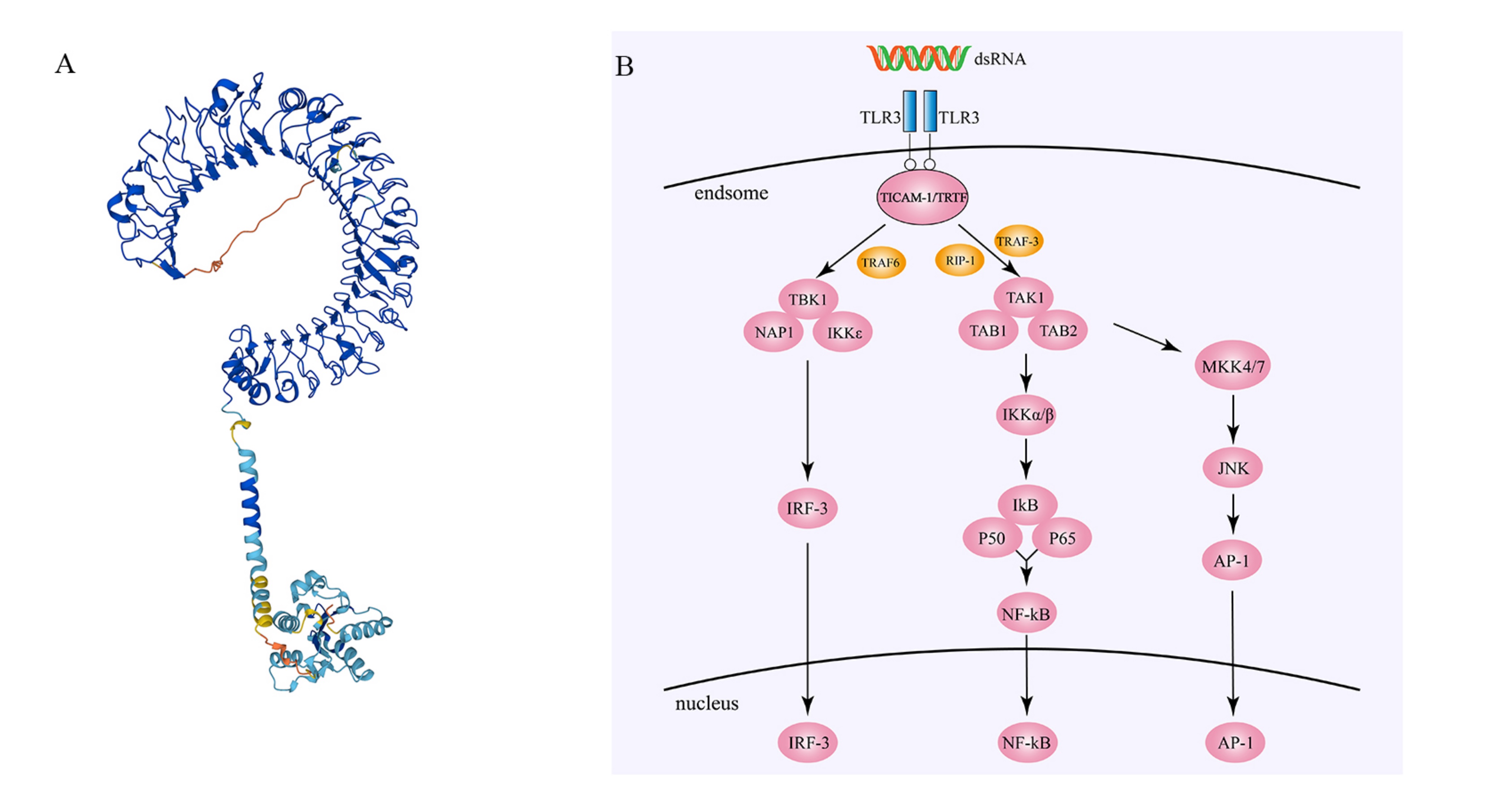
 β-sheet and an α-helix, which enable it to recognize and bind to dsRNA [43]. Below the LRR domain, the transmembrane region contains a membrane helix that anchors TLR3 in the cell membrane and transmits the signal from the extracellular region to the cytoplasm. In the cytoplasmic region, TLR3 includes a Toll/IL-1 receptor (TIR) domain, which mediates immune signaling by binding to downstream signaling molecules (Fig. 1) [44].

Fig. 1.

Structure and regulatory mechanisms of Toll-like receptor 3 (TLR3). (A) The three-dimensional structure of Toll-like receptor 3 (source: https://alphafold.ebi.ac.uk/entry/O15455, Copyright [2023], permission granted). (B) After double-stranded RNA (dsRNA) binds to two TLR3 molecules, the TIR domain of TLR3 binds to the TIR domain of TICAM-1/TRIF, and TRAF3 links TBK1 to TICAM-1/TRIF. TBK1, NAP1, and IKKϵ then form an activated complex, which mediates the activation of IRF3. Activated IRF3 translocates into the nucleus. The RHIM domain of RIP1 binds to the RHIM domain of TICAM-1/TRIF, leading to RIP1 activation. Activated RIP1 and TNF receptor-associated factor 6 (TRAF6) activate TGF-β-activated kinase 1 (TAK1), which then associates with TAK1-binding protein 1 (TAB1) and TAK1-binding protein 2 (TAB2) to form an activated complex. This complex activates IKKα and IKKβ, leading to the phosphorylation of IκB. The p50 and p65 subunits of nuclear factor kappa B (NF-κB) then form a heterodimer that translocates into the nucleus. When TAK1 associates with TAB1 and TAB2 to form an activated complex, it also activates mitogen-activated protein kinase kinase 4 (MKK4) and MKK7. Activated MKK4 and MKK7 then activate c-Jun N-terminal kinase (JNK), ultimately leading to the translocation of activated activator protein-1 (AP-1) into the nucleus.

2.2 Distribution and Expression of TLR3

TLR3 is expressed in various cell types, including dendritic cells, macrophages, epithelial cells, endothelial cells, and fibroblasts. In macrophages, fibroblasts, and certain epithelial cells, TRL3 is found both on the cell surface and within the cytoplasm. However, in dendritic cells, TLR3 is predominantly located in the endoplasmic reticulum (ER) and lysosomal membranes [45, 46]. In the ER, TLR3 forms a complex with other proteins, such as Unc-93 homolog B1 (UNC93B1), a transmembrane protein that helps TLR3-fold properly and localize correctly in the ER [47, 48, 49]. In lysosomes, TLR3 is involved in the recognition of viral RNA and regulates the immune response along with other proteins, such as TRIF and IRF3 [50]. Additionally, TLR3 can modulate its function by translocating to the cell membrane or nucleus, and upregulating UNC93B1 can promote TLR3 translocation to the cell membrane and activation by external dsRNA [51]. Some studies have also suggested that TLR3 can translocate from the ER to the nucleus after cellular stimulation, although the mechanism of this translocation is not yet clear [52]. In addition to the aforementioned subcellular locations, TLR3 also exists on the cell surface of cells such as in human fibroblasts and lung epithelial BEAS-2B cells [46, 53]. Despite some exploration of the subcellular localization of TLR3, further research is still needed.

2.3 Ligands for TLR3

The ligands of TLR3 are primarily double-stranded RNA (dsRNA), which is a double-stranded structure formed by complementary base pairing of two RNA strands. dsRNA is widely present both inside and outside of cells and is a structural component of the genomic RNA of many RNA viruses. dsRNA can enter cells through various pathways, such as viral infection, autophagy, and endocytosis. In addition to viral infection, dsRNA also has an endogenous source, as cellular RNA produced by tissue damage or cell death can serve as a potential source of dsRNA [54].

TLR3 exists as a monomer and is membrane-bound in resting cells. Upon binding of the extracellular domain (ECD) of TLR3 to dsRNA or other TLR3 agonists, dimerization occurs [55, 56]. The extracellular domain (ECD) of TLR3 undergoes dimerization upon binding to these dsRNAs [57]. Notably, the binding of TLR3-ECD to dsRNA requires an acidic environment [58]. TLR3-ECD binds to dsRNA at two sites located at the two ends of its “horseshoe-shaped” structure (N- and C-termini), forming a coordinated and stable dsRNA-TLR3 signaling complex composed of one dsRNA and two TLR3 molecules [59]. Subsequently, the oligomerization structure of TLR3 undergoes a change, causing TLR3 to be internalized with dsRNA into endoplasmic reticulum (ER) vesicles [60]. Inside ER vesicles, TLR3 binds to the TRIF adaptor protein. This thereby activates a cascade of signaling molecules, induces the expression of various cytokines and antiviral proteins, and promotes the activation of innate immune cells and immune responses.

2.4 The Signaling Pathways of TLR3

TLR3 is fully dependent on TICAM-1/TRIF for downstream signaling, and the TICAM-1/TRIF pathway ultimately activates AP-1, NF-κB, and IRF3, which together induce the expression of interferon (IFN)-β and inflammatory cytokines [61]. After activation by dsRNA, TLR3 undergoes dimerization and tyrosine phosphorylation. Subsequently, the Toll/interleukin-1 receptor (TIR) domain of TLR3 binds to the TIR domain of TICAM-1/TRIF. TICAM-1/TRIF contains an autoinhibitory N-terminal domain (NTD), a common TRAF6 binding motif, a TIR domain, and a C-terminal RHIM [27]. In this process, the NTD region of TICAM-1/TRIF is necessary for the activation of IRF3, and TRAF3 links TANK binding kinase 1 (TBK1) to TICAM-1/TRIF. TBK1, nucleosome assembly protein 1 (NAP1), and inhibitor of kappaB kinase epsilon (IKKε) form a complex that is activated, and activated TBK1 mediates the activation and phosphorylation of IRF3, which is translocated to the nucleus and activates IFN-β [29, 62]. The C-terminus of TICAM-1/TRIF is involved in the activation of NF-κB, and RIP1 binds to the C-terminus of TICAM-1/TRIF through the RHIM domain, leading to phosphorylation and polyubiquitination of RIP1. Together, RIP1 and TRAF6 act as activators of NF-κB. TRAF6 then activates TAK1, which binds with TAB1 and TAK1-binding protein 2 (TAB2) to form an activating complex that activates inhibitor of kappaB kinase (IKK)α/β. This results in the phosphorylation of IκB and the dissociation of NF-κB into the nucleus. NF-κB mainly exists as a p50-p65 dimer, and its activation in the nucleus plays a role in the immune response of the body, leading to a cascade of NF-κB reactions and the upregulation of the expression of inflammatory cytokines [11, 27]. In addition to activating IKKα/β, the activating complex can also activate AP-1 transcriptional responses mediated by MAPK. TAK1 phosphorylates the mitogen-activated protein kinase kinase (MKK) family, and phosphorylated MKK4 and MKK7 activate JNK, which ultimately activates AP-1 (Fig. 1) [27, 56].

The signaling pathway mediated by Toll-like receptor 3 is critical for host defense against pathogen invasion. However, excessive responses can also cause harmful damage to the host. It has been found that the E3 ubiquitin ligase TRIM32 can affect the recruitment of TBK1 to TRIF, thereby preventing further signal transduction by TRIF [63]. In addition, suppressor of cytokine signaling 1 (SOCS1) has been found to inhibit the cytokine receptor-associated signal transduction (JAK-STAT) pathway in the TLR3 signaling pathway [64], thus suppressing immune responses. It is noteworthy that SOCS1 can also be induced by cytokines (such as IFN-β) activated by the TLR3 signaling pathway and further inhibit the activation of the TLR3 signaling pathway [65]. Therefore, regulating the TLR3 signaling pathway through negative regulatory mechanisms plays an important role in maintaining immune system homeostasis and regulating immune function.

In addition to its own regulatory mechanisms, TLR3 can also cross-regulate other signaling pathways and participate in various immune responses [66]. Retinoic acid-inducible gene I (RIG-I)-like receptors (RLRs) can also recognize dsRNA and trigger interferon responses to inhibit viral replication and spread. Studies have shown that there is cross-regulation between the TLR3 and RIG-I pathways. TLR3 and RIG-I can mutually activate each other, thereby enhancing interferon production and synergistically acting against viral infections [67]. In addition, TLR3 and TLR4 act together on the TIR domain. It has been found that the TIR domains of TLR3 and TLR4 can form heterodimers and coactivate downstream signaling pathways, thereby enhancing host immune responses against pathogens [68]. However, the TLR3 and TLR4 signaling pathways can activate both the TRIF pathway and the MyD88 pathway, playing important roles in host immune responses [69].

TLR3 expression in tumors is not limited to the endoplasmic reticulum membrane. Upon activation in tumors, TLR3 triggers the activation of IRF3. This leads to the production of IFN-β, which inhibits tumor growth and angiogenesis. It also enhances the activation and proliferation of tumor-specific T cells while simultaneously activating caspase-dependent cell apoptosis. However, these effects of TLR3 may predominantly occur in the endoplasmic reticulum and endosomes. The study by Kentaro Yoneda et al. [70] suggested that in hepatocellular carcinoma (HCC) cells, membrane-bound TLR3 is responsible for the activation of NF-κB, which promote tumor metastasis; however, intracellular TLR3 is involved in tumor cell apoptosis. Similar conclusions have been drawn in metastatic intestinal epithelial cells (metastatic IECs), where the activation of surface-bound TLR3 by TLR3 agonists induces the production of C-X-C motif chemokine ligand 10 (CXCL10), which promotes tumor cell infiltration [71]. TLR3 in tumor cells acts as a double-edged sword, but its benefits outweigh its drawbacks. Thus, the application of various TLR3 agonists should be considered. Due to the potential toxic side effects of TLR3 agonists, they are often used in combination with other drugs to enhance efficacy, reduce drug resistance, and minimize the protumorigenic effects of TLR3 [34]. Poly(I:C), one of the most widely studied TLR3 agonists, in combination with paclitaxel inhibits the proliferation of paclitaxel-resistant colorectal cancer cells in vitro [72]. In oral squamous cell carcinoma, the combination of poly(I:C) and cisplatin reduces the dose of cisplatin and minimizes adverse reactions [73]. Other TLR agonists, such as poly(IC:LC), when combined with sorafenib and cetuximab, enhance tumor control and local immunity in vivo [74, 75]. TLR3 agonists can also contribute to the enhanced efficacy of immune checkpoint blockade [76]. Due to the high toxicity and short half-life of poly(I:C), nanomaterial packaging of poly(I:C), such as using Poly(lactic-co-glycolic acid) (PLGA) microspheres or liposomes, can increase drug uptake and improve drug delivery to target organs [35, 77]. The widespread distribution of TLR3 in various tumors forms the basis for its utilization in antitumor strategies.

Studying the regulatory mechanisms of TLR3 is of great significance for a deeper understanding of how the immune system fights against pathogens and for providing a scientific basis for the development of more effective vaccines and antitumor drugs. Furthermore, by investigating the regulatory mechanisms of the TLR3 signaling pathway, we can also better understand the pathogenesis of autoimmune diseases, cancer immunotherapy, and other fields and provide a scientific basis for the development of new treatment strategies, which has important clinical implications.

2.5 TLR3 and Cancer Immunity

The tumor immune microenvironment (TME) refers to a complex system composed of various cells and molecules adjacent to cancer cells, including immune cells, vascular endothelial cells, fibroblasts, matrix molecules, cytokines, and chemokines, among others [78]. These components interact with each other within the tumor microenvironment, influencing cancer occurrence, development, and treatment outcomes. TLR3 is expressed on various immune cells, including epithelial cells, T cells, B cells, NK cells, and dendritic cells (DCs) [79, 80, 81]. Several studies have shown that TLR3 induces the production of IL-12 and IFN via the TICAM-1 (TRIF) signaling pathway. Thus, it promotes the maturation of CD8α+ cDCs in mice and promotes the activation of NK cells and cytotoxic T lymphocytes (CTLs) to inhibit cancer growth [82, 83, 84, 85]. In patients with FPR1 deficiency, the antigen presentation function mediated by DCs is impaired. As a result, DCs are unable to closely interact with apoptotic cancer cells and fail to efficiently cross-present antigens. Consequently, these patients develop resistance to tumor chemotherapy. However, the use of the TLR3 agonist poly(I:C) has been shown to completely restore the immune deficiency caused by ANXA1/FPR1 impairment [86]. Furthermore, the study by Lanxiang Huang et al. [87] highlighted that TLR3 agonists and immunogenic cell death (ICD) inducers have a synergistic effect in enhancing the maturation of DCs within the tumor microenvironment. Thus, the exposure of tumor antigens is accelerated, and the uptake of dying tumor cells by DCs is promoted. This further enhances the immunogenicity of tumor cells [87]. To address potential inflammation caused by TLR3 agonists, a novel TLR3 agonist called ARNAX has been proposed. By binding to tumor-associated antigens (TAAs), it promotes the generation of TAA-specific CTLs, offering a potential therapeutic option for patients who are unresponsive to PD-1/PD-L1 inhibitors [88]. Macrophages play complex roles in cancer biology, and their presence in the tumor microenvironment may be associated with an increase in tumor size [89]. Research by Hiroaki Shime et al. [90] has shown that TLR3 can induce tumor-associated macrophages (Mfs) to switch from a state that promotes tumor growth to one with anticancer activity. Aurobind Vidyarthi et al. [91] found that TLR3 can shift M2 macrophages toward the M1 phenotype via IFN-αβ signaling. Macrophages of the M1 subtype display anticancer activity, while M2 macrophages can promote cancer development [92, 93]. These studies demonstrate the crucial role of TLR3 in regulating macrophage polarization and function. Recent research has been demonstrated that the frequency of myeloid-derived suppressor cells (MDSCs), which promote cancer growth and suppress the immune system, can be decreased by the activation of TLR3 [94]. Additionally, the ROS/RNS generated via the TICAM-1 pathway downstream of TLR3 activation are critical for enhancing the anticancer activity of MDSCs [95]. Furthermore, Zhongying Guo et al. [96] found that treatment of mice with dsRNA analogs resulted in a significant increase in the levels of INF-γ in the liver. INF-γ is an effective inhibitor of endothelial cell proliferation and cancer-associated angiogenesis. Thus, TLR3 may inhibit angiogenesis and alter the tumor microenvironment [96]. These findings provide insights into the potential therapeutic applications of TLR3 modulation in cancer immunotherapy.

In summary, TLR3 plays a critical role in the tumor microenvironment (TME), but research on the immune microenvironment of digestive system tumors involving TLR3 is currently limited. Using The Cancer Genome Atlas Program (TCGA) database and ssGSEA method, we evaluated the expression levels of TLR3 in 22 immune cell subtypes and presented its distribution in digestive system-related tumors in Fig. 2. Additionally, we applied the ESTIMATE algorithm to estimate the stromal score and immune score of TLR3 in digestive system tumors and demonstrated the correlation between TLR3 and the tumor microenvironment in Fig. 2. We found that TLR3 is correlated with the immune microenvironment of most digestive system tumors. These findings offer insight into the potential therapeutic applications of TLR3 modulation in digestive system tumors.
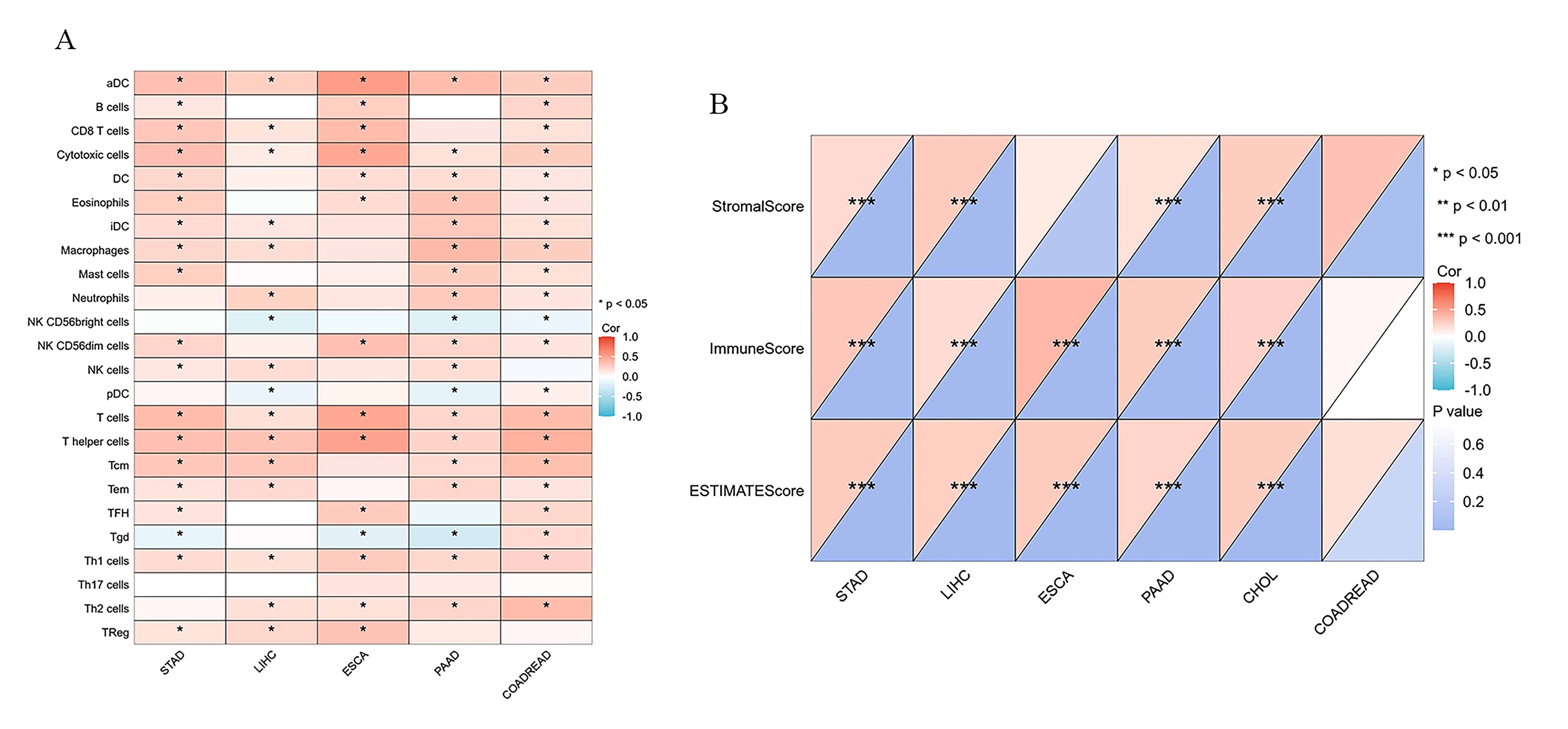
Fig. 2.

Correlation of TLR3 with immune infiltration in digestive system tumors. (A) The relationship between TLR3 and immune cell subtype infiltration in digestive system tumors was analyzed using the ssGSEA algorithm based on The Cancer Genome Atlas Program (TCGA) database. (B) The correlation between TLR3 and tumor immune score, stromal score, and ESTIMATE score was analyzed based on the ESTIMATE algorithm. Correlations were determined using the Pearson method, with *p < 0.05, **p < 0.01, and ***p < 0.001.

Therefore, we infer that the activation of the TLR3 signaling pathway may promote the infiltration and activation of immune cells to enhance the expression and presentation of tumor antigens, thus enhancing the antitumor immune response in most digestive system tumors. These findings provide a potential therapeutic target for improving the immune microenvironment of digestive system tumors.

3. Digestive System Tumors
3.1 Esophageal Cancer

Esophageal cancer (EC) is divided into two major types: esophageal adenocarcinoma (EAC) and esophageal squamous cell carcinoma (ESCC). Among all ECs, 87% are ESCC, but EAC is spreading in developed countries in Europe and America [97, 98]. Although the survival rate of esophageal cancer has significantly increased over the past fifty years, the five-year survival rate remains below 20% [98]. Early detection difficulties and limitations in late-stage treatments are obstacles to improving survival for patients with esophageal cancer [99]. Chronic inflammatory stimuli, such as smoking, alcohol consumption, spicy foods, Barrett’s esophagus, and gastroesophageal reflux disease (GERD), may contribute to the development of esophageal cancer [100].

The TLR family is involved in the inflammatory response in esophageal cancer. The expression of TLR3 gradually increases as the disease progresses, from esophageal simple hyperplasia (ESSH) and intraepithelial neoplasia (IEN) to esophageal squamous cell carcinoma, and TLR3 expression gradually increases in tumor tissue. Cases with high TLR3 expression also have a higher rate of lymph node metastasis [101, 102, 103]. Patients with high expression of TLR3 in esophageal cancer tissues demonstrate a better 5-year survival rate than those with low expression, and they also exhibit increased sensitivity to chemotherapy. This outcome is associated with the correlation between high TLR3 expression and infiltration and activation of immune effector cells, as well as the involvement of apoptotic pathways. Therefore, for esophageal cancer patients with high TLR3 expression, adjuvant chemotherapy following surgery may further improve the prognosis of advanced-stage patients [100, 101]. The activation of the TLR3-mediated inflammatory response by necrotic esophageal epithelial cells upregulates the expression of interleukin-8 and NF-κB, which are believed to play an important role in the development and progression of Barret’s esophagus [104]. TLR3-activated cancer cells release cytokines and chemokines during esophageal cancer development, recruiting and stimulating immune cells to release additional cytokines and chemokines. Due to the increased expression of TLR3 in esophageal cancer tissues, TLR3 has been identified as a potential therapeutic target. TLR3 agonists can serve as adjuvants in tumor vaccines and have demonstrated promising results in phase I clinical trials. Combination therapy with TLR3 agonists has the potential to overcome the inhibitory tumor microenvironment (TME), restoring the balance of TMEs and exerting antitumor effects [105]. This may also be the reason why patients with high TLR3 expression in esophageal cancer have a better prognosis [100]. However, the specific function and potential mechanisms of TLR3 in the pathogenesis of esophageal cancer are still not fully understood. Further research on the role of TLR3 in the progression of esophageal cancer may provide a new perspective for the diagnosis and treatment of this disease.

3.2 Gastric Cancer

Gastric cancer is the fifth most common cancer and the third leading cause of cancer-related deaths worldwide [106]. Among the various pathological types, gastric adenocarcinoma (GAC) is the most prevalent and is typically treated with a multidisciplinary approach involving surgery and chemotherapy. Ning Wang and Dingsheng Liu [107] found that TLR3 mRNA expression in GAC tissues is significantly lower than that in normal gastric tissues. Furthermore, copy number variation (CNV) analysis indicated a significant deletion of TLR3 DNA copies [107]. In contrast, Belen Fernandez-Garcia et al. [108] indicated that the expression of TLR3 in gastric cancer cells, as detected by immunohistochemistry (IHC), is relatively higher than that in stromal cells. Furthermore, high expression levels of TLR3 in gastric cancer cells are significantly associated with unfavorable pathological types and lower overall survival rates in patients [108]. A retrospective study involving 564 patients with gastric adenocarcinoma revealed that patients with high nuclear expression of TLR3 had a lower 5-year survival rate. However, there was no significant difference in the expression of TLR3 in the cytoplasm [109]. Zhihao Huang et al. [110] also noted a positive correlation between TLR3 expression levels and various immune biomarkers, including CD8+ T cells, CD4+ T cells, macrophages, neutrophils, dendritic cells, and others. Additionally, they found that high expression of TLR3, compared to that in normal tissues, was associated with increased resistance to treatment in patients [110]. In addition, another study found that the TLR3 agonist poly(I:C) can induce apoptosis of human gastric adenocarcinoma cells through endogenous delivery and inhibit the growth of human gastric adenocarcinoma in a nude mouse model [111]. Therefore, further investigation of the role played by TLR3 in the progression of gastric cancer is expected to provide a promising prognostic marker and a potential viable target for immunotherapy in the treatment of gastric cancer.

3.3 Hepatocellular Carcinoma

Hepatocellular carcinoma (HCC) accounts for approximately 90% of primary liver cancers. Risk factors associated with HCC include viral, metabolic, and immune-related diseases [112, 113]. The incidence of HCC in the middle-aged population (30–59 years old) has significantly decreased worldwide, mainly due to the successful implementation of the hepatitis B virus (HBV) vaccination program. Liver transplantation and surgical resection are early treatment options for HCC. However, most patients are diagnosed at an advanced stage [114]. Therefore, exploring new treatment directions is necessary. Researchers have found substantial expression levels of TLR3 in the cell membrane and cytoplasm of HCC cells, suggesting the need to investigate the role of TLR3 in HCC [115, 116].

Marc Bonnin et al. [115] found through multimodal experiments that in primary liver cancer tissues, the mRNA and protein expression levels of TLR3 were lower than those in normal tissues. Chronic infection with HBV and HCV can also lead to decreased expression of TLR3 in liver cells, which may be attributed to the suppression of the hepatic pro-inflammatory system. However, there are differing views on the expression changes of TLR3 during the stages of chronic hepatitis, liver cirrhosis, and hepatocellular carcinoma, which may be influenced by the use of antiviral medications [117]. In addition, Cheng Zhou et al. [118] also discovered that HCC patients with high TLR3 expression have a more complete capsule. High expression of TLR3 can promote the activation of natural killer (NK) cells and increase their cytotoxicity. This finding suggests that HCC patients with high TLR3 expression may have a longer overall survival [119]. Weiyun Li et al. [120] found that serine/threonine protein kinase 4 (STK4) could regulate the TLR3-mediated inflammatory response in macrophages, thereby inhibiting the progression of chronic inflammation to hepatocellular carcinoma. Additionally, in a study conducted by Cheng Zhou et al. [118], lenvatinib enhanced tumor apoptosis by upregulating TLR3 expression in Huh 7 and MHCC-97H cells. In a transgenic mouse study by Marc Bonnin et al. [115], the absence of TLR3 led to a reduction in apoptosis rates in precancerous liver cells and accelerated liver cancer development without affecting tumor malignancy and tumor size. In addition, it did not affect tumor immune infiltration. These findings suggest a potential beneficial role of TLR3 in HCC [115]. However, not all studies support this notion. One study suggested that low TLR3 expression may be associated with smaller tumor size. Moreover, TLR3 mutations can influence the progression from precancerous lesions to liver cancer, indicating that the TLR3+1234CT and TLR3+1234TT genotypes may be risk factors for the development of HCC in HBV- and HCV-related liver cirrhosis [121, 122].

Most HCC occurs in patients with chronic liver diseases, especially viral hepatitis (i.e., HBV and HCV) and liver cirrhosis caused by various etiologies [123]. Studies have shown that using TLR3 agonists can significantly inhibit HBV replication [116, 124, 125], and the upregulation of TLR3 can promote apoptosis of HBV-positive liver cancer cells [116]. We speculate that TLR3 may enhance the control of HBV by the hepatic intrinsic immune system by recognizing HBV virus RNA, thereby increasing the resistance of liver cells to HBV. Similarly, regarding HCV, spontaneous clearance of HCV infection was found in treated patients with elevated TLR3 expression, indicating a role for TLR3 in the existing antiviral state against HCV infection [117, 126].

dsRNA, as an important ligand of TLR3, plays a crucial role in host defense and disease prevention. BM-06 and poly(I:C) are two analogs of dsRNA, and their stimulation of liver cancer cells can significantly inhibit apoptosis and migration while inducing autophagy [96, 127, 128, 129]. However, the inhibitory effects of TLR3 agonists alone are limited. Peng Shen et al. [130] found that poly(I:C) can inhibit HCC proliferation and induce apoptosis through TLR3-dependent mechanisms. Furthermore, when used in combination with arsenic trioxide (ATO), poly(I:C) can enhance ATO-induced apoptosis in SMMC7721 cells by promoting ROS-dependent mitochondrial membrane potential loss (Dym loss) Nevertheless, some studies have shown that low concentrations of poly(I:C) not only fail to significantly inhibit the proliferation of liver cancer cells and induce cell apoptosis but also increase their migration and invasion ability.

3.4 Pancreatic Cancer

Pancreatic cancer (PC) is a highly malignant tumor and the seventh leading cause of cancer-related deaths [131]. Current treatment options, such as the modified FOLFIRINOX regimen, have limited efficacy in improving patient survival rates. In recent years, immunotherapy has emerged as a new treatment option for pancreatic cancer [132]. Spas Dimitrov Markov et al. [133] utilized a vaccine targeting MUC1 with IgE in combination with a TLR3 agonist and immune checkpoint inhibitors to induce NK cell and CD8+ T-cell-dependent antitumor responses in mice. Studies have shown that poly(I:C) can enhance the cytotoxic effects of tyrosine kinase inhibitors (TKIs) on human pancreatic cancer cell lines (hPDA) and can induce hPDA cell line lysis in vitro by enhancing the cytotoxic activity of γδT cells. Knockdown of TLR3 results in a weakened cytotoxic effect of interferon-alpha (IFN-α) combined with regorafenib [134, 135]. Rintatolimod (Ampligen®) is a TLR3 agonist that has been investigated for cancer treatment. In a single-center study involving 27 patients with locally advanced pancreatic cancer (LAPC), the combination therapy of Folfirinox and Rintatolimod demonstrated significant reductions in the neutrophil-to-lymphocyte ratio (NLR) and systemic immune inflammation index (SIII) among long-term survivors and increased levels of circulating B cells, leading to extended overall survival [136]. Furthermore, the combination of a poly(IC:LC)-activated autologous dendritic cell vaccine with peptides has been shown to induce the generation of tumor-specific T-cell responses. This treatment prolonged the survival of patients with advanced pancreatic cancer and exhibited good tolerability [137]. TLR3 agonists have also been combined with other vaccines, such as the ISCOMATRIX vaccine, to inhibit the development of pancreatic cancer [138].

However, some studies have shown that the expression of TLR3 in PC cells is significantly higher than that in normal pancreatic ductal epithelial cells, and the overexpression of TLR3 promotes malignant behaviors, such as proliferation, migration, and invasion of PC cells [139]. Additionally, it has been reported that poly(I:C) can promote the occurrence of PC in KRAS mutant mice [140]. Benzimidazole (C10) is a TLR3 inhibitor that does not rely on Wnt5a, and it can significantly inhibit the ability of hPDA cells to proliferate, migrate, and invade in vitro [141]. Therefore, despite the varying roles of TLR3 observed in PC in different studies, the potential of TLR3 agonists in the treatment of pancreatic cancer is evident. Consequently, TLR3 agonists hold promise as a future therapeutic option for pancreatic cancer.

3.5 Colorectal Cancer

Colorectal cancer (CRC) is the second leading cause of cancer-related deaths worldwide, and the inflammatory pathway promotes its development [142]. Patients with inflammatory bowel diseases, including Crohn’s disease and ulcerative colitis, have a significantly increased risk of developing CRC [143]. In CRC research, the efficacy of TLR3 agonists is diminished when facing tumors with KRAS mutations. However, Shiqi Long et al. [144] proposed a solution to this problem by combining respiratory syncytial virus and cetuximab. Both respiratory syncytial virus and poly(I:C) serve as TLR3 agonists and can enhance antibody-dependent cell-mediated cytotoxicity (ADCC) against CRC cells by activating TLR3 and affecting the IKKε/IBK signaling pathway. The combined use of respiratory syncytial virus and cetuximab can reduce drug resistance in CRC cells with wild-type KRAS and low EGFR expression [144]. While respiratory syncytial virus faces obstacles in the form of neutralizing antibody (nAb) clearance upon entering the human body, carrying it via peripheral blood mononuclear cells (PBMCs) or dendritic cells (DCs) shows promise in overcoming this issue. Shania Marie Corry et al. [145] also demonstrated that the TLR3 agonist poly(I:C) can reduce recurrence and liver metastasis of matrix-rich subtypes in stage II/III colon cancer. Poly(I:C) can enhance the cytotoxicity of paclitaxel through the TLR3-UNC93B1-IFN-β signaling pathway and synergize with 5Z-7-oxozeaenol and IDN-6556 (P5I) to produce a potent proapoptotic effect in CRC [72, 146]. These results further demonstrate that TLR3 agonists can inhibit the development of CRC alone or in combination with other drugs via the TLR3 signaling pathway [147]. TLR3 activation can suppress CRC, but the consumption of its agonists in vivo severely hinders its potential applications. Yingying Li et al. [148] extended the half-life of poly(I:C) in vivo by delivering it with polyethylene glycol-coated nanoscale coordination polymer (NCP). This provides new possibilities for the treatment of CRC with TLR3 agonists. TLR3 can serve not only as a critical node for treating CRC but also as an independent prognostic marker for early malignant transformation of colonic polyps and stage II CRC [149, 150].

3.6 Cholangiocarcinoma

Cholangiocarcinoma (CCA) is a malignant tumor that originates from the epithelium of the bile duct, accounting for 3% of gastrointestinal tumors. In recent years, the incidence of CCA has increased, but the specific etiology has not been determined [151]. Chronic inflammation (such as cirrhosis, viral hepatitis, primary sclerosing cholangitis, biliary infections, and parasitic infections) and an immunosuppressive tumor microenvironment are generally considered risk factors for CCA [152]. Research on TLR3 in CCA is not yet fully developed, and only Thanpisit Lomphithak et al. [153] mentioned TLR3 and its activators in a new CCA treatment direction in 2020. Using in vitro experiments, Thanpisit Lomphithak et al. [153] confirmed that the combination of poly(I:C) with an SMAC mimetic can induce apoptosis and necroptosis in cholangiocarcinoma cells. While poly(I:C) induces apoptosis and enhances invasion, the SMAC mimetic inhibits cIAP to release RIPK1-dependent cell death and reduce invasion. Although the exact mechanism of reduced invasion is unknown, in vitro experiments support this finding. Therefore, this study presents a novel treatment approach of combining poly(I:C) and SMAC mimetics in patients with tumors overexpressing RIPK1 to inhibit tumor invasion and increase cancer cell apoptosis and necroptosis. This idea provides a new option for CCA treatment, although it may only be effective in patients with high RIPK1 expression in tumor tissue [153].

4. Discussion

TLR3 is generally expressed in tumors, but its role and mechanism in different cancers are very diverse. In digestive tumors, the expression of TLR3 in tumor cells is often higher than that in normal tissues (Table 1, Ref. [97, 100, 105, 108, 111, 115, 116, 118, 121, 127, 129, 130, 134, 135, 139, 149, 153]). High expression of TLR3 can serve as a prognostic indicator in cancer patients, as it may increase the invasive ability of tumors. However, it is also associated with a favorable response to chemotherapy. High expression of TLR3 may be associated with a longer overall survival, which could be attributed to the intrinsic antitumor response of TLR3. In liver cancer tissues, the expression of TLR3 gradually increases during the progression of hepatitis and liver cirrhosis but decreases in liver cancer tissues. However, it remains higher than in normal tissues. This phenomenon may be attributed to the immune evasion mechanisms of liver cancer [115]. In the tumor microenvironment, dendritic cells (DCs) act as the most effective antigen-presenting cells (APCs) to initiate and modulate innate and adaptive immunity. However, the tumor microenvironment suppresses the maturation of DCs. Therefore, using TLR3 agonists to enhance the anticancer effect to treat tumors seems to be a feasible direction for conventional DCs (cDCs). In a study conducted by Huang et al. [87], an in situ dendritic cell (DC) vaccine, HELA-Exos, was constructed by loading the human neutrophil elastase protein (ELANE) and the TLR3 agonist Hiltonol into exosomes. It can specifically induce the immunogenic cell death (ICD) of breast cancer cells, thereby activating the antigen presentation function of cDC1s and cross-activating tumor-reactive CD8+ T cells. Thus it plays an anticancer role [87]. The use of vaccines or drugs that transport TLR3 agonists and other anticancer drugs through specific carriers has been studied and even applied in many types of cancers. TLR3 agonists have been combined with 5-fluorouracil or IFN-α to treat colorectal cancer [147]. Combined therapy with TLR3 agonists and other drugs has also been preliminarily validated in pancreatic and liver cancer. Several TLR3 agonists, such as poly(I:C), poly(A:U), BM-06, Ampligen®, and Hiltonol®, can be utilized as immune adjuvants in the human body to enhance the efficacy of cancer vaccines. They can also be combined with radiation therapy, chemotherapy, targeted therapy, and immunotherapy to improve the efficiency and tolerability of gastrointestinal tumor treatments.

Table 1.The clinical pathological characteristics and prognostic significance of TLR3 in digestive system tumors.
Cancer type Sample Alteration Related clinical and pathological features Significance Reference
Esophageal cancer Esophageal cancer tissue / / Potential immunologic adjuvant [105]
Esophageal cancer tissue / High expression has better overall and disease specific survival at 5 years; high expression has stronger sensitivity to chemotherapy Independent prognostic factors [100]
KYSE140 KYSE150 KYSE180 KYSE220 KYSE270 Upregulation / / [100]
Gastric cancer Gastric cancer tissue Upregulation Patients with high TLR3 expression have poor prognosis Negative regulator [108]
BGC-823 / / Antitumor activity [111]
Hepatocellular carcinoma Primary liver cancer tissue Downregulation High expression has better recurrence free survival Antitumor activity [115]
Primary liver cancer tissue / High expression has better prognosis and smaller tumors Antitumor activity [118]
SK-HEP-1 Hep3B PLC/PRF/5 HepG2 Focus HUh7 Downregulation / Antitumor activity [115]
Primary liver cancer tissue No difference The positive rate of TLR3 is negatively correlated with serum AFP levels, while HBsAg infection is positively correlated Antitumor activity [116]
Huh 7 / / Antitumor activity [97, 127, 129, 130]
MHCC-97H SMMC7721
HepG2 HepG2.2.15
Primary liver cancer tissue / Positive correlation with tumor size Negative regulator [121]
Pancreatic cancer BXPC-3 PAC1 Upregulation / Negative regulator [139]
HPDA / / Antitumor activity [134]
Panc 89 PancTul Colo 357 / / Antitumor activity [135]
Colorectal cancer Colorectal adenocarcinoma tissue Downregulation / / [149]
Cholangiocarcinoma Cholangiocarcinoma tissue Upregulation / / [153]

HPDA, human pancreatic cancer cell lines.

Furthermore, the mechanisms of TLR3 agonist therapy for digestive system tumors are not yet fully understood. Their interactions with other signaling pathways, differential expression and functions in different types and stages of tumors and their regulatory role in the tumor microenvironment require further exploration. Additionally, the toxicity of TLR3 agonists should be carefully considered. TLR3 has emerged as a new breakthrough in the treatment of digestive system tumors, and in the future, TLR3 agonists may extend their remarkable potential beyond digestive system tumors.

5. Conclusion

TLR3 is widely expressed in digestive system tumors, and its activation generally promotes tumor cell apoptosis and inhibits tumor progression. However, the effects of TLR3 may vary depending on its subcellular localization. This highlights that TLR3 agonists are not suitable for standalone therapy in digestive system tumors but rather as adjuvants in combination with other chemotherapy drugs, targeted agents, immune checkpoint inhibitors, etc. This combination approach allows for maximizing the impact on tumors and human tolerance. In the case of precancerous lesions in the digestive system, TLR3 expression undergoes a dynamic process, generally showing an increase compared to normal tissues. This may be attributed to the occurrence of chronic inflammation, leading to prolonged activation of the TLR3 pathway. However, research on TLR3 agonists in the context of precancerous lesions is still limited. Overall, TLR3 agonists have promising prospects in the treatment of digestive system tumors, as several are already in clinical trial stages and showing positive outcomes. Currently, TLR3 agonists have been used in human cancer treatment, but challenges remain in terms of their appropriate use and the development of combination treatment strategies.

Abbreviations

AP-1, activating protein-1; CNV, copy number variation; dsRNA, double-stranded RNA; GSEA, gene set enrichment analysis; IFN-α, interferon-alpha; IKKα, inhibitor of kappaB kinase alpha; IKKβ, inhibitor of kappaB kinase beta; IKKε, inhibitor of kappaB kinase epsilon; IL-1, interleukin-1; IL-12, interleukin-12; IRAK, IL-1R-associated kinase; IRF3, interferon regulatory factor 3; JNK, JUN N-terminal kinase; HIF-1α, hypoxia-inducible factor 1-alpha; Mal, MyD88-adaptor-like; MAPK, mitogen-activated protein kinase; MKK4, mitogen-activated protein kinase kinase 4; MyD88, MyD88 innate immune signal transduction adaptor; NAP1, nucleosome assembly protein 1; NF-κB, nuclear factor kappa B; p50, nuclear factor NF kappa B p50 subunit; p65, nuclear factor NF kappa B p65 subunit; PAMP, pathogen associated molecular patterns; Smac, second mitochondria-derived activator of caspases; SOCS1, suppressor of cytokine signaling 1; TAB1, TGF-Beta activated kinase 1 binding protein 1; TAB2, TGF-Beta activated kinase 1 binding protein 2; TAK1, transforming growth factor beta-activated kinase 1; TBK1, TANK binding kinase 1; TCGA, the cancer genome atlas; TICAM-1, TIR domain-containing adaptor molecule; TIRAP, TIR domain-containing adaptor protein; TIRAM1, TIR domain-containing adapter molecule-1; TRIF, TIR domain-containing adapter inducing interferon-β; TRAF3, TNF receptor associated factor 3; TRAF6, TNF receptor associated factor 6; TRAM, TRIF-related adaptor molecule; RIP1, receptor interacting serine/threonine protein kinase 1; Wnt, wingless/integrated.

Availability of Data and Materials

The data used in this study were obtained from publicly available databases, specifically, TCGA datasets.

Author Contributions

BH, LZ, TL, JH, and HZ conceptualized the study idea for the review. BH and CZ performed the article’s editing. BH and XW created all figures. CZ and HS analyzed and interpreted the data from TCGA. BH and CZ wrote the manuscript. LZ, TL, JH, and HZ made significant revisions to the manuscript. All authors contributed to editorial changes in the manuscript. All authors read and approved the final version. All authors participated sufficiently in the work and agreed to be accountable for all aspects of the study.

Ethics Approval and Consent to Participate

Not applicable.

Acknowledgment

The author thanks current members of the laboratory for their valuable contributions.

Funding

This article was supported by the National Natural Science Foundation of China (Grant No. 82073090), Human Resources and Social Security Department System of Shanxi Province (Grant No. 20210001), Research Project Supported by Shanxi Scholarship Council of China (Grant No. 2021-116), Shanxi ‘136’ Leading Clinical Key Specialty (Grant No. 2019XY002), and Key Laboratory of Hepatobiliary and Pancreatic Diseases of Shanxi Province (Preparatory).

Conflict of Interest

The authors declare that the research was conducted in the absence of any commercial or financial relationships that could be construed as a potential conflict of interest.

References
[1]
Organization WH. CANCER TODAY. 2020. Available at: https://gco.iarc.fr/today (Accessed: 30 April 2023).
[2]
Organization WH. Cancer. 2022. Available at: https://www.who.int/news-room/fact-sheets/detail/cancer (Accessed: 30 April 2023).
[3]
Elinav E, Nowarski R, Thaiss CA, Hu B, Jin C, Flavell RA. Inflammation-induced cancer: crosstalk between tumours, immune cells and microorganisms. Nature Reviews. Cancer. 2013; 13: 759–771.
[4]
Mantovani A, Allavena P, Sica A, Balkwill F. Cancer-related inflammation. Nature. 2008; 454: 436–444.
[5]
Coussens LM, Werb Z. Inflammation and cancer. Nature. 2002; 420: 860–867.
[6]
Zou X, Guo Y, Mo Z. TLR3 serves as a novel diagnostic and prognostic biomarker and is closely correlated with immune microenvironment in three types of cancer. Frontiers in Genetics. 2022; 13: 905988.
[7]
Orlacchio A, Mazzone P. The Role of Toll-like Receptors (TLRs) Mediated Inflammation in Pancreatic Cancer Pathophysiology. International Journal of Molecular Sciences. 2021; 22: 12743.
[8]
Chen JQ, Szodoray P, Zeher M. Toll-Like Receptor Pathways in Autoimmune Diseases. Clinical Reviews in Allergy & Immunology. 2016; 50: 1–17.
[9]
Vidya MK, Kumar VG, Sejian V, Bagath M, Krishnan G, Bhatta R. Toll-like receptors: Significance, ligands, signaling pathways, and functions in mammals. International Reviews of Immunology. 2018; 37: 20–36.
[10]
Hashimoto C, Hudson KL, Anderson KV. The Toll gene of Drosophila, required for dorsal-ventral embryonic polarity, appears to encode a transmembrane protein. Cell. 1988; 52: 269–279.
[11]
Blasius AL, Beutler B. Intracellular toll-like receptors. Immunity. 2010; 32: 305–315.
[12]
Marks KE, Cho K, Stickling C, Reynolds JM. Toll-like Receptor 2 in Autoimmune Inflammation. Immune Network. 2021; 21: e18.
[13]
Lu BL, Williams GM, Brimble MA. TLR2 agonists and their structure-activity relationships. Organic & Biomolecular Chemistry. 2020; 18: 5073–5094.
[14]
Shetab Boushehri MA, Lamprecht A. TLR4-Based Immunotherapeutics in Cancer: A Review of the Achievements and Shortcomings. Molecular Pharmaceutics. 2018; 15: 4777–4800.
[15]
Jin S, Nepal N, Gao Y. The role of toll-like receptors in peptic ulcer disease. Immunological Medicine. 2022; 45: 69–78.
[16]
Kawai T, Akira S. The role of pattern-recognition receptors in innate immunity: update on Toll-like receptors. Nature Immunology. 2010; 11: 373–384.
[17]
Iwasaki A, Medzhitov R. Toll-like receptor control of the adaptive immune responses. Nature Immunology. 2004; 5: 987–995.
[18]
Ishida H, Asami J, Zhang Z, Nishizawa T, Shigematsu H, Ohto U, et al. Cryo-EM structures of Toll-like receptors in complex with UNC93B1. Nature Structural & Molecular Biology. 2021; 28: 173–180.
[19]
Gnjatic S, Sawhney NB, Bhardwaj N. Toll-like receptor agonists: are they good adjuvants? Cancer Journal (Sudbury, Mass.). 2010; 16: 382–391.
[20]
Takeda K, Kaisho T, Akira S. Toll-like receptors. Annual Review of Immunology. 2003; 21: 335–376.
[21]
Bell JK, Mullen GED, Leifer CA, Mazzoni A, Davies DR, Segal DM. Leucine-rich repeats and pathogen recognition in Toll-like receptors. Trends in Immunology. 2003; 24: 528–533.
[22]
Botos I, Segal DM, Davies DR. The structural biology of Toll-like receptors. Structure (London, England: 1993). 2011; 19: 447–459.
[23]
Kaisho T, Akira S. Toll-like receptor function and signaling. The Journal of Allergy and Clinical Immunology. 2006; 117: 979–987; quiz 988.
[24]
Loiarro M, Ruggiero V, Sette C. Targeting TLR/IL-1R signalling in human diseases. Mediators of Inflammation. 2010; 2010: 674363.
[25]
Sea Urchin Genome Sequencing Consortium, Sodergren E, Weinstock GM, Davidson EH, Cameron RA, Gibbs RA, et al. The genome of the sea urchin Strongylocentrotus purpuratus. Science (New York, N.Y.). 2006; 314: 941–952.
[26]
Bernard NJ, O’Neill LA. Mal, more than a bridge to MyD88. IUBMB Life. 2013; 65: 777–786.
[27]
Ullah MO, Sweet MJ, Mansell A, Kellie S, Kobe B. TRIF-dependent TLR signaling, its functions in host defense and inflammation, and its potential as a therapeutic target. Journal of Leukocyte Biology. 2016; 100: 27–45.
[28]
Rast JP, Smith LC, Loza-Coll M, Hibino T, Litman GW. Genomic insights into the immune system of the sea urchin. Science (New York, N.Y.). 2006; 314: 952–956.
[29]
Takeda K, Akira S. TLR signaling pathways. Seminars in Immunology. 2004; 16: 3–9.
[30]
Yamamoto M, Sato S, Mori K, Hoshino K, Takeuchi O, Takeda K, et al. Cutting edge: a novel Toll/IL-1 receptor domain-containing adapter that preferentially activates the IFN-beta promoter in the Toll-like receptor signaling. Journal of Immunology (Baltimore, Md.: 1950). 2002; 169: 6668–6672.
[31]
Kawai T, Akira S. Signaling to NF-kappaB by Toll-like receptors. Trends in Molecular Medicine. 2007; 13: 460–469.
[32]
Shafeghat M, Kazemian S, Aminorroaya A, Aryan Z, Rezaei N. Toll-like receptor 7 regulates cardiovascular diseases. International Immunopharmacology. 2022; 113: 109390.
[33]
Squillace S, Salvemini D. Toll-like receptor-mediated neuroinflammation: relevance for cognitive dysfunctions. Trends in Pharmacological Sciences. 2022; 43: 726–739.
[34]
Yang Y, Li H, Fotopoulou C, Cunnea P, Zhao X. Toll-like receptor-targeted anti-tumor therapies: Advances and challenges. Frontiers in Immunology. 2022; 13: 1049340.
[35]
Tran TH, Tran TTP, Truong DH, Nguyen HT, Pham TT, Yong CS, et al. Toll-like receptor-targeted particles: A paradigm to manipulate the tumor microenvironment for cancer immunotherapy. Acta Biomaterialia. 2019; 94: 82–96.
[36]
Zhang Y, Liu J, Wang C, Liu J, Lu W. Toll-Like Receptors Gene Polymorphisms in Autoimmune Disease. Frontiers in Immunology. 2021; 12: 672346.
[37]
Martín-Medina A, Cerón-Pisa N, Martinez-Font E, Shafiek H, Obrador-Hevia A, Sauleda J, et al. TLR/WNT: A Novel Relationship in Immunomodulation of Lung Cancer. International Journal of Molecular Sciences. 2022; 23: 6539.
[38]
Xun Y, Yang H, Kaminska B, You H. Toll-like receptors and toll-like receptor-targeted immunotherapy against glioma. Journal of Hematology & Oncology. 2021; 14: 176.
[39]
Liu Z, Xie Y, Xiong Y, Liu S, Qiu C, Zhu Z, et al. TLR 7/8 agonist reverses oxaliplatin resistance in colorectal cancer via directing the myeloid-derived suppressor cells to tumoricidal M1-macrophages. Cancer Letters. 2020; 469: 173–185.
[40]
Ye J, Mills BN, Qin SS, Garrett-Larsen J, Murphy JD, Uccello TP, et al. Toll-like receptor 7/8 agonist R848 alters the immune tumor microenvironment and enhances SBRT-induced antitumor efficacy in murine models of pancreatic cancer. Journal for Immunotherapy of Cancer. 2022; 10: e004784.
[41]
Anfray C, Mainini F, Digifico E, Maeda A, Sironi M, Erreni M, et al. Intratumoral combination therapy with poly(I:C) and resiquimod synergistically triggers tumor-associated macrophages for effective systemic antitumoral immunity. Journal for Immunotherapy of Cancer. 2021; 9: e002408.
[42]
GeneCards. TLR3 Gene. 2023. Available at: https://www.genecards.org/cgi-bin/carddisp.pl?gene=TLR3 (Accessed: 30 April 2023).
[43]
Bell JK, Botos I, Hall PR, Askins J, Shiloach J, Segal DM, et al. The molecular structure of the Toll-like receptor 3 ligand-binding domain. Proceedings of the National Academy of Sciences of the United States of America. 2005; 102: 10976–10980.
[44]
Choe J, Kelker MS, Wilson IA. Crystal structure of human toll-like receptor 3 (TLR3) ectodomain. Science (New York, N.Y.). 2005; 309: 581–585.
[45]
Kadowaki N, Ho S, Antonenko S, Malefyt RW, Kastelein RA, Bazan F, et al. Subsets of human dendritic cell precursors express different toll-like receptors and respond to different microbial antigens. The Journal of Experimental Medicine. 2001; 194: 863–869.
[46]
Matsumoto M, Funami K, Tanabe M, Oshiumi H, Shingai M, Seto Y, et al. Subcellular localization of Toll-like receptor 3 in human dendritic cells. Journal of Immunology (Baltimore, Md.: 1950). 2003; 171: 3154–3162.
[47]
He Z, Ye S, Xing Y, Jiu Y, Zhong J. UNC93B1 curbs cytosolic DNA signaling by promoting STING degradation. European Journal of Immunology. 2021; 51: 1672–1685.
[48]
Brinkmann MM, Spooner E, Hoebe K, Beutler B, Ploegh HL, Kim YM. The interaction between the ER membrane protein UNC93B and TLR3, 7, and 9 is crucial for TLR signaling. The Journal of Cell Biology. 2007; 177: 265–275.
[49]
Kim YM, Brinkmann MM, Paquet ME, Ploegh HL. UNC93B1 delivers nucleotide-sensing toll-like receptors to endolysosomes. Nature. 2008; 452: 234–238.
[50]
Miyake K, Saitoh SI, Sato R, Shibata T, Fukui R, Murakami Y. Endolysosomal compartments as platforms for orchestrating innate immune and metabolic sensors. Journal of Leukocyte Biology. 2019; 106: 853–862.
[51]
Pohar J, Pirher N, Benčina M, Manček-Keber M, Jerala R. The role of UNC93B1 protein in surface localization of TLR3 receptor and in cell priming to nucleic acid agonists. The Journal of Biological Chemistry. 2013; 288: 442–454.
[52]
Johnsen IB, Nguyen TT, Ringdal M, Tryggestad AM, Bakke O, Lien E, et al. Toll-like receptor 3 associates with c-Src tyrosine kinase on endosomes to initiate antiviral signaling. The EMBO Journal. 2006; 25: 3335–3346.
[53]
Matsumoto M, Kikkawa S, Kohase M, Miyake K, Seya T. Establishment of a monoclonal antibody against human Toll-like receptor 3 that blocks double-stranded RNA-mediated signaling. Biochemical and Biophysical Research Communications. 2002; 293: 1364–1369.
[54]
Chen YG, Hur S. Cellular origins of dsRNA, their recognition and consequences. Nature Reviews. Molecular Cell Biology. 2022; 23: 286–301.
[55]
Cheng K, Wang X, Yin H. Small-molecule inhibitors of the TLR3/dsRNA complex. Journal of the American Chemical Society. 2011; 133: 3764–3767.
[56]
Chen Y, Lin J, Zhao Y, Ma X, Yi H. Toll-like receptor 3 (TLR3) regulation mechanisms and roles in antiviral innate immune responses. Journal of Zhejiang University. Science. B. 2021; 22: 609–632.
[57]
Botos I, Liu L, Wang Y, Segal DM, Davies DR. The toll-like receptor 3:dsRNA signaling complex. Biochimica et Biophysica Acta. 2009; 1789: 667–674.
[58]
Leonard JN, Ghirlando R, Askins J, Bell JK, Margulies DH, Davies DR, et al. The TLR3 signaling complex forms by cooperative receptor dimerization. Proceedings of the National Academy of Sciences of the United States of America. 2008; 105: 258–263.
[59]
Liu L, Botos I, Wang Y, Leonard JN, Shiloach J, Segal DM, et al. Structural basis of toll-like receptor 3 signaling with double-stranded RNA. Science (New York, N.Y.). 2008; 320: 379–381.
[60]
Alexopoulou L, Holt AC, Medzhitov R, Flavell RA. Recognition of double-stranded RNA and activation of NF-kappaB by Toll-like receptor 3. Nature. 2001; 413: 732–738.
[61]
Chattopadhyay S, Sen GC. dsRNA-activation of TLR3 and RLR signaling: gene induction-dependent and independent effects. Journal of Interferon & Cytokine Research. 2014; 34: 427–436.
[62]
Sen GC, Sarkar SN. Transcriptional signaling by double-stranded RNA: role of TLR3. Cytokine & Growth Factor Reviews. 2005; 16: 1–14.
[63]
Yang Q, Liu TT, Lin H, Zhang M, Wei J, Luo WW, et al. TRIM32-TAX1BP1-dependent selective autophagic degradation of TRIF negatively regulates TLR3/4-mediated innate immune responses. PLoS Pathogens. 2017; 13: e1006600.
[64]
Bhattacharyya S, Zhao Y, Kay TWH, Muglia LJ. Glucocorticoids target suppressor of cytokine signaling 1 (SOCS1) and type 1 interferons to regulate Toll-like receptor-induced STAT1 activation. Proceedings of the National Academy of Sciences of the United States of America. 2011; 108: 9554–9559.
[65]
Huang S, Huang J, Cui M, Wu X, Wang M, Zhu D, et al. Duck Tembusu Virus Inhibits Type I Interferon Production through the JOSD1-SOCS1-IRF7 Negative-Feedback Regulation Pathway. Journal of Virology. 2022; 96: e0093022.
[66]
Zhang ZQ, Ji SM, Yang LY, Mei XP. Triptolide Alleviates Neuropathic Pain by Inhibiting the Activation of Microglial Toll-Like Receptor 3. Journal of Integrative Neuroscience. 2022; 21: 150.
[67]
Zuo W, Wakimoto M, Kozaiwa N, Shirasaka Y, Oh SW, Fujiwara S, et al. PKR and TLR3 trigger distinct signals that coordinate the induction of antiviral apoptosis. Cell Death & Disease. 2022; 13: 707.
[68]
Kawai T, Akira S. Toll-like receptors and their crosstalk with other innate receptors in infection and immunity. Immunity. 2011; 34: 637–650.
[69]
Janssens S, Beyaert R. A universal role for MyD88 in TLR/IL-1R-mediated signaling. Trends in Biochemical Sciences. 2002; 27: 474–482.
[70]
Yoneda K, Sugimoto K, Shiraki K, Tanaka J, Beppu T, Fuke H, et al. Dual topology of functional Toll-like receptor 3 expression in human hepatocellular carcinoma: differential signaling mechanisms of TLR3-induced NF-kappaB activation and apoptosis. International Journal of Oncology. 2008; 33: 929–936.
[71]
Bugge M, Bergstrom B, Eide OK, Solli H, Kjønstad IF, Stenvik J, et al. Surface Toll-like receptor 3 expression in metastatic intestinal epithelial cells induces inflammatory cytokine production and promotes invasiveness. The Journal of Biological Chemistry. 2017; 292: 15408–15425.
[72]
Zhao J, Xue Y, Pan Y, Yao A, Wang G, Li D, et al. Toll-like receptor 3 agonist poly I:C reinforces the potency of cytotoxic chemotherapy via the TLR3-UNC93B1-IFN-β signaling axis in paclitaxel-resistant colon cancer. Journal of Cellular Physiology. 2019; 234: 7051–7061.
[73]
Ding L, Ren J, Zhang D, Li Y, Huang X, Ji J, et al. The TLR3 Agonist Inhibit Drug Efflux and Sequentially Consolidates Low-Dose Cisplatin-Based Chemoimmunotherapy while Reducing Side Effects. Molecular Cancer Therapeutics. 2017; 16: 1068–1079.
[74]
Ho V, Lim TS, Lee J, Steinberg J, Szmyd R, Tham M, et al. TLR3 agonist and Sorafenib combinatorial therapy promotes immune activation and controls hepatocellular carcinoma progression. Oncotarget. 2015; 6: 27252–27266.
[75]
Ming Lim C, Stephenson R, Salazar AM, Ferris RL. TLR3 agonists improve the immunostimulatory potential of cetuximab against EGFR+ head and neck cancer cells. Oncoimmunology. 2013; 2: e24677.
[76]
Lee WS, Kim DS, Kim JH, Heo Y, Yang H, Go EJ, et al. Intratumoral immunotherapy using a TLR2/3 agonist, L-pampo, induces robust antitumor immune responses and enhances immune checkpoint blockade. Journal for Immunotherapy of Cancer. 2022; 10: e004799.
[77]
Komal A, Noreen M, El-Kott AF. TLR3 agonists: RGC100, ARNAX, and poly-IC: a comparative review. Immunologic Research. 2021; 69: 312–322.
[78]
Binnewies M, Roberts EW, Kersten K, Chan V, Fearon DF, Merad M, et al. Understanding the tumor immune microenvironment (TIME) for effective therapy. Nature Medicine. 2018; 24: 541–550.
[79]
Fillatreau S. Novel regulatory functions for Toll-like receptor-activated B cells during intracellular bacterial infection. Immunological Reviews. 2011; 240: 52–71.
[80]
Smyth MJ, Dunn GP, Schreiber RD. Cancer immunosurveillance and immunoediting: the roles of immunity in suppressing tumor development and shaping tumor immunogenicity. Advances in Immunology. 2006; 90: 1–50.
[81]
Sandig H, Bulfone-Paus S. TLR signaling in mast cells: common and unique features. Frontiers in Immunology. 2012; 3: 185.
[82]
Akazawa T, Ebihara T, Okuno M, Okuda Y, Shingai M, Tsujimura K, et al. Antitumor NK activation induced by the Toll-like receptor 3-TICAM-1 (TRIF) pathway in myeloid dendritic cells. Proceedings of the National Academy of Sciences of the United States of America. 2007; 104: 252–257.
[83]
McCartney S, Vermi W, Gilfillan S, Cella M, Murphy TL, Schreiber RD, et al. Distinct and complementary functions of MDA5 and TLR3 in poly(I:C)-mediated activation of mouse NK cells. The Journal of Experimental Medicine. 2009; 206: 2967–2976.
[84]
Miyake T, Kumagai Y, Kato H, Guo Z, Matsushita K, Satoh T, et al. Poly I:C-induced activation of NK cells by CD8 alpha+ dendritic cells via the IPS-1 and TRIF-dependent pathways. Journal of Immunology (Baltimore, Md.: 1950). 2009; 183: 2522–2528.
[85]
Azuma M, Ebihara T, Oshiumi H, Matsumoto M, Seya T. Cross-priming for antitumor CTL induced by soluble Ag + polyI:C depends on the TICAM-1 pathway in mouse CD11c(+)/CD8α(+) dendritic cells. Oncoimmunology. 2012; 1: 581–592.
[86]
Le Naour J, Liu P, Zhao L, Adjemian S, Sztupinszki Z, Taieb J, et al. A TLR3 Ligand Reestablishes Chemotherapeutic Responses in the Context of FPR1 Deficiency. Cancer Discovery. 2021; 11: 408–423.
[87]
Huang L, Rong Y, Tang X, Yi K, Qi P, Hou J, et al. Engineered exosomes as an in situ DC-primed vaccine to boost antitumor immunity in breast cancer. Molecular Cancer. 2022; 21: 45.
[88]
Matsumoto M, Takeda Y, Seya T. Targeting Toll-like receptor 3 in dendritic cells for cancer immunotherapy. Expert Opinion on Biological Therapy. 2020; 20: 937–946.
[89]
Lewis CE, Pollard JW. Distinct role of macrophages in different tumor microenvironments. Cancer Research. 2006; 66: 605–612.
[90]
Shime H, Matsumoto M, Oshiumi H, Tanaka S, Nakane A, Iwakura Y, et al. Toll-like receptor 3 signaling converts tumor-supporting myeloid cells to tumoricidal effectors. Proceedings of the National Academy of Sciences of the United States of America. 2012; 109: 2066–2071.
[91]
Vidyarthi A, Khan N, Agnihotri T, Negi S, Das DK, Aqdas M, et al. TLR-3 Stimulation Skews M2 Macrophages to M1 Through IFN-αβ Signaling and Restricts Tumor Progression. Frontiers in Immunology. 2018; 9: 1650.
[92]
Sica A, Larghi P, Mancino A, Rubino L, Porta C, Totaro MG, et al. Macrophage polarization in tumour progression. Seminars in Cancer Biology. 2008; 18: 349–355.
[93]
Biswas SK, Sica A, Lewis CE. Plasticity of macrophage function during tumor progression: regulation by distinct molecular mechanisms. Journal of Immunology (Baltimore, Md.: 1950). 2008; 180: 2011–2017.
[94]
Zhou H, Jiang M, Yuan H, Ni W, Tai G. Dual roles of myeloid-derived suppressor cells induced by Toll-like receptor signaling in cancer. Oncology Letters. 2021; 21: 149.
[95]
Shime H, Matsumoto M, Seya T. Double-stranded RNA promotes CTL-independent tumor cytolysis mediated by CD11b+Ly6G+ intratumor myeloid cells through the TICAM-1 signaling pathway. Cell Death and Differentiation. 2017; 24: 385–396.
[96]
Guo Z, Chen L, Zhu Y, Zhang Y, He S, Qin J, et al. Double-stranded RNA-induced TLR3 activation inhibits angiogenesis and triggers apoptosis of human hepatocellular carcinoma cells. Oncology Reports. 2012; 27: 396–402.
[97]
Arnold M, Soerjomataram I, Ferlay J, Forman D. Global incidence of oesophageal cancer by histological subtype in 2012. Gut. 2015; 64: 381–387.
[98]
Uhlenhopp DJ, Then EO, Sunkara T, Gaduputi V. Epidemiology of esophageal cancer: update in global trends, etiology and risk factors. Clinical Journal of Gastroenterology. 2020; 13: 1010–1021.
[99]
Bolger JC, Donohoe CL, Lowery M, Reynolds JV. Advances in the curative management of oesophageal cancer. British Journal of Cancer. 2022; 126: 706–717.
[100]
Sato Y, Motoyama S, Wakita A, Kawakita Y, Liu J, Nagaki Y, et al. TLR3 expression status predicts prognosis in patients with advanced thoracic esophageal squamous cell carcinoma after esophagectomy. American Journal of Surgery. 2018; 216: 319–325.
[101]
Su R, Cai L, Xiong P, Liu Z, Chen S, Liu X, et al. TLR3 Expression is a Potential Prognosis Biomarker and Shapes the Immune-Active Tumor Microenvironment in Esophageal Squamous Cell Carcinoma. Journal of Inflammation Research. 2022; 15: 1437–1456.
[102]
Davakis S, Kapelouzou A, Liakakos T, Mpoura M, Stergiou D, Sakellariou S, et al. The Role of Toll-like Receptors in Esophageal Cancer. Anticancer Research. 2022; 42: 2813–2818.
[103]
Davakis S, Kapelouzou A, Sakellariou S, Kyriakoulis KG, Mylonakis A, Papadakos SP, et al. Clinical and Oncological Impact of the Toll-like Receptor-3 and -4 in Esophageal and Gastro-esophageal Junction Adenocarcinoma. Anticancer Research. 2023; 43: 3183–3191.
[104]
Helminen O, Huhta H, Lehenkari PP, Saarnio J, Karttunen TJ, Kauppila JH. Nucleic acid-sensing toll-like receptors 3, 7 and 8 in esophageal epithelium, barrett’s esophagus, dysplasia and adenocarcinoma. Oncoimmunology. 2016; 5: e1127495.
[105]
Ishikawa T, Kageyama S, Miyahara Y, Okayama T, Kokura S, Wang L, et al. Safety and antibody immune response of CHP-NY-ESO-1 vaccine combined with poly-ICLC in advanced or recurrent esophageal cancer patients. Cancer Immunology, Immunotherapy: CII. 2021; 70: 3081–3091.
[106]
Liu J, Dai Y, Lu Y, Liu X, Deng J, Lu W, et al. Identification and validation of a new pyroptosis-associated lncRNA signature to predict survival outcomes, immunological responses and drug sensitivity in patients with gastric cancer. Mathematical Biosciences and Engineering: MBE. 2023; 20: 1856–1881.
[107]
Wang N, Liu D. Identification and Validation a Necroptosis related Prognostic Signature and Associated Regulatory Axis in Stomach Adenocarcinoma. OncoTargets and Therapy. 2021; 14: 5373–5383.
[108]
Fernandez-Garcia B, Eiró N, González-Reyes S, González L, Aguirre A, González LO, et al. Clinical significance of toll-like receptor 3, 4, and 9 in gastric cancer. Journal of Immunotherapy (Hagerstown, Md.: 1997). 2014; 37: 77–83.
[109]
Eskuri M, Kemi N, Helminen O, Huhta H, Kauppila JH. Toll-like receptors 3, 7, 8, and 9 in gastric cancer. APMIS: Acta Pathologica, Microbiologica, et Immunologica Scandinavica. 2023; 131: 92–99.
[110]
Huang Z, He A, Wang J, Lu H, Xu X, Zhang R, et al. Toll-like receptor 3 is a potential prognosis marker and associated with immune infiltration in stomach adenocarcinoma. Cancer Biomarkers: Section a of Disease Markers. 2022; 34: 77–93.
[111]
Qu J, Hou Z, Han Q, Zhang C, Tian Z, Zhang J. Poly(I:C) exhibits an anti-cancer effect in human gastric adenocarcinoma cells which is dependent on RLRs. International Immunopharmacology. 2013; 17: 814–820.
[112]
Rinaldi L, Guarino M, Perrella A, Pafundi PC, Valente G, Fontanella L, et al. Role of Liver Stiffness Measurement in Predicting HCC Occurrence in Direct-Acting Antivirals Setting: A Real-Life Experience. Digestive Diseases and Sciences. 2019; 64: 3013–3019.
[113]
Sung H, Ferlay J, Siegel RL, Laversanne M, Soerjomataram I, Jemal A, et al. Global Cancer Statistics 2020: GLOBOCAN Estimates of Incidence and Mortality Worldwide for 36 Cancers in 185 Countries. CA: a Cancer Journal for Clinicians. 2021; 71: 209–249.
[114]
Yarchoan M, Agarwal P, Villanueva A, Rao S, Dawson LA, Llovet JM, et al. Recent Developments and Therapeutic Strategies against Hepatocellular Carcinoma. Cancer Research. 2019; 79: 4326–4330.
[115]
Bonnin M, Fares N, Testoni B, Estornes Y, Weber K, Vanbervliet B, et al. Toll-like receptor 3 downregulation is an escape mechanism from apoptosis during hepatocarcinogenesis. Journal of Hepatology. 2019; 71: 763–772.
[116]
Chen XL, Xu YY, Chen L, Wang GL, Shen Y. TLR3 Plays Significant Roles against HBV-Associated HCC. Gastroenterology Research and Practice. 2015; 2015: 572171.
[117]
Lebossé F, Testoni B, Fresquet J, Facchetti F, Galmozzi E, Fournier M, et al. Intrahepatic innate immune response pathways are downregulated in untreated chronic hepatitis B. Journal of Hepatology. 2017; 66: 897–909.
[118]
Zhou C, Yang ZF, Sun BY, Yi Y, Wang Z, Zhou J, et al. Lenvatinib Induces Immunogenic Cell Death and Triggers Toll-Like Receptor-3/4 Ligands in Hepatocellular Carcinoma. Journal of Hepatocellular Carcinoma. 2023; 10: 697–712.
[119]
Sun L, Dai JJ, Hu WF, Wang J. Expression of toll-like receptors in hepatic cirrhosis and hepatocellular carcinoma. Genetics and Molecular Research: GMR. 2016; 15: 3.
[120]
Li W, Xiao J, Zhou X, Xu M, Hu C, Xu X, et al. STK4 regulates TLR pathways and protects against chronic inflammation-related hepatocellular carcinoma. The Journal of Clinical Investigation. 2015; 125: 4239–4254.
[121]
Li G, Zheng Z. Toll-like receptor 3 genetic variants and susceptibility to hepatocellular carcinoma and HBV-related hepatocellular carcinoma. Tumour Biology. 2013; 34: 1589–1594.
[122]
El-Sharawy S, Negm OES, Abd-Elsalam S, El-Sorogy HA, Shehata MAH. Study of Toll-like Receptor 3 Gene Polymorphism as a Novel Risk Factor for HCV-related Hepatocellular Carcinoma in Egypt. Current Cancer Drug Targets. 2020; 20: 382–389.
[123]
El-Serag HB, Rudolph KL. Hepatocellular carcinoma: epidemiology and molecular carcinogenesis. Gastroenterology. 2007; 132: 2557–2576.
[124]
McClary H, Koch R, Chisari FV, Guidotti LG. Relative sensitivity of hepatitis B virus and other hepatotropic viruses to the antiviral effects of cytokines. Journal of Virology. 2000; 74: 2255–2264.
[125]
Wu J, Lu M, Meng Z, Trippler M, Broering R, Szczeponek A, et al. Toll-like receptor-mediated control of HBV replication by nonparenchymal liver cells in mice. Hepatology (Baltimore, Md.). 2007; 46: 1769–1778.
[126]
Medhi S, Deka M, Deka P, Swargiary SS, Hazam RK, Sharma MP, et al. Promoter region polymorphism & expression profile of toll like receptor-3 (TLR-3) gene in chronic hepatitis C virus (HCV) patients from India. The Indian Journal of Medical Research. 2011; 134: 200–207.
[127]
Eiró N, Altadill A, Juárez LM, Rodríguez M, González LO, Atienza S, et al. Toll-like receptors 3, 4 and 9 in hepatocellular carcinoma: Relationship with clinicopathological characteristics and prognosis. Hepatology Research. 2014; 44: 769–778.
[128]
Xu YY, Chen L, Zhou JM, Wu YY, Zhu YY. Inhibitory effect of dsRNA TLR3 agonist in a rat hepatocellular carcinoma model. Molecular Medicine Reports. 2013; 8: 1037–1042.
[129]
Wang G, Zhang M, Li Y, Zhou J, Chen L. Studying the Effect of Downregulating Autophagy-Related Gene LC3 on TLR3 Apoptotic Pathway Mediated by dsRNA in Hepatocellular Carcinoma Cells. Cancer Research and Treatment. 2017; 49: 230–245.
[130]
Shen P, Jiang T, Lu H, Han H, Luo R. Combination of Poly I:C and arsenic trioxide triggers apoptosis synergistically via activation of TLR3 and mitochondrial pathways in hepatocellular carcinoma cells. Cell Biology International. 2011; 35: 803–810.
[131]
Ilic M, Ilic I. Epidemiology of pancreatic cancer. World Journal of Gastroenterology. 2016; 22: 9694–9705.
[132]
Schizas D, Charalampakis N, Kole C, Economopoulou P, Koustas E, Gkotsis E, et al. Immunotherapy for pancreatic cancer: A 2020 update. Cancer Treatment Reviews. 2020; 86: 102016.
[133]
Markov SD, Caffrey TC, O’Connell KA, Grunkemeyer JA, Shin S, Hanson R, et al. IgE-Based Therapeutic Combination Enhances Antitumor Response in Preclinical Models of Pancreatic Cancer. Molecular Cancer Therapeutics. 2021; 20: 2457–2468.
[134]
Fujisawa M, Kanda T, Shibata T, Sasaki R, Masuzaki R, Matsumoto N, et al. Involvement of the Interferon Signaling Pathways in Pancreatic Cancer Cells. Anticancer Research. 2020; 40: 4445–4455.
[135]
Shojaei H, Oberg HH, Juricke M, Marischen L, Kunz M, Mundhenke C, et al. Toll-like receptors 3 and 7 agonists enhance tumor cell lysis by human gammadelta T cells. Cancer Research. 2009; 69: 8710–8717.
[136]
El Haddaoui H, Brood R, Latifi D, Oostvogels AA, Klaver Y, Moskie M, et al. Rintatolimod (Ampligen®) Enhances Numbers of Peripheral B Cells and Is Associated with Longer Survival in Patients with Locally Advanced and Metastasized Pancreatic Cancer Pre-Treated with FOLFIRINOX: A Single-Center Named Patient Program. Cancers. 2022; 14: 1377.
[137]
Mehrotra S, Britten CD, Chin S, Garrett-Mayer E, Cloud CA, Li M, et al. Vaccination with poly(IC:LC) and peptide-pulsed autologous dendritic cells in patients with pancreatic cancer. Journal of Hematology & Oncology. 2017; 10: 82.
[138]
Silva A, Mount A, Krstevska K, Pejoski D, Hardy MP, Owczarek C, et al. The combination of ISCOMATRIX adjuvant and TLR agonists induces regression of established solid tumors in vivo. Journal of Immunology (Baltimore, Md.: 1950). 2015; 194: 2199–2207.
[139]
Bai Z, Xu F, Feng X, Wu Y, Lv J, Shi Y, et al. Pyroptosis regulators exert crucial functions in prognosis, progression and immune microenvironment of pancreatic adenocarcinoma: a bioinformatic and in vitro research. Bioengineered. 2022; 13: 1717–1735.
[140]
Ochi A, Nguyen AH, Bedrosian AS, Mushlin HM, Zarbakhsh S, Barilla R, et al. MyD88 inhibition amplifies dendritic cell capacity to promote pancreatic carcinogenesis via Th2 cells. The Journal of Experimental Medicine. 2012; 209: 1671–1687.
[141]
Vaz J, Andersson R. Intervention on toll-like receptors in pancreatic cancer. World Journal of Gastroenterology. 2014; 20: 5808–5817.
[142]
Keum N, Giovannucci E. Global burden of colorectal cancer: emerging trends, risk factors and prevention strategies. Nature Reviews. Gastroenterology & Hepatology. 2019; 16: 713–732.
[143]
Nadeem MS, Kumar V, Al-Abbasi FA, Kamal MA, Anwar F. Risk of colorectal cancer in inflammatory bowel diseases. Seminars in Cancer Biology. 2020; 64: 51–60.
[144]
Long S, Gu Y, An Y, Lin X, Chen X, Wang X, et al. Reovirus enhances cytotoxicity of natural killer cells against colorectal cancer via TLR3 pathway. Journal of Translational Medicine. 2021; 19: 185.
[145]
Corry SM, McCorry AM, Lannagan TR, Leonard NA, Fisher NC, Byrne RM, et al. Activation of innate-adaptive immune machinery by poly(I:C) exposes a therapeutic vulnerability to prevent relapse in stroma-rich colon cancer. Gut. 2022; 71: 2502–2517.
[146]
Cai J, Hu D, Sakya J, Sun T, Wang D, Wang L, et al. ABIN-1 is a key regulator in RIPK1-dependent apoptosis (RDA) and necroptosis, and ABIN-1 deficiency potentiates necroptosis-based cancer therapy in colorectal cancer. Cell Death & Disease. 2021; 12: 140.
[147]
Taura M, Fukuda R, Suico MA, Eguma A, Koga T, Shuto T, et al. TLR3 induction by anticancer drugs potentiates poly I:C-induced tumor cell apoptosis. Cancer Science. 2010; 101: 1610–1617.
[148]
Li Y, Jiang X, Luo T, Xia J, Lee MJ, Weichselbaum RR, et al. TLR3 agonist nanoscale coordination polymer synergizes with immune checkpoint blockade for immunotherapy of cancer. Biomaterials. 2022; 290: 121831.
[149]
Xiang L, Wang S, Jin X, Duan W, Ding X, Zheng C. Expression of BMP2, TLR3, TLR4 and COX2 in colorectal polyps, adenoma and adenocarcinoma. Molecular Medicine Reports. 2012; 6: 973–976.
[150]
Castro FA, Försti A, Buch S, Kalthoff H, Krauss C, Bauer M, et al. TLR-3 polymorphism is an independent prognostic marker for stage II colorectal cancer. European Journal of Cancer (Oxford, England: 1990). 2011; 47: 1203–1210.
[151]
Khan SA, Tavolari S, Brandi G. Cholangiocarcinoma: Epidemiology and risk factors. Liver International: Official Journal of the International Association for the Study of the Liver. 2019; 39: 19–31.
[152]
Razumilava N, Gores GJ. Cholangiocarcinoma. Lancet (London, England). 2014; 383: 2168–2179.
[153]
Lomphithak T, Choksi S, Mutirangura A, Tohtong R, Tencomnao T, Usubuchi H, et al. Receptor-interacting protein kinase 1 is a key mediator in TLR3 ligand and Smac mimetic-induced cell death and suppresses TLR3 ligand-promoted invasion in cholangiocarcinoma. Cell Communication and Signaling: CCS. 2020; 18: 161.

Publisher’s Note: IMR Press stays neutral with regard to jurisdictional claims in published maps and institutional affiliations.

Share
Back to top