IMR Press / FBL / Volume 28 / Issue 10 / DOI: 10.31083/j.fbl2810260
Open Access Review
HMGB1 Signaling-Mediated Tumor Immunity in Cancer Progress
Show Less
1 Beijing Institute of Dental Research, Beijing Stomatological Hospital & School of Stomatology, Capital Medical University, 100050 Beijing, China
*Correspondence: xinyanzhangzh@126.com (Xinyan Zhang)
Front. Biosci. (Landmark Ed) 2023, 28(10), 260; https://doi.org/10.31083/j.fbl2810260
Submitted: 26 December 2022 | Revised: 4 April 2023 | Accepted: 27 April 2023 | Published: 24 October 2023
Copyright: © 2023 The Author(s). Published by IMR Press.
This is an open access article under the CC BY 4.0 license.
Abstract

Tumor immunity is a cycle that begins with the release of antigens from tumor cells and ends with the destruction of tumor cells. High mobility group box 1 (HMGB1) is a nonhistone protein widely present in the nucleus of mammalian cells and can be released by immune cells or tumor cells. As a proinflammatory mediator or alarm protein, the activity and function of HMGB1 are determined by the environment, binding receptors, redox status and posttranslational modifications (PTMs), and HMGB1 plays a key role in inflammation and tumor immune processes. In this review, we summarize in detail the current studies on the dual role of HMGB1 in tumor immunity, focusing mainly on immunosuppressive effects, such as regulatory T cells (Tregs), myeloid-derived suppressor cells (MDSCs) and tumor-associated macrophages (TAMs), as well as antitumor immunoenhancement effects, such as immunogenic cell death (ICD). Finally, we discuss the potential and challenges of HMGB1 in antitumor immunotherapy.

Keywords
HMGB1
tumor immunity
MDSCs
Tregs
TAMs
ICD
1. Introduction

The burden of cancer incidence and mortality is rapidly increasing and is becoming a serious threat to human survival and quality of life, especially in developing countries [1]. Immunotherapy is gaining increasing attention as a powerful clinical strategy for combating cancer. However, these therapies also have serious side effects, including autoimmunity and nonspecific inflammation, preventing the further application of cancer immunotherapy [2]. Therefore, it is especially important to further investigate the related mechanisms of tumor immunity. Chen DS et al. [3] proposed that the cyclic process of tumor immunity can be divided into seven major steps, beginning with the release of antigen by tumor cells (step 1) and finishing with the killing of tumor cells (step 7). Antigen-presenting cells (APCs), such as dendritic cells (DCs), recognize cancer-associated antigens (step 2), initiate and activate cytotoxic T lymphocyte (CTL) responses (step 3 and step 4), and infiltrate the tumor stroma to recognize and kill tumor cells (step 5 and step 6). Then, T-cell-mediated cytotoxic responses release new tumor antigens and promote the tumor-immune cycle. However, there is negative feedback to impede the above steps during cancer development. Therefore, it is crucial to study the related mechanisms and processes activating the tumor-immune cycle, including immunogenic cell death (ICD), APC maturation, T-cell response activation, immune cell infiltration, attenuation of immunosuppression, and enhancement of effector T-cell activity [4].

High mobility group box 1 (HMGB1), a nonhistone protein, plays an important role in cancer immunity. HMGB1 consists of three distinct structural domains, including two crosstalk HMG box structural domains (A and B boxes) and one 30 amino acid-long acidic C-terminal tail [5]. Among these, the B box regulates cytokine activity and induces the secretion of proinflammatory cytokines by macrophages [6], which can be antagonized by the A box [7]. The C-tail can bind both the A and B boxes and contribute to the regulation of HMGB1 binding to DNA [8]. The functions of intracellular HMGB1 (as a DNA binding protein) and extracellular HMGB1 (as a cytokine) are very different. Intracellular HMGB1, a highly conserved nuclear protein, maintains nucleosome and telomere homeostasis, regulates DNA repair and gene transcription by binding transcription factors, and plays the role of a tumor suppressor gene [9, 10, 11]. For example, studies have shown that HMGB1 exerts an antitumor role during K-Ras-driven pancreatic carcinogenesis and chemotherapy in breast cancer [12, 13]. Extracellular HMGB1 acts as an inflammatory factor and exerts pro- or antitumor effects in different ways. For instance, released HMGB1 not only contributes to ICD-induced anticancer immunity in early chemotherapy but also promotes the survival and recurrence of residual cancer cells in late chemotherapy. In this review, we focus on the Janus-faced roles of HMGB1 as an immunomodulator in tumor immunity and then provide additional possibilities for targeting HMGB1 in tumor immunotherapy. Specifically, we focus on the functions of secreted HMGB1 in regulatory T cells (Tregs), myeloid-derived suppressor cells (MDSCs), tumor-associated macrophages (TAMs) and ICD.

2. Release Pathway of Alarmin Protein HMGB1

HMGB1 is normally transferred to the extracellular environment as an alarm protein in response to multiple stimuli (inflammation, hypoxia and stress). However, due to the absence of signal peptide sequences, HMGB1 is not secreted by the classical protein secretion pathway but is translocated from the nucleus to the cytoplasm and then secreted via secretory lysosome-mediated exocytosis [14]. There are usually two secretory pathways. On the one hand, as one of damage-associated molecular patterns (DAMPs), HMGB1 is passively and directly released out of cells during apoptosis or necrosis and can become an adjuvant or danger signal for the immune system [15]. On the other hand, many cancer cells and immunoreactive cells also actively secrete HMGB1, which can be detected in cancer patient serum [16]. Furthermore, multiple forms of posttranslational modifications (PTMs) can also regulate the secretion of HMGB1, such as phosphorylation, methylation, acetylation, glycosylation and oxidation. For example, methylation alters the conformation of HMGB1, attenuating its ability to bind to DNA [17]. Additionally, extracellular HMGB1 delivers danger signals to surrounding cells and performs a variety of pathological and physiological functions by binding to classical receptors including receptor for advanced glycation end products (RAGE) and Toll-like receptor 2/4 (TLR2/4) [18].

2.1 Passive Release of HMGB1

HMGB1 can be passively released after various types of cell death, including apoptosis, necrosis, ferroptosis, pyroptosis, and other types of cell death, in response to various stimuli or injuries. The passive release of HMGB1 can be influenced by various factors and conditions, such as poly ADP-ribose polymerase 1 (PARP1), receptor-interacting serine-threonine kinase 3 (RIPK3), cathepsin, antioxidant enzymes, caspases, and deoxyribonucleases (DNases) [19].

Studies have reported that DNA-damaging drugs can induce HMGB1 release from necrotic cells by activating PARP1. In contrast, knockout of PARP1 in mouse fibroblasts attenuated the release of HMGB1 during the above process [20, 21]. In addition, RIPK1, a key promoter of necrosis, plays an important role in the formation of necrosomes. In RIPK3-/- mice, HMGB1 release as well as the degree of necrosis and inflammation have been shown to be attenuated [22]. Several members of the cathepsin family, such as cathepsin B and cathepsin D, also play important roles in the cell death-induced release of HMGB1 [19]. Another mediator affecting HMGB1 release is reactive oxygen species (ROS), which plays an important role in oxidative stress, and excessive ROS accumulation leads to cellular damage and cell death. Therefore, antioxidant enzymes such as peroxiredoxin reductase and glutathione reductase may inhibit the passive release of HMGB1 in an environment-dependent manner [23, 24]. In addition, the caspase family is mainly involved in apoptosis and pyroptosis. One study showed that serum HMGB1 levels were significantly lower in caspase-1/caspase-11 double knockout mice than in wild-type mice during lethal endotoxemia [25]. Finally, during neutrophil activation, neutrophil extracellular trap (NET) degradation induced by DNase significantly increases HMGB1 release [26].

2.2 Active Secretion of HMGB1

Immune cells or cancer cells can actively secrete HMGB1 in response to endogenous or exogenous stimuli, such as lipopolysaccharide (LPS), interferon-γ (IFN-γ), and tumor necrosis factor-α (TNF-α). Similar to passive release, the active secretion of HMGB1 is also regulated by many factors.

First, ROS not only affect the passive release of HMGB1 but also regulate its active secretion. For example, paclitaxel can upregulate the levels of ROS and activate p38 mitogen-activated protein kinase 1 (MAPK1)-nuclear factor-κB (NF-κB) signaling, resulting in HMGB1 secretion by macrophages [27]. Second, calcium-mediated activation of protein kinases, particularly Ca2+/calmodulin-dependent protein kinases (CaMKK), can affect the phosphorylation and secretion of HMGB1 [28]. Third, exportin 1 (XPO1), a member of the nuclear transporter receptor importin β superfamily, is involved in the extranuclear transportation of proteins or RNA. Therefore, the translocation of HMGB1 from the nucleus to the cytoplasm could be attenuated by XPO1 inhibitors [29]. Fourth, NF-κB, a classical inflammatory signaling pathway, is involved in HMGB1 release, and inhibition of the typical NF-κB pathway could suppress the secretion of HMGB1 from activated immune cells [30]. Fifth, it has been shown that Notch signaling activation facilitates HMGB1 release induced by LPS [31]. Sixth, MAPK family members can differentially enhance the secretion of HMGB1 in models of inflammation and injury [32]. Seventh, it has been shown that the activation of Janus kinase (JAK)- signal transducer and activator of transcription (STAT) signaling facilitates HMGB1 release in models of inflammation and injury, whereas the abrogation of JAK-STAT inhibits HMGB1 release and exerts a protective effect in the sepsis model [33]. Eighth, activation of P53 can induce HMGB1 release after treatment with carcinogens [34]. Ninth, activation of inflammatory vesicles can promote HMGB1 release from cancer cells or immune cells through different signaling pathways, while inhibition of inflammatory vesicle signaling pathways can attenuate HMGB1 release [35, 36, 37].

3. Role of HMGB1 in Suppressing Tumor Immunity
3.1 HMGB1 Promotes the Proliferation, Activation and Survival of MDSCs

MDSCs, also known as immature myeloid cells, are heterogeneous cells derived from bone marrow (BM) and play a pivotal role in immunosuppression. MDSCs can impair protective T-cell immunity in cancers and participate in tumor immune escape, as well as promote tumor angiogenesis and invasion and enhance tumor cell stemness [38, 39]. The expansion and activation of MDSCs are controlled by multiple growth factors and cytokines derived from the stroma, tumor cells and immune cells. Among them, granulocyte macrophage colony stimulating factor (GM-CSF), vascular endothelial growth factor (VEGF) and granulocyte colony stimulating factor (G-CSF) are the most critical cytokines for MDSC expansion [40]. In addition, interleukin 6 (IL-6) and tumor necrosis factor-α (TNF-α) can facilitate the proliferation, activation, accumulation and expansion of MDSCs [41, 42]. Additionally, Interleukin receptor-associated kinase 1 (IRAK1) can induce IFN-γ signaling in leukemic cells to activate MDSCs [43].

Secreted HMGB1 is an essential cytokine in the TME and can also affect the differentiation, activation and survival of MDSCs. Hubert P et al. [44] examined tissue specimens of human invasive breast, lung and cervical cancers and found that HMGB1 was mostly observed in the cytoplasm, while the nucleus was stained in normal tissues, suggesting that malignant tumors may secrete HMGB1. Targeting extracellular HMGB1 significantly reduced MDSCs and inhibited reactivated antitumor immune responses. Li J et al. [45] provided evidence that HMGB1 may mediate renal cell carcinoma immune escape by promoting MDSC proliferation. The authors found that downregulation of HMGB1 significantly inhibited tumor growth and prolonged survival in Renca-bearing mice treated with αHMGB1 antibody. In addition, the differentiation and proliferation of MDSCs were also suppressed in vitro and in vivo, and the pro-tumor ability of HMGB1 was impaired when MDSCs were abolished with Gr-1 antibody in vivo. In mice with colorectal cancer, abdominal surgical trauma induced a large release of peritoneal HMGB1 and recruited large amounts of MDSCs, resulting in tumor peritoneal metastasis. Blocking HMGB1 signaling with HMGB1-Box-A reduced MDSC recruitment and alleviated the burden of peritoneal metastasis in mice [46]. Resveratrol may ameliorate Lewis lung carcinoma by downregulating HMGB1 in tumor cells, not only by inducing apoptosis to reduce granulocytic MDSC (G-MDSC) accumulation but also by inhibiting monocytic MDSC (M-MDSC) migration and facilitating the differentiation of M-MDSCs to CD11c+ or macrophages [47]. In clinical research on ipilimumab for metastatic melanoma, serum S100A8/A9 and HMGB1 levels were elevated in nonresponding patients, which attracted and activated MDSCs [48]. RAGE and TLR4, as the main functional receptors of HMGB1, are involved in HMGB1 signaling to regulate the growth and activation of MDSCs. Tang Q et al. [49] found that blocking HMGB1-RAGE signaling inhibited mouse tumor growth and reduced the release of the cytokines IL-23 and IL-17. The reduction in IL-17 inhibited melanoma size, attenuated cell proliferation and angiogenesis, and decreased the number of CD11b+ Gr-1+ MDSCs in tumor tissues. In another study, HMGB1 in the microenvironment of mouse tumors (including colon carcinoma, melanoma and mammary carcinoma) regulated MDSC proliferation and development by activating NF-κB signaling and inhibited antigen-driven CD4+and CD8+ T-cell activation. Additionally, HMGB1 may enhance the differentiation of BM progenitor cells toward MDSCs, promote the crosstalk between macrophages and MDSCs by increasing MDSC-generated IL-10, and downregulate the T-cell homing receptor L-selectin. An HMGB1 antagonist (A Box) partially increased the expression of L-selectin on T cells, suggesting that the effect of HMGB1 on MDSCs may be regulated through the receptor RAGE [50]. A recent study showed that complement C5a induced the generation of NET by increasing the expression of HMGB1 and its receptors RAGE and TLR4 in polymorphonuclear MDSCs (PMN-MDSCs), ultimately leading to Lewis lung carcinoma metastasis [51].

Tumor-derived exosomes are extracellular vesicles that can send signals to immune cells in the TME, leading to immune escape and metastasis of tumor cells [52]. HMGB1 is also expressed on exosomes, and exosomes carrying HMGB1 are also involved in the regulation of MDSCs. Hepatocellular carcinoma (HCC) cell exosome-derived HMGB1 promotes the activation of B cells and the expansion of T-cell Ig and mucin domain (TIM)-1+ regulatory B cells (Breg) through the TLR2/4 and MAPK pathways, resulting in tumor immune escape [53]. HMGB1 in tumor cell-released autophagosomes (TRAP) induces Breg differentiation by activating the TLR2-myeloid differentiation factor 88 (MyD88)-NF-κB signaling pathway in B cells [54]. It has been reported that gastric cancer cell-derived exosomes activates neutrophils by the HMGB1/TLR4/NF-κB pathway leading to tumor metastasis (Table 1, Ref. [45, 46, 47, 48, 50, 53, 54, 55]).

Table 1.Extracellular HMGB1 promotes the proliferation, activation and survival of MDSCs in cancers.
Cancer types Immune cells HMGB1 effect HMGB1 inhibitor Signaling pathway Ref
Renal cell carcinoma MDSC proliferation and recruitment Immune escape Gr-1 antibody - [45]
Colorectal cancer MDSC recruitment Peritoneal metastasis A Box - [46]
Lewis lung carcinoma Inhibition of M-MDSC migration and facilitation of M-MDSC differentiation Improved prognosis Resveratrol Apoptosis [47]
Melanoma Attracted and activated MDSCs Tumor metastasis - - [48]
Mouse tumors Enhanced the differentiation of BM progenitor cells toward MDSCs Tumor progression A Box RAGE [50]
HCC Activation of B cells and expansion of TIM-1+ Breg Tumor immune escape - TLR2/4 and MAPK pathways [53]
Mouse thymoma Breg differentiation Tumor growth MyD88-NF-κB [54]
Gastric cancer Activated neutrophils Tumor metastasis TLR4/NF-κB [55]

HMGB1, high mobility group box 1; MDSC, myeloid-derived suppressor cells; BM, bone marrow; HCC, hepatocellular carcinoma; TIM, T-cell Ig and mucin domain.

HMGB1 not only affects the activation and differentiation of MDSCs, but also regulates MDSC survival through autophagy. Autophagy is a basic lysosomal catabolic pathway that prolongs MDSC survival and is a key pathway for MDSC-mediated immunosuppression [56]. Parker KH et al. [57] cultured MDSCs under starvation conditions and treated them with an autophagy antagonist (bafilomycin) or HMGB1 antagonist (ethyl pyruvate) and found that HMGB1 promoted autophagic MDSC survival and modulated the autophagic flux of MDSCs. However, autophagy in the TME also attenuated the inhibitory activity of MDSCs on CD4+ and CD8+ T cells. It has been reported that autophagy can maintain cellular metabolism by reutilizing amino acids generated from the degradation of nonessential cytoplasmic components [58]. Thus, autophagy attenuates the inhibition of T-cell activation by MDSCs, possibly due to the elimination of some nonessential cellular functions by autophagic survival. However, more evidence is still needed to confirm this hypothesis. Furthermore, since PTMs have an important role in the function of HMGB1, it remains unclear whether PTMs of HMGB1 can participate in the differentiation, activation and survival of MDSCs. This may be one of the focuses of future research.

3.2 HMGB1 Induces the Differentiation and Activation of Tregs

Tregs represent another important immunosuppressive cell type in the TME. In the 1970s, Tregs were originally defined by Gershon et al. [59] and described as suppressor T cells that functionally inhibit the immune response by impacting the activity of other cells [60]. Among these, classical Tregs are thymus-derived CD4+CD25+Forkhead box protein P3+ (FOXP3+) T cells. Studies have shown that Treg-mediated immunosuppression is one of the main factors contributing to tumor immune evasion and antitumor immunotherapy failure [61, 62]. It was reported that the expression of HMGB1 receptors (RAGE and TLR4) was detected on head and neck squamous cell carcinoma (HNSCC) patient-derived Tregs and that HMGB1 could counteract anticancer immune responses by promoting Tregs migration and immunosuppressive functions [63]. In another study, tumor cell (mouse breast and lung cancer cell)-derived HMGB1 did not affect Tregs proliferation in vitro but may inhibit naturally acquired CD8 T-cell-dependent anticancer immunity by promoting IL-10 production by Tregs [64]. Additionally, both tumor-derived HMGB1 and thymic stromal lymphopoietin (TSLP) were essential for the regulation of DC-activated Tregs in a TSLP receptor (TSLPR)-dependent manner. In a mouse breast cancer model, silencing HMGB1 in tumor cells reduced tumor-associated Tregs activation, enhanced tumor-specific CD8 T-cell responses, and initiated CD8α+/CD103+ DC- and T-cell-dependent therapeutic antitumor immunotherapy [65]. Vanichapol T et al. [66] also found that human peripheral blood mononuclear cells (PMSCs) treated with neuroblastoma cell medium increased the ratio of CD4+CD25+FOXP3+ Tregs in vitro, and neutralizing HMGB1 inhibited Tregs differentiation in a dose-dependent manner. In a coculture system of cervical/vulvar cancer cells and plasmacytoid DCs (pDCs), adding anti-HMGB1 antibody restored the phenotype of pDCs and suppressed the induction of Tregs by pDCs [67]. Conversely, in another study, HMGB1 expression in the nucleus was found to be significantly negatively correlated with intratumoral and peritumoral infiltration of FOXP3+ Tregs in samples from breast cancer patients [68]. This may be due to the different functions of intranuclear and extracellular HMGB1, which also illustrates that cell-intrinsic stress signals can influence (or be influenced by) the complex properties of different tumor immune infiltrates. Although HMGB1 can affect the differentiation and activation of Tregs (Table 2, Ref. [63, 64, 65, 66]), the specific receptors and associated pathways remain unclear. In addition, intracellular and extracellular HMGB1 may play different roles in the induction of Tregs, which may be a focus for future studies.

Table 2.HMGB1 induces the differentiation and activation of Tregs in cancers.
Cancer types Immune cells HMGB1 effect HMGB1 inhibitor Ref
HNSCC Promoted Tregs migration Counteracted anticancer immuninty - [63]
Mouse breast and lung cancer Promoted IL-10 production by Tregs Inhibited anticancer immunity - [64]
Mouse breast cancer Tumor-associated Tregs activation Inhibited immunotherapy - [65]
Neuroblastoma Tregs differentiation Suppression of antitumor immunity Anti-HMGB1 antibody [66]

HNSCC, head and neck squamous cell carcinoma; IL, interleukin.

3.3 HMGB1 Promotes TAM Polarization and Recruitment

Macrophages are common immune cells and can be activated into proinflammatory macrophages (M1) by the classical pathway and pro-tumor macrophages (M2) by the nonclassical pathway [69]. M1 and M2 subtypes are dynamic and modifiable. TAMs, high jacked by tumors, are a major component of the TME and play an important role in tumor growth, metastasis, angiogenesis and immunosuppression. It has been reported that TAMs resemble M2 macrophages but are not completely M2 isoforms and consist of different proportions of M1 and M2 macrophages at different stages [70]. In many solid tumors, TAMs have been reported to be associated with poor patient prognosis and to facilitate tumor progression [70, 71, 72]. In epithelial ovarian cancer (EOC), HMGB1 overexpression is associated with TAM infiltration and lymph node metastasis, and in vitro HMGB1 may combine with TAMs and promote lymphangiogenic potential [73]. A retrospective study found that the perineural HMGB1 levels in HCC positively correlated with TAM infiltration and may have important prognostic value [74]. In another study, the HMGB1/TLR2/NADPH oxidase 2 (NOX2)/autophagy axis was reported to activate M2 macrophages in HCC [75]. Chen H et al. [76] found that cancer-released HMGB1 may act on CD68- and CD163-positive M2-like macrophages to promote tumor progression by staining human fibroblast sarcoma tissues. In esophageal adenocarcinoma, HMGB1 binding to TLR2 activated M2 macrophages in vitro [77]. Additionally, HMGB1 activated RAGE signaling to enhance the anti-inflammatory and protumor effects of M2-like TAMs, which was different from activating the classical NF-κB pathway to enhance the proinflammatory effects of the M1 subtype [78]. When confronted with complex mediators released by tumor cells, TAMs may not be purely polarized M1 or M2 subtypes but rather a mixture (M1 and M2) that can exert proinflammatory or pro-tumor effects at different cancer stages [79, 80]. Therefore, HMGB1 affects not only macrophage M2 polarization but also M1 polarization and regulates the M1/M2 ratio in cancers. In a mouse breast cancer model, the application of an HMGB1 inhibitor increased the M1/M2 ratio in treated tumors without affecting the overall number of macrophages [44]. In glioblastoma, HMGB1 enhanced M1-like polarization of TAMs via the RAGE-NF-κB-NLRP3 inflammatory pathway [81]. In a mouse bladder cancer model, radiotherapy combined with glycyrrhizin (an HMGB1 inhibitor) increased the frequency of antitumor M1-like TAMs compared to radiotherapy alone, but did not affect M2-like TAMs [82]. The results also suggested that HMGB1-mediated TAMs may consist of different ratios of M1 and M2 macrophages.

HMGB1 is also closely associated with TAM-mediated treatment tolerance. In cervical cancer, septin 9 (SEPT9) can interact with HMGB1 to modulate macrophage polarization and promote irradiation resistance [11]. In prostate cancer, HMGB1 is essential for enzalutamide-resistant TAM activation and neuroendocrine differentiation. HMGB1-activated macrophages secrete IL-6, which directly promotes HMGB1 transcription by STAT3 signaling [83]. In pancreatic ductal adenocarcinoma (PDAC), TAMs mediate drug resistance by upregulating HMGB1 expression [84]. In colorectal cancer, activated HIF1α signaling may drive HMGB1 transcription and promote macrophage infiltration and GDF15 production, leading to chemoresistance through the enhancement of fatty acid β-oxidation [85]. The above results suggest that HMGB1 can drive tumor development by affecting the polarization and infiltration of TAMs in the TME (Table 3, Ref. [75, 76, 77, 81, 82]), but the specific mechanisms need to be further investigated. Furthermore, there are still other questions that need further investigation, such as how the HMGB1 gene regulates different proportions of M1 and M2 isoforms in TAMs, and whether PTMs are involved in this process.

Table 3.HMGB1 promotes TAM polarization and recruitment in cancers.
Cancer types Immune cells HMGB1 effect Effect of HMGB1 inhibitor Signaling pathway Ref
HCC Activated M2 macrophages Induced poor prognosis - TLR2/NOX2/autophagy [75]
Fibroblast sarcoma M2-like macrophages Tumor progression - - [76]
Esophageal adenocarcinoma Activated M2 macrophages Tumor progression - TLR2 [77]
Glioblastoma Enhanced M1-like polarization of TAMs Enhanced temozolomide sensitivity - RAGE-NF-κB-NLRP3 [81]
Mouse bladder cancer Increased M1-like TAMs - Glycyrrhizin, antitumor - [82]

TAM, tumor-associated macrophages; M1, proinflammatory macrophages; M2, pro-tumor macrophages.

4. Role of HMGB1 in ICD
4.1 Extracellular HMGB1 is a Marker of ICD

ICD is one of the main approaches to kill tumor cells in tumor therapy. Specifically, ICD is a specific functional type of regulated cell death (RCD) that activates an adaptive immune response to antigens expressed by dying cells. ICD can be triggered by viral infections, radiotherapy, chemotherapy and photodynamic therapy (PDT). The molecular hallmark of ICD is the release of DAMPs from dying cancer cells, which are recognized by innate pattern recognition receptors (PRRs), such as TLRs, thereby activating an antitumor-specific immune response [86]. HMGB1, as an important immunostimulatory DAMP, plays an important role in ICD released from the nucleus to surrounding dead cells. However, the functions of intranuclear HMGB1 and released HMGB1 are completely different. Vaes RDW et al. [87] indicated that upregulated levels of intranuclear HMGB1 may perform an important function in radiotherapy-induced resistance and therefore cannot serve as a suitable marker for radiotherapy-induced ICD. However, many studies have shown that chemoradiotherapy leads to the release of HMGB1 in the TME, and released HMGB1 is essential for ICD immunogenicity [88]. A prospective study of patients with advanced lung cancer showed that systemic anticancer treatment (specifically platinum-based chemotherapy) increased the serum levels of HMGB1 in patients, possibly slowing tumor growth by promoting ICD [89]. Bains SJ et al. [90] showed that increased HMGB1 expression during neoadjuvant chemotherapy was positively associated with distant metastasis-free survival (DMFS) in colorectal cancers. In esophageal squamous cell carcinoma (ESCC), chemoradiotherapy elevated HMGB1 levels in patient serum and increased HMGB1 expression in the TME, thereby promoting patient survival. The improved prognosis may be due to the ability of HMGB1 to mature DCs and further induce tumor antigen-specific T-cell responses [91]. Similarly, it has been reported that overexpression of mitochondrial translation initiation factor 2 (MTIF2) in HCC cells reduces the release of HMGB1, inhibites DC maturation and CD8+ T-cell proliferation, and impairs 5-fluorouracil-induced ICD [92]. Intensive research indicates that during chemoradiotherapy-induced ICD, TLR4 and adapter MyD88 are critical for DCs to process and present antigens from dead tumor cells. In rectal cancer, patients with high cytoplasmic translocation of HMGB1 have a low metastasis rate, which may be caused by released HMGB1 triggering DC maturation through TLR4 and inducing T-cell immune responses [93]. Furthermore, in TLR4 mutant (TLR4-/-) mice, immune responses were not induced after treatment of tumor cells with oxaliplatin, doxorubicin or radiation compared to wild-type (WT) mice. Correspondingly, TLR4-/- breast cancer patients had a higher metastasis rate at 5 years postoperatively and lower DMFS than patients carrying the normal TLR4 gene [88]. These results suggest that HMGB1/TLR4/MyD88 may play an important role in ICD-mediated antitumor immunity. Additionally, HMGB1 can also interact with another receptor RAGE to activate the MAPK and NF-κB signaling pathways, leading to DC maturation [94]. In addition to chemoradiotherapy and neoadjuvant chemotherapy, which induce ICD, PDT is also used as a method to induce ICD and kill tumors utilizing the action of photosensitizers and intracellular oxygen. Porphyrazin-mediated PDT has been reported to induce HMGB1 release from mouse fibrosarcoma cells, and released HMGB1 can be phagocytized by BM-derived DCs (BMDCs), which subsequently lead to the maturation and activation of BMDCs. Additionally, dead tumor cells produced by PDT may serve as a protective effect of prophylactic vaccination in mice [95]. Similar to the aforementioned inducers, viral infections can also stimulate the production of ICD. Newcastle disease virus (NDV), a type of oncolytic virus (OV), is a common ICD inducer in some tumors [96, 97]. NDV has been reported to induce HMGB1 release in melanoma cells, leading to ICD, which can be attenuated by inhibitors of multiple forms of cell death (apoptosis, autophagy and necroptosis), endoplasmic reticulum (ER) stress and STAT3 [98]. The results also suggested that STAT3 may play a key role in NDV-induced melanoma ICD.

However, in some other studies, HMGB1 was released following tumor treatment (radiotherapy or chemotherapy) and played the converse role. For example, serum HMGB1 levels were evaluated during chemoradiotherapy, but were not associated with the prognosis of patients with non-small cell lung cancer (NSCLC) [99]. In a mouse colon cancer model, serum HMGB1 levels were evaluated in mice after doxorubicin treatment, which promoted tumor cell regeneration and metastasis that could be inhibited through treatment with anti-HMGB1 antibody [100]. In another study, HMGB1 was negatively associated with chemotherapy-induced antitumor responses by interacting with DC-specific T-cell immunoglobulin domain and mucin domain 3 (TIM-3) (Table 4, Ref. [91, 92, 93, 94, 101]). The combination of a STAT3 inhibitor (stattic) and doxorubicin increased HMGB1 levels in melanoma and colon cancer cell media and promoted the secretion of the cytokine IL-12 in DCs, a marker of DC maturation and induction of the Th1 response, compared to monotherapies in vitro. This result suggests that inhibiting STAT3 promoted doxorubicin-induced ICD [102].

Table 4.Role of HMGB1 in ICD.
Cancer types HMGB1 effect Effect of HMGB1 inhibitor Signaling pathway Ref
ESCC Matured DCs and induced tumor antigen-specific T-cell responses - - [91]
HCC - MTIF2 overexpression, impaired 5-Fluorouracil-induced ICD Reduced the release of HMGB1 [92]
Rectal cancer Inducing T-cell immune responses - TLR4 [93]
- DC maturation Neutralizing Abs RAGE, MAPK and NF-κB signaling [94]
Mouse Lewis lung cancer, colorectal adenocarcinoma Diminishing the immunogenicity from dying tumor cells - Interacting with TIM-3 [101]

DC, dendritic cells; ESCC, esophageal squamous cell carcinoma; ICD, immunogenic cell death.

4.2 Possible Factors Influencing the Role of Extracellular HMGB1 in ICD

These contradictory results may be related to the different levels of HMGB1 in patients after treatment, where HMGB1 levels may be elevated or decreased. For example, in a clinical study of 11 patients with advanced HNSCC, tumor recurrence was observed in patients with downregulated HMGB1 levels after radiotherapy, but not in patients with upregulated HMGB1 levels. Moreover, HMGB1 was negatively correlated with gross tumor volume during treatment and positively correlated with grade three toxicity (RTOG), infection and C-reactive protein (CRP) levels [103]. Additionally, different froms of cell death induced by HMGB1 may contribute to the opposite effects. For instance, it has been reported that reduced HMGB1 can bind to RAGE and induce Beclin1-dependent autophagy to further promote tumor tolerance to chemoradiotherapy. On the other hand, oxidized HMGB1 can increase drug cytotoxicity by inducing caspase-9/-3-dependent apoptosis [104].

Next, the redox status of HMGB1 may also be another important influencing factor. HMGB1, a redox-sensitive DAMP, contains three cysteines (Cys23, Cys45 and Cys106) located within the binding regions of the A box and B box , and the redox status of each of these residues is critical for the secretion and function of HMGB1. First, the fully reduced state of HMGB1 is defined as the presence of all three cysteines in a fully reduced state in inactive cells with thiol groups (all-thiol HMGB1). All-thiol HMGB1 can exert chemotactic effects by binding to C-X-C Motif Chemokine Ligand 12 (CXCL12) and enhancing C-X-C chemokine receptor type 4 (CXCR4, CXCL12 receptor) signaling [105]. Second, the mildly oxidized state of HMGB1 refers to the disulfide bond between Cys23 and Cys45 while maintaining the reduced form of Cys106 (disulfide HMGB1). Disulfide HMGB1 is the single redox form of HMGB1 that is able to bind to the myeloid differentiation factor 2 (MD-2, TLR4 coreceptor)/TLR4 receptor complex to exert cytokine effects [106]. For example, HMGB1 released by neurons is likely the disulfide form that induces inflammation and pain by TLR4 [107, 108]. Third, the fully oxidized state of HMGB1 is defined as the full oxidation of all cysteine residues into sulfonyl groups (sulfonyl HMGB1). Sulfonyl HMGB1 was often considered an inactive molecule in the past, but a recent study has shown that sulfonyl HMGB1 acts as a tolerance signal on antigen-presenting cells in a RAGE-dependent manner and is involved in the recruitment of immune cells that inhibit cytotoxic cells, thereby attenuating their ability to kill tumor cells. Further studies revealed that sulfonyl HMGB1 can promote the accumulation of Tregs and MDSCs, increase the ratio of M2/M1 macrophages, and enhance the tolerance of dendritic cells [44]. Thus, it can be concluded that the fully reduced form of HMGB1 in three cysteines acted as the chemoattractant, whereas the disulfide-bonded HMGB1 acted as a proinflammatory cytokine, and further oxidation of cysteine to sulfonate (mediated by ROS) inhibited immunostimulatory activity and promoted dead cell-induced immune tolerance [109, 110]. Additionally, the extracellular redox state in tumors is highly variable and different extracellular redox states might occur in different cancers or in different stages of the same cancer type [111]. Thus, these conflicting results were mostly observed in established tumors, probably due to the higher basal levels of ROS in tumor tissues than in normal tissues [112]. ROS induce the release of HMGB1 [113] and may oxidize it to the sulfonate form. This may be partly responsible for the suppression of antitumor immunity by HMGB1 in some cancers. However, there are still aspects that need to be further investigated, such as PTMs, which can likewise affect the activity and function of HMGB1.

5. HMGB1 is a Potential Prognostic Biomarker

The identification of prognostic biomarkers is important to distinguish high-risk patients from others on individualized therapy. HMGB1 exerts both pro- and antitumor effects in tumorigenesis and immunotherapy, depending on the environment, binding receptors, redox states, and PTMs. Thus, the role of HMGB1 in the prognosis of tumor patients is complex. One meta-analysis of seven studies reporting six different tumor types showed that high expression of HMGB1 is associated with poor progression-free survival (PFS) in patients with different tumors, including HCC, nasopharyngeal carcinoma, HNSCC, bladder cancer, prostate cancer, and cervical cancer [114]. Other studies reported that HMGB1 expression is negatively associated with the prognosis in patients with gastric cancer, neuroblastoma, and fibroblast sarcoma [55, 66, 76]. In contrast, high HMGB1 expression is positively correlated with the prognosis of patients with glioblastoma, rectal cancer, esophageal squamous cell carcinoma, and Lewis lung carcinoma [81, 91, 93]. Since HMGB1 is not only located intracellularly, it can also function as a cytokine in the extracellular environment. Further studies revealed that elevated HMGB1 levels in patient serum are highly associated with advanced stages of cervical cancer, pancreatic cancer, and malignant pleural mesothelioma [115, 116, 117, 118]. This suggests that both HMGB1 expression in cancer tissue and cancer patient serum could be a sensitive prognostic factor.

6. Conclusions

The TME is a highly complex and dynamic microenvironment that consists of tumor cells, innate cells, endothelial cells, lymphocytes and stromal cells [119]. The aim of tumor immunotherapy is to eliminate tumor cells in the TME by reactivating and maintaining the tumor-immune cycle and restoring host antitumor immune responses. Herein, we summarized the dual role of HMGB1 as an immunotherapeutic target. In brief, HMGB1 can exert immunosuppressive effects by promoting the proliferation, activation and survival of MDSCs, inducing the differentiation and activation of Tregs, and promoting the polarization and recruitment of TAMs. On the other hand, HMGB1 can also enhance antitumor immune responses in the host by participating in ICD produced by chemoradiotherapy, infections and PDT (Fig. 1). HMGB1 has been demonstrated to be a promising treatment target in experimental models for multiple inflammatory and infectious diseases thus far [120]. However, clinical trials targeting HMGB1 have been limited to glycyrrhizic acid (HMGB1 inhibitor) for the treatment of depression [121], and there are no clinical trials of drugs directly targeting HMGB1 for the treatment of cancers. Therefore, some main issues, which are also challenges for basic research or tumor immunotherapy, remain that need to be studied further. First, since the passive release and active secretion of HMGB1 can be adjusted at different levels, it is necessary to distinguish the active secretion mechanism and passive release mechanism of HMGB1 and whether they affect the role of HMGB1 in the tumor immunity. Second, it is unclear whether PTMs and redox states affect the role of HMGB1 in tumor immunity. Third, autophagy is a strictly catabolic pathway that plays an important role in cancer. However, it is unclear how HMGB1 is involved in the regulation of both MDSC survival and functional activity in the tumor microenvironment through autophagy. Therefore, the specific mechanism of autophagy in the regulation of MDSC survival and function through HMGB1 needs to be further investigated, which is important for understanding immunosuppression in tumorigenesis. Fourth, since HMGB1 localized at different locations plays different roles in the induction of Tregs, it is important to study the specific mechanisms involved separately. Fifth, it is unclear whether HMGB1 regulation of both M1 and M2 polarization in TAMs generated from the same cancer is associated with tumor stage. Sixth, the specific pathway of HMGB1, an important marker of ICD, that drives the occurrence and progression of ICD remains unclear. Since HMGB1 has a seemingly contradictory role in ICD, we speculate whether it is related to different cancer types or ICD inducers. The dosage of ICD-inducing agents is also a factor that needs to be focused on to avoid the occurrence of cytotoxicity. Therefore, the role of HMGB1 in ICD may be one of the main research directions for tumor immunotherapy in the future. Finally, considering that HMGB1 exerts different functions by binding to different partners or at different stages, the specific receptors and application conditions need to be taken into account when designing drugs targeting HMGB1.
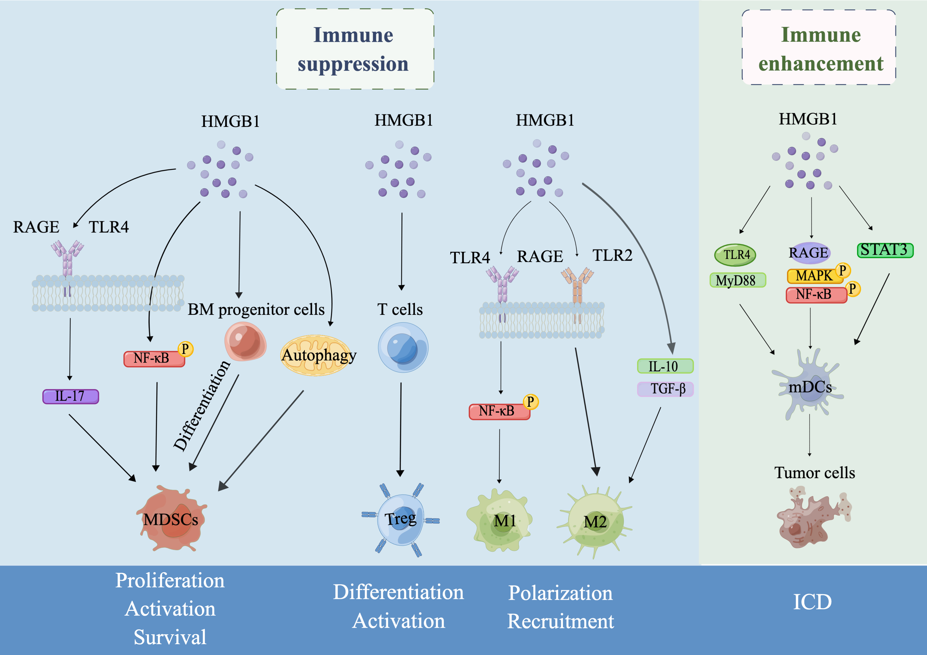
Fig. 1.

Schematic illustration of the dual role of HMGB1 in tumor immunity drawn by Figdraw. In the TME, HMGB1 can exert an immunosuppressive role by regulating the proliferation, activation and survival of MDSCs, the differentiation and activation of Tregs, and the polarization and recruitment of TAMs. On the other hand, HMGB1 can also enhance antitumor immune responses by inducing ICD. Specifically, first, secreted HMGB1 induces MDSC differentiation from BM progenitor cells, increases MDSC proliferation by promoting IL-17 secretion by binding to the receptors RAGE or TLR4, enhances MDSC development by activating nuclear factor-κB (NF-κB) signaling, and promotes MDSC survival by regulating autophagy. Second, HMGB1 can promote the differentiation and activation of Tregs. Third, HMGB1 may promote M1 polarization of TAMs by binding to receptors the TLR4 or RAGE to activate NF-κB signaling, contribute to M2 polarization by binding to the receptors RAGE or TLR2 or by the secretion of IL-10/transforming growth factor-β (TGF-β), and enhance macrophage recruitment by IL-6/STAT3 signaling. Finally, HMGB1 may mature DCs by TLR4/MyD88, RAGE/MAPK/NF-κB or STAT3 signaling to further induce ICD.

Abbreviations

TNF-α, tumor necrosis factor α; NF-κB, nuclear factor kappa B; TGF-β, transforming growth factor-β; NET, neutrophil extracellular trap; HCC, hepatocellular carcinoma; TIM, T-cell Ig and mucin domain; Breg, regulatory B cells; MAPK, mitogen-activated protein kinase; TRAP, tumor cell-released autophagosomes; FOXP3, forkhead box protein P3; TSLP, thymic stromal lymphopoietin; PMSCs, peripheral blood mononuclear cells; EOC, epithelial ovarian cancer; OSCC, oral squamous cell carcinoma; SEPT9, septin 9; MTIF2, mitochondrial translation initiation factor 2; ESCC, esophageal squamous cell carcinoma; DMFS, distant metastasis-free survival; RCD, regulated cell death; PDT, photodynamic therapy; WT, wild-type; APCs, antigen-presenting cells; DCs, dendritic cells; CTL, cytotoxic T lymphocyte; ICD, immunogenic cell death; HMGB1, high mobility group box 1; DAMPs, damage-associated molecular patterns; PTMs, posttranslational modifications; RAGE, receptor for advanced glycation end products; TLR2/4, Toll-like receptor 2/4; MDSCs, myeloid-derived suppressor cells; Tregs, regulatory T cells; TAMs, tumor-associated macrophages; BM, bone marrow; VEGF, vascular endothelial growth factor; GM-CSF, granulocyte macrophage colony stimulating factor; G-CSF, granulocyte colony stimulating factor; OV, oncolytic virus; ER, endoplasmic reticulum; TIM-3, T-cell immunoglobulin domain and mucin domain 3; NDV, newcastle disease virus; PRRs, recognition receptors; PDAC, pancreatic ductal adenocarcinoma; RTOG, grade three toxicity; CRP, C-reactive protein; ROS, reactive oxygen species; PARP1, poly ADP-ribose polymerase 1; RIPK3, receptor-interacting serine-threonine kinase 3; DNases, deoxyribonucleases; IFN-γ, interferon-γ; LPS, lipopolysaccharide; TNF-α, tumor necrosis factor-α; CaMKK, calmodulin-dependent protein kinases; XPO1, exportin 1; JAK, Janus kinase; STAT, signal transducer and activator of transcription; CXCL12, C-X-C Motif Chemokine Ligand 12; CXCR4, C-X-C chemokine receptor type 4; MD-2, myeloid differentiation factor 2; PFS, progression-free survival.

Author Contributions

JW finalized the visualization, acquiring, analyzing, or interpreting data from the reviewed literature. XZ put forward the idea, supervised this study and revised the manuscript. All authors read and approved the final version of this manuscript.

Ethics Approval and Consent to Participate

Not applicable.

Acknowledgment

Not applicable.

Funding

This study was funded by Beijing (China) municipal Natural Science Foundation (7202057) and China National Nature Science Foundation (81772868).

Conflict of Interest

The authors declare no conflict of interest.

References
[1]
Sung H, Ferlay J, Siegel RL, Laversanne M, Soerjomataram I, Jemal A, et al. Global Cancer Statistics 2020: GLOBOCAN Estimates of Incidence and Mortality Worldwide for 36 Cancers in 185 Countries. CA: a Cancer Journal for Clinicians. 2021; 71: 209–249.
[2]
Riley RS, June CH, Langer R, Mitchell MJ. Delivery technologies for cancer immunotherapy. Nature Reviews. Drug Discovery. 2019; 18: 175–196.
[3]
Chen DS, Mellman I. Oncology meets immunology: the cancer-immunity cycle. Immunity. 2013; 39: 1–10.
[4]
Pio R, Ajona D, Ortiz-Espinosa S, Mantovani A, Lambris JD. Complementing the Cancer-Immunity Cycle. Frontiers in Immunology. 2019; 10: 774.
[5]
Sims GP, Rowe DC, Rietdijk ST, Herbst R, Coyle AJ. HMGB1 and RAGE in inflammation and cancer. Annual Review of Immunology. 2010; 28: 367–388.
[6]
Li J, Kokkola R, Tabibzadeh S, Yang R, Ochani M, Qiang X, et al. Structural basis for the proinflammatory cytokine activity of high mobility group box 1. Molecular Medicine (Cambridge, Mass.). 2003; 9: 37–45.
[7]
Ellerman JE, Brown CK, de Vera M, Zeh HJ, Billiar T, Rubartelli A, et al. Masquerader: high mobility group box-1 and cancer. Clinical Cancer Research: an Official Journal of the American Association for Cancer Research. 2007; 13: 2836–2848.
[8]
Musumeci D, Roviello GN, Montesarchio D. An overview on HMGB1 inhibitors as potential therapeutic agents in HMGB1-related pathologies. Pharmacology & Therapeutics. 2014; 141: 347–357.
[9]
Kang R, Zhang Q, Zeh HJ, 3rd, Lotze MT, Tang D. HMGB1 in cancer: good, bad, or both? Clinical Cancer Research: an Official Journal of the American Association for Cancer Research. 2013; 19: 4046–4057.
[10]
Zuo Z, Che X, Wang Y, Li B, Li J, Dai W, et al. High mobility group Box-1 inhibits cancer cell motility and metastasis by suppressing activation of transcription factor CREB and nWASP expression. Oncotarget. 2014; 5: 7458–7470.
[11]
Jiao X, Zhang S, Jiao J, Zhang T, Qu W, Muloye GM, et al. Promoter methylation of SEPT9 as a potential biomarker for early detection of cervical cancer and its overexpression predicts radioresistance. Clinical Epigenetics. 2019; 11: 120.
[12]
Kang R, Xie Y, Zhang Q, Hou W, Jiang Q, Zhu S, et al. Intracellular HMGB1 as a novel tumor suppressor of pancreatic cancer. Cell Research. 2017; 27: 916–932.
[13]
He Q, Liang CH, Lippard SJ. Steroid hormones induce HMG1 overexpression and sensitize breast cancer cells to cisplatin and carboplatin. Proceedings of the National Academy of Sciences of the United States of America. 2000; 97: 5768–5772.
[14]
Gardella S, Andrei C, Ferrera D, Lotti LV, Torrisi MR, Bianchi ME, et al. The nuclear protein HMGB1 is secreted by monocytes via a non-classical, vesicle-mediated secretory pathway. EMBO Reports. 2002; 3: 995–1001.
[15]
Krysko DV, Garg AD, Kaczmarek A, Krysko O, Agostinis P, Vandenabeele P. Immunogenic cell death and DAMPs in cancer therapy. Nature Reviews. Cancer. 2012; 12: 860–875.
[16]
Aria H, Rezaei M. Immunogenic cell death inducer peptides: A new approach for cancer therapy, current status and future perspectives. Biomedicine & Pharmacotherapy. 2023; 161: 114503.
[17]
Richard SA, Jiang Y, Xiang LH, Zhou S, Wang J, Su Z, et al. Post-translational modifications of high mobility group box 1 and cancer. American Journal of Translational Research. 2017; 9: 5181–5196.
[18]
Paudel YN, Angelopoulou E, Piperi C, Balasubramaniam VRMT, Othman I, Shaikh MF. Enlightening the role of high mobility group box 1 (HMGB1) in inflammation: Updates on receptor signalling. European Journal of Pharmacology. 2019; 858: 172487.
[19]
Chen R, Kang R, Tang D. The mechanism of HMGB1 secretion and release. Experimental & Molecular Medicine. 2022; 54: 91–102.
[20]
Ni SY, Zhong XL, Li ZH, Huang DJ, Xu WT, Zhou Y, et al. Puerarin Alleviates Lipopolysaccharide-Induced Myocardial Fibrosis by Inhibiting PARP-1 to Prevent HMGB1-Mediated TLR4-NF-κB Signaling Pathway. Cardiovascular Toxicology. 2020; 20: 482–491.
[21]
Qin WD, Mi SH, Li C, Wang GX, Zhang JN, Wang H, et al. Low shear stress induced HMGB1 translocation and release via PECAM-1/PARP-1 pathway to induce inflammation response. PLoS ONE. 2015; 10: e0120586.
[22]
Murakami Y, Matsumoto H, Roh M, Giani A, Kataoka K, Morizane Y, et al. Programmed necrosis, not apoptosis, is a key mediator of cell loss and DAMP-mediated inflammation in dsRNA-induced retinal degeneration. Cell Death and Differentiation. 2014; 21: 270–277.
[23]
Shichita T, Hasegawa E, Kimura A, Morita R, Sakaguchi R, Takada I, et al. Peroxiredoxin family proteins are key initiators of post-ischemic inflammation in the brain. Nature Medicine. 2012; 18: 911–917.
[24]
Chiang HS, Maric M. Lysosomal thiol reductase negatively regulates autophagy by altering glutathione synthesis and oxidation. Free Radical Biology & Medicine. 2011; 51: 688–699.
[25]
Lamkanfi M, Sarkar A, Vande Walle L, Vitari AC, Amer AO, Wewers MD, et al. Inflammasome-dependent release of the alarmin HMGB1 in endotoxemia. Journal of Immunology (Baltimore, Md.: 1950). 2010; 185: 4385–4392.
[26]
Tohme S, Yazdani HO, Al-Khafaji AB, Chidi AP, Loughran P, Mowen K, et al. Neutrophil Extracellular Traps Promote the Development and Progression of Liver Metastases after Surgical Stress. Cancer Research. 2016; 76: 1367–1380.
[27]
Sekiguchi F, Domoto R, Nakashima K, Yamasoba D, Yamanishi H, Tsubota M, et al. Paclitaxel-induced HMGB1 release from macrophages and its implication for peripheral neuropathy in mice: Evidence for a neuroimmune crosstalk. Neuropharmacology. 2018; 141: 201–213.
[28]
Quan H, Bae HB, Hur YH, Lee KH, Lee CH, Jang EA, et al. Stearoyl lysophosphatidylcholine inhibits LPS-induced extracellular release of HMGB1 through the G2A/calcium/CaMKKβ/AMPK pathway. European Journal of Pharmacology. 2019; 852: 125–133.
[29]
Chen Y, Li G, Liu Y, Werth VP, Williams KJ, Liu ML. Translocation of Endogenous Danger Signal HMGB1 From Nucleus to Membrane Microvesicles in Macrophages. Journal of Cellular Physiology. 2016; 231: 2319–2326.
[30]
Wang CM, Jiang M, Wang HJ. Effect of NF κB inhibitor on high mobility group protein B1 expression in a COPD rat model. Molecular Medicine Reports. 2013; 7: 499–502.
[31]
Tsao PN, Wei SC, Huang MT, Lee MC, Chou HC, Chen CY, et al. Lipopolysaccharide-induced Notch signaling activation through JNK-dependent pathway regulates inflammatory response. Journal of Biomedical Science. 2011; 18: 56.
[32]
Huang W, Xie W, Gong J, Wang W, Cai S, Huang Q, et al. Heat stress induces RIP1/RIP3-dependent necroptosis through the MAPK, NF-κB, and c-Jun signaling pathways in pulmonary vascular endothelial cells. Biochemical and Biophysical Research Communications. 2020; 528: 206–212.
[33]
Imbaby S, Matsuda N, Tomita K, Hattori K, Palikhe S, Yokoo H, et al. Beneficial effect of STAT3 decoy oligodeoxynucleotide transfection on organ injury and mortality in mice with cecal ligation and puncture-induced sepsis. Scientific Reports. 2020; 10: 15316.
[34]
Yan HX, Wu HP, Zhang HL, Ashton C, Tong C, Wu H, et al. p53 promotes inflammation-associated hepatocarcinogenesis by inducing HMGB1 release. Journal of Hepatology. 2013; 59: 762–768.
[35]
Barlan AU, Griffin TM, McGuire KA, Wiethoff CM. Adenovirus membrane penetration activates the NLRP3 inflammasome. Journal of Virology. 2011; 85: 146–155.
[36]
Miller JM, Thompson JK, MacPherson MB, Beuschel SL, Westbom CM, Sayan M, et al. Curcumin: a double hit on malignant mesothelioma. Cancer Prevention Research (Philadelphia, Pa.). 2014; 7: 330–340.
[37]
Xiang M, Shi X, Li Y, Xu J, Yin L, Xiao G, et al. Hemorrhagic shock activation of NLRP3 inflammasome in lung endothelial cells. Journal of Immunology (Baltimore, Md.: 1950). 2011; 187: 4809–4817.
[38]
Gabrilovich DI, Ostrand-Rosenberg S, Bronte V. Coordinated regulation of myeloid cells by tumours. Nature Reviews. Immunology. 2012; 12: 253–268.
[39]
Solito S, Pinton L, Damuzzo V, Mandruzzato S. Highlights on molecular mechanisms of MDSC-mediated immune suppression: paving the way for new working hypotheses. Immunological Investigations. 2012; 41: 722–737.
[40]
Jin S, Yang Z, Hao X, Tang W, Ma W, Zong H. Roles of HMGB1 in regulating myeloid-derived suppressor cells in the tumor microenvironment. Biomarker Research. 2020; 8: 21.
[41]
Weber R, Groth C, Lasser S, Arkhypov I, Petrova V, Altevogt P, et al. IL-6 as a major regulator of MDSC activity and possible target for cancer immunotherapy. Cellular Immunology. 2021; 359: 104254.
[42]
Hu X, Li B, Li X, Zhao X, Wan L, Lin G, et al. Transmembrane TNF-α promotes suppressive activities of myeloid-derived suppressor cells via TNFR2. Journal of Immunology (Baltimore, Md.: 1950). 2014; 192: 1320–1331.
[43]
Cai B, Liu Y, Chong Y, Zhang H, Matsunaga A, Fang X, et al. IRAK1-regulated IFN-γ signaling induces MDSC to facilitate immune evasion in FGFR1-driven hematological malignancies. Molecular Cancer. 2021; 20: 165.
[44]
Hubert P, Roncarati P, Demoulin S, Pilard C, Ancion M, Reynders C, et al. Extracellular HMGB1 blockade inhibits tumor growth through profoundly remodeling immune microenvironment and enhances checkpoint inhibitor-based immunotherapy. Journal for Immunotherapy of Cancer. 2021; 9: e001966.
[45]
Li J, Sun J, Rong R, Li L, Shang W, Song D, et al. HMGB1 promotes myeloid-derived suppressor cells and renal cell carcinoma immune escape. Oncotarget. 2017; 8: 63290–63298.
[46]
Li W, Wu K, Zhao E, Shi L, Li R, Zhang P, et al. HMGB1 recruits myeloid derived suppressor cells to promote peritoneal dissemination of colon cancer after resection. Biochemical and Biophysical Research Communications. 2013; 436: 156–161.
[47]
Zhao Y, Shao Q, Zhu H, Xu H, Long W, Yu B, et al. Resveratrol ameliorates Lewis lung carcinoma-bearing mice development, decreases granulocytic myeloid-derived suppressor cell accumulation and impairs its suppressive ability. Cancer Science. 2018; 109: 2677–2686.
[48]
Gebhardt C, Sevko A, Jiang H, Lichtenberger R, Reith M, Tarnanidis K, et al. Myeloid Cells and Related Chronic Inflammatory Factors as Novel Predictive Markers in Melanoma Treatment with Ipilimumab. Clinical Cancer Research: an Official Journal of the American Association for Cancer Research. 2015; 21: 5453–5459.
[49]
Tang Q, Li J, Zhu H, Li P, Zou Z, Xiao Y. Hmgb1-IL-23-IL-17-IL-6-Stat3 axis promotes tumor growth in murine models of melanoma. Mediators of Inflammation. 2013; 2013: 713859.
[50]
Parker KH, Sinha P, Horn LA, Clements VK, Yang H, Li J, et al. HMGB1 enhances immune suppression by facilitating the differentiation and suppressive activity of myeloid-derived suppressor cells. Cancer Research. 2014; 74: 5723–5733.
[51]
Ortiz-Espinosa S, Morales X, Senent Y, Alignani D, Tavira B, Macaya I, et al. Complement C5a induces the formation of neutrophil extracellular traps by myeloid-derived suppressor cells to promote metastasis. Cancer Letters. 2022; 529: 70–84.
[52]
Whiteside TL. Exosomes and tumor-mediated immune suppression. The Journal of Clinical Investigation. 2016; 126: 1216–1223.
[53]
Ye L, Zhang Q, Cheng Y, Chen X, Wang G, Shi M, et al. Tumor-derived exosomal HMGB1 fosters hepatocellular carcinoma immune evasion by promoting TIM-1+ regulatory B cell expansion. Journal for Immunotherapy of Cancer. 2018; 6: 145.
[54]
Zhou M, Wen Z, Cheng F, Ma J, Li W, Ren H, et al. Tumor-released autophagosomes induce IL-10-producing B cells with suppressive activity on T lymphocytes via TLR2-MyD88-NF-κB signal pathway. Oncoimmunology. 2016; 5: e1180485.
[55]
Zhang X, Shi H, Yuan X, Jiang P, Qian H, Xu W. Tumor-derived exosomes induce N2 polarization of neutrophils to promote gastric cancer cell migration. Molecular Cancer. 2018; 17: 146.
[56]
Alissafi T, Hatzioannou A, Mintzas K, Barouni RM, Banos A, Sormendi S, et al. Autophagy orchestrates the regulatory program of tumor-associated myeloid-derived suppressor cells. The Journal of Clinical Investigation. 2018; 128: 3840–3852.
[57]
Parker KH, Horn LA, Ostrand-Rosenberg S. High-mobility group box protein 1 promotes the survival of myeloid-derived suppressor cells by inducing autophagy. Journal of Leukocyte Biology. 2016; 100: 463–470.
[58]
Levine B, Klionsky DJ. Development by self-digestion: molecular mechanisms and biological functions of autophagy. Developmental Cell. 2004; 6: 463–477.
[59]
Gershon RK, Kondo K. Infectious immunological tolerance. Immunology. 1971; 21: 903–914.
[60]
Shevach EM. Fatal attraction: tumors beckon regulatory T cells. Nature Medicine. 2004; 10: 900–901.
[61]
Sakaguchi S. Naturally arising Foxp3-expressing CD25+CD4+ regulatory T cells in immunological tolerance to self and non-self. Nature Immunology. 2005; 6: 345–352.
[62]
Zou W. Immunosuppressive networks in the tumour environment and their therapeutic relevance. Nature Reviews. Cancer. 2005; 5: 263–274.
[63]
Wild CA, Brandau S, Lotfi R, Mattheis S, Gu X, Lang S, et al. HMGB1 is overexpressed in tumor cells and promotes activity of regulatory T cells in patients with head and neck cancer. Oral Oncology. 2012; 48: 409–416.
[64]
Liu Z, Falo LD, Jr, You Z. Knockdown of HMGB1 in tumor cells attenuates their ability to induce regulatory T cells and uncovers naturally acquired CD8 T cell-dependent antitumor immunity. Journal of Immunology (Baltimore, Md.: 1950). 2011; 187: 118–125.
[65]
Zhang Y, Liu Z, Hao X, Li A, Zhang J, Carey CD, et al. Tumor-derived high-mobility group box 1 and thymic stromal lymphopoietin are involved in modulating dendritic cells to activate T regulatory cells in a mouse model. Cancer Immunology, Immunotherapy: CII. 2018; 67: 353–366.
[66]
Vanichapol T, Chiangjong W, Panachan J, Anurathapan U, Chutipongtanate S, Hongeng S. Secretory High-Mobility Group Box 1 Protein Affects Regulatory T Cell Differentiation in Neuroblastoma Microenvironment In Vitro. Journal of Oncology. 2018; 2018: 7946021.
[67]
Demoulin S, Herfs M, Somja J, Roncarati P, Delvenne P, Hubert P. HMGB1 secretion during cervical carcinogenesis promotes the acquisition of a tolerogenic functionality by plasmacytoid dendritic cells. International Journal of Cancer. 2015; 137: 345–358.
[68]
Ladoire S, Enot D, Senovilla L, Ghiringhelli F, Poirier-Colame V, Chaba K, et al. The presence of LC3B puncta and HMGB1 expression in malignant cells correlate with the immune infiltrate in breast cancer. Autophagy. 2016; 12: 864–875.
[69]
Brown JM, Recht L, Strober S. The Promise of Targeting Macrophages in Cancer Therapy. Clinical Cancer Research: an Official Journal of the American Association for Cancer Research. 2017; 23: 3241–3250.
[70]
Martínez VG, Rubio C, Martínez-Fernández M, Segovia C, López-Calderón F, Garín MI, et al. BMP4 Induces M2 Macrophage Polarization and Favors Tumor Progression in Bladder Cancer. Clinical Cancer Research: an Official Journal of the American Association for Cancer Research. 2017; 23: 7388–7399.
[71]
Daurkin I, Eruslanov E, Stoffs T, Perrin GQ, Algood C, Gilbert SM, et al. Tumor-associated macrophages mediate immunosuppression in the renal cancer microenvironment by activating the 15-lipoxygenase-2 pathway. Cancer Research. 2011; 71: 6400–6409.
[72]
Nishie A, Ono M, Shono T, Fukushi J, Otsubo M, Onoue H, et al. Macrophage infiltration and heme oxygenase-1 expression correlate with angiogenesis in human gliomas. Clinical Cancer Research: an Official Journal of the American Association for Cancer Research. 1999; 5: 1107–1113.
[73]
Zhang W, Tian J, Hao Q. HMGB1 combining with tumor-associated macrophages enhanced lymphangiogenesis in human epithelial ovarian cancer. Tumour Biology: the Journal of the International Society for Oncodevelopmental Biology and Medicine. 2014; 35: 2175–2186.
[74]
Zhang QB, Jia QA, Wang H, Hu CX, Sun D, Jiang RD, et al. High-mobility group protein box1 expression correlates with peritumoral macrophage infiltration and unfavorable prognosis in patients with hepatocellular carcinoma and cirrhosis. BMC Cancer. 2016; 16: 880.
[75]
Shiau DJ, Kuo WT, Davuluri GVN, Shieh CC, Tsai PJ, Chen CC, et al. Hepatocellular carcinoma-derived high mobility group box 1 triggers M2 macrophage polarization via a TLR2/NOX2/autophagy axis. Scientific Reports. 2020; 10: 13582.
[76]
Chen H, Lin X, Liu H, Huang C, Li R, Ai J, et al. HMGB1 Translocation is Associated with Tumor-Associated Myeloid Cells and Involved in the Progression of Fibroblastic Sarcoma. Pathology Oncology Research: POR. 2021; 27: 608582.
[77]
Flis E, Barber G, Nulty C, Keogh B, McGuirk P, Anand A, et al. Identification of TLR2 Signalling Mechanisms Which Contribute to Barrett’s and Oesophageal Adenocarcinoma Disease Progression. Cancers. 2021; 13: 2065.
[78]
Rojas A, Delgado-López F, Perez-Castro R, Gonzalez I, Romero J, Rojas I, et al. HMGB1 enhances the protumoral activities of M2 macrophages by a RAGE-dependent mechanism. Tumour Biology: the Journal of the International Society for Oncodevelopmental Biology and Medicine. 2016; 37: 3321–3329.
[79]
Sica A, Larghi P, Mancino A, Rubino L, Porta C, Totaro MG, et al. Macrophage polarization in tumour progression. Seminars in Cancer Biology. 2008; 18: 349–355.
[80]
Wei C, Yang C, Wang S, Shi D, Zhang C, Lin X, et al. Crosstalk between cancer cells and tumor associated macrophages is required for mesenchymal circulating tumor cell-mediated colorectal cancer metastasis. Molecular Cancer. 2019; 18: 64.
[81]
Li Z, Fu WJ, Chen XQ, Wang S, Deng RS, Tang XP, et al. Autophagy-based unconventional secretion of HMGB1 in glioblastoma promotes chemosensitivity to temozolomide through macrophage M1-like polarization. Journal of Experimental & Clinical Cancer Research: CR. 2022; 41: 74.
[82]
Ayoub M, Shinde-Jadhav S, Mansure JJ, Alvarez F, Connell T, Seuntjens J, et al. The immune mediated role of extracellular HMGB1 in a heterotopic model of bladder cancer radioresistance. Scientific Reports. 2019; 9: 6348.
[83]
Wang C, Peng G, Huang H, Liu F, Kong DP, Dong KQ, et al. Blocking the Feedback Loop between Neuroendocrine Differentiation and Macrophages Improves the Therapeutic Effects of Enzalutamide (MDV3100) on Prostate Cancer. Clinical Cancer Research: an Official Journal of the American Association for Cancer Research. 2018; 24: 708–723.
[84]
Xian G, Zhao J, Qin C, Zhang Z, Lin Y, Su Z. Simvastatin attenuates macrophage-mediated gemcitabine resistance of pancreatic ductal adenocarcinoma by regulating the TGF-β1/Gfi-1 axis. Cancer Letters. 2017; 385: 65–74.
[85]
Zheng H, Yu S, Zhu C, Guo T, Liu F, Xu Y. HIF1α promotes tumor chemoresistance via recruiting GDF15-producing TAMs in colorectal cancer. Experimental Cell Research. 2021; 398: 112394.
[86]
Galluzzi L, Vitale I, Aaronson SA, Abrams JM, Adam D, Agostinis P, et al. Molecular mechanisms of cell death: recommendations of the Nomenclature Committee on Cell Death 2018. Cell Death and Differentiation. 2018; 25: 486–541.
[87]
Vaes RDW, Hendriks LEL, Vooijs M, De Ruysscher D. Biomarkers of Radiotherapy-Induced Immunogenic Cell Death. Cells. 2021; 10: 930.
[88]
Apetoh L, Ghiringhelli F, Tesniere A, Obeid M, Ortiz C, Criollo A, et al. Toll-like receptor 4-dependent contribution of the immune system to anticancer chemotherapy and radiotherapy. Nature Medicine. 2007; 13: 1050–1059.
[89]
Inoue H, Tsutsumi H, Tanaka K, Iwama E, Shiraishi Y, Hirayama A, et al. Increased plasma levels of damage-associated molecular patterns during systemic anticancer therapy in patients with advanced lung cancer. Translational Lung Cancer Research. 2021; 10: 2475–2486.
[90]
Bains SJ, Abrahamsson H, Flatmark K, Dueland S, Hole KH, Seierstad T, et al. Immunogenic cell death by neoadjuvant oxaliplatin and radiation protects against metastatic failure in high-risk rectal cancer. Cancer Immunology, Immunotherapy: CII. 2020; 69: 355–364.
[91]
Suzuki Y, Mimura K, Yoshimoto Y, Watanabe M, Ohkubo Y, Izawa S, et al. Immunogenic tumor cell death induced by chemoradiotherapy in patients with esophageal squamous cell carcinoma. Cancer Research. 2012; 72: 3967–3976.
[92]
Xu D, Wang Y, Wu J, Zhang Z, Chen J, Xie M, et al. MTIF2 impairs 5 fluorouracil-mediated immunogenic cell death in hepatocellular carcinoma in vivo: Molecular mechanisms and therapeutic significance. Pharmacological Research. 2021; 163: 105265.
[93]
Huang CY, Chiang SF, Ke TW, Chen TW, Lan YC, You YS, et al. Cytosolic high-mobility group box protein 1 (HMGB1) and/or PD-1+ TILs in the tumor microenvironment may be contributing prognostic biomarkers for patients with locally advanced rectal cancer who have undergone neoadjuvant chemoradiotherapy. Cancer Immunology, Immunotherapy: CII. 2018; 67: 551–562.
[94]
Dumitriu IE, Baruah P, Valentinis B, Voll RE, Herrmann M, Nawroth PP, et al. Release of high mobility group box 1 by dendritic cells controls T cell activation via the receptor for advanced glycation end products. Journal of Immunology (Baltimore, Md.: 1950). 2005; 174: 7506–7515.
[95]
Turubanova VD, Mishchenko TA, Balalaeva IV, Efimova I, Peskova NN, Klapshina LG, et al. Novel porphyrazine-based photodynamic anti-cancer therapy induces immunogenic cell death. Scientific Reports. 2021; 11: 7205.
[96]
Koks CA, Garg AD, Ehrhardt M, Riva M, Vandenberk L, Boon L, et al. Newcastle disease virotherapy induces long-term survival and tumor-specific immune memory in orthotopic glioma through the induction of immunogenic cell death. International Journal of Cancer. 2015; 136: E313–E325.
[97]
Ye T, Jiang K, Wei L, Barr MP, Xu Q, Zhang G, et al. Oncolytic Newcastle disease virus induces autophagy-dependent immunogenic cell death in lung cancer cells. American Journal of Cancer Research. 2018; 8: 1514–1527.
[98]
Shao X, Wang X, Guo X, Jiang K, Ye T, Chen J, et al. STAT3 Contributes To Oncolytic Newcastle Disease Virus-Induced Immunogenic Cell Death in Melanoma Cells. Frontiers in Oncology. 2019; 9: 436.
[99]
Suwinski R, Giglok M, Galwas-Kliber K, Idasiak A, Jochymek B, Deja R, et al. Blood serum proteins as biomarkers for prediction of survival, locoregional control and distant metastasis rate in radiotherapy and radio-chemotherapy for non-small cell lung cancer. BMC Cancer. 2019; 19: 427.
[100]
Luo Y, Chihara Y, Fujimoto K, Sasahira T, Kuwada M, Fujiwara R, et al. High mobility group box 1 released from necrotic cells enhances regrowth and metastasis of cancer cells that have survived chemotherapy. European Journal of Cancer (Oxford, England: 1990). 2013; 49: 741–751.
[101]
Chiba S, Baghdadi M, Akiba H, Yoshiyama H, Kinoshita I, Dosaka-Akita H, et al. Tumor-infiltrating DCs suppress nucleic acid-mediated innate immune responses through interactions between the receptor TIM-3 and the alarmin HMGB1. Nature Immunology. 2012; 13: 832–842.
[102]
Jafari S, Lavasanifar A, Hejazi MS, Maleki-Dizaji N, Mesgari M, Molavi O. STAT3 inhibitory stattic enhances immunogenic cell death induced by chemotherapy in cancer cells. Daru: Journal of Faculty of Pharmacy, Tehran University of Medical Sciences. 2020; 28: 159–169.
[103]
Clasen K, Welz S, Faltin H, Zips D, Eckert F. Dynamics of HMBG1 (High Mobility Group Box 1) during radiochemotherapy correlate with outcome of HNSCC patients. Strahlentherapie Und Onkologie. 2022; 198: 194–200.
[104]
Tang D, Kang R, Cheh CW, Livesey KM, Liang X, Schapiro NE, et al. HMGB1 release and redox regulates autophagy and apoptosis in cancer cells. Oncogene. 2010; 29: 5299–5310.
[105]
Schiraldi M, Raucci A, Muñoz LM, Livoti E, Celona B, Venereau E, et al. HMGB1 promotes recruitment of inflammatory cells to damaged tissues by forming a complex with CXCL12 and signaling via CXCR4. The Journal of Experimental Medicine. 2012; 209: 551–563.
[106]
Yang H, Wang H, Ju Z, Ragab AA, Lundbäck P, Long W, et al. MD-2 is required for disulfide HMGB1-dependent TLR4 signaling. The Journal of Experimental Medicine. 2015; 212: 5–14.
[107]
Kong ZH, Chen X, Hua HP, Liang L, Liu LJ. The Oral Pretreatment of Glycyrrhizin Prevents Surgery-Induced Cognitive Impairment in Aged Mice by Reducing Neuroinflammation and Alzheimer’s-Related Pathology via HMGB1 Inhibition. Journal of Molecular Neuroscience: MN. 2017; 63: 385–395.
[108]
Sun X, Zeng H, Wang Q, Yu Q, Wu J, Feng Y, et al. Glycyrrhizin ameliorates inflammatory pain by inhibiting microglial activation-mediated inflammatory response via blockage of the HMGB1-TLR4-NF-kB pathway. Experimental Cell Research. 2018; 369: 112–119.
[109]
Venereau E, Casalgrandi M, Schiraldi M, Antoine DJ, Cattaneo A, De Marchis F, et al. Mutually exclusive redox forms of HMGB1 promote cell recruitment or proinflammatory cytokine release. The Journal of Experimental Medicine. 2012; 209: 1519–1528.
[110]
Kazama H, Ricci JE, Herndon JM, Hoppe G, Green DR, Ferguson TA. Induction of immunological tolerance by apoptotic cells requires caspase-dependent oxidation of high-mobility group box-1 protein. Immunity. 2008; 29: 21–32.
[111]
Chaiswing L, Oberley TD. Extracellular/microenvironmental redox state. Antioxidants & Redox Signaling. 2010; 13: 449–465.
[112]
Tong L, Chuang CC, Wu S, Zuo L. Reactive oxygen species in redox cancer therapy. Cancer Letters. 2015; 367: 18–25.
[113]
Tsung A, Klune JR, Zhang X, Jeyabalan G, Cao Z, Peng X, et al. HMGB1 release induced by liver ischemia involves Toll-like receptor 4 dependent reactive oxygen species production and calcium-mediated signaling. The Journal of Experimental Medicine. 2007; 204: 2913–2923.
[114]
Wu T, Zhang W, Yang G, Li H, Chen Q, Song R, et al. HMGB1 overexpression as a prognostic factor for survival in cancer: a meta-analysis and systematic review. Oncotarget. 2016; 7: 50417–50427.
[115]
Chung HW, Lim JB, Jang S, Lee KJ, Park KH, Song SY. Serum high mobility group box-1 is a powerful diagnostic and prognostic biomarker for pancreatic ductal adenocarcinoma. Cancer Science. 2012; 103: 1714–1721.
[116]
Wittwer C, Boeck S, Heinemann V, Haas M, Stieber P, Nagel D, et al. Circulating nucleosomes and immunogenic cell death markers HMGB1, sRAGE and DNAse in patients with advanced pancreatic cancer undergoing chemotherapy. International Journal of Cancer. 2013; 133: 2619–2630.
[117]
Tabata C, Shibata E, Tabata R, Kanemura S, Mikami K, Nogi Y, et al. Serum HMGB1 as a prognostic marker for malignant pleural mesothelioma. BMC Cancer. 2013; 13: 205.
[118]
Sheng X, Du X, Zhang X, Li D, Lu C, Li Q, et al. Clinical value of serum HMGB1 levels in early detection of recurrent squamous cell carcinoma of uterine cervix: comparison with serum SCCA, CYFRA21-1, and CEA levels. Croatian Medical Journal. 2009; 50: 455–464.
[119]
Sautès-Fridman C, Cherfils-Vicini J, Damotte D, Fisson S, Fridman WH, Cremer I, et al. Tumor microenvironment is multifaceted. Cancer Metastasis Reviews. 2011; 30: 13–25.
[120]
Ulloa L, Messmer D. High-mobility group box 1 (HMGB1) protein: friend and foe. Cytokine & Growth Factor Reviews. 2006; 17: 189–201.
[121]
Cao ZY, Liu YZ, Li JM, Ruan YM, Yan WJ, Zhong SY, et al. Glycyrrhizic acid as an adjunctive treatment for depression through anti-inflammation: A randomized placebo-controlled clinical trial. Journal of Affective Disorders. 2020; 265: 247–254.

Publisher’s Note: IMR Press stays neutral with regard to jurisdictional claims in published maps and institutional affiliations.

Share
Back to top