IMR Press / FBL / Volume 28 / Issue 10 / DOI: 10.31083/j.fbl2810237
Open Access Review
Apigenin’s Therapeutic Potential Against Viral Infection
Show Less
1 Department of Life Science, Chung-Ang University, 06974 Seoul, Republic of Korea
2 Department of Microbiology, College of Medicine, Ewha Womans University, 07804 Seoul, Republic of Korea
*Correspondence: shhong13@ewha.ac.kr (So-Hee Hong); yjseo@cau.ac.kr (Young-Jin Seo)
Front. Biosci. (Landmark Ed) 2023, 28(10), 237; https://doi.org/10.31083/j.fbl2810237
Submitted: 10 July 2023 | Revised: 24 August 2023 | Accepted: 6 September 2023 | Published: 17 October 2023
(This article belongs to the Special Issue Research on Influenza Viruses)
Copyright: © 2023 The Author(s). Published by IMR Press.
This is an open access article under the CC BY 4.0 license.
Abstract

Several antiviral drugs are clinically approved to treat influenza that is a highly prevalent acute respiratory disease. However, emerging drug-resistant virus strains undermine treatment efficacy, highlighting the exigency for novel antiviral drugs to counter these drug-resistant strains. Plants and their derivates have been historically utilized as medicinal remedies, and extensive studies have evidenced the antiviral potential of phytochemicals. Notably, apigenin is a predominant flavonoid with minimal toxicity and substantial therapeutic effects in various disease models. Despite its many anti-inflammatory, anti-oxidant, anti-cancer, anti-bacterial, and other beneficial bioactivities, existing reviews have yet to focus on apigenin’s antiviral effects. Therefore, this review elucidates apigenin’s therapeutic and antiviral properties in vitro and in vivo, discussing its mode of action and future prospects. Apigenin’s remarkable inhibition by modulating multiple mechanisms against viruses has promising potential for novel plant-derived antiviral drugs and further clinical study developments.

Keywords
flavonoids
4′,5,7-trihydroxyflavone
antiviral drugs
influenza
phytochemicals
1. Introduction

Influenza is a viral acute respiratory disease, and a prominent recurrent threat to human health. Several influenza pandemics have transpired in recent history, such as the Spanish flu in 1918, the Hong Kong flu in 1968, and the Swine flu in 2009 [1]. Approximately 500 million people were infected, and 50 million died during the most severe 1918 Spanish pandemic [2]. Although anti-influenza drugs and vaccines have significantly prevented and treated influenza viral infection, they remain limited due to the virus’ high mutation rate and subsequent subtype generation [3]. Thus, influenza vaccines must be updated annually to stay ahead of rapidly mutating viruses [4]. Discordance between influenza vaccine strains and circulating viruses may engender a more severe endemic or pandemic due to reduced vaccine efficacy [5]. Furthermore, emerging drug-resistant influenza strains threaten current antiviral drug applications [6], emphasizing the urgency for new antiviral drugs.

Nearly 90 drugs have been approved for nine human viral disease treatments [7]. Plants have been used as traditional remedies for thousands of years, and many studies have revealed plant derivative and metabolite therapeutic potentials against diverse diseases. An estimated 34% of approved small molecule drugs in the past four decades are natural products or their derivatives, and 25% are from plants exclusively [8, 9]. Due to recent recurrent viral pandemics, phytochemicals have garnered attention as promising antiviral drug agents [10]. Plant derivatives or phytochemicals maintain significant structural complexity and can target viral and host proteins in various ways [11].

Apigenin is a natural plant flavonoid found in fruits, herbs, and various plants. As part of the flavone class, apigenin maintains a flavone structure with hydroxyl group substitutions at positions 4,5, and 7, as indicated by its chemical name (4,5,7,-trihydroxyflavone). Apigenin has low solubility in water and high permeability, notable Biopharmaceutics Classification System Class II drug features [12]. Many studies have verified apigenin’s anti-inflammatory, antioxidant, and anti-apoptotic activities [13, 14, 15], showcasing its therapeutic potential for diverse human diseases, such as cancer, cardiometabolic disorders, and skin inflammatory conditions [16, 17, 18]. This review details apigenin’s therapeutic effects by focusing on its anti-influenza properties and mode of action.

2. Apigenin’s Therapeutic Effects on Human Diseases

Apigenin’s potential as a therapeutic drug was discovered in 1960 when it inhibited basophil’s histamine release. Apigenin significantly reduced production of IgE, IgG2a, IgG1, histamine, and hexosaminidase in a mouse allergic rhinitis model by inhibiting the TLR4/MyD88/NF-κB signaling pathway [19, 20]. Many studies have also corroborated apigenin’s anti-cancer properties. Apigenin suppresses the proliferation and migration of various cancer types, including colorectal, breast, liver, lung, and prostate [21, 22], inducing autophagy and cell cycle arrest to prompt cancer cell apoptosis [23, 24]. Concurrently, apigenin indirectly represses cancer cells by enhancing the immune response. For example, apigenin’s immune stimulation incorporates PI3K/AKT, MAPK/ERK, JAK/STAT, NF-κB, and Wnt/β-catenin signaling pathways [25]. Furthermore, apigenin provides neuroprotection. In an Alzheimer’s mouse model, apigenin reduced cytokine production and nitric oxide release in inflammatory cells while elevating neuronal cell viability by hindering caspase-mediated apoptosis signaling, thereby protecting against neuritis [26].

In addition, apigenin ameliorated streptozotocin-induced diabetic nephropathy in rats. Apigenin treatment mitigated renal dysfunction, fibrosis, and inflammation in diabetic rats by significantly reducing MAPK activation, which prevented apoptosis and production of Tumor-necrosis factor α (TNF-α) and Interleukin 6 (IL-6) [27]. Interestingly, apigenin attenuated skin inflammation and prevented UV-generated pyrimidine dimers by downregulating cyclooxygenase-2 (COX2) expressions [28] and obstructing the MAPK/ AP-1 signaling pathway [29]. As such, apigenin is a promising drug candidate for allergy, cancers, neurological disorders, and autoimmune diseases (Table 1, Ref. [19, 20, 21, 22, 23, 24, 25, 26, 27, 28, 29]).

Table 1.Therapeutic effects and incorporated signaling pathways of apigenin in human diseases.
Disease Therapeutic effects Mechanism Ref
Allergy Production of IgE, IgG2a, IgG1, histamine and hexosaminidase Suppress NF-κB, MAPK [19, 20]
Cancer Apoptosis, autophagy, cell cycle arrest Modulate PI3K, AKT, MAPK, ERK, JAK, STAT, NF-κB, and Wnt/β-catenin signaling [21, 22, 23, 24, 25]
Proliferation, migration and invasion
Immune response
Alzheimer’s disease Neuronal cell viability Inhibiting caspase-mediated apoptosis signaling [26]
Proinflammatory cytokine and nitric oxide
Diabetes Renal dysfunction, fibrosis, and inflammation Suppress of MAPK activity [27]
Proinflammatory cytokine (TNF-α and IL-6) level in serum
Skin inflammation UV-induced skin inflammation Suppress MAPK/AP-1 signaling pathways [28, 29]
UV-generated pyrimidine dimer, COX2 expression

Ig, Immunoglobulin; TNF, Tumor-necrosis factor; IL, Interleukin; UV, Ultra-violet; COX, Cyclooxygenase-2; NF-κB, Nuclear factor kappa-light-chain-enhancer of activated B cells; PI3K, Phosphoinositide 3-kinases; AKT, Protein kinase B; MAPK, Mitogen-activated protein kinase; ERK, Extracellular signal-regulated protein kinase; JAK, Janus kinase; STAT, Signal transducer and activator of transcription; AP, Activator protein; , increase; , decrease.

3. Antiviral Effects of Apigenin

Apigenin has exerted remarkable antiviral activity against diverse viruses such as herpes simplex virus (HSV) [30, 31, 32, 33, 34], enterovirus 71 (EV71) [35, 36, 37, 38, 39], hepatitis C virus (HCV) [40, 41, 42, 43], dengue virus (DENV) [44, 45, 46, 47], severe acute respiratory syndrome coronavirus (SARS-CoV) [48, 49, 50, 51, 52, 53], and influenza virus [54, 55, 56, 57, 58, 59, 60, 61, 62, 63, 64, 65, 66, 67, 68]. Its antiviral activity is theorized to be mediated by multiple mechanisms, such as inhibiting viral replication, suppressing viral gene expression, and modulating host immune responses.

3.1 Herpes Virus

Asteraceae plant extracts, from which apigenin is abundantly isolated, were found to have an antiviral effect against HSV-1 in a previous report [30]. Similarly, chamomile extract is one of the richest natural apigenin sources, exhibiting anti-HSV-1 and -HSV-2 activity in vitro. Moreover, chamomile extract inhibits HSV-1 and HSV-2 during or after viral absorption; the most effective viral inhibition stage was when the extract was provided during viral absorption. Chamomile extract also presented the most substantial virucidal activity on both HSV-1 and HSV-2 particles than other plant extracts, completely inactivating viral particles within 3 and 1 hours against HSV-1 and HSV-2, respectively, indicating apigenin’s potent antiviral activity against HSV [31]. Also, Rittà et al. [32] discovered that Arisaema tortuosum leaf extract exhibited anti-HSV-2 activity even against acyclovir-resistant HSV-1 and HSV-2. Among extract components, apigenin produced the most inhibitory activity against HSV-1 (EC50 = 7.04 µg/mL) and HSV-2 (EC50 = 0.05 ± 0.02 µg/mL). Through this study, the authors demonstrated that apigenin inhibited the post-entry step of the virus replication cycle, interfering with its proliferation and transmittance [32]. Likewise, the anti-HSV-1 activity of apigenin was reported by other investigators (EC50 = 5 µg/mL) [33]. Interestingly, vitexin, an apigenin flavone glucoside, also exhibited anti-HSV-1 activity with EC50 value of 18 ± 3.3 µg/mL [34], corroborating the inhibitory activity of apigenin against HSV.

3.2 Enterovirus 71

Apigenin’s antiviral properties were also effective against Enterovirus 71 (EV71). Apigenin from dried Paulownia tomentosa flowers inhibited EV71 infection with an 11.0 µM EC50 by disrupting the internal ribosome EV71 entry site and heterogenous nuclear ribonucleoprotein (hnRNPs) interaction required for viral translation [35]. The antiviral effect against EV71 was more efficient with high dose of apigenin, indicating apigenin inhibits EV71 in a dose-dependent manner [36, 37, 38]. Of note, the underlying mechanism of anti-EV71 activity of apigenin has been established. Pre-treatment of host cells with apigenin significantly reduced EV71 infection, indicating that apigenin targeted the host cells for its anti-EV71 effect [35]. Further studies confirmed that apigenin targeted heterogenous nuclear ribonucleoprotein (hnRNPs). HnRNP A1 and A2 are known to interact with internal ribosome entry site, required for viral translation, thus considered as essential factors for initiation of EV71 polypeptide translation [39]. Apigenin treatment resulted in significantly reduced EV71 RNA in hnRNP A1 and A2 by disrupting viral RNA association with hnRNPs while not affecting the redistribution of those proteins [35, 36]. The antiviral effect in vitro was corroborated in vivo showing 88.89% of survival protection against lethal dose of EV71 in newborn mouse, suggesting apigenin as promising antiviral agents [38].

3.3 Hepatitis C Virus

Similarly, several studies have observed apigenin’s effect on HCV. As an Eclipta alba component, apigenin substantially interfered with HCV non-structure protein (NS) 5B’s RNA-dependent-RNA polymerase (RdRp) activity, exhibiting a 175.5 µM IC50. Concomitantly, apigenin treatment reduced HCV replication by 90% in a cell culture system. Interestingly, apigenin directly binds to HCV NS5B, inhibiting its enzymatic activity [40]. Likewise, in vitro system using Huh7 cells expressing HCV replicons, apigenin treatment with very low concentrations (0.1 µM) significantly reduced the HCV RNA copy number up to 40% [41]. Furthermore, Shibata et al. [42] reported that apigenin impeded HCV replication by depleting mature miR122 expression levels, which are essential for HCV RNA propagation and stability [69]. Inhibiting mature miR122 expression levels with apigenin is likely due to inhibiting TAR RNA binding protein (TRBP) phosphorylation. In previous reports by Ohno et al. [43], apigenin suppressed TRMP phosphorylation, an integral factor for the maturation of miRNA subsets by binding to Dicer [70], modulating ERK activity, and impairing miRNA subset maturation.

3.4 Dengue Virus

Apigenin is also known to inhibit the DENV protein NS5. In a cell culture system antagonized by DENV’s NS5 during infection [44], apigenin obstructed DENV2 replication by restoring STAT2 phosphorylation. Notably, apigenin stimulated STAT2 even without infection by promoting STAT2 Tyr 689 phosphorylation and activation, indicating a low probability for viral escape mutations affecting apigenin [45]. Concomitant to the inhibitory activity of apigenin against DENV protein, apigenin treatment reduced DENV titer in vitro. Apigenin inhibited DENV-3 infection in macrophage (U937-DC-SIGN) up to 50% at 40 µM [46]. Also, apigenin inhibited DENV2 infectivity with EC50 value of 10.55 ± 3.36 µM. Interestingly, by using alkyne-tagged apigenin to visualize the compound localization inside the cells, they found that apigenin was localized at the perinuclear region and colocalized with a dengue protein in the early phase of DENV infection, suggesting further insight for underlying mechanisms of anti-DENV activity of apigenin [47].

3.5 SARS-CoV

SARS-CoV has recently emerged as a predominantly threatening virus, prompting researchers to seek novel antiviral agents. Therefore, as a prominent plant-derived antiviral material, apigenin’s antiviral effect against SARS-CoVs has been increasingly investigated. Chaves and colleagues revealed apigenin’s antiviral activity against SARS-CoV-2 B.1 lineage in Calu-3 cells with a 5.11 ± 0.26 µM EC50. Moreover, apigenin treatment reduced inflammatory cytokine TNF-alpha production in SARS-CoV-2 infected Calu-3 cells [48]. Thus, an enzyme inhibition assay was performed in silico and in vitro to ascertain how apigenin inhibits coronavirus replication. In silico docking analysis revealed that apigenin potentially interferes with the main SARS-CoV protease (Mpro), essential for SARS-CoV replication [49]. Molecular docking analysis evidenced potent binding energies of –7.2 Kcal/mol (PDB: 6WTT) [50] and –6.7 Kcal/mol (PDB: 6LU7) [51]. However, in vitro enzyme inhibition activity was not as efficient, resulting in a 280.8 ± 21.4 IC50 [52]. Apigenin also interacts with host factors SARS-CoVs target, such as the angiotensin-converting enzyme (ACE)-2 receptor and transmembrane protease serine subtype 2 (TMRPSS2). The in silico molecular docking assay demonstrated that apigenin has a high binding energy to ACE-2 receptor (–8.5 Kcal/mol) and TMPRSS2 (–7.7 Kcal/mol) [51]. Apigenin’s high-binding affinity to host factors may explain its antiviral effect on SARS-CoV replication. Interestingly, rhoifolin, an apigenin glycoside, inhibited Mpro activity at 20 µM with a high binding affinity (–9.565 Kcal/mol) [53].

These findings substantiate that apigenin has immense potential for antiviral therapeutics against diverse viruses such as HSV, EV71, HCV, and DNV (Table 2, Ref. [30, 31, 32, 33, 34, 35, 36, 37, 38, 39, 40, 41, 42, 43, 44, 45, 46, 47, 48, 49, 50, 51, 52, 53]).

Table 2.The antiviral effects of apigenin against HSV, EV71, HCV, and DENV.
Virus Antiviral effects Mechanism Ref
HSV EC50 = 5 µg/mL (HSV-1), EC50 = 0.05 ± 0.02 µg/mL (HSV-2) Exert virucidal activity, Interfere with viral absorption, Inhibit the post-entry step of the virus replication [30, 31, 32, 33, 34]
EV71 EC50 = 11.0 µM Inhibit interaction between internal ribosome entry site of EV71 and hnRNP A1 and A2 [35, 36, 37, 38, 39]
HCV Reduced RNA copy number by 0.1 µM of apigenin, Inhibition of HCV replication by 5 µM of apigenin Rapidly bind to NS5B and inhibit RdRp activity, Decrease miR122 expression levels, Suppress the phosphorylation of TRBP [40, 41, 42, 43]
DENV EC50 = 10.55 ± 3.36 µM (DENV2), 50% inhibition of virus infection at 40 µM (DENV3) Restore STAT2 Tyr 689 phosphorylation and activation, Colocalized with a DENV protein in the early phase of DENV [44, 45, 46, 47]
SARS-CoV EC50 = 5.11 ± 0.26 µM (SARS-CoV-2) Reduce the production of proinflammatory cytokine in response to virus, Interact with viral protein (Mpro) and host factor (ACE-2 receptor, TMPRSS2) [48, 49, 50, 51, 52, 53]

HSV, Herpes simplex virus; EV, Enterovirus; HCV, Hepatitis C virus; DENV, Dengue virus; SARS-CoV, Severe acute respiratory syndrome coronavirus; EC, Effective concentration; NS, Non-structure protein; hnRNP, Heterogenous nuclear ribonucleoprotein; RdRp, RNA-dependent-RNA polymerase; TRBP, TAR RNA binding protein; STAT, Signal transducer and activator of transcription; Mpro, main protease; ACE, Angiotensin-converting enzyme; TMPRSS2, Transmembrane protease serine subtype 2.

4. Apigenin’s Antiviral Effects against the Influenza Virus

Several studies have explored apigenin’s antiviral effect on influenza (Table 3, Ref. [54, 55, 56, 59, 60, 61, 62]). Apigenin inhibited replication of influenza A virus (IAV) H1N1 strains through diverse mechanisms such as inhibition of NA, viral attachment/entry, mRNA expression, RdBP activity, viral particle production, nucleoprotein production, lessening cytopathic effects caused by the virus infection [54, 56, 59, 60, 61, 62]. Similarly, apigenin inhibited the replication of the avian influenza H5N1 virus strain A/Thailand/Kan-1/04 (IC50 = 16.01 ± 4.33 µM, selectivity index = 2.53) through NA inhibition [55] and IAV H3N2 (Jinan/15/90) through reduction of nucleoproteins. Furthermore, apigenin suppressed the replication of Influenza B virus strain (B/Jiangsu/10/2003) (IC50 = 12.35 ± 3.71 µg/mL) by inhibition of viral NA, indicating its potent antiviral effect on the influenza virus [56].

Table 3.The antiviral effects of apigenin against influenza virus.
Type Subtype Strain EC50 or IC50 value Mechanism Ref
A H1N1 California/07/2009 EC50 = 1.438 µg/mL NA inhibition, Viral attachment and entry inhibition, Inhibition of viral mRNA expression, Inhibition of IAV RdRP activity, Reduction of viral particle production, Nucleoprotein reduction [54, 56, 59, 60, 61, 62]
PR/8/34 IC50 = 9.3 ± 2.2 µM
EC50 = 15.3 ± 3.0 µg/mL
Toyama/129/2011 EC50 = 17.8 ± 8.7 µg/mL
Toyama/26/2011 EC50 = 8.1 ± 4.7 µg/mL
Osaka2024/2009 IC50 = 23.3 ± 7.4 µM
Osaka71/2011 IC50 = 76.7 ± 7.8
H3N2 Jinan/15/90 IC50 = 1.43 µg/mL NA inhibition (IC50 = 7.81 ± 1.70 µg/mL) [56]
H5N1 Thailand/1(Kan-1)/04 - Nucleoprotein reduction (IC50 = 16.01 ± 4.33 µM) [55]
B Jiangsu/10/2003 - NA inhibition (IC50 = 12.35 ± 3.71 µg/mL) [56]

PR, Puerto Rico; EC, Effective concentration; IC, Inhibitory concentration; CC, Cytotoxic concentration; CPE, Cytopathic effect; NA, Neuriminidase; IAV, Influenza A virus.

4.1 Apigenin’s Antiviral Activity during Early-Stage Influenza Viral Infection

Upon replication onset (0–4 hours post-infection, hpi), influenza viruses attach to host cells and enter by endocytosis [57, 58]. Apigenin treatment before viral infection (cell protection assay) did not significantly reduce IAV mRNA expression [54] or cytopathic effect [59, 60], whereas apigenin pre-incubation with IAV did [54, 60]. These results demonstrate apigenin’s disruptive activity against IAV and provide insight into IAV particle and apigenin interactions. Moreover, apigenin inhibited replication when cells were simultaneously treated with apigenin and the influenza virus. Similarly, apigenin post-treatment (2 and 4 hpi) effectively reduced virus-induced cytopathic effects and IAV mRNA expression in host cells. Therefore, these results following apigenin administration 0–4 hours post-infection (hpi) indicate interference with the influenza A virus’ attachment and penetration. Apigenin’s anti-influenza viral activity during early-stage infection was also observed for other IAV strains, including H1N1 (A/California/07/2009, A/PR/8/34) and H3N2 (A/Jinan/15/90, A/Minfang/151/2000) [54, 56, 59, 60, 61, 62]. Collectively, these results prove that apigenin can inhibit early-stage IAV replication (Fig. 1).
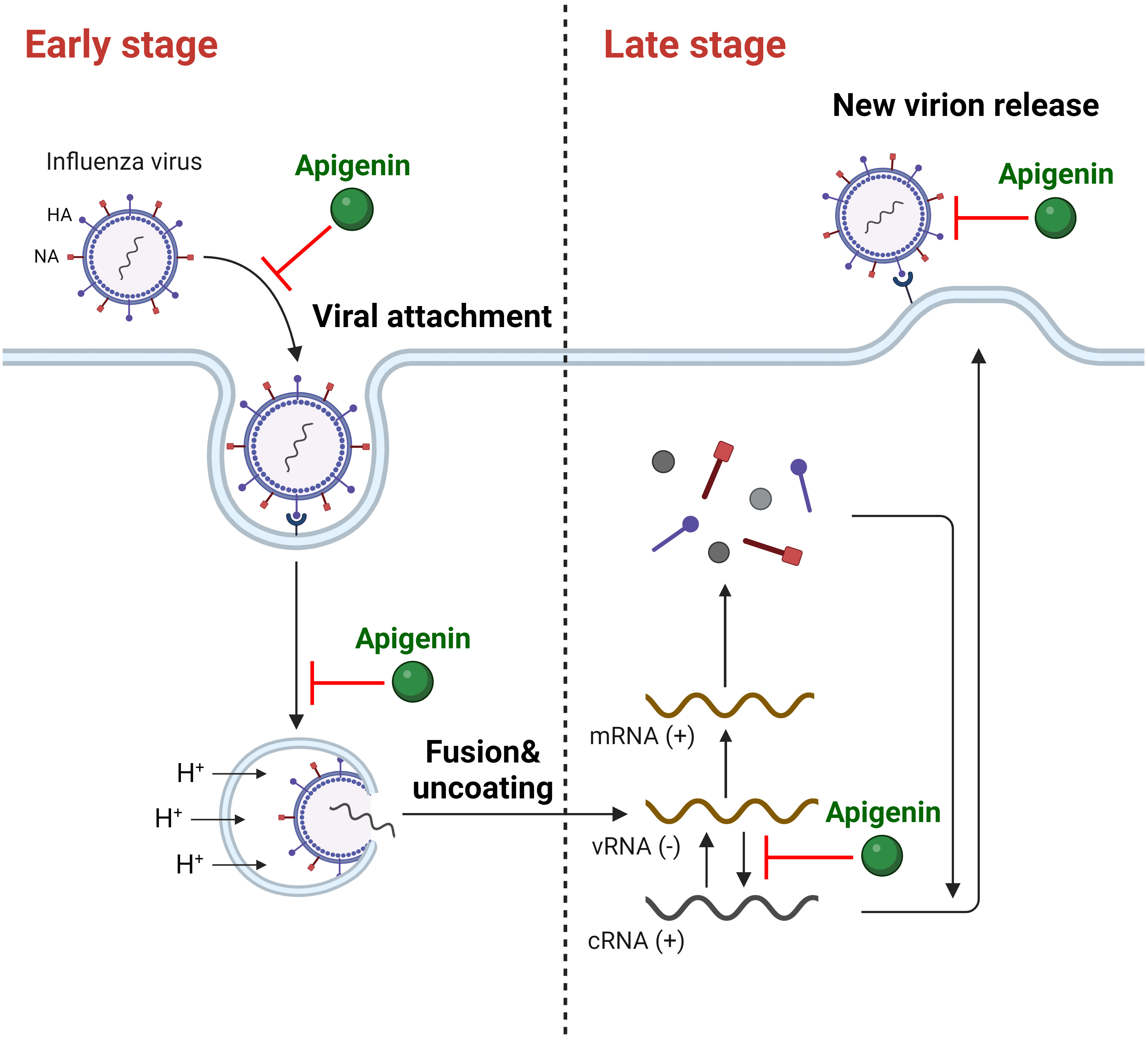
Fig. 1.

The inhibitory activity of apigenin against IAV on multiple stages of infection. (+), positive strand; (-), negative strand. Created with https://www.biorender.com/.

4.2 Apigenin’s Anti-Influenza Viral Activity during Late-Stage Infection

Viral mRNA translation, protein packaging, egress, and other critical propagation processes occur during late-stage viral replication (4–8 hpi). The influenza virus forms a complete structure by assembling its progeny particles and releasing them from the host cell for propagation [57, 58]. Notably, apigenin also inhibited late-stage IAV replication. When cells were treated with apigenin 5 hpi, (+)-strand viral RNA synthesis was attenuated, supporting apigenin’s potential for IAV RNA-dependent-RNA polymerase (RdRP) inhibition [54]. Similarly, viral particle production was considerably reduced when apigenin was administered 4–8 hpi [59, 60, 61]. Apigenin treatment after virus infection decreased the level of viral NP after 8 hpi with inhibited NA activity, indicating the inhibitory role of apigenin in virus assembly and release stages [41]. Thus, apigenin suppresses IAV replication by obstructing multiple IAV replication stages (Fig. 1).

4.3 Apigenin’s Antiviral Activity against Drug-Resistant Influenza Strains

Apigenin’s antiviral effect on drug-resistant IAV strains was also investigated. Morimoto et al. [61] noted that apigenin exhibited antiviral effects on oseltamivir-resistant strains (Osaka/2024/2009 and Osaka/71/2011) with 23.3 ± 7.4 and 76.7 ± 7.8 µM (selectivity index = 40 and 12, respectively) IC50 values. Moreover, this effect was not strain-specific, as it was also observed in A/HebeiXinhua/SWL1106/2017 (oseltamivir & amantadine-resistant H1N1) and A/FujianXinluo/SWL2457/2014 (Amantadine-resistant H1N1) strains [60]. Apigenin’s striking antiviral effects on drug-resistant IAV strains may be due to its many inhibitory methods compared to existing antiviral drugs. Thus, apigenin could substantially improve treatments against novel mutated IAVs that threaten severe pandemics worldwide.

4.4 The Regulation of Influenza Virus-Induced Signaling Pathways by Apigenin

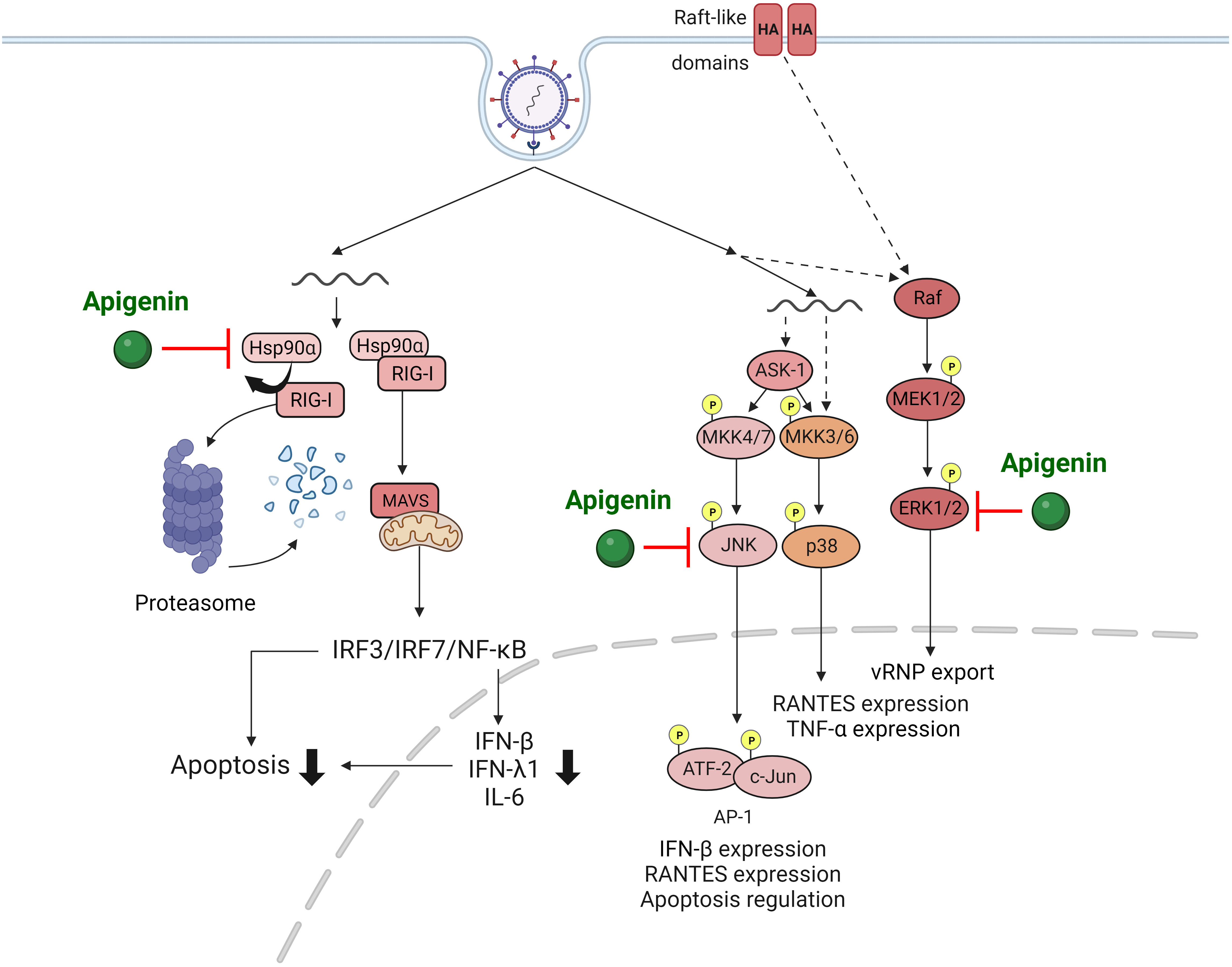
IAV infection activates various signaling pathways within the host cell, such as mitogen-activated protein kinase (MAPK) [63, 64] and retinoic acid-inducible gene-I (RIG-I) pathways [65]. Assessing alterations in signaling pathways up-regulated by IAV provides invaluable insights for antiviral drug developments targeting these components. Joo et al. [54] revealed that apigenin effectively suppressed IAV infection-induced MAPK signaling pathway activations that are critical for virus replication and cytokine production. Importantly, apigenin severely suppressed the elevation of extracellular signal-regulated (ERK) and stress-activated (SAPK) protein kinases phosphorylation upon IAV infection, subsequently reducing viral replication.

RIG-I is a prominent innate RNA sensor that recognizes influenza viral RNA and activates antiviral host responses, including cytokine cascade [65]. However, uncontrolled overproduction of cytokines such as interferons (IFNs) is a major factor that exacerbates immunopathologic lung injury upon IAV infection [66, 67, 68]. Therefore, regulating cytokine production induced by RIG-I pathway activation is vital for managing IAV infection outcomes. Xu et al. [59] revealed that apigenin treatment significantly suppressed IAV-induced RIG-I activation and proinflammatory cytokines such as IFNs. Interestingly, apigenin-mediated suppression of RIG-I was due to ubiquitin-mediated RIG-I protein degradation, leading to disruption of interactions between RIG-I and heat shock protein 90α. In addition, apigenin’s RIG-I inhibition was RIG-I pathway-specific as it minimally affected other PRRs, such as melanoma differentiation-associated gene 5 (MDA5), cyclic GAM-AMP synthase (cGAS), and Toll-like receptors (TLRs). The authors also proved that apigenin inhibited viral replication, virus-induced cell death, and A/PR/8/34 (H1N1) virus NA activity in vitro, independently from RIG-I suppression [59]. Therefore, apigenin inhibits signaling pathway activation induced by IAV infection, further illuminating apigenin’s underlying mechanisms against viruses (Fig. 2).

Fig. 2.

Regulation of apigenin in IAV-induced signaling pathways. Created with BioRender.com.

4.5 In Vivo Anti-Influenza Viral Potential of Apigenin

Lastly, apigenin’s anti-IAV activity strongly suggests its potential as an antiviral drug in vitro, inspiring many in vivo studies. Intranasally administered apigenin significantly reduced viral loads in mouse lungs infected with the IAV H1N1 strain (A/California/07/2009), corroborating apigenin’s IAV suppression in vitro. Furthermore, oral administration of an aqueous Agrimonia pilosa and Galla rhois extracts mixture (APRG64), where apigenin is the main antiviral component, considerably curtailed IAV-infected mice morbidity and mortality. In addition, proinflammatory cytokine expressions, such as IFN-γ, TNF-α, and IL-6, in IAV-infected mice lungs and LPS-stimulated splenocytes were notably suppressed by APRG64 treatment. The observed therapeutic effects regarding IAV’s mortality and APRG immunopathogenesis were comparable to oseltamivir phosphate (Tamiflu) [54]. However, the oral administration of 30 mg/kg apigenin in another study failed to exhibit anti-IAV (A/PR/8/34) efficacy in mice [62]. Therefore, despite apigenin’s promising anti-IAV activity, the administration route and dose must be carefully considered and require further investigation for new herbal antiviral drug development.

5. Concluding Remarks

Influenza has continuously threatened human health, invoking unpredictable and recurrent pandemics; four influenza A pandemics have occurred within the last century. Although most influenza A and B strains are susceptible to oseltamivir or zanamivir (neuraminidase inhibitors), resistant strains were observed frequently since the 2009 H1N1 influenza A pandemic. Therefore, developing novel anti-influenza drugs that effectively target current drug-resistant strains is urgently needed. Apigenin is a plant flavonoid component with substantial antiviral effects against various viruses, including influenza virus, evidenced through numerous in vitro and in vivo experiments. As summarized in the present review, apigenin exerts anti-viral activity against HSV, HCV, EV71, DENV, SARS-CoV, and influenza by disrupting virus particles, interfering with multiple viral replication stages by inhibiting viral proteins, regulating virus-induced signaling pathways, and interrupting virus interactions with host factors, such as RdRP, the ACE-2 receptor, and TMPRSS2.

Apigenin directly inhibits influenza virus and host factor interactions while alleviating pathogenic inflammatory responses caused by infection, similar to clinically used anti-IAV drugs. Furthermore, apigenin has also proven effective against drug-resistant influenza strains. These results indicated that apigenin is a promising candidate for anti-influenza therapy, as it can directly inhibit influenza infectivity and replication while reducing clinical symptoms associated with robust inflammatory responses. However, despite its potential as an anti-influenza drug, further studies using in vivo and nonhuman primate models are needed to reveal apigenin’s intricate anti-influenza mechanisms for clinical use, as current findings are limited to in vitro and in vivo mouse experiments.

Various studies have studied apigenin’s pharmacological features, reporting its nontoxic and nonmutagenic nature with various biological benefits, including anti-inflammatory, anti-cancer, anti-oxidant, anti-bacterial, and anti-depressant. However, high apigenin doses may induce slight side effects [71, 72, 73]. Apigenin exerts biological effects through numerous signaling molecule interactions, including ERK, c-Jun N-terminal kinase (JNK), p38, enzymes like cytochrome P450s, and the benzodiazepine receptor [73, 74, 75]. Based on their remarkable beneficial effects, apigenin’s pharmacokinetics has been investigated. Researchers found that oral apigenin administration in a rat model was absorbed, resulting in deficient blood levels [76, 77]. Apigenin absorption primarily undergoes Phase I to produce luteolin, scutellarein, and iso-scutellarein by utilizing Phase I enzymes and Phase II metabolism through glucuronidation and sulfation of apigenin in enteric and enterohepatic cycling [72, 77, 78, 79, 80]. Up to 63% of apigenin was excreted through urinary and fecal routes, as corroborated in clinical trials [77, 81]. Also, apigenin elimination was reported as considerably slow; 24.8% of radiolabeled apigenin was observed in the body ten days post-administration [77]. Apigenin’s higher absorption and accumulation extent than other dietary flavonoids, such as luteolin and quercetin, indicate better oral bioavailability and pharmacokinetics [77, 79, 82, 83]. However, apigenin’s clinical developments are severely limited due to its low solubility in water and organic solvents. Therefore, new technologies or formulations must be developed to improve apigenin’s bioavailability.

Similar to other plant extracts, apigenin’s slow production rate or inefficient processing techniques could hamper its clinical applications, particularly during an influenza pandemic. Therefore, a systemic mass-production manufacturing system must be developed.

Abbreviations

Ig, Immunoglobulin; TLR, Toll-like receptor; MyD, Myeloid differentiation primary response protein; NF-κB, Nuclear factor kappa-light-chain-enhancer of activated B cells; PI3K, Phosphoinositide 3-kinases; AKT, Protein kinase B; MAPK, Mitogen-activated protein kinase; ERK, Extracellular signal-regulated protein kinase; JAK, Janus kinase; STAT, Signal transducer and activator of transcription; TNF, Tumor-necrosis factor; IL, Interleukin; AP, Activator protein; COX2, Cyclooxygenase-2; EC, Effective concentration; HSV, Herpes simplex virus; HCV, Hepatitis C virus; DENV, Dengue virus; EV71, Enterovirus 71; SARS-CoV, Severe acute respiratory syndrome coronavirus; hnRNPs, Heterogenous nuclear ribonucleoprotein; NS, Non-structure protein; RdRp, RNA-dependent-RNA polymerase; TRBP, TAR RNA binding protein; Mpro, Main protease; ACE, Angiotensin-converting enzyme; TMPRSS2, Transmembrane protease serine subtype 2; hpi, Hours post-infection; RIG-I, Retinoic acid-inducible gene-I; SAPK, Stress-activated protein kinase; IFNs, Interferons; MDA5, Melanoma differentiation-associated gene 5; cGAS, Cyclic GAM-AMP synthase; JNK, c-Jun N-terminal kinase.

Author Contributions

Conceptualization: YS, SH, IL, JL; writing/original draft preparation: IL, JL, YS, SH; writing-review and editing; YS, SH. All authors read and approved the final version of the manuscript. All authors have participated sufficiently in the work and agreed to be accountable for all aspects of the work.

Ethics Approval and Consent to Participate

Not applicable.

Acknowledgment

The figures were created with Biorender.com.

Funding

This work was supported by the Chung-Ang University Research Scholarship Grants in 2023 and by the National Research Foundation of Korea (NRF) grant funded by the Korean government [grant number: NRF-2018R1A5A1025077].

Conflict of Interest

The authors declare no conflict of interest.

References
[1]
Taubenberger JK, Morens DM. Influenza: the once and future pandemic. Public Health Reports. 2010; 125: 16–26.
[2]
Taubenberger JK, Morens DM. 1918 Influenza: the mother of all pandemics. Emerging Infectious Diseases. 2006; 12: 15–22.
[3]
White MC, Lowen AC. Implications of segment mismatch for influenza A virus evolution. The Journal of General Virology. 2018; 99: 3–16.
[4]
Rajão DS, Pérez DR. Universal Vaccines and Vaccine Platforms to Protect against Influenza Viruses in Humans and Agriculture. Frontiers in Microbiology. 2018; 9: 123.
[5]
Tricco AC, Chit A, Soobiah C, Hallett D, Meier G, Chen MH, et al. Comparing influenza vaccine efficacy against mismatched and matched strains: a systematic review and meta-analysis. BMC Medicine. 2013; 11: 153.
[6]
Hussain M, Galvin HD, Haw TY, Nutsford AN, Husain M. Drug resistance in influenza A virus: the epidemiology and management. Infection and Drug Resistance. 2017; 10: 121–134.
[7]
De Clercq E, Li G. Approved Antiviral Drugs over the Past 50 Years. Clinical Microbiology Reviews. 2016; 29: 695–747.
[8]
Calixto JB. The role of natural products in modern drug discovery. Anais Da Academia Brasileira De Ciencias. 2019; 91: e20190105.
[9]
Newman DJ, Cragg GM. Natural Products as Sources of New Drugs over the Nearly Four Decades from 01/1981 to 09/2019. Journal of Natural Products. 2020; 83: 770–803.
[10]
Thomas E, Stewart LE, Darley BA, Pham AM, Esteban I, Panda SS. Plant-Based Natural Products and Extracts: Potential Source to Develop New Antiviral Drug Candidates. Molecules (Basel, Switzerland). 2021; 26: 6197.
[11]
Ghildiyal R, Prakash V, Chaudhary VK, Gupta V, Gabrani R. Phytochemicals as Antiviral Agents: Recent Updates. Plant-Derived Bioactives: Production, Properties and Therapeutic Applications. 2020; 12: 279–295.
[12]
Zhang J, Liu D, Huang Y, Gao Y, Qian S. Biopharmaceutics classification and intestinal absorption study of apigenin. International Journal of Pharmaceutics. 2012; 436: 311–317.
[13]
Lee JH, Zhou HY, Cho SY, Kim YS, Lee YS, Jeong CS. Anti-inflammatory mechanisms of apigenin: inhibition of cyclooxygenase-2 expression, adhesion of monocytes to human umbilical vein endothelial cells, and expression of cellular adhesion molecules. Archives of Pharmacal Research. 2007; 30: 1318–1327.
[14]
Lim R, Barker G, Wall CA, Lappas M. Dietary phytophenols curcumin, naringenin and apigenin reduce infection-induced inflammatory and contractile pathways in human placenta, foetal membranes and myometrium. Molecular Human Reproduction. 2013; 19: 451–462.
[15]
Safari M, Parsaie H, Sameni HR, Aldaghi MR, Zarbakhsh S. Anti-Oxidative and Anti-Apoptotic Effects of Apigenin on Number of Viable and Apoptotic Blastomeres, Zona Pellucida Thickness and Hatching Rate of Mouse Embryos. International Journal of Fertility & Sterility. 2018; 12: 257–262.
[16]
Shankar E, Goel A, Gupta K, Gupta S. Plant flavone apigenin: An emerging anticancer agent. Current Pharmacology Reports. 2017; 3: 423–446.
[17]
Xu Y, Li X, Wang H. Protective Roles of Apigenin Against Cardiometabolic Diseases: A Systematic Review. Frontiers in Nutrition. 2022; 9: 875826.
[18]
Yoon JH, Kim MY, Cho JY. Apigenin: A Therapeutic Agent for Treatment of Skin Inflammatory Diseases and Cancer. International Journal of Molecular Sciences. 2023; 24: 1498.
[19]
Li H, Zhang H, Zhao H. Apigenin attenuates inflammatory response in allergic rhinitis mice by inhibiting the TLR4/MyD88/NF-κB signaling pathway. Environmental Toxicology. 2023; 38: 253–265.
[20]
Chen F, He D, Yan B. Apigenin Attenuates Allergic Responses of Ovalbumin-Induced Allergic Rhinitis Through Modulation of Th1/Th2 Responses in Experimental Mice. Dose-Response. 2020; 18: 1559325820904799.
[21]
Xu M, Wang S, Song YU, Yao J, Huang K, Zhu X. Apigenin suppresses colorectal cancer cell proliferation, migration and invasion via inhibition of the Wnt/β-catenin signaling pathway. Oncology Letters. 2016; 11: 3075–3080.
[22]
Lee YM, Lee G, Oh TI, Kim BM, Shim DW, Lee KH, et al. Inhibition of glutamine utilization sensitizes lung cancer cells to apigenin-induced apoptosis resulting from metabolic and oxidative stress. International Journal of Oncology. 2016; 48: 399–408.
[23]
Gupta S, Afaq F, Mukhtar H. Involvement of nuclear factor-kappa B, Bax and Bcl-2 in induction of cell cycle arrest and apoptosis by apigenin in human prostate carcinoma cells. Oncogene. 2002; 21: 3727–3738.
[24]
Zhao G, Han X, Cheng W, Ni J, Zhang Y, Lin J, et al. Apigenin inhibits proliferation and invasion, and induces apoptosis and cell cycle arrest in human melanoma cells. Oncology Reports. 2017; 37: 2277–2285.
[25]
Yan X, Qi M, Li P, Zhan Y, Shao H. Apigenin in cancer therapy: anti-cancer effects and mechanisms of action. Cell & Bioscience. 2017; 7: 50.
[26]
Balez R, Steiner N, Engel M, Muñoz SS, Lum JS, Wu Y, et al. Neuroprotective effects of apigenin against inflammation, neuronal excitability and apoptosis in an induced pluripotent stem cell model of Alzheimer’s disease. Scientific Reports. 2016; 6: 31450.
[27]
Malik S, Suchal K, Khan SI, Bhatia J, Kishore K, Dinda AK, et al. Apigenin ameliorates streptozotocin-induced diabetic nephropathy in rats via MAPK-NF-κB-TNF-α and TGF-β1-MAPK-fibronectin pathways. American Journal of Physiology. Renal Physiology. 2017; 313: F414–F422.
[28]
Tong X, Van Dross RT, Abu-Yousif A, Morrison AR, Pelling JC. Apigenin prevents UVB-induced cyclooxygenase 2 expression: coupled mRNA stabilization and translational inhibition. Molecular and Cellular Biology. 2007; 27: 283–296.
[29]
Hwang YP, Oh KN, Yun HJ, Jeong HG. The flavonoids apigenin and luteolin suppress ultraviolet A-induced matrix metalloproteinase-1 expression via MAPKs and AP-1-dependent signaling in HaCaT cells. Journal of Dermatological Science. 2011; 61: 23–31.
[30]
Visintini Jaime MF, Redko F, Muschietti LV, Campos RH, Martino VS, Cavallaro LV. In vitro antiviral activity of plant extracts from Asteraceae medicinal plants. Virology Journal. 2013; 10: 245.
[31]
Yucharoen R, Chansakaow S, Tragoolpua Y. Inhibitory effect of aromatic herbs, lavender, sage and chamomile against herpes simplex virus infection. African Journal of Biotechnology. 2011; 10: 15394–15401.
[32]
Rittà M, Marengo A, Civra A, Lembo D, Cagliero C, Kant K, et al. Antiviral Activity of a Arisaema Tortuosum Leaf Extract and Some of its Constituents against Herpes Simplex Virus Type 2. Planta Medica. 2020; 86: 267–275.
[33]
Lyu SY, Rhim JY, Park WB. Antiherpetic activities of flavonoids against herpes simplex virus type 1 (HSV-1) and type 2 (HSV-2) in vitro. Archives of Pharmacal Research. 2005; 28: 1293–1301.
[34]
Fahmy NM, Al-Sayed E, Moghannem S, Azam F, El-Shazly M, Singab AN. Breaking Down the Barriers to a Natural Antiviral Agent: Antiviral Activity and Molecular Docking of Erythrina speciosa Extract, Fractions, and the Major Compound. Chemistry & Biodiversity. 2020; 17: e1900511.
[35]
Ji P, Chen C, Hu Y, Zhan Z, Pan W, Li R, et al. Antiviral activity of Paulownia tomentosa against enterovirus 71 of hand, foot, and mouth disease. Biological & Pharmaceutical Bulletin. 2015; 38: 1–6.
[36]
Zhang W, Qiao H, Lv Y, Wang J, Chen X, Hou Y, et al. Apigenin inhibits enterovirus-71 infection by disrupting viral RNA association with trans-acting factors. PLoS ONE. 2014; 9: e110429.
[37]
Lv X, Qiu M, Chen D, Zheng N, Jin Y, Wu Z. Apigenin inhibits enterovirus 71 replication through suppressing viral IRES activity and modulating cellular JNK pathway. Antiviral Research. 2014; 109: 30–41.
[38]
Dai W, Bi J, Li F, Wang S, Huang X, Meng X, et al. Antiviral Efficacy of Flavonoids against Enterovirus 71 Infection in Vitro and in Newborn Mice. Viruses. 2019; 11: 625.
[39]
Shih SR, Stollar V, Li ML. Host factors in enterovirus 71 replication. Journal of Virology. 2011; 85: 9658–9666.
[40]
Manvar D, Mishra M, Kumar S, Pandey VN. Identification and evaluation of anti hepatitis C virus phytochemicals from Eclipta alba. Journal of Ethnopharmacology. 2012; 144: 545–554.
[41]
Pisonero-Vaquero S, García-Mediavilla MV, Jorquera F, Majano PL, Benet M, Jover R, et al. Modulation of PI3K-LXRα-dependent lipogenesis mediated by oxidative/nitrosative stress contributes to inhibition of HCV replication by quercetin. Laboratory Investigation; a Journal of Technical Methods and Pathology. 2014; 94: 262–274.
[42]
Shibata C, Ohno M, Otsuka M, Kishikawa T, Goto K, Muroyama R, et al. The flavonoid apigenin inhibits hepatitis C virus replication by decreasing mature microRNA122 levels. Virology. 2014; 462-463: 42–48.
[43]
Ohno M, Shibata C, Kishikawa T, Yoshikawa T, Takata A, Kojima K, et al. The flavonoid apigenin improves glucose tolerance through inhibition of microRNA maturation in miRNA103 transgenic mice. Scientific Reports. 2013; 3: 2553.
[44]
Mazzon M, Jones M, Davidson A, Chain B, Jacobs M. Dengue virus NS5 inhibits interferon-alpha signaling by blocking signal transducer and activator of transcription 2 phosphorylation. The Journal of Infectious Diseases. 2009; 200: 1261–1270.
[45]
Acchioni C, Acchioni M, Mancini F, Amendola A, Marsili G, Tirelli V, et al. A cellular screening platform, stably expressing DENV2 NS5, defines a novel anti-DENV mechanism of action of Apigenin based on STAT2 activation. Virology. 2023; 583: 1–13.
[46]
Jasso-Miranda C, Herrera-Camacho I, Flores-Mendoza LK, Dominguez F, Vallejo-Ruiz V, Sanchez-Burgos GG, et al. Antiviral and immunomodulatory effects of polyphenols on macrophages infected with dengue virus serotypes 2 and 3 enhanced or not with antibodies. Infection and Drug Resistance. 2019; 12: 1833–1852.
[47]
Hengphasatporn K, Kaewmalai B, Jansongsaeng S, Badavath VN, Saelee T, Chokmahasarn T, et al. Alkyne-Tagged Apigenin, a Chemical Tool to Navigate Potential Targets of Flavonoid Anti-Dengue Leads. Molecules. 2021; 26: 6967.
[48]
Chaves OA, Fintelman-Rodrigues N, Wang X, Sacramento CQ, Temerozo JR, Ferreira AC, et al. Commercially Available Flavonols Are Better SARS-CoV-2 Inhibitors than Isoflavone and Flavones. Viruses. 2022; 14: 1458.
[49]
Zhang L, Lin D, Sun X, Curth U, Drosten C, Sauerhering L, et al. Crystal structure of SARS-CoV-2 main protease provides a basis for design of improved α-ketoamide inhibitors. Science. 2020; 368: 409–412.
[50]
Farhat A, Ben Hlima H, Khemakhem B, Ben Halima Y, Michaud P, Abdelkafi S, et al. Apigenin analogues as SARS-CoV-2 main protease inhibitors: In-silico screening approach. Bioengineered. 2022; 13: 3350–3361.
[51]
Alzaabi MM, Hamdy R, Ashmawy NS, Hamoda AM, Alkhayat F, Khademi NN, et al. Flavonoids are promising safe therapy against COVID-19. Phytochemistry Reviews. 2022; 21: 291–312.
[52]
Ryu YB, Jeong HJ, Kim JH, Kim YM, Park JY, Kim D, et al. Biflavonoids from Torreya nucifera displaying SARS-CoV 3CL(pro) inhibition. Bioorganic & Medicinal Chemistry. 2010; 18: 7940–7947.
[53]
Jo S, Kim S, Shin DH, Kim MS. Inhibition of SARS-CoV 3CL protease by flavonoids. Journal of Enzyme Inhibition and Medicinal Chemistry. 2020; 35: 145–151.
[54]
Joo YH, Lee YG, Lim Y, Jeon H, Lee IG, Cho YB, et al. Anti-influenza A virus activity by Agrimonia pilosa and Galla rhois extract mixture. Biomedicine & Pharmacotherapy. 2022; 155: 113773.
[55]
Sithisarn P, Michaelis M, Schubert-Zsilavecz M, Cinatl J, Jr. Differential antiviral and anti-inflammatory mechanisms of the flavonoids biochanin A and baicalein in H5N1 influenza A virus-infected cells. Antiviral Research. 2013; 97: 41–48.
[56]
Liu AL, Liu B, Qin HL, Lee SM, Wang YT, Du GH. Anti-influenza virus activities of flavonoids from the medicinal plant Elsholtzia rugulosa. Planta Medica. 2008; 74: 847–851.
[57]
Krammer F. The human antibody response to influenza A virus infection and vaccination. Nature Reviews. Immunology. 2019; 19: 383–397.
[58]
Gaush CR, Smith TF. Replication and plaque assay of influenza virus in an established line of canine kidney cells. Applied Microbiology. 1968; 16: 588–594.
[59]
Xu X, Miao J, Shao Q, Gao Y, Hong L. Apigenin suppresses influenza A virus-induced RIG-I activation and viral replication. Journal of Medical Virology. 2020. (online ahead of print)
[60]
Xu L, Jiang W, Jia H, Zheng L, Xing J, Liu A, et al. Discovery of Multitarget-Directed Ligands Against Influenza A Virus From Compound Yizhihao Through a Predictive System for Compound-Protein Interactions. Frontiers in Cellular and Infection Microbiology. 2020; 10: 16.
[61]
Morimoto R, Hanada A, Matsubara C, Horio Y, Sumitani H, Ogata T, et al. Anti-influenza A virus activity of flavonoids in vitro: a structure-activity relationship. Journal of Natural Medicines. 2023; 77: 219–227.
[62]
Kai H, Obuchi M, Yoshida H, Watanabe W, Tsutsumi S, Park YK, et al. In vitro and in vivo anti-influenza virus activities of flavonoids and related compounds as components of Brazilian propolis (AF-08). Journal of Functional Foods. 2014; 8: 214–223.
[63]
Olschläger V, Pleschka S, Fischer T, Rziha HJ, Wurzer W, Stitz L, et al. Lung-specific expression of active Raf kinase results in increased mortality of influenza A virus-infected mice. Oncogene. 2004; 23: 6639–6646.
[64]
Pleschka S, Wolff T, Ehrhardt C, Hobom G, Planz O, Rapp UR, et al. Influenza virus propagation is impaired by inhibition of the Raf/MEK/ERK signalling cascade. Nature Cell Biology. 2001; 3: 301–305.
[65]
Chow KT, Gale M, Jr, Loo YM. RIG-I and Other RNA Sensors in Antiviral Immunity. Annual Review of Immunology. 2018; 36: 667–694.
[66]
Wang X, Wu W, Zhang W, Leland Booth J, Duggan ES, Tian L, et al. RIG-I overexpression decreases mortality of cigarette smoke exposed mice during influenza A virus infection. Respiratory Research. 2017; 18: 166.
[67]
Chawla-Sarkar M, Lindner DJ, Liu YF, Williams BR, Sen GC, Silverman RH, et al. Apoptosis and interferons: role of interferon-stimulated genes as mediators of apoptosis. Apoptosis: an International Journal on Programmed Cell Death. 2003; 8: 237–249.
[68]
Lee S, Hirohama M, Noguchi M, Nagata K, Kawaguchi A. Influenza A Virus Infection Triggers Pyroptosis and Apoptosis of Respiratory Epithelial Cells through the Type I Interferon Signaling Pathway in a Mutually Exclusive Manner. Journal of Virology. 2018; 92: e00396–18.
[69]
Jopling CL, Yi M, Lancaster AM, Lemon SM, Sarnow P. Modulation of hepatitis C virus RNA abundance by a liver-specific MicroRNA. Science. 2005; 309: 1577–1581.
[70]
Paroo Z, Ye X, Chen S, Liu Q. Phosphorylation of the human microRNA-generating complex mediates MAPK/Erk signaling. Cell. 2009; 139: 112–122.
[71]
Ross JA, Kasum CM. Dietary flavonoids: bioavailability, metabolic effects, and safety. Annual Review of Nutrition. 2002; 22: 19–34.
[72]
Wang M, Firrman J, Liu L, Yam K. A Review on Flavonoid Apigenin: Dietary Intake, ADME, Antimicrobial Effects, and Interactions with Human Gut Microbiota. BioMed Research International. 2019; 2019: 7010467.
[73]
Salehi B, Venditti A, Sharifi-Rad M, Kręgiel D, Sharifi-Rad J, Durazzo A, et al. The Therapeutic Potential of Apigenin. International Journal of Molecular Sciences. 2019; 20: 1305.
[74]
Li Y, Ning J, Wang Y, Wang C, Sun C, Huo X, et al. Drug interaction study of flavonoids toward CYP3A4 and their quantitative structure activity relationship (QSAR) analysis for predicting potential effects. Toxicology Letters. 2018; 294: 27–36.
[75]
Avallone R, Zanoli P, Puia G, Kleinschnitz M, Schreier P, Baraldi M. Pharmacological profile of apigenin, a flavonoid isolated from Matricaria chamomilla. Biochemical Pharmacology. 2000; 59: 1387–1394.
[76]
Ding SM, Zhang ZH, Song J, Cheng XD, Jiang J, Jia XB. Enhanced bioavailability of apigenin via preparation of a carbon nanopowder solid dispersion. International Journal of Nanomedicine. 2014; 9: 2327–2333.
[77]
Gradolatto A, Basly JP, Berges R, Teyssier C, Chagnon MC, Siess MH, et al. Pharmacokinetics and metabolism of apigenin in female and male rats after a single oral administration. Drug Metabolism and Disposition: the Biological Fate of Chemicals. 2005; 33: 49–54.
[78]
Gradolatto A, Canivenc-Lavier MC, Basly JP, Siess MH, Teyssier C. Metabolism of apigenin by rat liver phase I and phase ii enzymes and by isolated perfused rat liver. Drug Metabolism and Disposition. 2004; 32: 58–65.
[79]
Tang D, Chen K, Huang L, Li J. Pharmacokinetic properties and drug interactions of apigenin, a natural flavone. Expert Opinion on Drug Metabolism & Toxicology. 2017; 13: 323–330.
[80]
Chen J, Lin H, Hu M. Metabolism of flavonoids via enteric recycling: role of intestinal disposition. The Journal of Pharmacology and Experimental Therapeutics. 2003; 304: 1228–1235.
[81]
Nielsen SE, Young JF, Daneshvar B, Lauridsen ST, Knuthsen P, Sandström B, et al. Effect of parsley (Petroselinum crispum) intake on urinary apigenin excretion, blood antioxidant enzymes and biomarkers for oxidative stress in human subjects. The British Journal of Nutrition. 1999; 81: 447–455.
[82]
Chen Z, Tu M, Sun S, Kong S, Wang Y, Ye J, et al. The exposure of luteolin is much lower than that of apigenin in oral administration of Flos Chrysanthemi extract to rats. Drug Metabolism and Pharmacokinetics. 2012; 27: 162–168.
[83]
Ueno I, Nakano N, Hirono I. Metabolic fate of [14C] quercetin in the ACI rat. The Japanese Journal of Experimental Medicine. 1983; 53: 41–50.

Publisher’s Note: IMR Press stays neutral with regard to jurisdictional claims in published maps and institutional affiliations.

Share
Back to top