


 , Sergio S. Mühlen 2,‡, Cora Roehlecke 1
1 Department of Anatomy, Medical Theoretical Center, TU-Dresden, 01069 Dresden, Germany
2 Center and Department of Biomedical Engineering/Unicamp, 13083-872 Campinas-SP, Brazil
3 Department of Pharmacy-DIFAR, Biochemistry and Physiology Lab, University of Genoa, Via le Benedetto 16126 Genova, Italy
4 Deep Sequencing Group SFB 655, Biotechnology Center, TU-Dresden, 01069 Dresden, Germany
†These authors contributed equally.
Academic Editor: Graham Pawelec
Abstract
Introduction: Studies show that electric fields are used as therapy
during nerve and tissue injuries along with trans-retinal stimulation. However,
cellular and molecular changes induced by such treatments remain largely unknown
especially in retinal photoreceptor cells. In vitro studies show that
direct current electric fields (dcEF) were known to influence cell division,
polarity, shape, and motility. Here we could characterize for the first time the
reactions of 661W, a retinal cone photoreceptor especially regarding organelle
polarization, membrane polarization of mitochondria, O
Keywords
- cell migration
- polarization
- retinal photoreceptor
- cytoskeleton
- O2 consumption
- ATP production
- electric fields
Photoreceptor dystrophies and degenerative diseases of the retina are major causes of blindness worldwide, ranging till 5–14% in age-related macular degeneration [1]. In recent studies, electric fields (EF, e.g., via transcorneal electric stimulation - TCES) have been successfully applied to treat such retinal diseases [2, 3, 4, 5, 6] as well of degenerative diseases of the optic nerve [7, 8].
Regarding pathophysiology, it could be demonstrated that TCES treatment inhibits the expression of proinflammatory cytokines, while upregulating the expression of IL-10, protected retinal cell survival through downregulation of NF-kappaB signaling pathway or at least partly via anti-inflammation mechanism and attenuating microglial activation [9]. In general, electric fields (endogenous as well as externally applied) play an important role for directing cell migration, for wound healing, development, and regeneration. In cultures, applied direct current electric fields (EF) influence cell division, polarity, shape, and motility. As an example of retinal cells, EF direct retinal ganglion cell axon growth in vitro [8]. Also, neural stem cells could be guided by EF [10]. This electrical signal overrides all other biochemical signaling in the migration pathway, offering a tool in brain stem cell therapies [11].
In general, cell migration is a complex process that involves membrane protrusion, adhesion and retraction [12, 13]. The forces driving the process of migration in various physiological and pathological conditions are dependent on chemical signals and extracellular environment. Apart from other cues like chemical gradients and mechanical forces which induce cell migration, the significance of endogenous EF as candidate signals for numerous physiological processes has been confirmed repeatedly [14, 15].
Endogenous electric signals play significant role in various biological processes such as development, wound healing, and regeneration [16, 17, 18]. Endogenous ionic currents detected by vibrating probe experiments in developing embryos [19, 20] amputated limbs [21] and at wound sites [17] were in the same order of magnitude as currents produced by electric field strengths required to induce directional cell migration in vitro [22, 23].
Various studies confirm that numerous cell types migrate towards cathode or anode in the presence of EF through a process known as electrotaxis or galvanotaxis [24, 25, 26, 27, 28, 29]. Cellular responses to EF either at trailing or leading edge, ultimately polarize the cells with asymmetric distribution of signaling molecules and redistribution of focal adhesions to induce persistent directional migration [17, 24, 26, 30]. Those cellular activities like polarization and persistent directional migration might involve multiple signaling pathways such as focal adhesion kinase [31] that orchestrate with each other due to asymmetric localization of cell membrane receptors which initiate signal transduction to downstream effectors upon EF stimulation [32, 33, 34, 35]. However, directed migration and polarization requires cytoskeletal reorganization and its interaction with ion transporters along with variety of intracellular response [36, 37]. Yet the mechanism of how these electrical signals are interpreted by the cell remains unclear, but they should involve asymmetric ionic flow through voltage gated channels [38].
In the present study, we looked for basic mechanisms of EF effects on photoreceptor-like cell polarity and polarization of intracellular structures. Using a well-established migration assay, photoreceptor cone-like 661W mouse retina cells. This cell line was taken because of positive clinical results after electrical stimulation in retinal degenerative diseases (see above) and because retinal photoreceptor cells must orient within the eye exactly in the direction of the light beam. In addition, these cells exhibit cellular and biochemical characteristics of cone photoreceptor cells [39]. To achieve the objectives and parameters mentioned above, we conducted a series of different experiments with different dcEF intensities (of physiological levels) and times of exposition using a steady electric field during the whole stimulation period. Cells were exposed to dcEF strength of 3 V/cm and 5 V/cm, consistent with physiological dcEF and also with those applied in similar studies in other cell types [35, 36]. We used dcEF because in most cases of endogenous wound healing steady (direct) currents are involved [13, 14, 15, 16]. Also, in neuroprotection studies of the eye often dcEF were applied [7].
Using immunofluorescence techniques, we have investigated repositioning of important organelles and cytoskeletal proteins following stimulation. Furthermore, we investigated local changes in plasma and mitochondrial membrane potentials using ion reporter dyes in the presence and absence of an applied EF.
In response to the directional stimulus dcEF, cells extended membrane protrusions and formed a leading edge towards the cathode becoming elongated perpendicular to the EF. Directional migration occurred towards the cathode (averaged displacement 155% of control for 3 V/cm and 235% for 5 V/cm; average speed increased by 142% and 243% respectively).
Cytoskeleton proteins and organelles moved asymmetrically in the cells. Actin cytoskeleton, microtubule organizing center (MTOC) and Golgi apparatus (GA) were reoriented in the direction of the leading edge, while microtubules (MTs) accumulated in the rear edge of the cells. The nucleus was translocated to the rear edge. After EF exposure, both plasma and mitochondrial membranes were depolarized, especially at the cathodal side of the cells. Upon exposure to EF, there is increase in mitochondrial respiration and ATP synthesis, especially under prolonged stimulation (absolute and percentage data are given in chapter “results”).
Because an exact positioning of photoreceptors is a key issue in retinal architecture, our study can give hints to achieve this goal by directed EF. Here, for the first time the migration, rearrangement of the inner architecture of such retinal cells and the energetic expense of such changes, could be monitored in single cells.
We used an immortalized mouse retinal cell line 661W, kindly provided by Dr.
Muayyad Al-Ubaidi (Department of Cell Biology, University of Oklahoma Health
Sciences Center, USA). The cells were cultured in Dulbecco’s modified Eagle
Medium (DMEM) (Gibco, Germany) supplemented with 10% fetal calf serum (FCS)
(Biochrom, Germany). Cultures were maintained at 37 °C in humidified air
and 5% CO
The setup for EF stimulation is shown in Figs. 1,2. Cells were seeded with
density of 5
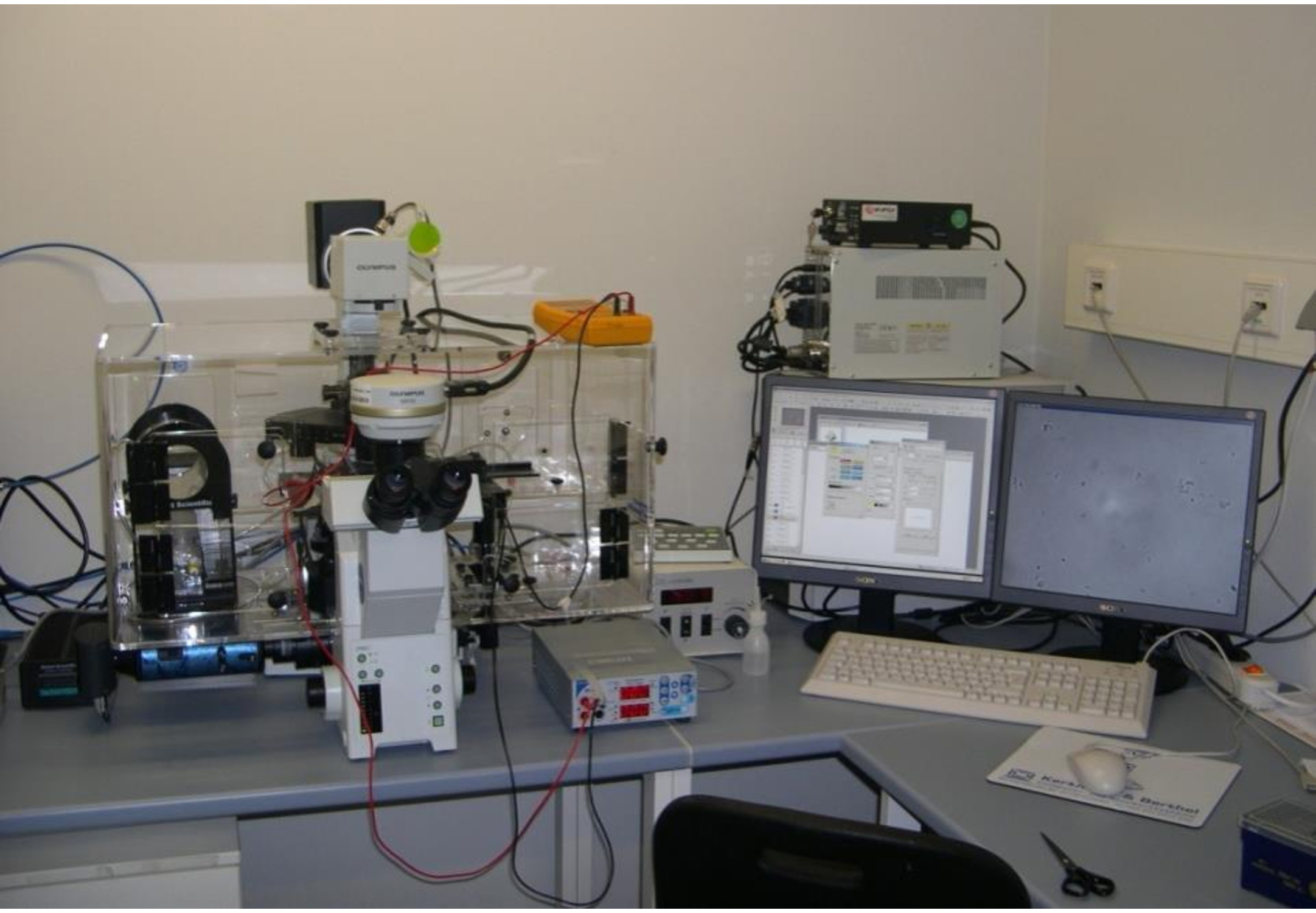 Fig. 1.
Setup for life cell imaging and computerized data acquisition.
Cells are placed within the medium reservoir (Ibidi
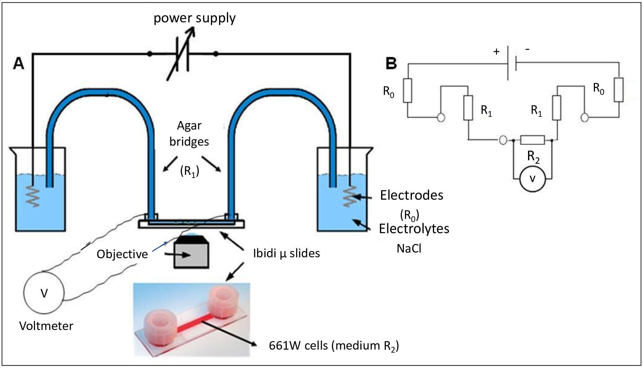 Fig. 2.
Schematic drawing of the experimental set up used in the
migration assay. (A) Consisting of a power supply, 2 beakers with saline
solution (0.9% NaCl), 2 agar-bridges (2% agar), vital microscope (Olympus IX81)
and cultivated cells in
To determine whether the physiological dcEF is causing cellular death or not, we evaluated the viability of the cells after the exposure to EF. We used the viability/cytotoxicity assay (LIVE/DEAD® Molecular Probes) to determine the death rate of cells 5 h after exposure to dcEF. The assay was conducted in each different group studied: control, 30 min and 5 h 3 V/cm, 5 h 5 V/cm, 5 h 8 V/cm. The viability was checked directly after dcEF and 5 h after the dcEF. Death rate was 5.4% for 3 V/cm and 6% for 5 V/cm (N = 5, Ncell = 500) at 5 h of exposure. In controls (no dcEF) 4% of dead cells were found. Further control experiments after 5 h showed that in mitochondria the red/green ratio of the JC1 was the same as in the stimulation period, using flow cytometry.
For plasma and mitochondrial analysis, control cells were divided into two groups: (1) untreated control cells (2) positive control cells (treated with Gramicidin and CCCP for plasma and mitochondrial membrane potential, respectively). For cell motility experiments, migration was monitored via an Olympus IX81 (Olympus, Hamburg/Germany) inverted microscope and the acquisition of images was controlled by Xcellence software (Olympus, Germany, version 1.2) (Fig. 1).
For microscopy experiments, 2 mL of 0.1
For microscopy, 1.5
Following EF treatment, cells were fixed with 4% paraformaldehyde (5 min),
permeabilized (6 min in 10
For RNA isolation, EF stimulated 661W cells and control cells in Ibidi slides
were trypsinized and centrifuged at 150 g for 5 min Supernatants were aspirated
and later total RNA isolation was done according to manufacturer’s instructions
(Qiagen, Germany). The RNA integrity was checked using 2100 bioanalyzer. mRNA was
isolated from 1
An amperometric electrode (Unisense-Micros respiration, Unisense A/S, Denmark)
was used to measure the oxygen consumption. The experiment was performed in a
closed chamber at 23 °C. For each experiment, around 2
ATP formation from ADP and inorganic phosphate (Pi) in 661W cells was measured
by the luciferin/luciferase chemiluminescent method (Roche Applied Science), as
described previously [38]. Cells (5
0.2
Images were taken every 1 min during 5 h. An average number of 125 cells were
scored for each condition (n = 10). The cell movement during application of EF,
to cathode (–) or anode (+), was tracked using the Olympus imaging software
Cell^R. Manual settings were used for cell tracking. In order to
quantify the cell speed (
To quantify polarization, the cell body from 661W cells was divided into a cathodal half and an anodal half by a line perpendicular to the applied EF and through the center of the cell. The percentage of fluorescence intensity in each half of the cells was calculated considering the total fluorescence exhibited in the cell by using ImageJ software (Open source software; National Institutes of Health, Bethesda, MD, USA). This criterion was used for scoring polarization of Golgi apparatus (GA), microtubules organizing center (MTOC), microtubules (MTs), actin and nucleus and also for the accumulation of the dyes for plasma and mitochondria membrane potentials. All measurements were made considering reduction of background and normalization by the area. Flow cytometry analysis was done using CellQuest software (Version 5.2.1 BD Biosciences, San Jose, CA, USA).
Alignment of the short reads to the mm10 transcriptome was performed with GSNAP [40] and a table of read counts per gene was created based on the overlap of the uniquely mapped reads with the Ensembl Genes annotation v. 81 (European Molecular Biology Laboratory’s European Bioinformatics Institute, Hinxton, United Kingdom) (July 2015) for mm10, using feature Counts (v. 1.4.6) [41]. The raw read counts were normalized based on the library size and the testing for differential expression between the different cell treatments was performed with the DESeq2 R package (v.1.8.1) [42]. Based on the normalized gene expression level, sample to sample Euclidean distance as well as Pearson’s correlation coefficient (r) were computed to explore correlation between biological replicates and different libraries. Differential expression was tested by fitting the count data to the negative binomial distribution. The p-values for the statistical significance of the fold change were adjusted for multiple testing with the Benjamini-Hochberg correction for controlling the false discovery rate [43]. Accepting a maximum of 10% false detections, this resulted in significantly upregulated and downregulated genes.
Each set of data analyzed was previously tested for normality using the Lillie
test on Matlab (version 9.3, Matlab is a program of MathWorks, Inc. Natick, MA, USA
). The data was then normalized by the average of the control group.
Statistics were calculated using ANOVA (normal data) and Kruskall-Wallis test
(non-normal data). The considered
We investigated the effects of direct current electric fields on migration of 661W cells. Cells were stimulated with different voltages (3 V/cm, 5 V/cm) and were monitored with time-lapse video imaging software for 5 h. The displacement of migrating cells at different conditions (control, EF 3 V/cm, EF 5 V/cm) was analyzed. The trajectory of cell migration is random in controls (no dcEF) (Fig. 3A); during EF stimulation (3 V/cm) (Fig. 3B) it is directed towards cathode, more directed with increased field strength (Fig. 3C). In absence of EF, cells migrated randomly with decreased displacement and migration velocity (Fig. 3D). Depending upon EF strengths, cells migrated more actively with significant increase in displacement and speed (Fig. 3D,E). In Fig. 3A–C, each dot represents a cell, and its relative position represents the distance travelled by cells from the center of the plot (coordinates 0, 0). The morphological changes of the cells at different time frames during 5 h duration was monitored in the presence and absence of EF (Supplementary Fig. 1). In an applied EF, cells facing the cathode exhibited protrusive activity and are strongly aligned perpendicularly to the field when compared to control cells which kept an amorphous form. Experiments reversing the polarity of the EF applied were done to verify the reliability of the directional response. Thus, upon reversing the field after 2.5 h, the cells responses were fully reversed (Supplementary Video 1).
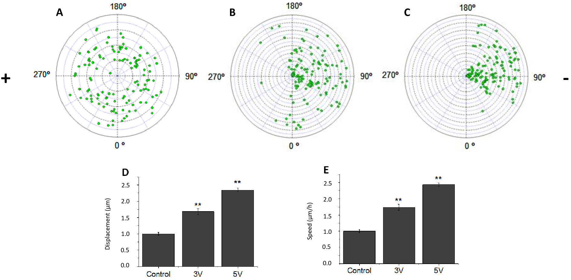 Fig. 3.
Electrotaxis of 661W cells in the presence and absence
of EF. (A) Polar plot shows random cell migration in the absence of EF. (B)
Polar plot shows the cathodal migration of the cells at 3 V/cm EF. (C) Polar plot
shows cathode directed migration of cells at 5 V/cm EF. (D) Averaged displacement
of cells and (E) Speed of the cells. Controls show smaller displacement, while EF
stimulated cells show significant increase in cell displacement and speed. Signs
(+/–) show the polarity of the electric field. n = 10, ** p
To monitor the changes of plasma membrane potential in the presence and absence
of EF, we used plasma membrane specific-dye, DiBAC
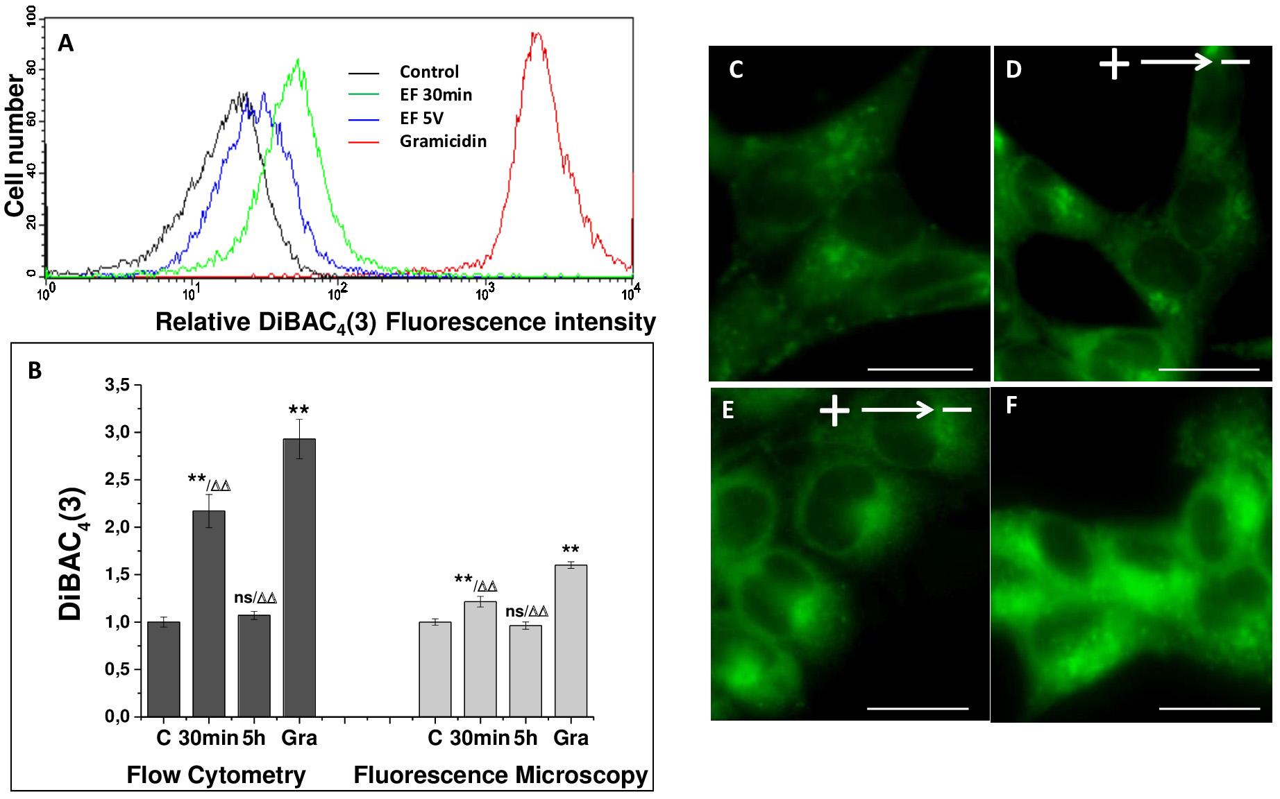 Fig. 4.
Response of DiBAC
Control cells displayed fluorescence typical of mitochondria, corresponding to high potential. In contrast, positive control cells treated with CCCP presented an only green fluorescence pattern (Fig. 5A–E). In cytometric analysis, cells treated with EF exhibited mitochondrial membrane potential loss with 20% decreased fluorescence ratio in relation to control. There is an increased mitochondrial localization in the EF treated cells at the anodal side compared to the control cells where the distribution of mitochondria is random. In addition, the mitochondrial membrane potential is slightly higher (more red than green fluorescence) at the anodal side of the cell; also, the number of mitochondria is higher on this side. Furthermore, we analyzed the changes in mitochondrial calcium in the presence and absence of EF by transfecting the cells with mito-GcaMP2. We observed in the cells treated with EF an increased amount of mitochondrial calcium towards anodal side of the cells compared to control (Supplementary Fig. 2).
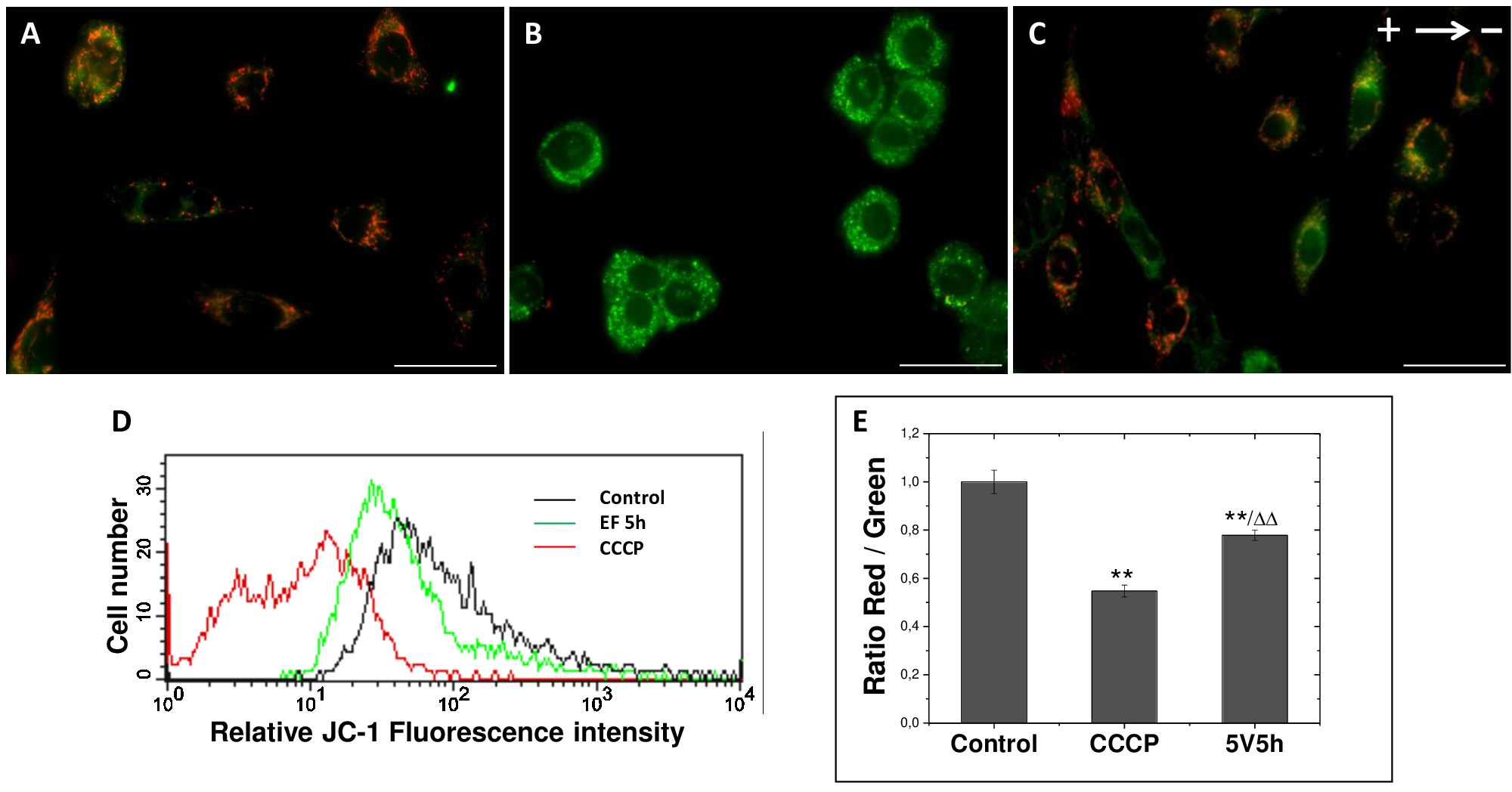 Fig. 5.
Changes in mitochondrial membrane potential of 661W
cells with EF stimulation. Mitochondrial membrane potential changes were
monitored using JC-1 at different conditions. The ionophore CCCP was used to
dissipate the mitochondrial membrane potential and to define the baseline for the
analyses of mitochondrial potential. (A–C) Representative images of cells
stained with JC-1 at different treated conditions: (A) control, (B) CCCP
(positive control) and (C) EF (5 h 5 V/cm). Scale bar, 100
Upon exposure to EF, there is slight increase in mitochondrial consumption of oxygen in the presence of malate and pyruvate. As reported in Table 1 ADP stimulated oxygen consumption increased in time dependent manner in the presence of EF whereas in the presence of pyruvate/malate the consumption decreased after 1 h and then gradually increased after 3 h. Table 2 shows ATP synthesis by the same samples used in oxymetric analysis. ATP production by 661W cells is in the same order of magnitude that of control samples, when treated with EF for short time. Prolonged stimulation, however, leads to increased ATP synthesis.
| Samples | nmol O/min /mg protein | |
| Pyruvate/malate | ADP | |
| Control | 42.6 |
55.8 |
| EF (1 h) | 38.4 |
64.63 |
| EF (3 h) | 48.3 |
69.7 |
| Data are the mean | ||
| Samples | pmol ATP produced/min/mg protein |
| Control | 201.2 |
| EF (1 h) | 207.5 |
| EF (3 h) | 231.8 |
| Data
are the mean | |
Polarization of organelles and cytoskeletal structures was random in absence of
EF (Fig. 6A,C,E,G). However, after EF stimulation there is a significant
redistribution of microtubules, Golgi, MTOC and actin towards cathode or anode
(Fig. 6B,D,F,H). Microtubules which were reoriented in the direction of migration
(cathodal side) initially were aligned perpendicular to the EF and positioned
themselves at the rear (anodal side) after 5 h. (Fig. 6A,B). EF stimulation
resulted in repositioning of 60.2%
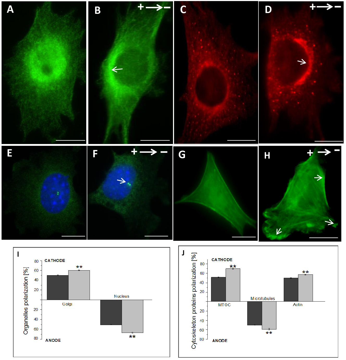 Fig. 6.
EF induced organelle and cytoskeletal polarization in
661W cells. Immunostainings of microtubules (A,B), Golgi apparatus (C,D), MTOC
and nucleus (E,F) and of actin transfected cells (G,H) showing morphological
polarization in control and EF treated cells. Signs (+/–) show the polarity of
the EF. Organelles became polarized after 5 V/cm 5 h EF exposure. (I) Percentage
of organelles facing cathode (Golgi apparatus) or anode (nucleus) quadrants in
cells stimulated with EF is significantly higher than in control cells (no EF).
(J) Percentage of reoriented cytoskeletal proteins towards cathode (Actin, MTOC)
and anode (Microtubules) quadrants is significantly higher in EF stimulated cells
than in control cells. n = 4. ** p
To understand the association of EF stimulation on cells, mRNA sequencing was performed. Our analysis shows that differential expression of genes due to EF govern several important molecular and cellular function. Of several functions, the major events altered were cellular movement, cell to cell signaling, molecular transport, morphology, assembly and organization, and cell signaling (Fig. 7). The respective genes involved in the functional networks are listed in Supplementary Table 1. Further studies are being carried out to study the effects of EF on different pathways in this cell line.
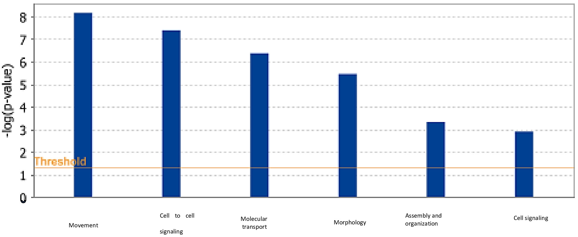 Fig. 7.
The relative changes obtained through IPA analysis for
bio-functions for the dataset of differentially expressed genes (p
Although the effect of EF has been previously shown in many different cell types [23, 26, 31, 44, 45], we could show here for the first-time direct EF effects on retinal photoreceptor-like cells. EF when applied had caused alterations in (i) migration, characterized by the active formation of lamellipodia which were clearly visible under light microscope (Supplementary Fig. 1). (ii) Directionality, which is cathodal oriented in this cell type (Fig. 3A–C) and, (iii) increased displacement and speed, which is dependent on voltage and time (Fig. 3D,E). The cells displayed positioned themselves perpendicular to the field with a wide lamellipodia at the leading edge and a small trailing edge that may be smooth (Supplementary Fig. 1A–D). Maximum alignment of 661W cells was reached within 5 h of stimulation at constant voltage.
In an EF, perpendicular cell positioning to the field occurs in order to minimize the stress due to the potential changes across the cell plasma membrane due to the constant voltage [46]. The transmembrane potential is affected by various electrical and geometrical properties of the cells such as membrane conductivity, shape and orientation (Fig. 4A–D). In our experiments, depolarization upon EF stimulation was characterized by an increased influx of dye initially which was later stabilized. This transient influx of dye accumulated at the cathodal side of the cells in the presence of EF compared with control cells where the dye accumulation was all over the cell (Fig. 4C–F). This inhomogeneity observed in the plasma membrane potential may be due to an imbalance in ion fluxes through activation of voltage gated ion channels [47]. However, in sum we observed a clear cathodal orientation of the depolarization.
Directed migration involves the initiation of cell polarity which is regulated by internal polarity effectors that promote the reorganization of cytoskeleton with well-defined front and rear ends [48]. Our findings indicate that actin, microtubules and the microtubule-organizing center (MTOC) are actively involved in the migratory response of 661W cells by being polarized to the cathodal or anodal side of the cells. In this work, we showed that actin selectively accumulated towards cathodal (leading) side of cells when stimulated by EF. In the absence of EF, the actin network was randomly distributed and extended in multiple directions wherein under certain circumstances the nucleus organizes actin structures [46]. From front to rear, actin-mediated forces sequentially promote cell protrusions, adhesion, contraction and retraction [49, 50, 51, 52, 53].
In our experiments, short term stimulation caused accumulation of microtubules in the cell front (cathodal side) and an increased accumulation towards anodal side of cells after long exposure times. Even though more microtubules grow towards the cell front, the density of microtubules close to the cell cortex is lower at the protruding front than in the retracting rear. This is probably caused by the speed of membrane protrusion exceeding that of microtubule polymerization together with the active rearward transport of microtubules by actin retrograde flow [54, 55, 56].
Our results also show reorientation of MTOC towards cathodal side. Microtubules
and actin cytoskeleton can affect MTOC positioning through cytoskeletal linkers
such as plectin that couple microtubules to myosin-powered actin retrograde flow
[50, 54, 55, 56, 57, 58, 59]. Relative levels of the actin isoforms can regulate microtubule
dynamics where
We observed an increased mitochondrial calcium on anodal side of the cells after EF exposure and a high mitochondrial distribution at the anodal side (Supplementary Fig. 2). Increased mitochondrial calcium at the anodal side and decreased at the cathodal side could give rise to push-pull effects, causing net movement of cells towards the cathode. Here, it is well-known that mitochondria are involved in cellular calcium homeostasis [69, 70, 71]. Indeed, electric field-directed cell shape changes, displacement, and cytoskeletal reorganization were shown to be calcium dependent [72]. Regarding directed migration, increased localized intracellular calcium is involved together with various possible calcium pathways [73].
Our results suggest that increases in intracellular mitochondrial calcium may be a result of rapid changes in mitochondrial membrane potentials that surpass the membrane threshold due to applied electric fields. Furthermore, increased mitochondrial calcium drives an adaptive metabolic boost in stress situations like ER stress. Accordingly, blocking calcium transfer impaired a metabolic response, rendering cells more vulnerable to, e.g., ER stress [74]. Furthermore, calcium, which is released from ER by the inositol 1,4,5-triphosphate receptor is taken up by mitochondria where it is required for efficient oxygen consumption and ATP production [75].
In our experiments, we could directly monitor the increased energetic expense of
concomitant changes in organelle cytoskeletal rearrangement and migration in the
presence of EF. Here, we have found that ADP stimulated oxygen consumption
increased in time dependent manner in the presence of EF whereas in the presence
of pyruvate/malate the consumption decreased after 1 h and then gradually
increased. We also showed that prolonged EF stimulation led to an increased ATP
production. Regarding this, it is described that EF induced directed movement or
migration in rigid media leads to a higher O
Though EF activates the downstream signaling pathways through plasma membrane to recruit the components of cytoskeleton and polarity machinery, the exact signaling events between the external and internal cues remain unclear. Our present findings from sequencing will allow us to further study about directionality and polarization in the presence of EF. Fig. 7 and Supplementary Table 1 show a few bio-functions and their possible candidate genes, differentially regulated in the presence of EF. Chemokine ligands and solute carrier transporters along with transmembrane reporters might activate the downstream signaling pathways for persistent directed migration and polarization in the EF stimulated cells, because chemokines and other genes mentioned are required in cell motility, assembly, and organization and signaling [77, 78, 79]. In this line, certain chemokines are known to induce calcium influx into the cells [80]. Also, the transporters expressed are involved in transport of cationic amino acids at the plasma membrane, phosphate linked antiporters at the ER, sodium independent, anionic amino acid transporters and also to maintain cellular iron ion homeostasis [81, 82, 83].
However, much more work has to be done to elucidate the detailed signaling mechanisms in all the here mentioned processes activated by EF.
Retinal 661W cells, are susceptible to EF like other retinal cells treated in in vitro, in vivo and in patients with transcorneal electrostimulation of the retina. Regarding polarization in the EF, we found the following events: directed migration towards the cathode with over two-fold increase in speed and displacement as well as significant depolarization of the plasma membrane at the leading edge of the cell (cathodal side). Then redistribution of microtubules into the direction of migration (cathodal side), initially aligned perpendicular to the EF. Also, the MTOC re-oriented into this direction. Concomitantly with the microtubules, actin oriented along the field and cell membrane and reorganized in an asymmetrical fashion with an increased fluorescence at the cathodal side. The cytoskeletal elements and the MTOC redistributed significantly from 50% (random) to 60–70% into the dcEF-induced direction. The Golgi apparatus, which is involved in many steps of actin synthesis, moved to the cathodal side by 65%. Later the microtubules positioned themselves at the rear (anodal side), like the nucleus by 70% compared to the random 50% in controls without EF.
Interestingly, mitochondria and also the mitochondrial calcium moved to the
anodal side, possibly because of energy consuming processes at the site of the
nucleus and ER. We also measured an overall 115.2% increase in ATP production
and 113.3% increase in O
In sum, we could demonstrate that this cell type can easily be addressed by EF. The mechanisms found can enhance our understanding regarding beneficial effects of EF treatment in retinal diseases.
JG-H. and SB performed most of the cell and molecular biologic experiments. DC did the experiments regarding metabolism. DA did the gene expression analysis. RHWF, SSM and CR designed the experiments and have written the manuscript together with JG-H and SB.
Not applicable.
Not applicable.
This research received no external funding.
The authors declare no conflict of interest. RHWF is serving as one of the Editorial Board members and Guest Editors of this journal. We declare that RHWF had no involvement in the peer review of this article and has no access to information regarding its peer review. Full responsibility for the editorial process for this article was delegated to GP.
Supplementary material associated with this article can be found, in the online version, at https://doi.org/10.31083/j.fbl2709273.
References
Publisher’s Note: IMR Press stays neutral with regard to jurisdictional claims in published maps and institutional affiliations.