IMR Press / RCM / Volume 23 / Issue 7 / DOI: 10.31083/j.rcm2307233
Open Access Original Research
Efficacy and Safety of a Polytetrafluoroethylene Membrane Wrapped a Single Layer of Sirolimus-Eluting Stent in a Porcine Coronary Perforation Model
Show Less
1 Department of Cardiology, Xijing Hospital, Fourth Military Medical University, 710032 Xi’an, Shaanxi, China
*Correspondence: lingtao@fmmu.edu.cn (Ling Tao); rutaowang5@gmail.com (Rutao Wang)
These authors contributed equally.
Academic Editor: Carlo Briguori
Rev. Cardiovasc. Med. 2022, 23(7), 233; https://doi.org/10.31083/j.rcm2307233
Submitted: 13 April 2022 | Revised: 17 May 2022 | Accepted: 23 May 2022 | Published: 24 June 2022
Copyright: © 2022 The Author(s). Published by IMR Press.
This is an open access article under the CC BY 4.0 license.
Abstract

Background: Covered stents are effective in treating coronary artery perforation (CAP), however, the high rate of immediate device deployment failure and in-stent restenosis have limited the application of the currently covered 
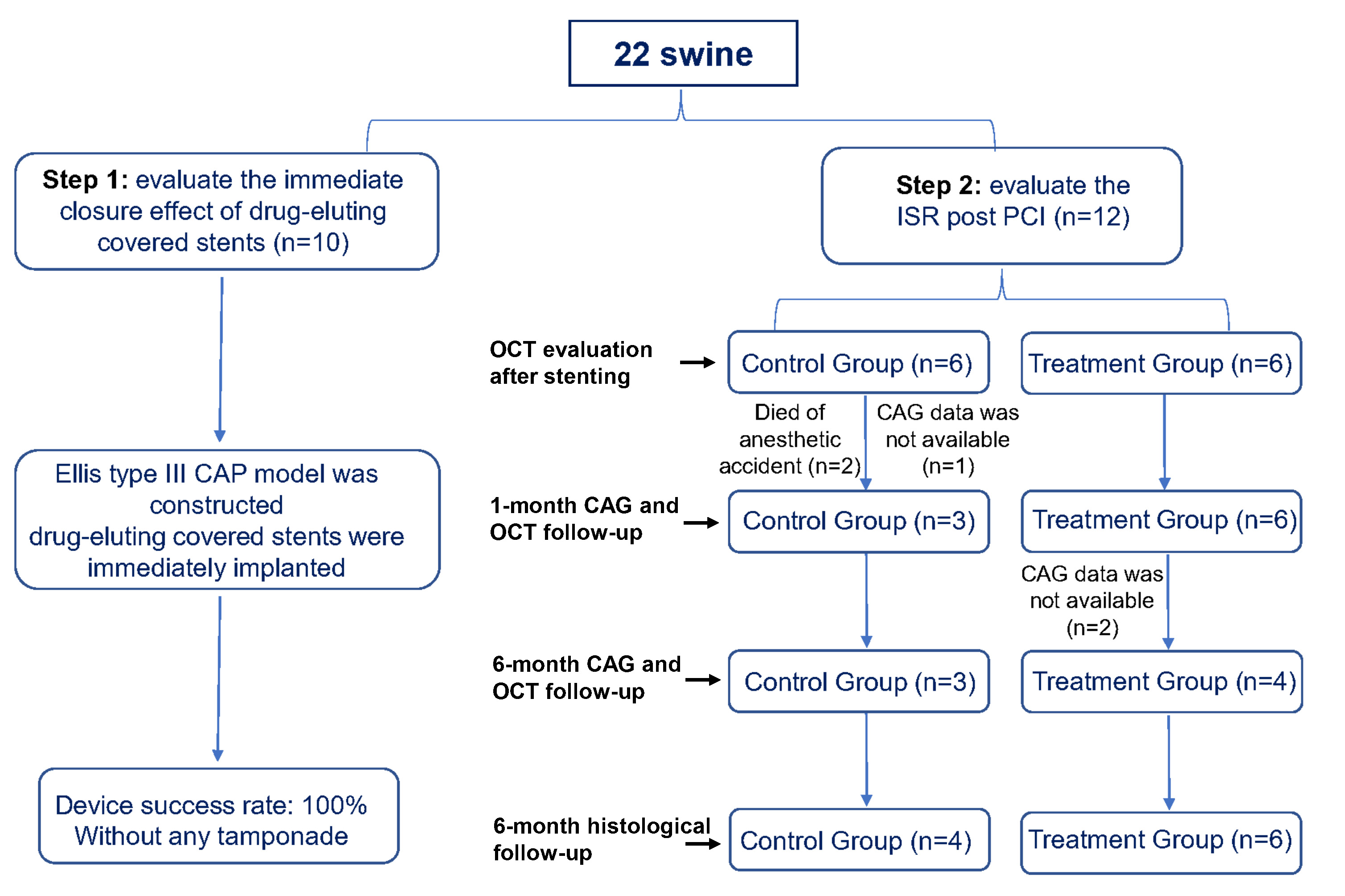
stents. Methods: We designed a covered stent system consisting of a single layer of drug-eluting stent and a layer of polytetrafluoroethylene (PTFE) membrane wrapped at the outer layer of the stent. The immediate sealing effect of our novel covered stent was observed by using an Ellis type III CAP model. The device’s success was defined as its ability to seal the perforation, assessed by visual estimation and final thrombolysis in myocardial infarction (TIMI) 3 flow. The antiproliferative effect was evaluated in 12 swine, which were randomly assigned to treatment (sirolimus-eluting covered stents) and control (bare metal covered stents) groups. Coronary angiography and optical coherence tomography (OCT) were performed at index procedure, 1- and 6-month after stent implantation. All swine were sacrificed for histopathological analyses at 6-month. Results: The device success rate was 100%. All swine were alive at 6-month follow-up. At 1-month, the treatment group had a larger minimal luminal diameter (MLD) (1.89 ± 0.29 mm vs. 0.63 ± 0.65 mm, p = 0.004) and lower late luminal loss (LLL) (0.47 ± 0.15 mm vs. 1.80 ± 0.34 mm, p < 0.001) compared with control group. At 6-month, the treatment group had a numerically higher MLD (0.94 ± 0.75 mm vs. 0.26 ± 0.46 mm; p = 0.230) and lower LLL (1.43 ± 0.85 mm vs. 2.17 ± 0.28 mm; p = 0.215) compared with control group. Histological analyses revealed the mean plaque area was lower in the treatment group (2.99 ± 0.81 mm2 vs. 4.29 ± 0.77 mm2, p = 0.035) than in the control group. No in-stent thrombosis was observed in either group. Conclusions: In the porcine model of coronary perforation, the PTFE membrane wrapped sirolimus-eluting stent showed a high device success rate in sealing the perforation. The drug-eluting covered stent demonstrated a relatively sustained antiproliferative effect up to 6 months post-implantation.

Keywords
covered stent
coronary artery perforation
percutaneous coronary intervention
Funding
2018YFA0107400/National Key R&D Program of China
82070853/Program for National Science Funds of China
81730011/Program for National Science Funds of China
81970721/Program for National Science Funds of China
81927805/Program for National Science Funds of China
PCSIRT-14R08/Program for Changjiang Scholars and Innovative Research Team in University
Figures
Fig. 1.
Share
Back to top