IMR Press / JIN / Volume 22 / Issue 1 / DOI: 10.31083/j.jin2201019
Open Access Review
The Role of Ferroptosis in Nervous System Disorders
Show Less
1 Department of Anaesthesiology and Perioperative Medicine and Department of Intensive Care Unit, Xijing Hospital, The Fourth Military Medical University, 710032 Xi’an, Shaanxi, China
2 College of Life Sciences, Northwest University, 710069 Xi’an, Shaanxi, China
*Correspondence: zongping03@163.com (Zongping Fang); xjzhang0806@163.com (Xijing Zhang)
These authors contributed equally.
Academic Editor: Drozdstoy Stoyanov
J. Integr. Neurosci. 2023, 22(1), 19; https://doi.org/10.31083/j.jin2201019
Submitted: 24 June 2022 | Revised: 29 July 2022 | Accepted: 1 August 2022 | Published: 16 January 2023
(This article belongs to the Special Issue Advances in Depression Research)
Copyright: © 2023 The Author(s). Published by IMR Press.
This is an open access article under the CC BY 4.0 license.
Abstract

Ferroptosis is distinct from other apoptotic forms of programmed cell death and is characterized by the accumulation of iron and lipid peroxidation. Iron plays a crucial role in the oxidation of lipids via the Fenton reaction with oxygen. Hence, iron accumulation causes phospholipid peroxidation which induces ferroptosis. Moreover, detoxification by glutathione is disrupted during ferroptosis. A growing number of studies have implicated ferroptosis in nervous system disorders such as depression, neurodegenerative disease, stroke, traumatic brain injury, and sepsis-associated encephalopathy. This review summarizes the pathogenesis of ferroptosis and its relationship with various nervous system disorders.

Keywords
ferroptosis
iron
lipid peroxidation
glutathione
nervous system disorders
1. Introduction

Nervous system disorders include depression, neurodegenerative disease, stroke, traumatic brain injury (TBI), and sepsis-associated encephalopathy (SAE). These conditions seriously affect the quality of life and significantly increase the economic and social burden [1, 2, 3, 4]. A common feature of nervous system disorders is the abnormal activation of cell death [5, 6]. A better understanding of the mechanism of neuronal death should therefore lead to new approaches for the prevention and treatment of nervous system disorders.

Programmed cell death is classified into apoptosis, pyroptosis, autophagy, necroptosis, and ferroptosis according to the different molecular mechanisms and morphological features (Table 1) [7, 8, 9]. Ferroptosis was first proposed in 2012 as a novel form of programmed cell death that results from the accumulation of ferrous ions and the reduction of Glutathione (GSH) synthesis [10, 11, 12]. GSH is comprised of glutamate, cysteine, and glycine. This sulfhydryl-containing tripeptide is essential for the protection of the brain from oxidative stress through its action as a free radical scavenger and lipid peroxidation inhibitor [13]. Transferrin-bound iron is transported into the cell by transferrin receptor 1 (TFR1) and converted into ferrous ions. The accumulation of ferrous ions leads to the production of lipid-reactive oxygen species and eventually to ferroptosis [14]. Additionally, impaired generation of GSH reduces the synthesis of glutathione peroxidase 4 (GPX4), thereby blocking the conversion of lipid hydroperoxides into non-toxic lipid alcohols and leading to the accumulation of phospholipid hydroperoxides (PLOOHs) [15, 16]. PLOOHs are lipid-based forms of reactive oxygen species (ROS) that are essential for the inhibition of ferroptosis [17]. The accumulation of PLOOHs causes rapid and irreparable damage to the membrane and directly catalyzes the chain reaction of peroxidation to induce lipid peroxide-dependent cell death [18].

Table 1.Comparison of several types of programmed cell death.
Cell death forms Cell membrane Cytoplasm Cell nucleus Biochemical characteristics Immune characteristics
Ferroptosis Breakage and bubble Increase mitochondrial membrane density Normal The increase of iron-dependent lipid peroxides Promote inflammation
Apoptosis Integrity No significant change Nuclear shrinkage chromatin condensation DNA fragmentation decreases the mitochondrial membrane potential Inhibit inflammation
Autophagy Integrity Lysosomal vacuoles are double membranes Normal LC3 changed from LC3I to LC3II Promote inflammation
Necroptosis Membrane rupture Swelling of organelles Partial condensation of chromatin Enrichment of kinase and decrease of ATP level Promote inflammation
Pyroptosis Loss of membrane integrity Loss of organelles DNA condensation and fragmentation Formation of inflammasomes activation of caspase-1 and release of a large number of pro-inflammatory factors Promote inflammation

The link between ferroptosis and a wide range of disorders including cardiovascular disease, cancer, and metabolic disease has been studied extensively [19, 20, 21]. For example, cardiomyopathy was improved following the upregulation of GPX4. Furthermore, an iron chelating agent was able to reduce the level of the ferrous ion, ameliorate the side effects of cardiomyopathy, and prevent ferroptosis [22]. Recent studies have also shown a close relation between ferroptosis and various nervous system disorders. The aim of this review is to summarize the development and mechanism of ferroptosis and its role in nervous system disorders, thereby suggesting possible new options for the treatment of nervous system disorders.

2. The Development of Ferroptosis

Ferroptosis is a form of programmed cell death caused by the accumulation of iron-dependent lipid peroxide [23]. In 2003, Dolma et al. [24] reported that Erastin and RSL3 (RAS-selective lethal agents) could induce cell death in RAS mutated cells. In 2007, Yagoda et al. [25] found that Erastin-induced cell death was distinct from apoptosis, autophagy, necroptosis, and other forms of cell death. The morphology of this new type of cell death was characterized by a loss of mitochondrial integrity. The following year, two compounds similar to Erastin (RSL5 and RSL3) were identified that could induce iron-dependent, non-apoptotic cell death in tumor cells [26]. This form of cell death could be inhibited by iron-chelating agents (DFOM) and antioxidants (vitamin E) [27]. In 2012, Dixon et al. [10] formally named this as “ferroptosis”. In contrast to other types of cell death, the cell membrane is broken during ferroptosis while the mitochondrial membrane density is increased. The concept of “ferroptosis” was further developed in 2012 [28].

3. Mechanisms of Ferroptosis
3.1 Iron Metabolism and Ferroptosis

Iron is the most abundant transition metal element in the Earth’s crust and plays a critical role in oxygen transport, cellular energy metabolism, and many enzymatic reactions [29, 30]. Under normal conditions, iron is absorbed from food in the duodenum and mucosa of the upper jejunum [31]. The ferrous ions released from the ferric transporter on the outer surface of the capillary endothelial cell are oxidized by ceruloplasmin into ferric ions, which are then captured by transferrin (Tf) in the intercellular fluid and cerebrospinal fluid. Tf binds the ferric ions and forms a Tf-TFR1 complex with transferrin receptor 1 (TFR1), which is then endocytosed via clathrin-coated pits at the cell surface [32, 33]. In the acidic endosomes, ferric ions are reduced into ferrous ions and transported by divalent metal transporter 1 (DMT1) to labile iron pools in the cytoplasm. Here, the ferrous ions are stored in ferritin or imported into mitochondria via mitotic ferritin. Non-transferrin-bound iron at the cell surface is directly introduced into the labile iron pool by DMT1 [34, 35, 36]. The accumulation of intracellular ferrous ions promotes lipid peroxidation, leading to the production of ROS and ultimately to ferroptosis (Fig. 1) [37].
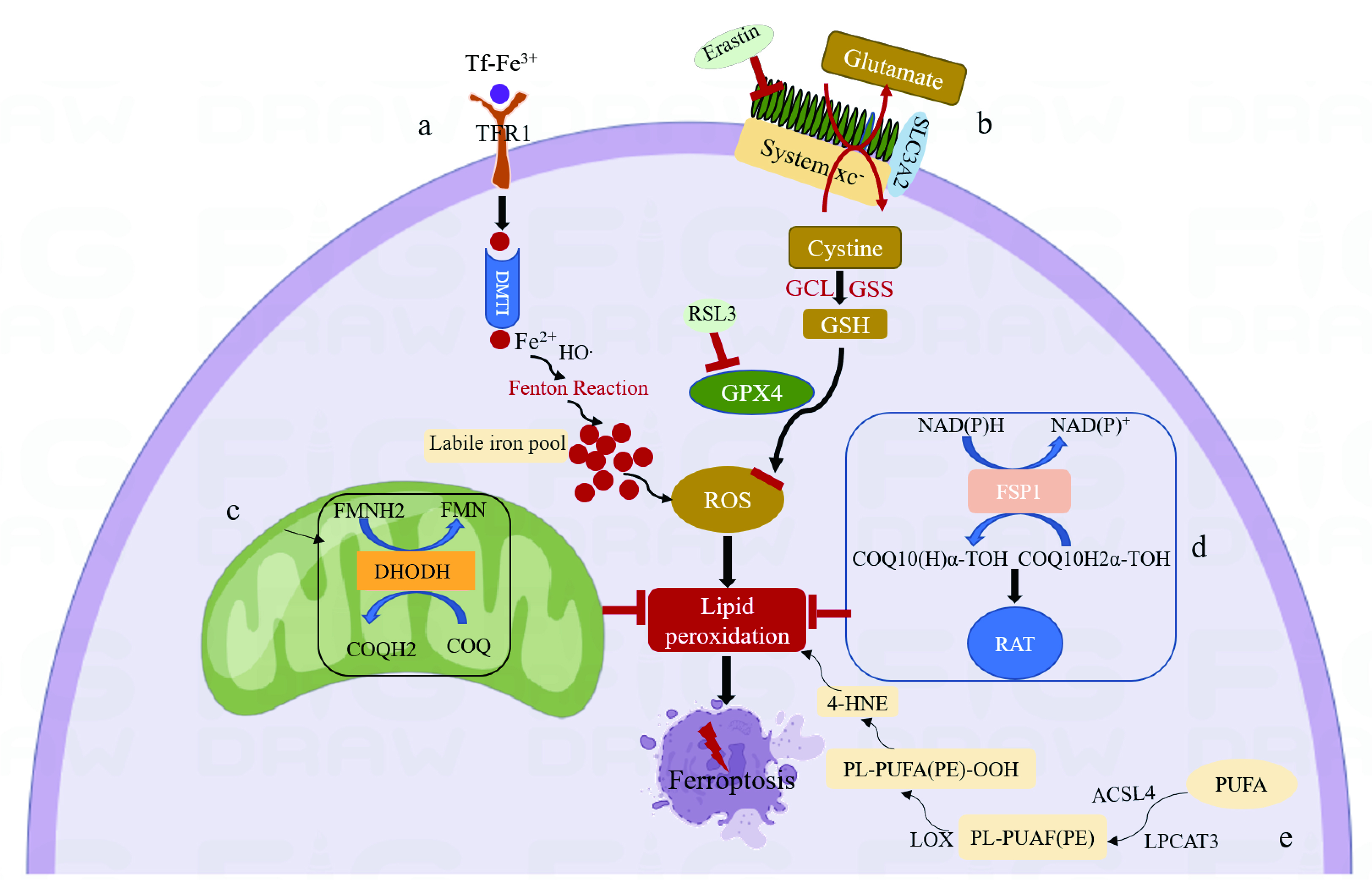
Fig. 1.

Mechanism of ferroptosis. (a) Tf binds ferric ions and forms a Tf-TFR1 complex with TFR1. The following endocytosis into acidic endosomes, ferric ions are reduced into ferrous ions and transported by DMT1 to labile iron pools in the cytoplasm. The accumulation of intracellular ferrous ions promotes lipid peroxidation, leading to the production of ROS and ultimately to ferroptosis. (b) System xc-exchanges intracellular glutamate for extracellular cystine in a 1:1 ratio, which is then synthesized into GSH by GCL and GSS. GSH is a reducing cofactor for glutathione peroxidase (GPX4), a membrane lipid repair enzyme that eliminates lipid hydroperoxides to protect against ferroptosis. (c) Dihydroorotate Dehydrogenase (DHODH) in the mitochondrial membrane resists ferroptosis by oxidizing dihydroorotate (DHO) to orotic acid (OA) and reducing ubiquinone (CoQ10) to Dihydroubiquinone (CoQH2). (d) Ferroptosis suppressor protein 1 (FSP1) converts ubiquinone into ubiquinol and generates a lipophilic, radical-trapping antioxidant (RTA) that prevents lipid peroxide formation and inhibits ferroptosis. (e) Polyunsaturated fatty acids (PUFA) are substrates for lipid peroxidation and react with acyl-CoA synthetase long-chain family member-4 (ACSL4) to generate Malondialdehyde (MDA) and 4-hydroxynonenal (4-HNE), eventually leading to ferroptosis.

3.2 Lipid Peroxidation and Ferroptosis

Lipid peroxidation is the hallmark of ferroptosis. In the initial stage, lipid peroxidation can occur through both enzymatic and non-enzymatic reaction processes [38, 39]. However, which of these two is essential for ferroptosis remains controversial. Several studies have suggested that non-enzymatic lipid peroxidation is responsible for ferroptosis [40]. This is driven by the Fenton reaction, which uses iron and oxygen to catalyze a chain reaction that leads to the production of phospholipid hydroperoxides (PLOOHs) [40]. Polyunsaturated fatty acid (PUFA) phospholipids are substrates for lipid peroxidation. Enzymes such as acyl-CoA synthetase long-chain family member-4 (ACSL4) inhibit ferroptosis by reducing PUFA phospholipid synthesis. ACSL4 is therefore a marker of sensitivity to ferroptosis [41]. Mechanistically, ACSL4 enriches cellular membranes with long polyunsaturated ω6 fatty acids. The peroxidation of polyunsaturated fatty acids (PUFAs) and unlimited lipid peroxidation generates products such as malondialdehyde (MDA) and the formation of reactive aldehydes, especially 4-hydroxynonenal (4-HNE). Eventually, this leads to the loss of cell membrane integrity and to ferroptosis (Fig. 1) [42, 43].

3.3 System xc--GSH-GPX4 and Ferroptosis

System xc- is an important intracellular antioxidant system consisting of two subunits: SLC7A11 and SLC3A2 [44]. SLC7A11 is mainly responsible for transport activity and is highly specific for cystine and glutamate, while SLC3A2 acts as a chaperone protein [45]. System xc- exchanges intracellular glutamate for extracellular cystine (Cys2) in a 1:1 ratio. The cystine is then synthesized into glutathione (GSH) by glutamate-cysteine ligase (GCL) and glutathione synthetase (GSS) [46, 47, 48]. GSH is a reducing cofactor for glutathione peroxidase (GPX4), which is a membrane lipid repair enzyme [16]. Inhibition of system xc- leads to the depletion of intracellular cystine which is the rate-limiting substrate for the synthesis of the antioxidant glutathione (GSH) [49]. Due to the antioxidant properties of GSH, its depletion causes an imbalance between the cellular oxidation and antioxidant systems, as well as the inhibition of glutathione-dependent enzymes such as GPX4. GPX4 is a member of the glutathione peroxidase family [40]. These can reduce hydroperoxides produced from phospholipids and cholesterol to their corresponding alcohols by using two electrons from the GPX4 catalytic cysteine residues [16]. Genetic studies have established that GPX4 is a key regulator of ferroptosis [50]. In the normal state, PUFAs are frequently oxidized by lipoxygenases such as ACSL4 but are then immediately reduced by GPX4 and its GSH cofactor [51]. Hence, the loss of GPX4 leads to peroxidation chain reactions that cause lipid peroxidation-dependent cell death by blocking hydroperoxide conversion into non-toxic lipid alcohols [11, 52, 53]. In conclusion, disruption of the System xc--GSH-GPX4 axis results in the inability to eliminate PLOOH, the accumulation of lipid peroxides, and finally to ferroptosis (Fig. 1).

3.4 Ferroptosis Suppressor Protein 1 (Fsp1) and Ferroptosis

Previous studies on ferroptosis defense mechanisms have focused on GPX4 [54]. Recently, two articles published in 2019 reported on a novel ferroptosis suppressor, FSP1, that was independent of GSH [55, 56]. FSP1 was originally named AIFM2 and has homology to apoptosis-inducing factor (AIF) [55]. However, it lacks the N-terminal mitochondrial targeting sequence present in AIF and does not promote apoptosis. AIFM2 was therefore renamed FSP1 [55]. It is located in lipid droplets (LDs) and the plasma membrane [57]. It can convert ubiquinone on cell membranes into its reduced form, panthenol, which inhibits peroxidation and prevents ferroptosis [55]. By employing a synthetic lethal CRISPR/Cas9 screen, Doll et al. [55] found that FSP1 was an oxidoreductase that can reduce coenzyme Q10 (CoQ) to generate a lipophilic, radical trapping antioxidant (RTA) that prevents lipid peroxide formation (Fig. 1). Venkatesh et al. [58] reported that inhibition of MDM2 and MDMX resulted in increased levels of FSP1 protein, which in turn led to increased levels of the endogenous lipophilic antioxidant CoQ, thereby inhibiting ferroptosis. Song et al. [59] showed that miR-4443 regulates the expression of FSP1 through METLL3 in an m6A manner, thus regulating ferroptosis.

3.5 Mitochondrial Dihydroorotate Dehydrogenase (DHODH) and Ferroptosis

DHODH is a flavin-dependent enzyme located in the mitochondrial membrane [60]. By selectively inhibiting dihydroorotate dehydrogenase, thereby inhibiting pyrimidine synthesis [61]. This novel, GSH-independent ferroptosis defense system was first reported by Boyi’s team in 2021 [61]. They demonstrated that DHODH inhibits ferroptosis by oxidizing dihydroorotate (DHO) to orotic acid (OA) and reducing ubiquinone (CoQ10) to Dihydroubiquinone (CoQH2) in the mitochondrial membrane (Fig. 1). To our knowledge, there have been no further advances regarding the DHODH and ferroptosis mechanism. The molecular mechanisms that link ferroptosis with various diseases also warrant further exploration.

4. Ferroptosis and Nervous System Disorders
4.1 Ferroptosis and Depression

Depression is a major global health issue that affects about 300 million people worldwide [62]. It is characterized by persistent mood depression and restlessness and includes psychomotor disorders and cognitive impairments [63]. It also has the characteristics of being universal, chronic, and recurrent [64]. Unfortunately, completely effective treatment for depression has yet to be discovered. Currently, treatment for depression mainly involves drug therapy and psychotherapy. However, their efficacy is quite limited and drug therapy requires long-term medication, while psychotherapy is slow [64, 65, 66]. Therefore, it is very important to explore the pathophysiological mechanism of depression. Studies have shown that hippocampal volume is significantly reduced in both adults and adolescents with depression [67]. Patients who have recovered from depression also show a decreased hippocampal volume [67]. The destruction and atrophy of neurons during depression may be the cause of this reduced volume [68, 69]. Previous studies on depression and the death of hippocampal neurons have focused mainly on apoptosis [70]. For example, Zhao et al. [71] found that repetitive transcranial magnetic stimulation (RTMS) reduces depression-like behavior by inhibiting the apoptosis of hippocampal neurons. Dexmedetomidine can also reduce the apoptosis of hippocampal neurons in Sprague Dawley rats and decrease depressive behavior by inhibiting the p38 MAPK/c-Myc /CLIC4 signaling pathway [72]. As described above, ferroptosis is a newly discovered form of programmed cell death that is caused mainly by the accumulation of iron and lipid peroxides [73]. Ferroptosis offers a new treatment strategy for depression. Cao et al. [74] confirmed that mice with chronic unpredictable mild stress (CUMS) could be used as an animal model for depression. Jiao et al. [75] found that GPX4 expression was decreased in this model whereas that of FTH1, ACSL4, and COX2 was increased, indicating that ferroptosis was activated. Wang et al. [76] found that sodium hydride (NaHS) can reduce iron deposition and oxidative stress and increase the protein level of GPX4 and SLC7A11 in the prefrontal cortex of type 1 diabetic mice, thereby reducing ferroptosis and alleviating anxiety-depressive behavior (Table 2, Ref. [76, 77, 78, 79, 80, 81, 82, 83, 84, 85]). Using the chronic social defeat stress (CSDS) mouse model, Dang et al. [77] found that edravone (EDA) could upregulate GPX4 expression to alleviate depressive behavior, but that EX527 and ML385 can reduce Nrf2, HO-1, and Gpx4 protein levels and thereby attenuate the antidepressant effects of EDA (Table 2). In conclusion, these results confirm that ferroptosis occurs in mouse models of depression. Although the underlying molecular mechanism remains unknown, the upregulation of GPX4 can inhibit ferroptosis and thereby reduce depressive behavior in mice.

Table 2.Ferroptosis effector and its mechanism in nervous system disorders.
Nervous system disorder Reagents Functions References
Depression Sodium hydride GPX4 and SLC7A11. Wang Y, et al. [76]
Edravone GPX4 Dang R, et al. [77]
EX527 and ML385 Nrf2, HO-1, and Gpx4
Neurodegenerative diseases
Alzheimer’s disease Lip-1 Trapping free radical Hambright WS, et al. [79]
Parkinson’s disease Iron ion chelating agents and ferroptosis inhibitor Fer-1 Iron and GPX4 Mahoney-Sanchez L, et al. [78]
ferric ammonium citrate Iron Zhang P, et al. [80]
Stroke
Ischemic stroke Carvacrol GPX4 Guan X, et al. [81]
Cerebral hemorrhage Iron ion chelating agent Deferoxamine Iron Okauchi M, et al. [85]
Traumatic Brain Injury Ferrostatin-1 (Fer‐1) Iron deposition Xie BS, et al. [82]
Sepsis-associated encephalopathy Iron ion chelating agents Iron Yao P, et al. [83]
miR-9-5p Transferrin receptor Wei XB, et al. [84]

In conclusion, ferroptosis is a potential target for the treatment of depression, but there is still no clinical evidence to support the use of ferroptosis inhibitors in humans.

4.2 Ferroptosis and Neurodegenerative Diseases

Neurodegenerative diseases are caused by the accumulation of oligomers and the aggregation of misfolded proteins [86, 87]. Due to the aging of society, neurodegenerative diseases have a considerable socio-economic impact. They include Alzheimer’s disease (AD), Parkinson’s disease (PD), and Huntington’s disease (HD) [88, 89], and are characterized by a loss of function of specific neuronal groups and of their connections [88]. Emerging evidence has shown that ferroptosis can disrupt neuronal viability and is involved in the pathological mechanism of neurodegenerative diseases [78, 90].

4.2.1 Ferroptosis and AD

AD is a complex neurodegenerative disease characterized by extracellular accumulation of the proteins amyloid-β and tau in the cerebral cortex and limbic regions [91, 92, 93]. It is caused by the degeneration of memory and cognitive neurons, with the main manifestations being memory loss, delusions and hallucinations. The inability of AD patients to live independently imposes a heavy burden on their family and on society [21]. Previous studies on neuronal loss have focused on apoptosis and necroptosis [94, 95, 96, 97]. More recent evidence has shown that ferroptosis is involved in the pathogenesis of AD, thus offering a new treatment strategy for this disease [90, 98, 99]. Aging is the major risk factor for neurodegenerative diseases. With older age, iron accumulates in the neurons of the brain and exacerbates oxidative damage, causing a variety of pathologies [100, 101, 102, 103]. This eventually leads to the significant cognitive impairment seen in AD. Rogers et al. [104] reported that a set of common-acting, iron-responsive 5-untranslated region (5UTR) motifs can be folded into RNA stem-loops that have a significant impact on AD. Iron is a positive regulator of amyloid precursor protein (APP) translation via the iron-responsive-like element (IRE-like) RNA stem-loops in the 5UTR of APP mRNA [104, 105, 106]. Studies have shown that Aβ plays an important role in the reduction of divalent iron, with this transformational process leading to excess free radical production and neuronal damage [107]. Stoyanovsky et al. [39] reported that GPX4-knockout mice showed AD-like hippocampal neurodegeneration and increased levels of lipid peroxidation, which is a marker of ferroptosis. This could be attenuated by the ferroptosis inhibitor Liproxstatin-1 [79] (Table 2). In conclusion, inhibition of ferroptosis alleviates neurodegeneration in a mouse model of AD, suggesting that ferroptosis could be a potential therapeutic target for this disease.

4.2.2 Ferroptosis and PD

PD is the most common severe movement disorder in the world and one of the most disabling diseases of the central nervous system [48]. Attributed to a selective loss of neurons in the substantia nigra, the symptoms of PD include tremors, stiffness, slow movement, postural instability, and difficulty in walking [108, 109]. Previous studies on PD have focused on dopamine-based therapies, but these are only effective during the early course of the disease [78]. Further exploration of novel treatment methods is therefore needed. The traditionally recognized pathological features of PD are dopaminergic neuronal death and intracellular α-synuclein (α-syn) deposition [110, 111, 112]. In addition, iron accumulation, elevated oxidative stress, and lipid peroxidation damage have also been implicated in the pathological processes of PD [113], suggesting that ferroptosis could be a potential therapeutic target. Tian et al. [114] reported that the expression of FTH1 and GPX4 were decreased in the rat 6-hydroxydopamine (6-OHDA) model of PD, while the level of the ferrous ion was increased. Moreover, these workers showed that iron-chelating agents and the ferroptosis inhibitor Fer-1 could reverse PD caused by the reduction of FTH1 (Table 2). In vitro experiments showed that knockdown of FTH1 in PC-12 cells significantly inhibited cell viability and resulted in mitochondrial dysfunction [114]. These results demonstrate that 6-OHDA-induced ferroptosis is FTH1-mediated and that overexpression of FTH1 in a cell model of PD can attenuate ferroptosis. Mahoney-Sanchez et al. [78] reported that α-syn was functionally related to the metabolism of iron and lipids. Zhang P et al. [80] used ferric ammonium citrate (FAC) to treat dopaminergic cells and thus mimic iron overload during the progression PD (Table 2).

The main focus to date for the clinical management of PD and AD is the iron chelator deferiprone, which inhibits ferroptosis in vitro [115, 116]. Deferiprone significantly reduced brain iron levels in phase II clinical trials of PD, with phase III and IV trials still ongoing. Phase II clinical trials of deferiprone are also currently underway for AD [117, 118].

4.3 Ferroptosis and Stroke

Stroke is an acute disorder of blood circulation in the brain tissue caused by arteriovenous stenosis, occlusion, or rupture due to various inducing factors [119, 120]. It is also one of the most important causes of disability [121, 122]. The clinical symptoms of a stroke are language disorder and cognitive impairment [123]. Stroke can be divided into ischemic stroke and cerebral hemorrhage (ICH) stroke [124]. It has been reported that ferroptosis plays an important role in the pathophysiological processes of various cancer types [125, 126]. In recent years there has also been substantial evidence that ferroptosis is associated with the progression of stroke [127].

4.3.1 Ferroptosis and Ischemic Stroke

Ischemic stroke is characterized by the inadequate blood supply to specific areas of the brain, leading to permanent disability [128]. Insufficiency of blood in the brain activates the ischemic cascade reaction of neurons, which involves excitatory toxicity, oxidative stress, post-ischemic inflammation, and eventually cell death. Recently, ferroptosis has been found to play an important role in the development of ischemic stroke by influencing iron metabolism and lipid peroxidation, while the inhibition of ferroptosis successfully reverses ischemic damage [129]. During acute ischemia, the blood-brain barrier is destroyed and a large number of ferrous ions enter the brain parenchyma, resulting in the Fenton reaction and ferroptosis [130]. Moreover, Cui et al. [41] found that ACSL4 plays a key role in stroke. Knockdown of ACSL4 protected mice from ischemic injury by inhibiting proinflammatory cytokine production in the microglia, whereas the overexpression of ACSL4 exacerbated ischemic brain injury by promoting lipid peroxidation and facilitating neuronal death. ACSL4 is a regulator of neural death and neuroinflammation, and interfering with its expression may be a novel therapeutic approach for ischemic stroke [41]. Guan et al. [81] showed that carvacrol inhibits ferroptosis by increasing the expression of GPX4, thereby providing protection against cognitive dysfunction in ischemic-reperfusion gerbils (Table 2). These findings suggest that the inhibition of ferroptosis improves the outcome after stroke, thus offering a potentially novel treatment strategy.

4.3.2 Ferroptosis and ICH

ICH refers to bleeding caused by the rupture or leakage of blood vessel in the brain, the compression of brain tissue, and damage to neurons [131]. Bao et al. [132] reported that excess iron is present in the brain tissue following ICH and this iron accumulation can lead to oxidative stress and neuronal damage. The iron chelating agent DFX mitigates brain atrophy and the neurological deficits caused by ICH without causing any detectable side effects [85] (Table 2). This provides useful information for DFX-ICH clinical trials. Chen et al. [133] found that PTGS2, MDA, and COX-2 expression levels all increased post-ICH in mice and that Fer-1 treatment reduced neurological deficits, memory impairment, and brain atrophy.

The above results indicate that ferroptosis may be a potential target for the treatment of ICH, but as yet there is no clinical evidence to support the treatment of stroke with ferroptosis inhibitors.

4.4 Ferroptosis and Traumatic Brain Injury (TBI)

TBI is a series of pathological changes to normal brain function caused by external physical forces [134]. It often leads to death and disability and can be divided into primary and secondary impairments [135]. Several types of programmed cell death occur during TBI pathophysiology, including necrosis, apoptosis, necroptosis, pyroptosis, and ferroptosis [136, 137]. Research has shown that TBI is followed by several characteristics of ferroptosis during the secondary impairment stage, including iron accumulation, dysfunction of iron metabolism, and up-regulation of ferroptosis-related genes [135, 82]. GPX4 activity decreases significantly at 6 h, 1 d, and 3 d after TBI, while the MDA concentration and lipid ROS levels increase significantly at 6 h, peak at 3 d, and return to baseline levels at 7 d [134, 138]. Mitochondrial shrinking was observed by transmission electron microscopy three days after TBI. Ferrostatin-1 (Fer-1) injection through the ventricle significantly reduced iron deposition and neurodegeneration, while also alleviating injury damage and improving long-term motor and cognitive performance [82] (Table 2). This strongly suggests the involvement of ferroptosis after TBI, but the precise mechanism underlying ferroptosis in TBI remains unknown [139]. Reduction of iron accumulation and inhibition of lipid peroxidation may therefore be potential treatments for TBI.

4.5 Ferroptosis and SAE

Sepsis-associated encephalopathy (SAE) can be defined as a diffuse brain dysfunction caused by an inflammatory response but without an accompanying infection of the central nervous system (CNS) [140]. Research suggests that ferroptosis is induced during sepsis and is involved in the pathogenesis of SAE [141]. In support of this, the iron concentration and GPX4 expression level are increased in the hippocampal neurons of SAE mice. Furthermore, iron chelating agents can alleviate SAE-associated cognitive dysfunction [83] (Table 2). Wei et al. [84] found that inhibition of miR-9-5p led to increased expression of transferrin receptor (TFRC) and exacerbation of SAE (Table 2). More recent research has also shown that inhibiting ferroptosis after SAE can reduce glutamatergic excitability [142]. Hence, ferroptosis may offer a new strategy for the treatment of SAE. In summary, an imbalance of the antioxidant system results in a large accumulation of lipid peroxidation. By reducing the iron concentration and the expression level of GPX4, ferroptosis can be inhibited in SAE mice, thereby improving cognitive dysfunction.

5. Conclusions

This review has outlined the development and mechanism of ferroptosis. We have also highlighted the potential role of ferroptosis in various disorders of the nervous system.

Many studies now support the contention that ferroptosis represents a potential target for the treatment of nervous system disorders. Inhibiting ferroptosis can effectively alleviate depression, neurodegenerative diseases, stroke, TBI, and SAE [41, 76, 84]. For example, EDA can improve depression in CSDS mice by upregulating GPX4 protein expression [77]. GPX4-knockout mice showed AD-like hippocampal neurodegeneration and increased levels of lipid peroxidation, which is a marker of ferroptosis. This could be attenuated by the ferroptosis inhibitor Liproxstatin-1 [79]. 6-hydroxydopamine -induced ferroptosis is FTH1-mediated, and that overexpression of FTH1 in a cell model of PD can attenuate ferroptosis in the rat [114]. In a mouse model of stroke, ACSL4 knockout protects against ischemic injury by inhibiting the production of microglial pro-inflammatory cytokines [41]. In a TBI mouse model, the injection of Fer-1, a ferroptosis inhibitor, effectively reduced iron deposition and neurodegeneration while alleviating injury and improving long-term motor and cognitive abilities [82]. The use of an iron chelator in SAE mice reduced iron accumulation and improved cognitive dysfunction in SAE mice [83]. Therefore, regulation of the ferroptosis defense System xc--GSH-GPX4 signal axis can improve neurogenerative disease. FSP1 and DHODH are also potential targets for the treatment of ferroptosis [55, 61]. Iron-chelating agents can effectively reduce the iron concentration, thereby reducing ferroptosis and providing an effective target for the treatment of disease [143]. Increased expression of ACSL4 accelerates the development of ferroptosis, thus also providing a potential therapeutic target. Conversely, the induction of ferroptosis is a potentially novel cancer treatment strategy [125, 144, 145, 146].

Abbreviations

SAE, sepsis-associated encephalopathy; TFR1, transferrin receptors 1; GPX4, Glutathione peroxidase 4; DMT1, divalent metal transporter 1; PLOOHs, phosphColipid peroxidation; PUFA, Polyunsaturated fatty acid; ACSL4, acyl-CoA synthetase long-chain family member-4; MDA, malondialdehyde; 4-HNE, 4-hydroxynonenal; GCL, glutamate-cysteine ligase; GSS, glutathione synthetase; CoQ10, Coenzyme Q10; DHODH, Mitochondrial Dihydroorotate Dehydrogenase; CSDS, chronic social defeat stress; CNS, central nervous system; TBI, Traumatic brain injury; Fer-1, Ferrostatin-1; AD, Alzheimer’s disease; PD, Parkinson’s disease.

Author Contributions

XZ designed an overview. ZFang provided help and advice on writing. LD wrote the manuscript. YW contributed to editorial changes in the manuscript. ZFan and XG searched a great deal of literature. YL modified the manuscript. All authors contributed to editorial changes in the manuscript. All authors read and approved the final manuscript.

Ethics Approval and Consent to Participate

Not applicable.

Acknowledgment

The authors would like to thank our mentor and colleagues.

Funding

This study was supported by National Nature Science Foundation Grant (No. 81871603) and Discipline Boost Program of the First Affiliated Hospital of Air Force Military Medical University (No. XJZT21J08).

Conflict of Interest

The authors declare no conflict of interest.

References
[1]
Zhang X, Zhou J, Gu Z, Zhang H, Gong Q, Luo K. Advances in nanomedicines for diagnosis of central nervous system disorders. Biomaterials. 2021; 269: 120492.
[2]
Linnerbauer M, Rothhammer V. Protective Functions of Reactive Astrocytes Following Central Nervous System Insult. Frontiers in Immunology. 2020; 11: 573256.
[3]
Fox ME, Lobo MK. The molecular and cellular mechanisms of depression: a focus on reward circuitry. Molecular Psychiatry. 2019; 24: 1798–1815.
[4]
Peng X, Luo Z, He S, Zhang L, Li Y. Blood-Brain Barrier Disruption by Lipopolysaccharide and Sepsis-Associated Encephalopathy. Frontiers in Cellular and Infection Microbiology. 2021; 11: 768108.
[5]
McKenzie BA, Dixit VM, Power C. Fiery Cell Death: Pyroptosis in the Central Nervous System. Trends in Neurosciences. 2020; 43: 55–73.
[6]
Chi H, Chang H, Sang T. Neuronal Cell Death Mechanisms in Major Neurodegenerative Diseases. International Journal of Molecular Sciences. 2018; 19: 3082.
[7]
Koren G, Kochavi-Atiya Y, Bentur Y, Olivieri NF. The effects of subcutaneous deferoxamine administration on renal function in thalassemia major. International Journal of Hematology. 1991; 54: 371–375.
[8]
Shi J, Gao W, Shao F. Pyroptosis: Gasdermin-Mediated Programmed Necrotic Cell Death. Trends in Biochemical Sciences. 2017; 42: 245–254.
[9]
Wei X, Yi X, Zhu X, Jiang D. Posttranslational Modifications in Ferroptosis. Oxidative Medicine and Cellular Longevity. 2020; 2020: 8832043.
[10]
Dixon SJ, Lemberg KM, Lamprecht MR, Skouta R, Zaitsev EM, Gleason CE, et al. Ferroptosis: an iron-dependent form of nonapoptotic cell death. Cell. 2012; 149: 1060–1072.
[11]
Imai H, Matsuoka M, Kumagai T, Sakamoto T, Koumura T. Lipid Peroxidation-Dependent Cell Death Regulated by GPx4 and Ferroptosis. Current Topics in Microbiology and Immunology. 2017; 403: 143–170.
[12]
Ursini F, Maiorino M. Lipid peroxidation and ferroptosis: The role of GSH and GPx4. Free Radical Biology & Medicine. 2020; 152: 175–185.
[13]
Owen JB, Butterfield DA. Measurement of oxidized/reduced glutathione ratio. Methods in Molecular Biology. 2010; 648: 269–277.
[14]
Wu Y, Jiao H, Yue Y, He K, Jin Y, Zhang J, et al. Ubiquitin ligase E3 HUWE1/MULE targets transferrin receptor for degradation and suppresses ferroptosis in acute liver injury. Cell Death and Differentiation. 2022; 29: 1705–1718.
[15]
Zhang Y, Swanda RV, Nie L, Liu X, Wang C, Lee H, et al. mTORC1 couples cyst(e)ine availability with GPX4 protein synthesis and ferroptosis regulation. Nature Communications. 2021; 12: 1589.
[16]
Maiorino M, Conrad M, Ursini F. GPx4, Lipid Peroxidation, and Cell Death: Discoveries, Rediscoveries, and Open Issues. Antioxidants & Redox Signaling. 2018; 29: 61–74.
[17]
Wang HP, Schafer FQ, Goswami PC, Oberley LW, Buettner GR. Phospholipid hydroperoxide glutathione peroxidase induces a delay in G1 of the cell cycle. Free Radical Research. 2003; 37: 621–630.
[18]
Sakamoto T, Maebayashi K, Tsunoda Y, Imai H. Inhibition of lipid peroxidation during the reproductive period extends the lifespan of Caenorhabditis elegans. Journal of Clinical Biochemistry and Nutrition. 2020; 66: 116–123.
[19]
Chen X, Li X, Xu X, Li L, Liang N, Zhang L, et al. Ferroptosis and cardiovascular disease: role of free radical-induced lipid peroxidation. Free Radical Research. 2021; 55: 405–415.
[20]
Mou Y, Wang J, Wu J, He D, Zhang C, Duan C, et al. Ferroptosis, a new form of cell death: opportunities and challenges in cancer. Journal of Hematology & Oncology. 2019; 12: 34.
[21]
Ajoolabady A, Aslkhodapasandhokmabad H, Libby P, Tuomilehto J, Lip GYH, Penninger JM, et al. Ferritinophagy and ferroptosis in the management of metabolic diseases. Trends in Endocrinology and Metabolism: TEM. 2021; 32: 444–462.
[22]
Hu H, Chen Y, Jing L, Zhai C, Shen L. The Link Between Ferroptosis and Cardiovascular Diseases: A Novel Target for Treatment. Frontiers in Cardiovascular Medicine. 2021; 8: 710963.
[23]
Stockwell BR, Friedmann Angeli JP, Bayir H, Bush AI, Conrad M, Dixon SJ, et al. Ferroptosis: A Regulated Cell Death Nexus Linking Metabolism, Redox Biology, and Disease. Cell. 2017; 171: 273–285.
[24]
Dolma S, Lessnick SL, Hahn WC, Stockwell BR. Identification of genotype-selective antitumor agents using synthetic lethal chemical screening in engineered human tumor cells. Cancer Cell. 2003; 3: 285–296.
[25]
Yagoda N, von Rechenberg M, Zaganjor E, Bauer AJ, Yang WS, Fridman DJ, et al. RAS-RAF-MEK-dependent oxidative cell death involving voltage-dependent anion channels. Nature. 2007; 447: 864–868.
[26]
Yang WS, Stockwell BR. Synthetic lethal screening identifies compounds activating iron-dependent, nonapoptotic cell death in oncogenic-RAS-harboring cancer cells. Chemistry & Biology. 2008; 15: 234–245.
[27]
Chen X, Yu C, Kang R, Tang D. Iron Metabolism in Ferroptosis. Frontiers in Cell and Developmental Biology. 2020; 8: 590226.
[28]
Hirschhorn T, Stockwell BR. The development of the concept of ferroptosis. Free Radical Biology & Medicine. 2019; 133: 130–143.
[29]
Liu Q, Barker S, Knutson MD. Iron and manganese transport in mammalian systems. Biochimica Et Biophysica Acta. Molecular Cell Research. 2021; 1868: 118890.
[30]
Lal A. Iron in Health and Disease: An Update. Indian Journal of Pediatrics. 2020; 87: 58–65.
[31]
Fuqua BK, Vulpe CD, Anderson GJ. Intestinal iron absorption. Journal of Trace Elements in Medicine and Biology: Organ of the Society for Minerals and Trace Elements (GMS). 2012; 26: 115–119.
[32]
Gao G, Li J, Zhang Y, Chang Y. Cellular Iron Metabolism and Regulation. Advances in Experimental Medicine and Biology. 2019; 1173: 21–32.
[33]
Candelaria PV, Leoh LS, Penichet ML, Daniels-Wells TR. Antibodies Targeting the Transferrin Receptor 1 (TfR1) as Direct Anti-cancer Agents. Frontiers in Immunology. 2021; 12: 607692.
[34]
Garrick MD, Singleton ST, Vargas F, Kuo H, Zhao L, Knöpfel M, et al. DMT1: which metals does it transport? Biological Research. 2006; 39: 79–85.
[35]
Li H, Li F, Sun H, Qian ZM. Membrane-inserted conformation of transmembrane domain 4 of divalent-metal transporter. The Biochemical Journal. 2003; 372: 757–766.
[36]
Anderson GJ, Frazer DM. Current understanding of iron homeostasis. The American Journal of Clinical Nutrition. 2017; 106: 1559S–1566S.
[37]
Park E, Chung SW. ROS-mediated autophagy increases intracellular iron levels and ferroptosis by ferritin and transferrin receptor regulation. Cell Death & Disease. 2019; 10: 822.
[38]
Conrad M, Pratt DA. The chemical basis of ferroptosis. Nature Chemical Biology. 2019; 15: 1137–1147.
[39]
Stoyanovsky DA, Tyurina YY, Shrivastava I, Bahar I, Tyurin VA, Protchenko O, et al. Iron catalysis of lipid peroxidation in ferroptosis: Regulated enzymatic or random free radical reaction? Free Radical Biology & Medicine. 2019; 133: 153–161.
[40]
Jiang X, Stockwell BR, Conrad M. Ferroptosis: mechanisms, biology and role in disease. Nature Reviews. Molecular Cell Biology. 2021; 22: 266–282.
[41]
Cui Y, Zhang Y, Zhao X, Shao L, Liu G, Sun C, et al. ACSL4 exacerbates ischemic stroke by promoting ferroptosis-induced brain injury and neuroinflammation. Brain, Behavior, and Immunity. 2021; 93: 312–321.
[42]
Tong Y, Wang S. Not All Stressors Are Equal: Mechanism of Stressors on RPE Cell Degeneration. Frontiers in Cell and Developmental Biology. 2020; 8: 591067.
[43]
Jaganjac M, Borovic Sunjic S, Zarkovic N. Utilizing Iron for Targeted Lipid Peroxidation as Anticancer Option of Integrative Biomedicine: A Short Review of Nanosystems Containing Iron. Antioxidants (Basel, Switzerland). 2020; 9: 191.
[44]
Lin W, Wang C, Liu G, Bi C, Wang X, Zhou Q, et al. SLC7A11/xCT in cancer: biological functions and therapeutic implications. American Journal of Cancer Research. 2020; 10: 3106–3126.
[45]
Tu H, Tang L, Luo X, Ai K, Peng J. Insights into the novel function of system Xc- in regulated cell death. European Review for Medical and Pharmacological Sciences. 2021; 25: 1650–1662.
[46]
Yan B, Ai Y, Sun Q, Ma Y, Cao Y, Wang J, et al. Membrane Damage during Ferroptosis Is Caused by Oxidation of Phospholipids Catalyzed by the Oxidoreductases POR and CYB5R1. Molecular Cell. 2021; 81: 355–369.e10.
[47]
Wang L, Liu Y, Du T, Yang H, Lei L, Guo M, et al. ATF3 promotes erastin-induced ferroptosis by suppressing system Xc. Cell Death and Differentiation. 2020; 27: 662–675.
[48]
Lewerenz J, Hewett SJ, Huang Y, Lambros M, Gout PW, Kalivas PW, et al. The cystine/glutamate antiporter system x(c)(-) in health and disease: from molecular mechanisms to novel therapeutic opportunities. Antioxidants & Redox Signaling. 2013; 18: 522–555.
[49]
Yang WS, Stockwell BR. Ferroptosis: Death by Lipid Peroxidation. Trends in Cell Biology. 2016; 26: 165–176.
[50]
Seibt TM, Proneth B, Conrad M. Role of GPX4 in ferroptosis and its pharmacological implication. Free Radical Biology & Medicine. 2019; 133: 144–152.
[51]
Doll S, Proneth B, Tyurina YY, Panzilius E, Kobayashi S, Ingold I, et al. ACSL4 dictates ferroptosis sensitivity by shaping cellular lipid composition. Nature Chemical Biology. 2017; 13: 91–98.
[52]
Yang WS, SriRamaratnam R, Welsch ME, Shimada K, Skouta R, Viswanathan VS, et al. Regulation of ferroptotic cancer cell death by GPX4. Cell. 2014; 156: 317–331.
[53]
Tang D, Kroemer G. Ferroptosis. Current Biology: CB. 2020; 30: R1292–R1297.
[54]
Gaschler MM, Andia AA, Liu H, Csuka JM, Hurlocker B, Vaiana CA, et al. FINO_2 initiates ferroptosis through GPX4 inactivation and iron oxidation. Nature Chemical Biology. 2018; 14: 507–515.
[55]
Doll S, Freitas FP, Shah R, Aldrovandi M, da Silva MC, Ingold I, et al. FSP1 is a glutathione-independent ferroptosis suppressor. Nature. 2019; 575: 693–698.
[56]
Bersuker K, Hendricks JM, Li Z, Magtanong L, Ford B, Tang PH, et al. The CoQ oxidoreductase FSP1 acts parallel to GPX4 to inhibit ferroptosis. Nature. 2019; 575: 688–692.
[57]
Cano-Ballesteros S, Palmquist-Gomes P, Marín-Sedeño E, Guadix JA, Pérez-Pomares JM. Fsp1 cardiac embryonic expression delineates atrioventricular endocardial cushion, coronary venous and lymphatic valve development. Journal of Anatomy. 2021; 238: 508–514.
[58]
Venkatesh D, O’Brien NA, Zandkarimi F, Tong DR, Stokes ME, Dunn DE, et al. MDM2 and MDMX promote ferroptosis by PPARα-mediated lipid remodeling. Genes & Development. 2020; 34: 526–543.
[59]
Song Z, Jia G, Ma P, Cang S. Exosomal miR-4443 promotes cisplatin resistance in non-small cell lung carcinoma by regulating FSP1 m6A modification-mediated ferroptosis. Life Sciences. 2021; 276: 119399.
[60]
Vyas VK, Ghate M. Recent developments in the medicinal chemistry and therapeutic potential of dihydroorotate dehydrogenase (DHODH) inhibitors. Mini Reviews in Medicinal Chemistry. 2011; 11: 1039–1055.
[61]
Mao C, Liu X, Zhang Y, Lei G, Yan Y, Lee H, et al. DHODH-mediated ferroptosis defence is a targetable vulnerability in cancer. Nature. 2021; 593: 586–590.
[62]
Zhou B, Zhu Z, Ransom BR, Tong X. Oligodendrocyte lineage cells and depression. Molecular Psychiatry. 2021; 26: 103–117.
[63]
Kandola A, Ashdown-Franks G, Hendrikse J, Sabiston CM, Stubbs B. Physical activity and depression: Towards understanding the antidepressant mechanisms of physical activity. Neuroscience and Biobehavioral Reviews. 2019; 107: 525–539.
[64]
Duman RS, Aghajanian GK, Sanacora G, Krystal JH. Synaptic plasticity and depression: new insights from stress and rapid-acting antidepressants. Nature Medicine. 2016; 22: 238–249.
[65]
Liu W, Ge T, Leng Y, Pan Z, Fan J, Yang W, et al. The Role of Neural Plasticity in Depression: From Hippocampus to Prefrontal Cortex. Neural Plasticity. 2017; 2017: 6871089.
[66]
Rakel RE. Depression. Primary Care. 1999; 26: 211–224.
[67]
Chan SWY, Harmer CJ, Norbury R, O’Sullivan U, Goodwin GM, Portella MJ. Hippocampal volume in vulnerability and resilience to depression. Journal of Affective Disorders. 2016; 189: 199–202.
[68]
Duman RS, Aghajanian GK. Synaptic dysfunction in depression: potential therapeutic targets. Science. 2012; 338: 68–72.
[69]
McEwen BS, Eiland L, Hunter RG, Miller MM. Stress and anxiety: structural plasticity and epigenetic regulation as a consequence of stress. Neuropharmacology. 2012; 62: 3–12.
[70]
Peng Z, Zhang C, Yan L, Zhang Y, Yang Z, Wang J, et al. EPA is More Effective than DHA to Improve Depression-Like Behavior, Glia Cell Dysfunction and Hippcampal Apoptosis Signaling in a Chronic Stress-Induced Rat Model of Depression. International Journal of Molecular Sciences. 2020; 21: 1769.
[71]
Zhao L, Ren H, Gu S, Li X, Jiang C, Li J, et al. rTMS ameliorated depressive-like behaviors by restoring HPA axis balance and prohibiting hippocampal neuron apoptosis in a rat model of depression. Psychiatry Research. 2018; 269: 126–133.
[72]
Chen Y, Li L, Zhang J, Cui H, Wang J, Wang C, et al. Dexmedetomidine Alleviates Lipopolysaccharide-Induced Hippocampal Neuronal Apoptosis via Inhibiting the p38 MAPK/c-Myc/CLIC4 Signaling Pathway in Rats. Molecular Neurobiology. 2021; 58: 5533–5547.
[73]
Qiu Y, Cao Y, Cao W, Jia Y, Lu N. The Application of Ferroptosis in Diseases. Pharmacological Research. 2020; 159: 104919.
[74]
Cao H, Zuo C, Huang Y, Zhu L, Zhao J, Yang Y, et al. Hippocampal proteomic analysis reveals activation of necroptosis and ferroptosis in a mouse model of chronic unpredictable mild stress-induced depression. Behavioural Brain Research. 2021; 407: 113261.
[75]
Jiao H, Yang H, Yan Z, Chen J, Xu M, Jiang Y, et al. Traditional Chinese Formula Xiaoyaosan Alleviates Depressive-Like Behavior in CUMS Mice by Regulating PEBP1-GPX4-Mediated Ferroptosis in the Hippocampus. Neuropsychiatric Disease and Treatment. 2021; 17: 1001–1019.
[76]
Wang Y, Wang S, Xin Y, Zhang J, Wang S, Yang Z, et al. Hydrogen sulfide alleviates the anxiety-like and depressive-like behaviors of type 1 diabetic mice via inhibiting inflammation and ferroptosis. Life Sciences. 2021; 278: 119551.
[77]
Dang R, Wang M, Li X, Wang H, Liu L, Wu Q, et al. Edaravone ameliorates depressive and anxiety-like behaviors via Sirt1/Nrf2/HO-1/Gpx4 pathway. Journal of Neuroinflammation. 2022; 19: 41.
[78]
Mahoney-Sánchez L, Bouchaoui H, Ayton S, Devos D, Duce JA, Devedjian J. Ferroptosis and its potential role in the physiopathology of Parkinson’s Disease. Progress in Neurobiology. 2021; 196: 101890.
[79]
Hambright WS, Fonseca RS, Chen L, Na R, Ran Q. Ablation of ferroptosis regulator glutathione peroxidase 4 in forebrain neurons promotes cognitive impairment and neurodegeneration. Redox Biology. 2017; 12: 8–17.
[80]
Zhang P, Chen L, Zhao Q, Du X, Bi M, Li Y, et al. Ferroptosis was more initial in cell death caused by iron overload and its underlying mechanism in Parkinson’s disease. Free Radical Biology & Medicine. 2020; 152: 227–234.
[81]
Guan X, Li X, Yang X, Yan J, Shi P, Ba L, et al. The neuroprotective effects of carvacrol on ischemia/reperfusion-induced hippocampal neuronal impairment by ferroptosis mitigation. Life Sciences. 2019; 235: 116795.
[82]
Xie B, Wang Y, Lin Y, Mao Q, Feng J, Gao G, et al. Inhibition of ferroptosis attenuates tissue damage and improves long-term outcomes after traumatic brain injury in mice. CNS Neuroscience & Therapeutics. 2019; 25: 465–475.
[83]
Yao P, Chen Y, Li Y, Zhang Y, Qi H, Xu W. Hippocampal neuronal ferroptosis involved in cognitive dysfunction in rats with sepsis-related encephalopathy through the Nrf2/GPX4 signaling pathway. Zhonghua Wei Zhong Bing Ji Jiu Yi Xue. 2019; 31: 1389–1394.
[84]
Wei X, Jiang W, Zeng J, Huang L, Ding H, Jing Y, et al. Exosome-Derived lncRNA NEAT1 Exacerbates Sepsis-Associated Encephalopathy by Promoting Ferroptosis Through Regulating miR-9-5p/TFRC and GOT1 Axis. Molecular Neurobiology. 2022; 59: 1954–1969.
[85]
Okauchi M, Hua Y, Keep RF, Morgenstern LB, Schallert T, Xi G. Deferoxamine treatment for intracerebral hemorrhage in aged rats: therapeutic time window and optimal duration. Stroke. 2010; 41: 375–382.
[86]
Menzies FM, Fleming A, Caricasole A, Bento CF, Andrews SP, Ashkenazi A, et al. Autophagy and Neurodegeneration: Pathogenic Mechanisms and Therapeutic Opportunities. Neuron. 2017; 93: 1015–1034.
[87]
Djajadikerta A, Keshri S, Pavel M, Prestil R, Ryan L, Rubinsztein DC. Autophagy Induction as a Therapeutic Strategy for Neurodegenerative Diseases. Journal of Molecular Biology. 2020; 432: 2799–2821.
[88]
Reith W. Neurodegenerative diseases. Der Radiologe. 2018; 58: 241–258.
[89]
Kovacs GG. Concepts and classification of neurodegenerative diseases. Handbook of Clinical Neurology. 2017; 145: 301–307.
[90]
Yan N, Zhang J. Iron Metabolism, Ferroptosis, and the Links With Alzheimer’s Disease. Frontiers in Neuroscience. 2020; 13: 1443.
[91]
Sayre LM, Perry G, Harris PL, Liu Y, Schubert KA, Smith MA. In situ oxidative catalysis by neurofibrillary tangles and senile plaques in Alzheimer’s disease: a central role for bound transition metals. Journal of Neurochemistry. 2000; 74: 270–279.
[92]
Blessed G, Tomlinson BE, Roth M. The association between quantitative measures of dementia and of senile change in the cerebral grey matter of elderly subjects. The British Journal of Psychiatry. 1968; 114: 797–811.
[93]
Tiwari S, Atluri V, Kaushik A, Yndart A, Nair M. Alzheimer’s disease: pathogenesis, diagnostics, and therapeutics. International Journal of Nanomedicine. 2019; 14: 5541–5554.
[94]
Caccamo A, Branca C, Piras IS, Ferreira E, Huentelman MJ, Liang WS, et al. Necroptosis activation in Alzheimer’s disease. Nature Neuroscience. 2017; 20: 1236–1246.
[95]
Shao X, Cui W, Xie X, Ma W, Zhan Y, Lin Y. Treatment of Alzheimer’s disease with framework nucleic acids. Cell Proliferation. 2020; 53: e12787.
[96]
Behl C. Apoptosis and Alzheimer’s disease. Journal of Neural Transmission. 2000; 107: 1325–1344.
[97]
Wang X, Zhao J. Neuroprotective effect of CPCGI on Alzheimer’s disease and its mechanism. Molecular Medicine Reports. 2020; 21: 115–122.
[98]
Lane DJR, Metselaar B, Greenough M, Bush AI, Ayton SJ. Ferroptosis and NRF2: an emerging battlefield in the neurodegeneration of Alzheimer’s disease. Essays in Biochemistry. 2021; 65: 925–940.
[99]
Bao W, Pang P, Zhou X, Hu F, Xiong W, Chen K, et al. Loss of ferroportin induces memory impairment by promoting ferroptosis in Alzheimer’s disease. Cell Death and Differentiation. 2021; 28: 1548–1562.
[100]
Ward RJ, Zucca FA, Duyn JH, Crichton RR, Zecca L. The role of iron in brain ageing and neurodegenerative disorders. The Lancet. Neurology. 2014; 13: 1045–1060.
[101]
Honda K, Smith MA, Zhu X, Baus D, Merrick WC, Tartakoff AM, et al. Ribosomal RNA in Alzheimer disease is oxidized by bound redox-active iron. The Journal of Biological Chemistry. 2005; 280: 20978–20986.
[102]
Yamamoto A, Shin R, Hasegawa K, Naiki H, Sato H, Yoshimasu F, et al. Iron (III) induces aggregation of hyperphosphorylated tau and its reduction to iron (II) reverses the aggregation: implications in the formation of neurofibrillary tangles of Alzheimer’s disease. Journal of Neurochemistry. 2002; 82: 1137–1147.
[103]
Zhang N, Yu X, Xie J, Xu H. New Insights into the Role of Ferritin in Iron Homeostasis and Neurodegenerative Diseases. Molecular Neurobiology. 2021; 58: 2812–2823.
[104]
Rogers JT, Cahill CM. Iron-responsive-like elements and neurodegenerative ferroptosis. Learning & Memory (Cold Spring Harbor, N.Y.). 2020; 27: 395–413.
[105]
Singh A, Kong Q, Luo X, Petersen RB, Meyerson H, Singh N. Prion protein (PrP) knock-out mice show altered iron metabolism: a functional role for PrP in iron uptake and transport. PLoS ONE. 2009; 4: e6115.
[106]
Singh A, Haldar S, Horback K, Tom C, Zhou L, Meyerson H, et al. Prion protein regulates iron transport by functioning as a ferrireductase. Journal of Alzheimer’s Disease: JAD. 2013; 35: 541–552.
[107]
McIntosh A, Mela V, Harty C, Minogue AM, Costello DA, Kerskens C, et al. Iron accumulation in microglia triggers a cascade of events that leads to altered metabolism and compromised function in APP/PS1 mice. Brain Pathology (Zurich, Switzerland). 2019; 29: 606–621.
[108]
Schneider RB, Iourinets J, Richard IH. Parkinson’s disease psychosis: presentation, diagnosis and management. Neurodegenerative Disease Management. 2017; 7: 365–376.
[109]
Reich SG, Savitt JM. Parkinson’s Disease. The Medical Clinics of North America. 2019; 103: 337–350.
[110]
Rocha EM, De Miranda B, Sanders LH. Alpha-synuclein: Pathology, mitochondrial dysfunction and neuroinflammation in Parkinson’s disease. Neurobiology of Disease. 2018; 109: 249–257.
[111]
Henderson MX, Trojanowski JQ, Lee VM. α-Synuclein pathology in Parkinson’s disease and related α-synucleinopathies. Neuroscience Letters. 2019; 709: 134316.
[112]
Dauer W, Przedborski S. Parkinson’s disease: mechanisms and models. Neuron. 2003; 39: 889–909.
[113]
Mochizuki H, Choong C, Baba K. Parkinson’s disease and iron. Journal of Neural Transmission. 2020; 127: 181–187.
[114]
Tian Y, Lu J, Hao X, Li H, Zhang G, Liu X, et al. FTH1 Inhibits Ferroptosis Through Ferritinophagy in the 6-OHDA Model of Parkinson’s Disease. Neurotherapeutics. 2020; 17: 1796–1812.
[115]
Rao SS, Portbury SD, Lago L, Bush AI, Adlard PA. The Iron Chelator Deferiprone Improves the Phenotype in a Mouse Model of Tauopathy. Journal of Alzheimer’s Disease: JAD. 2020; 78: 1783–1787.
[116]
Moreau C, Danel V, Devedjian JC, Grolez G, Timmerman K, Laloux C, et al. Could Conservative Iron Chelation Lead to Neuroprotection in Amyotrophic Lateral Sclerosis? Antioxidants & Redox Signaling. 2018; 29: 742–748.
[117]
Guo C, Wang T, Zheng W, Shan Z, Teng W, Wang Z. Intranasal deferoxamine reverses iron-induced memory deficits and inhibits amyloidogenic APP processing in a transgenic mouse model of Alzheimer’s disease. Neurobiology of Aging. 2013; 34: 562–575.
[118]
Devos D, Moreau C, Devedjian JC, Kluza J, Petrault M, Laloux C, et al. Targeting chelatable iron as a therapeutic modality in Parkinson’s disease. Antioxidants & Redox Signaling. 2014; 21: 195–210.
[119]
Benjamin EJ, Virani SS, Callaway CW, Chamberlain AM, Chang AR, Cheng S, et al. Heart Disease and Stroke Statistics-2018 Update: A Report From the American Heart Association. Circulation. 2018; 137: e67–e492.
[120]
Wu S, Wu B, Liu M, Chen Z, Wang W, Anderson CS, et al. Stroke in China: advances and challenges in epidemiology, prevention, and management. The Lancet. Neurology. 2019; 18: 394–405.
[121]
Datta A, Sarmah D, Mounica L, Kaur H, Kesharwani R, Verma G, et al. Cell Death Pathways in Ischemic Stroke and Targeted Pharmacotherapy. Translational Stroke Research. 2020; 11: 1185–1202.
[122]
Sekerdag E, Solaroglu I, Gursoy-Ozdemir Y. Cell Death Mechanisms in Stroke and Novel Molecular and Cellular Treatment Options. Current Neuropharmacology. 2018; 16: 1396–1415.
[123]
Tan Q, Fang Y, Gu Q. Mechanisms of Modulation of Ferroptosis and Its Role in Central Nervous System Diseases. Frontiers in Pharmacology. 2021; 12: 657033.
[124]
Benjamin EJ, Muntner P, Alonso A, Bittencourt MS, Callaway CW, Carson AP, et al. Heart Disease and Stroke Statistics-2019 Update: A Report From the American Heart Association. Circulation. 2019; 139: e56–e528.
[125]
Xu T, Ding W, Ji X, Ao X, Liu Y, Yu W, et al. Molecular mechanisms of ferroptosis and its role in cancer therapy. Journal of Cellular and Molecular Medicine. 2019; 23: 4900–4912.
[126]
Wang Y, Wei Z, Pan K, Li J, Chen Q. The function and mechanism of ferroptosis in cancer. Apoptosis. 2020; 25: 786–798.
[127]
Zhang Y, Lu X, Tai B, Li W, Li T. Ferroptosis and Its Multifaceted Roles in Cerebral Stroke. Frontiers in Cellular Neuroscience. 2021; 15: 615372.
[128]
Farina M, Vieira LE, Buttari B, Profumo E, Saso L. The Nrf2 Pathway in Ischemic Stroke: A Review. Molecules (Basel, Switzerland). 2021; 26: 5001.
[129]
Tuo Q, Lei P, Jackman KA, Li X, Xiong H, Li X, et al. Tau-mediated iron export prevents ferroptotic damage after ischemic stroke. Molecular Psychiatry. 2017; 22: 1520–1530.
[130]
She X, Lan B, Tian H, Tang B. Cross Talk Between Ferroptosis and Cerebral Ischemia. Frontiers in Neuroscience. 2020; 14: 776.
[131]
Xiong X, Wang J, Qian Z, Yang Q. Iron and intracerebral hemorrhage: from mechanism to translation. Translational Stroke Research. 2014; 5: 429–441.
[132]
Bao W, Zhou X, Zhou L, Wang F, Yin X, Lu Y, et al. Targeting miR-124/Ferroportin signaling ameliorated neuronal cell death through inhibiting apoptosis and ferroptosis in aged intracerebral hemorrhage murine model. Aging Cell. 2020; 19: e13235.
[133]
Chen B, Chen Z, Liu M, Gao X, Cheng Y, Wei Y, et al. Inhibition of neuronal ferroptosis in the acute phase of intracerebral hemorrhage shows long-term cerebroprotective effects. Brain Research Bulletin. 2019; 153: 122–132.
[134]
Geng Z, Guo Z, Guo R, Ye R, Zhu W, Yan B. Ferroptosis and traumatic brain injury. Brain Research Bulletin. 2021; 172: 212–219.
[135]
Tang S, Gao P, Chen H, Zhou X, Ou Y, He Y. The Role of Iron, Its Metabolism and Ferroptosis in Traumatic Brain Injury. Frontiers in Cellular Neuroscience. 2020; 14: 590789.
[136]
Thurman DJ, Alverson C, Dunn KA, Guerrero J, Sniezek JE. Traumatic brain injury in the United States: A public health perspective. The Journal of Head Trauma Rehabilitation. 1999; 14: 602–615.
[137]
Selassie AW, Zaloshnja E, Langlois JA, Miller T, Jones P, Steiner C. Incidence of long-term disability following traumatic brain injury hospitalization, United States, 2003. The Journal of Head Trauma Rehabilitation. 2008; 23: 123–131.
[138]
Wu Y, Song J, Wang Y, Wang X, Culmsee C, Zhu C. The Potential Role of Ferroptosis in Neonatal Brain Injury. Frontiers in Neuroscience. 2019; 13: 115.
[139]
Wenzel SE, Tyurina YY, Zhao J, St Croix CM, Dar HH, Mao G, et al. PEBP1 Wardens Ferroptosis by Enabling Lipoxygenase Generation of Lipid Death Signals. Cell. 2017; 171: 628–641.e26.
[140]
Molnár L, Fülesdi B, Németh N, Molnár C. Sepsis-associated encephalopathy: A review of literature. Neurology India. 2018; 66: 352–361.
[141]
Liu Q, Wu J, Zhang X, Wu X, Zhao Y, Ren J. Iron homeostasis and disorders revisited in the sepsis. Free Radical Biology & Medicine. 2021; 165: 1–13.
[142]
Xie Z, Xu M, Xie J, Liu T, Xu X, Gao W, et al. Inhibition of Ferroptosis Attenuates Glutamate Excitotoxicity and Nuclear Autophagy in a CLP Septic Mouse Model. Shock (Augusta, Ga.). 2022; 57: 694–702.
[143]
Gleason A, Bush AI. Iron and Ferroptosis as Therapeutic Targets in Alzheimer’s Disease. Neurotherapeutics. 2021; 18: 252–264.
[144]
Dixon SJ, Stockwell BR. The role of iron and reactive oxygen species in cell death. Nature Chemical Biology. 2014; 10: 9–17.
[145]
Friedmann Angeli JP, Krysko DV, Conrad M. Ferroptosis at the crossroads of cancer-acquired drug resistance and immune evasion. Nature Reviews. Cancer. 2019; 19: 405–414.
[146]
Liang C, Zhang X, Yang M, Dong X. Recent Progress in Ferroptosis Inducers for Cancer Therapy. Advanced Materials (Deerfield Beach, Fla.). 2019; 31: e1904197.

Publisher’s Note: IMR Press stays neutral with regard to jurisdictional claims in published maps and institutional affiliations.

Share
Back to top