

 , Maria-Valeria Karakasi 3, Marina-Kleopatra Boziki 1, Vasileios Siokas 4, Athina-Maria Aloizou 4, Efthimios Dardiotis 4, Nikolaos Grigoriadis 1
1 Second Department of Neurology, Aristotle University of Thessaloniki, 54621 Thessaloniki, Greece
2 Second Department of Psychiatry, Aristotle University of Thessaloniki, 56430 Thessaloniki, Greece
3 Third Department of Psychiatry, Aristotle University of Thessaloniki, 54621 Thessaloniki, Greece
4 Department of Neurology, University of Thessaly, 41110 Larissa, Greece
†These authors contributed equally.
Academic Editors: Gülgün Şengül and Nelson Torro
Abstract
Body dysmorphic disorder (BDD) is characterized by an individual’s preoccupation with a perceived defect in their appearance which to others may be barely noticeable or even completely unnoticed. It confers significant disturbances of everyday functioning in affected persons. The present review study provides an overview of neuroimaging findings on BDD. Literature on three platforms, PubMed, Google Scholar and PsycArticles of APA PsycNet, was searched for studies on patients with BBD compared with healthy controls (HCs), with a focus on neuroimaging findings. Out of an initial yield of 414 articles, 23 fulfilled inclusion criteria and were reviewed. Among the most remarkable findings were functional abnormalities in visual processing, frontostriatal and limbic systems, reduced global efficiency of White Matter (WM) connectivity, reduced cortical thickness in temporal and parietal lobes, and correlations between these neuroimaging findings and clinical variables such as symptom severity and degree of insight. Structural, volumetric and functional neuroimaging findings in BDD affected persons may help shed light on the pathophysiology and neurobiological underpinnings of this condition. Future studies should further investigate the use of imaging findings as potential prognostic biomarkers of treatment efficacy and disease outcome.
Keywords
- Body dysmorphic disorder
- Neuroimaging
- fMRI
- White matter
- Gray matter
- DTI
Body dysmorphic disorder (BDD) is a severe psychiatric condition that affects approximately 0.7–2.9% of the general population [1]. This disorder entails high lifetime hospitalization rates (48%) and an alarmingly elevated risk of mortality, as 24–28% of affected persons will at some point in their life attempt suicide [2]. BDD is characterized by persistent preoccupation with one or more perceived defects in one’s own physical appearance that are unnoticeable or slightly noticeable to others. Patients often have ideas of reference (i.e., the conviction that people are judging the perceived defect) with an estimated 27% to 39% having delusional beliefs. Therefore, they may engage in repetitive and excessive behaviors including repeated examination of the perceived flaw, excessive attempts to camouflage it, marked avoidance of circumstances that increase distress about the perceived flaw, or seeking reassurance from others about their appearance without satisfaction. Symptoms are adequately severe to cause individuals significant distress or functional impairment in important fields of professional, social and everyday life [3].
BDD often coexists with other psychiatric comorbidities, such as eating disorders as well as affective disorders such as major depression, dysthymia and anxiety disorders [4]. Phenomenological overlap has been revealed with some of those, e.g., Anorexia Nervosa (AN) and BDD [1]. As for treatment options, Cognitive Behavioral Therapy (CBT) and selective serotonin reuptake inhibitors (SSRIs) have been proposed as effective choices for BDD [5, 6].
In recent decades, research has approached BDD in search of its neurobiological bases, yielding some structural and functional findings that can be summarized below. Volumetric studies have provided inconsistent evidence of BDD patients exhibiting greater White Matter (WM) and thalamic volume [7, 8, 9], reduced cortical thickness in left temporal and parietal lobes [10], reduced total gray matter (GM) volumes [9], and correlations between these volumetric abnormalities and clinical variables, such as symptom severity, illness duration or degree of insight [11, 12].
Diffusion Tensor Imaging studies have shown abnormal white matter tracts connectivity, mainly involving visual and emotion processing systems, as well as whole brain network disorganization, which indicates inefficient information transfer in BDD patients [13, 14, 15, 16, 17]. Functional studies, employing resting state or task-based fMRI have revealed that BDD subjects exhibit functional abnormalities in visual processing1 (1Visual processing pertaining to the ability to perceive, analyze, synthesize, and think with visual patterns and involves the ability to store and recall visual representations via visual imagery and visual memory.), frontostriatal2 (2Frontostriatal circuits connecting frontal lobe regions with the basal ganglia (striatum) being part of the executive functions.) and limbic systems3 (3The limbic system, located on both sides of the thalamus, immediately beneath the medial temporal lobe of the cerebrum primarily in the forebrain mediating a variety of functions including emotion, behavior, long-term memory, and olfaction.) [2, 4, 18, 19, 20, 21, 22, 23, 24, 25]. More precisely, studies have shown aberrant functional connectivity within an occipito-temporal face-processing network4 (4Occipito-temporal face-processing network being specialized for face perception located on the lateral surface of the occipital lobe adjacent to the inferior occipital gyrus.) [18], hyperactivity in the frontostriatal system, orbitofrontal cortex (OFC)5 (5Orbitofrontal cortex receiving projections from the medial dorsal nucleus of the thalamus, representing emotion and reward in decision making.), caudate6 (6The caudate nucleus, one of the structures comprising the corpus striatum and one of the brain structures composing the reward system and functions as part of the cortico–basal ganglia–thalamic loop.) when viewing their own face [2], regional brain hyperactivity in the OFC and the basal ganglia during symptom provocation [4], abnormal brain activation patterns when visually processing own face [2], others’ faces [19], or bodies [21] or inanimate objects [20]. Furthermore, it has been observed that pastients tend to focus on local details rather than the global holistic picture [20]. BDD patients also demonstrate abnormal connectivity between amygdala and temporal lobe, compared to controls, in resting state fMRI [24]. A SPECT study has revealed dopaminergic system differences in BDD from healthy controls [26]. A functional neuroimaging study on BDD [27], utilizing SPECT but lacking control subjects, revealed perfusion deficits in occipital regions and involvement of parietal lobes, the latter being consistent with the characteristic altered body perception of BDD.
Since the research in this field has rapidly grown during the last years, the present study aims to present an updated overview of the knowledge acquired to date on the neuroimaging findings in individuals with BDD in comparison with HCs. To the authors’ knowledge, the present work is the first review study to be focusing specifically and exhaustively on the neuroimaging abnormalities of the BDD, despite the existence of various non-specific reviews on this disorder (among which the most recent having been published in 2017) [28].
A search of literature was conducted on PubMed, Google Scholar and PsycArticles of APA PsycNet with the terms “MRI” or “fMRI” or “MRS” or “magnetic resonance” or “CT” or “PET” or “SPECT” or “positron emission tomography” or “imaging” or “neuroimaging” or “tomography” or “voxel-based morphometry” or “VBM” or “single photon emission computed tomography” or “diffusion tensor imaging” or “DTI” AND “body dysmorphic disorder” or “dysmorphophobia” in titles and abstracts. We included only original studies written in English, conducted in humans, who compared neuroimaging findings in patients (adolescents or adults) with BDD and matched healthy controls (HCs). The diagnosis of BDD in each case was substantiated by the diagnostic classification systems used by each author as well as the edition used at the given time (cases with both DSM and ICD classification systems were included in the study). Investigation methods included structural or functional neuroimaging techniques (MRI, fMRI, MRS, CT scan, PET scan, SPECT) targeting the central nervous system. Reviews and meta-analyses were excluded. Case reports and case series were included only if comparison between the studied individual(s) and (a) matched HC(s) was provided. Studies investigating exclusively infants and children were also excluded. The exclusion of research data for infants and children was based on the fact that the brain of children differentiates from that of adults in terms of connectivity of functional networks and functional imaging [29]. Therefore, the authors tried as much as possible to remove parameters being possibly responsible for divergences not clearly BDD-related or age-related.
End-of-search date was the 17th April 2021. The search yielded 414 results, 30 of which were retained after title screening, whereas the remaining 384 were excluded based on topic irrelevance, study design and removal of duplicates. Abstracts of the 30 identified studies were screened and after application of the inclusion criteria, 23 suitable manuscripts were reviewed. The Prisma flow-chart is shown in Fig. 1.
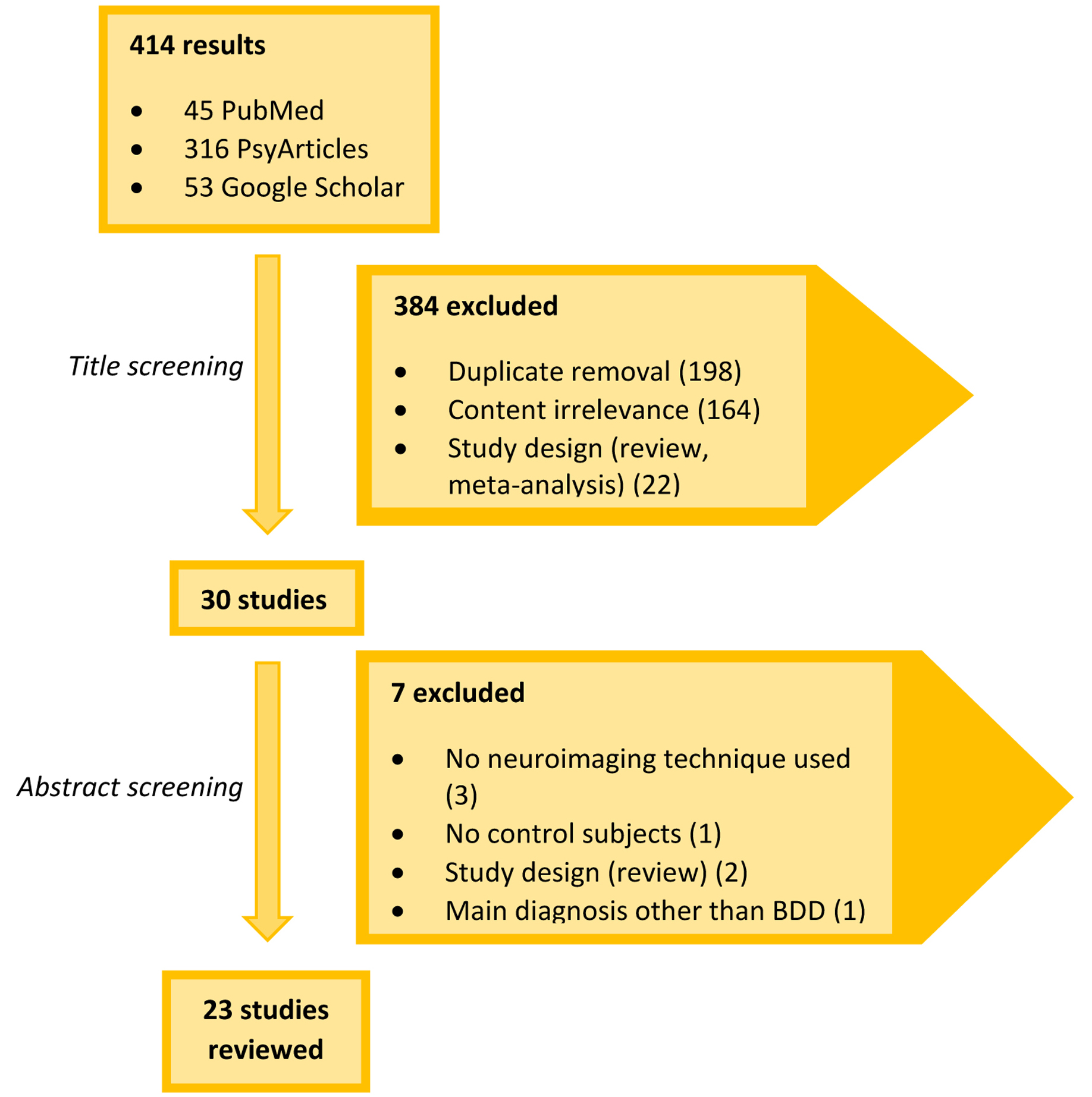 Fig. 1.
Flow chart of the study-selection procedure. Only original studies that used a healthy control group were included. Of the initial 414 search results, only 23 studies were finally reviewed.
Volumetric studies of the brain morphology have not yielded consistent results to date. The first one, by Rauch et al. [7] used morphometric MRI to compare regional brain volumes between BDD subjects and matched HCs, revealing a relative leftward shift in caudate asymmetry and greater total white matter volume in BDD group, pointing towards abnormal developmental processes. These findings support the conceptualization of BDD more as an Obsessive-Compulsive Related Disorder (OCRD) than an affective spectrum disorder, since the observed morphometric abnormalities involve brain regions that were also found abnormal in Obsessive-Compulsive Disorder (OCD) studies (will be further discussed in the “Discussion” section) [30, 31]. There have also been studies, however, that did not confirm these findings. Divergent results were found in Feusner et al. [11] who compared volumes in regions such as inferior frontal gyrus (IFG), amygdala, caudate, and total grey and white matter, finding no statistically significant volumetric differences between BDD versus controls, nor laterality. However, positive correlations were noted between BDD symptom severity -as represented by higher Yale-Brown Obsessive-Compulsive Scale Modified for Body Dysmorphic Disorder (BDD-YBOCS) score- and volumes of the left IFG and right amygdala, which are in concordance with the involvement (functional hyperactivity) of these regions in pathological face processing [32]. A third volumetric study by Atmaca et al. [8] revealed smaller OFC and anterior cingulate volumes (ACC) and larger white matter volumes and a trend for increased thalamic volume in BDD subjects versus the comparison group. There was a negative correlation between illness duration and OFC volumes. No differences were found in total brain, total GM and caudate volumes in patients versus controls. These findings are once again in line with the classification of BDD in OCRD, since there is evidence of OFC involvement in OFC both from neuroimaging [33, 34] and neuropsychological studies [35]. Buchanan et al. [9] revealed that BDD subjects had smaller total gray matter volume, smaller right OFC, bilateral thalamus, left ACC, left hippocampus and left amygdala compared with control subjects. As for clinical covariates, illness duration was negatively correlated with right orbitofrontal cortex volume and symptom severity was negatively correlated with the left amygdala volume. Investigating cortical thickness in BDD, Madsen et al. [12] reported no significant differences in cortical thickness, volume GM volume between BDD and HCs. Within the BDD group however, anxiety severity was associated with reduced GM thickness in the left superior temporal cortex, reduced GM volume in the right temporal fusiform cortex and greater GM volume in the right caudate nucleus. Discrepant results appeared in a study by Grace et al. [10] who found reduced GM cortical thickness in BDD versus control group in the left middle temporal and left inferior parietal gyrus, thus supporting the idea of their involvement in BDD pathophysiology. No significant correlation was found between cortical thinness and clinical variables such as symptom severity and insight.
With respect to Diffusion Tensor Imaging studies, a study by Feusner et al. [15] revealed fiber disorganization in white matter tracts connecting visual and emotion/memory processing systems. Affected tracts were the inferior longitudinal fasciculus (ILF), which is involved in object and face recognition processes [36] and the forceps major (FM), which connects right and left visual processing systems [15]. Poor integration of information between those regions may be correlated with poor insight and mediate the inability to accurately perceive and/or contextualize visual stimuli in individuals with BDD [37]. The same year, Buchanan et al. [14] investigated white matter properties with DTI and found reduced widespread FA in most major white matter tracts (superior longitudinal fasciculus, inferior fronto-occipital fasciculus and corpus callosum) in BDD subjects compared to controls. Lower FA reflects compromised white matter integrity and inefficient connections between various brain regions, thus may contribute to the emotional and cognitive dysregulation within BDD subjects [38]. As for clinical correlations, FA was not correlated with symptom severity, but it was negatively correlated with social anxiety scores.
Consistent results of abnormal white matter network organization were confirmed by Arienzo et al. [17] who found higher mean clustering coefficient (MCC). Their work suggests negative correlation between symptom severity and global efficiency. Furthermore, they provide evidence of aberrant connectivity -as reflected on higher edge betweenness centrality- between regions implicated in lower-order visual processing, higher-order visual processing, emotional processing and interhemispheric visual information transfer. Consistent with earlier work [20], Leow et al. [16] revealed that BDD subjects under-utilize several structural connections within the visual system, suggesting an abnormally low information transfer between primary and secondary visual cortical regions, and within higher order temporal lobe visual processing systems. Zhang et al. [13] compared brain network organization (“connectome”) in individuals with AN, BDD and HCs. They concluded that BDD and weight-restored Anorexia Nervosa (AN) individuals exhibit similar abnormal modular organization involving frontal, basal ganglia, and posterior cingulate nodes, when compared to HCs. BDD persons had similar path length with controls, while participants with AN had longer Normalized Path Length (NPL), which is a measure of global efficiency of white matter connectivity. Thus, longer NPL represents less efficient information transfer. Similar results appeared in a more recent study by Vaughn et al. [39] confirmed similar NPL in BDD and controls, while AN patients exhibit increased NPL, reflecting reduced white matter global efficiency, but better insight than BDD, as reflected on lower Brown Assessment of Beliefs Scale (BABS) score [40]. More specifically, they sought to develop a predictive model to distinguish AN, BDD and HCs, based on neuroimaging findings and psychometric evaluations. They found that distinguishing individuals with BDD or weight-restored AN from HCs can be accurate by using psychometric scores alone, namely Hamilton Anxiety Rating Scale (HAM-A) [41], and the Montgomery-Asberg Depression Scale (MADRS) [42], neuroimaging not being indispensable. On the other hand, in order to distinguish BDD from AN, the contribution of neuroimaging was considered important, as the researchers found higher NPL and better insight in AN than BDD.
Regarding functional MRI studies, Feusner et al. [19] concluded that patients with BDD have a different pattern of brain activation from that of healthy control subjects, such as different spatial frequencies when viewing others’ faces, including greater left hemisphere activity and bilateral activation of the amygdala. That indicates differences in visual processing beyond distortions of their own appearance. Feusner et al. [2] revealed abnormal patterns of brain activation in BDD versus controls when visually processing their own face. The key findings were greater activation in the left OFC and the bilateral head of the caudate for Normal Spatial Frequency (NSF) faces, hypoactivation in the left occipital cortex for low spatial frequency (LSF) faces and frontostriatal hyperactivity. They also observed that BDD symptom severity correlated with activation in frontostriatal and visual processing systems, specifically in the right visual cortex, bilateral head of the caudate, right precentral and postcentral gyri, right anterior cingulate gyrus, and right and left orbitofrontal cortex. Feusner et al. [20] found that BDD subjects have abnormal brain activation patterns when viewing non-symptom-related objects. More precisely, they showed lesser activity in visual association areas (in the parahippocampal gyrus, lingual gyrus, and precuneus) for configural and holistic (low detail-LSF) elements, and greater activations in medial prefrontal regions for details (high spatial frequency, HSF, images). Greater symptom severity was associated with lesser activity in dorsal occipital cortex and ventrolateral prefrontal cortex for NSF and HSF images. Bohon et al. [23] found a linear relationship between activity in the amygdala (region involved in anxiety) and ventral visual stream (VVS) (region involved in detailed visual processing) for BDD as well as controls. Within BDD subjects, the authors demonstrated a quadratic relationship between anxiety and activity in the right VVS and a linear relationship between anxiety and activity in the left VVS. Therefore, authors concluded that there seems to be an association between anxiety and activity in the VVS for own-face stimuli, in BDD. Furthermore, in the BDD group, anxiety was significantly mediated by right amygdala activation and HARS score predicted the right amygdala activity, whereas these correlations were not found for the left side. Moody et al. [18] provided evidence of aberrant functional connectivity in BDD within an occipito-temporal face-processing network, utilizing face-viewing task-based fMRI. Lower connectivity was correlated with increased symptom severity, as represented by higher BDD-YBOCS scores.
Wei Li et al. [22] used fMRI and event-related potentials to investigate for abnormal activity associated with early visual signaling. Indeed, their results showed hypoactivity in visual processing regions in BDD and AN patients when they were viewing faces and houses of various spatial frequencies. In details, they revealed hypoactivity in AN and BDD patients in early secondary visual processing regions when viewing LSF faces and LSF houses, hypoactivity in regions including the occipital fusiform cortex, lateral occipital cortex, and frontal pole for LSF houses, as well as hypoactivity in the dorsal visual stream (precuneus, lateral occipital cortex) for LSF faces. Furthermore, the BDD group showed hyperactivity in fusiform cortex when viewing HSF houses. Greater activity correlated with lower attractiveness ratings of faces. Taken together, there is evidence that AN and BDD share similar aberrant spatio-temporal activation for holistic information for appearance and nonappearance-related stimuli. The suggested abnormal early visual system functioning may contribute to distorted perception.
Beucke et al. [4] aimed to investigate aberrancies in the degree connectivity and the clinical correlates in BDD. Patients were found to present reduced local connectivity in the right amygdala than controls. This reduction was more pronounced for the LSF condition. As for brain-behavioral associations, there was a positive correlation between BDD symptom severity (as reflected on BDD-YBOCS score) and the degree of local and distant connectivity in the right posterior-lateral OFC. As studies have shown that the degree of connectivity of these areas is unusually high in OCD and also positively correlated with the severity of symptoms, these results are suggestive of high similarities in brain-behavioral associations across the OCRD spectrum, but deviance in connectivity abnormalities [4, 17]. Rangaprakash et al. [25] investigated the shared and distinct fronto-limbic connectivity patterns in AN and BDD, during repeated exposure to fearful faces. They found bidirectional medial prefrontal cortex (mPFC)-amygdala connectivity in HCs, unidirectional mPFC-to-amygdala connectivity in BDD (left mPFC-to-right amygdala) and no significant prefrontal-amygdala connectivity in AN in either direction. Also, BDD exhibited significant rACC-to-amygdala connectivity and AN exhibited significant rACC-to-mPFC while viewing fearful faces. Therefore, both clinical groups had abnormal connectivity between mPFC and amygdala. A double-blind study by Grace et al. [24] revealed greater resting state functional connectivity (rsFC) between the left amygdala and (a) left middle temporal gyrus and (b) left Inferior temporal gyrus, in BDD subjects as compared to controls. This abnormal connectivity was reversed with intranasal Oxytocin(OXT) administration. Moody et al. [21] acquired fMRI data from patients with AN, BDD and HCs while matching photos of others’ bodies. They found that BDD demonstrated hypoactivity in dorsal visual and parietal networks compared to controls. This is in line with previous findings of hypoactivity in BDD in visual systems for face perception [2]. Also, both BDD and AN showed hyperconnectivity in the dorsal visual network and hypoconnectivity in parietal network, as compared with controls. The affected regions were primarily within somatosensory components of the parietal network. In both disorders, aberrant activity and connectivity were associated with symptom severity and subjective appearance evaluations. For example, lower activation in dorsal visual network was associated with worse insight in BDD patients. As for the striatal network, there were no significant activation or connectivity differences between BDD, AN and HCs.
Lastly, a SPECT study by Vulink et al. [26] revealed an abnormal dopaminergic system in BDD subjects compared to controls. More specifically, BDD patients showed lower striatal dopamine D2/3 receptor availability in putamen and caudate. No correlations between receptor availability and clinical variables, meaning symptom severity as reflected in BDD-YBOCS score, or insight and delusionality as measured by BABS score, were detected. Key findings of the above discussed studies are summarized in the Supplementary Table 1.
BDD is a relatively common and potentially handicapping psychiatric disorder, where patients are extremely, sometimes even obsessively, preoccupied with a perceived flaw in their appearance, which is hardly noticeable to others. They ritualistically engage in behaviors to check and hide or camouflage the perceived defect and persistently seek reassurance. BDD may cause considerable functional impairment in various social, professional, or other everyday life aspects. It has been shown that BDD subjects exhibit a range of deficits in executive function, selective attention, information processing, verbal and non-verbal memory, recognition of others’ emotion and a bias to interpret neutral cues as negative, response inhibition and visual processing [9, 20], spatial working memory and thinking speed [43]. All in all, there are fundamental cognitive and perceptual impairments involved.
Interest to unravel the neurobiological underpinnings of BDD is recently gaining ground; a well-deserved interest considering its alarmingly high hospitalization and suicide-attempt rates. Based on the thus far research, a proposed model for the pathophysiology of BDD includes visual and emotional processing abnormalities and limbic and frontostriatal system dysfunction. The perceptual distortions, poor insight, obsessive thoughts and compulsive behaviors, may be to a certain extent attributable to the combination of the above factors [44]. Divergent results have emerged from volumetric studies. Increased total WM volume has been reported in some studies [7, 8] but was not confirmed by others [11]. Increased WM volume may reflect augmented volume of myelin per fiber or increased glia proportion, which may be attributed to primary developmental processes. As for GM volume or cortical thickness, it was reported reduced in some studies [9], specifically in the left middle temporal gyrus and left inferior parietal gyrus [10] while others found it not different from controls [8, 11, 12]. As is known from healthy cohorts, temporal and parietal network play a role in self-facial recognition, and the recognition of faces and basic emotions in others [45, 46]. That may explain the contribution of cortical thinning within the left inferior parietal and temporal lobe to face and self-perception distortion and emotion recognition disturbances [10]. The discrepant results between these studies could be attributed to the different mean age of participants (and consequently duration of illness), different exclusion criteria concerning current or past medication use and comorbidities, sample sizes and technical differences in the MRI machines and image acquisition parameters.
In BDD patients, regional reductions in brain volumes were identified within the bilateral [8] or right [9] orbitofrontal cortex (OFC), bilateral [8] and left [9] anterior cingulate cortex ACC, bilateral thalamus, left hippocampus and left amygdala, relative to controls [9]. Discrepant results were reported by other work, such as larger left caudate volumes [7], a trend for increased thalamic volume [8], or no significant differences in ROI between BDD and controls [11, 12].
As far as clinical variables are concerned, symptom severity -as estimated by higher BDD-YBOCS score- was positively correlated with volumes of the left IFG and right amygdala [11], while others report negative correlation with the left amygdala volume [9]. Illness duration was negatively correlated with OFC volumes [8, 9] and anxiety severity was associated with reduced GM thickness in the left superior temporal cortex and the right temporal fusiform cortex, and greater GM volume in the right caudate nucleus [12]. On the other hand, some studies found no significant correlations between cortical thickness and clinical variables [10].
The majority of findings, e.g., the larger white matter volume and aberrant GM cortical thickness, are in line with the classification of BDD among OCRD, as similar results have been reported in OCD in previous works [12]. Obsessive-Compulsive Disorder (OCD) is a psychiatric illness for which extensive and ongoing research has been being conducted since the 1980s in investigation of its neurobiological basis with initial findings suggesting hyperactivity in the prefrontal cortex, anterior cingulate cortex and caudate nucleus–thus leading to the cortico-striato-thalamo-cortical (CSTC) model–as well as subsequent findings indicating involvement of widespread associative networks, also including regions of the parietal cortex, limbic areas (e.g., the amygdala) and the cerebellum [47]. Volumetric studies of the brain morphology have not yielded consistent results to date. As already mentioned, some study findings have supported the conceptualization of BDD more as an OCRD since the observed morphometric abnormalities involved brain regions found abnormal also in corresponding studies on OCD. However, there are studies that did not confirm these findings as would be expected [12]. Rauch et al. [7] disclosed a relative leftward shift in caudate asymmetry and greater total white matter volume among BDD patients, whereas morphometric MRI studies on OCD suggested a rightward shift in striatal asymmetry [30, 31]. Furthermore, preliminary evidence of subtle volumetric abnormalities involving the OFC and thalamus are included among the characteristics that BDD and OCD have in common. Atmaca et al. [8] revealed smaller OFC and anterior cingulate volumes (ACC) as well as larger white matter volumes and a trend for increased thalamic volume in BDD subjects [35]. On the contrary, BDD has been associated with increased white matter volume, whereas reduced white matter volumes were reported in the majority of OCD studies. In addition, the smaller anterior cingulate volumes detected in BDD could also support for the conceptualization of BDD as an affective spectrum disorder [8]. It is noteworthy that ACC has an important inhibitory effect over emotional responses, and OFC is involved in decision-making, emotion regulation and self-focused thinking [48, 49, 50]. Thus, the reported reductions in left ACC and OFC volume may mediate emotion dysregulation in BDD [51]. Reduced OFC volume because of developmental defects may favor poorer outcomes and chronic BDD [9].
Diffusion Tensor Imaging studies have been convergent in reporting total and regional white matter tract disorganization, impaired white tract integrity, thus less efficient information transfer between various brain regions. Involved areas include the tracts connecting the right and left visual processing system, connecting the visual and emotional processing systems [15, 17], such as the inferior [15] and superior [14] longitudinal fasciculus, the forceps major (FM) [15], fronto-occipital fasciculus [14] and corpus callosum [14]. Reduced widespread FA has been revealed in BDD subjects [14], thus reflecting impaired white matter integrity and connection between different brain regions, thus may mediate emotional and cognitive dysregulation. Previous work [52] has shown that myelin abnormalities, which are largely under genetic control, can contribute to reduced FA. Therefore, genetically driven neurodevelopment abnormalities causing irregular myelination, may affect the predisposition to BDD.
Some studies have investigated brain network organization in parallel between BDD and AN individuals. They observed similar abnormalities in modular organization involving frontal, basal ganglia, and posterior cingulate nodes, compared with HCs [13]. Such abnormal modular associations in frontostriatal systems are also found in OCD and are theorized to be involved in establishing and maintaining motor and cognitive habits [52], which means they may mediate compulsive behaviors such as ritual formation [13]. Considered within the context of the frontostriatal hypothesis of OCD, dysfunction in frontostriatal loops leads to deficits in cognitive and inhibitory functions. AN and BDD also have some distinct characteristics, for example BDD has similar NPL with controls, while AN has longer NPL, reflecting inefficient information transfer, but better insight ascompared to BDD [1, 13].
Functional studies utilizing resting state or task-based fMRI, have congruently reported abnormal brain activation patterns in BDD patients as compared with controls, in limbic, frontostriatal and visual processing systems. More specifically, aberrant functional connectivity is revealed within an occipito-temporal face-processing network [18], as well as regional brain hyperactivity in the basal ganglia and orbitofrontal cortex (OFC) during symptom provocation [4]. Furthermore, studies showed abnormal brain activation patterns when visually processing own face [2], others’ faces [19], or bodies [21] or inanimate objects, like houses [20]. More precisely, when patients were viewing their own face, researchers observed hyperactivity in the frontostriatal system, OFC and caudate. Frontostriatal hyperactivity was positively correlated with symptom severity, and may also be associated with emotional reactions like aversiveness, or symptoms of obsessive thoughts and compulsive behaviors [2]. Dopaminergic abnormalities, that is, lower striatal dopamine D2/3 receptor availability, have also been reported [26], which is in accordance with similar findings in OCD. Compulsive behaviors, such as ritualistic mirror checking, control for appearance features, seeking reassurance, and compulsivity-related dysfunction of the reward system may be mediated by the striatal dopaminergic abnormality [26].
It is worth explaining that detailed analysis of facial traits (i.e., edges depicting contours of the nose, eyelashes, skin blemishes etc.) is conveyed byHSF visual information. On the contrary, configural aspects of faces (i.e., spatial relationships between facial features, general shape of the face) are primarily conveyed by low spatial frequency LSF visual information [19]. That said, clinical observation had previously supported what neuroimaging later came to explain and confirm, that patients are susceptible to focusing on regional details rather than the global holistic picture. The underlying mechanism might be the reduced activity in visual association areas (in the parahippocampal gyrus, lingual gyrus, and precuneus), the aberrant spatio-temporal activation and aberrant fronto-limbic connectivity patterns for configural elements, while increased activations in medial prefrontal regions for detailed elements [20, 22, 25]. The distortions in visual perception apply not only to details in human physical appearance but also to symptom-unrelated objects. Another congruent finding in fMRI is the predominant left hemisphere activity in BDD subjects and right predominance in the control group, as it is known that the left hemisphere subserves analytic processing, while the right hemisphere dominates for holistic or global processing [19].
The amygdala is extensively connected both anatomically and functionally to the OFC and ACC, also projecting strongly to the mediodorsal nucleus of the thalamus, the final relay station before the OFC/ACC/BG loops project back to the cortex, and it is thus critically positioned to influence the output of these loops [13]. The amygdala imparts an affective function to the fronto-striatal network, playing a crucial role in mediating normal fear and anxiety. The amygdala, as a region implicated in anxiety, seems to play an important role in BDD pathophysiology. It has been shown that anxiety severity, as quantified by the HARS, is mediated by and can predict the right amygdala activation in BDD patients. Additionally, there is a relationship between amygdala activation and activity in the VVS, which plays a role in detailed visual processing [23]. There seems to be a vicious circle as BDD patients report perceiving themselves as more disfigured when they are anxious, thus implying that anxiety itself may further accentuate perceptual distortions [23]. Reduced local connectivity in the right amygdala was disclosed among BDD patients, while this reduction was more pronounced for the LSF condition. There was a positive correlation between BDD symptom severity and the degree of local and distant connectivity in the right posterior-lateral OFC. Several researchers support fronto-striato-limbic models of OCD that attribute a specific role in mediating the anxiety symptoms to the amygdala and associated para-limbic regions [13]. As studies have shown that the degree of connectivity of these areas is unusually high in OCD and also positively correlated with the severity of symptoms, these results are suggestive of high similarities in brain-behavioral associations across the OCRD spectrum, but deviance in connectivity abnormalities [4, 17].
BDD subjects also demonstrate decreased local connectivity, more so for the LSF images, in the right amygdala than controls [4] and abnormal connectivity between mPFC and amygdala [25] which was also the case for AN patients. Also, in resting state fMRI, individuals with BDD exhibit abnormal connectivity between amygdala and temporal lobe in comparison with controls. Interestingly, this abnormal connectivity is eliminated with intranasal oxytocin administration [24], which indicates a potential perspective for future treatment options. Because, if oxytocin can decrease the connectivity between amygdala and temporal lobe, it could also dampen the fine-detail processing bias, and may be of therapeutic importance in BDD.
Some studies investigated functional abnormalities in BDD alongside AN [21, 22, 25], finding distinct and common anomalies in visual connectivity patterns, when encoding global features and body processing, as well as fronto-limbic connectivity patterns. Indeed, there is evidence of similar hypoactivity in visual processing regions, and aberrant spatio-temporal activation for configural information for appearance and nonappearance stimuli [22]. Another common finding in AN and BDD is the hyperconnectivity in the dorsal visual network and hypoconnectivity in parietal network compared with controls, when viewing others’ bodies [21]. This partially explains the shared trait of distorted perception in AN and BDD, because the parietal network is involved in body perception and the striatal network is involved in face processing, body perception and reward. Additionally, as mentioned above, both AN and BDD have aberrant connectivity between mPFC and amygdala upon repeated exposure to fearful faces [25]. As it is known that these regions and their in-between connectivity play a pivotal role in the fear expression and modulation, these findings provide grounds for understanding the aberrant fear processing circuits in BDD and AN.
Interestingly, it is highly likely that some of these abnormalities are amenable to change after successful treatment. For example, as mentioned above, the increased connectivity between left amygdala and temporal lobe in resting state fMRI, is restored with intranasal oxytocin [24]. The augmented thalamic volume in psychotropic-naive, pediatric OCD patients is reduced following efficient therapy with paroxetine [53]. Similarly, anterior cingulate cortex hyperactivity further increases with symptom provocation [54] and normalizes after successful treatment of OCD [55]. The enlarged left amygdala observed in OCD has been found reduced after treatment [56]. Given the common pathophysiologic grounds of the two entities, the neuroimaging alterations in BDD individuals may also be restored after pharmacologic treatment and psychotherapy, so as to provide a “countable” measure of treatment efficacy alongside the clinical state. Future studies could investigate toward that direction. It would also be of interest to investigate if there is a specific pattern of neuroimaging abnormalities correlated with higher suicide risk.
With respect to psychiatric and personality comorbidities in BDD, people BDD are more likely to experience narcissistic, histrionic, and avoidant personality disorders, as compared to OCD patients, who are more likely to present with obsessive-compulsive personality disorder traits. Social phobia, major depressive disorder, substance use disorders, and suicidal ideation are common in both BDD and OCD, but more prevalent in patients with BDD. Lifetime suicidal attempt rates among patients with BDD reach 22% in comparison with 8% in OCD patients. Symptom severity as well as marked psychosocial dysfunction are both risk factors associated with both suicidal ideation and attempted suicide. Risk factors for attempted suicide in BDD involve affective disorders, post-traumatic stress disorder, substance use disorder, and personality disorders especially within Cluster-B [57].
The possibility of using the neuroimaging abnormalities in follow-up, to assess therapy’s efficacy, as a co-determinant factor of remission, seems promising. Even more compelling appears the possibility of utilizing them as a prognostic factor of clinical course or severity of affected persons right from the moment of the diagnosis. This could help clinicians distinguish the most severe cases, more susceptible to self-harm and suicide attempt, and promote patient care.
There were several limitations in the present review. Importantly, most reviewed studies had divergent endpoints and objectives. Some aimed at the connectivity, others at the activation, whereas others at the volumes of Regions of Interest (ROIs) , while submitting participants to different tasks, or studying the resting state. This renders the comparability between the studies particularly precarious and risks drawing inaccurate conclusions. That is why a meta-analysis was not performed. Also, different studies used different inclusion and exclusion criteria. For instance, current or past medication use, current or past comorbidities, consisted exclusion criteria for some studies but not others. Depressive and anxiety disorders are so frequently comorbid in BDD that numerous studies decide to include these patients, as excluding them would not make a representative sample. On the other hand, these comorbidities may have played a role and contributed at least partially to the findings. At the same time, the causality relationship between findings-BDD cannot be proved, as they could equally be a predisposing factor or a result of BDD. Furthermore, the small sample sizes, and the divergent illness duration and mean patient age across the studies, also weaken the robustness of their comparison.
We present an overview of the neuroimaging findings acquired to date in BDD patients, and we attempt to explain their relationship with clinical manifestations. These observations may help designing further studies in order to investigate the potential implementation of neuroimaging in the clinical practice, as a monitoring tool of BBD patients.
ACC, Anterior Cingulate Cortex; BDD, Body Dysmorphic Disorder; ERP, event-related potentials; FM, forceps major; FA, fractional anisotropy; GM, Grey Matter; HC, healthy control; HSF, High Spatial Frequency; IFG, Inferior frontal gyrus; ILF, inferior longitudinal fasciculus; LSF, Low Spatial Frequency; MCC, mean clustering co-efficient; NSF, Normal Spatial Frequency; NPL, Normalized Path Length; OCRD, obsessive-compulsive and related disorders; OCSD, Obsessive Compulsive Spectrum Disorder; OFC, Orbitofrontal cortex; PCC, Posterior Cingulate Cortex; ROI, Region of interest; rsFC, resting state Functional Connectivity; VBM, Voxel-Based Morphometry; VVS, Ventral Visual Stream; WM, White Matter.
EM, CB, ED, NG—Conceptualization and design. EM, CB, VS, AMA—Data acquisition. MVK, MKB, AMA— Formal analysis and interpretation. EM, CB, VS—Writing original draft. MVK, MKB, ED, NG—Writing-review and editing. ED, NG—Supervision.
Not applicable.
The authors would like to thank Eleni Grigoriadou for her invaluable help in data handling.
This research received no external funding.
The authors declare no conflict of interest.
Supplementary material associated with this article can be found, in the online version, at https://www.imrpress.com/journal/JIN/21/2/10.31083/j.jin2102045.