IMR Press / FBL / Volume 29 / Issue 2 / DOI: 10.31083/j.fbl2902047
Open Access Original Research
Calcium Channel Inhibitory Effect of Marjoram (Origanum majorana L.): Its Medicinal Use in Diarrhea and Gut Hyperactivity
Show Less
1 Department of Pharmacology & Toxicology, College of Pharmacy, Prince Sattam Bin Abdulaziz University, Al-Kharj 11942, Saudi Arabia
2 Department of Pharmacy, Mohammed Al-Mana College for Medical Sciences, Dammam 34222, Saudi Arabia
3 Department of Pharmacognosy, College of Pharmacy, Taif University, Taif 21944, Saudi Arabia
*Correspondence: n_rehman5@hotmail.com (Najeeb Ur Rehman)
Front. Biosci. (Landmark Ed) 2024, 29(2), 47; https://doi.org/10.31083/j.fbl2902047
Submitted: 6 November 2023 | Revised: 30 December 2023 | Accepted: 5 January 2024 | Published: 4 February 2024
Copyright: © 2024 The Author(s). Published by IMR Press.
This is an open access article under the CC BY 4.0 license.
Abstract

Background: The leaves of Origanum majorana (O. majorana) are traditionally renowned for treating diarrhea and gut spasms. This study was therefore planned to evaluate its methanolic extract. Methods: Gas chromatography–mass spectrometry (GC-MS) was used to identify the phytochemicals, and Swiss albino mice were used for an in vivo antidiarrheal assay. Isolated rat ileum was used as an ex vivo assay model to study the possible antispasmodic effect and its mechanism(s). Results: The GC-MS analysis of O. majorana detected the presence of 21 compounds, of which alpha-terpineol was a major constituent. In the antidiarrheal experiment, O. majorana showed a substantial inhibitory effect on diarrheal episodes in mice at an oral dosage of 200 mg/kg, resulting in 40% protection. Furthermore, an oral dosage of 400 mg/kg provided even greater protection, with 80% effectiveness. Similarly, loperamide showed 100% protection at oral doses of 10 mg/kg. O. majorana caused complete inhibition of carbachol (CCh, 1 µM) and high K+ (80 mM)-evoked spasms in isolated ileal tissues by expressing significantly higher potency (p < 0.05) against high K+ compared to CCh, similar to verapamil, a Ca++ antagonist. The verapamil-like predominant Ca++ ion inhibitory action of O. majorana was further confirmed in the ileal tissues that were made Ca++-free by incubating the tissues in a physiological salt solution having ethylenediaminetetraacetic acid (EDTA) as a chelating agent. The preincubation of O. majorana at increasing concentrations (0.3 and 1 mg/mL) shifted towards the right of the CaCl2-mediated concentration-response curves (CRCs) with suppression of the maximum contraction. Similarly, verapamil also caused non-specific suppression of Ca++ CRCs towards the right, as expected. Conclusions: Thus, this study conducted an analysis to determine the chemical constituents of the leaf extract of O. majorana and provided a detailed mechanistic basis for the medicinal use of O. majorana in hyperactive gut motility disorders.

Keywords
O. majorana
antispasmodic
Ca++ channel blocker
GC-MS
verapamil
CRCs
1. Introduction

Most volatile oil-containing plants have been used as condiments to enhance the flavor of food since ancient times. Later on, a few species of volatile oil-containing plants were explored for managing diseases [1]. Plant families like Apiaceae, Asteraceae, Burseraceae, Lamiaceae, Lauraceae, Myrtaceae, and Zingiberaceae are known for their members containing volatile oils. In particular, members of the Lamiaceae (Labiatae) family have been explored to a massive extent during these days [2]. A few of the well-known members of the family Lamiaceae are marjoram, basil, mint, oregano, and thyme, which are used for culinary and medicinal purposes.

Marjoram (Origanum majorana L.) belongs to the family Lamiaceae, is widely recognized as sweet marjoram, and has been traditionally used for managing gastrointestinal and respiratory diseases [3, 4]. Marjoram is indigenous to the Mediterranean region and the Arabian Peninsula. The ancient Greeks and Romans believed this plant was a symbol of happiness. It is a perennial bushy plant that reaches a height of up to 12 to 24 inches. The plant bears oval leaves, which are oppositely arranged with white or red flowers. Marjoram leaves are widely used for garnishing purposes. The leaves contain essential oil, and the oil obtained from the leaves is used in mouthwashes and rubbed topically in cases of nasal congestion [5]. Marjoram has been used extensively in pharmaceuticals and food due to its flavor and medicinal properties.

Due to the dynamic uses of this plant, several studies have reported its antioxidant effect [6, 7], antibacterial effect [8], neurobiological activity [7], and skin protection effects [9]. Several studies related to its essential oil composition [10], Electrospray Ionisation Mass Spectrometry (ESI-MS) fingerprinting of essential oil [11], ratios of cis- and trans-Sabinene Hydrate [12], determination of polyphenolic compounds [8, 13, 14], and the effects of blanching on the stability of polyphenols [15] have been reported. The development of nanoemulsions as well as pectin films [16] was also done for the essential oil of marjoram.

The constituents and concentration of the compounds in the plant vary due to factors like geographical variations, soil, and climatic conditions [17]. The essential oil of marjoram majorly contributes to most of its activities. Similarly, the methanolic extract of marjoram possesses polyphenolic compounds, which majorly contribute to its antioxidant activity. Hence, for establishing a representative prototype of O. majorana L. from the Arabian Peninsula, it was worth carrying out a chemical analysis of the essential oil and the phenolic content of marjoram obtained from the Arabian Peninsula. The present study conducted a Gas chromatography–mass spectrometry (GC-MS)-assisted chemo profiling, an investigation of the antioxidants, and a detailed antidiarrheal evaluation of the methanolic extract of O. majorana leaves.

2. Materials and Methods
2.1 Chemicals

The chemicals utilized in this study were obtained from Sigma Company, located in St. Louis, MO, USA, including carbachol (CCh), acetylcholine perchlorate (ACh), and verapamil. The physiological buffer solution (Tyrode’s) was prepared using salts acquired from Merck (Darmstadt, Germany). Castor oil was acquired from a local pharmacy in Al-Kharj. All chemicals used were of analytical quality.

2.2 Animals

Wistar albino rats (weighing 200–250 g) were used for the ex vivo study, while Swiss albino mice (weighing 30–35 g) were used for the in vivo study. These animals were obtained from the Animal Care Unit at the College of Pharmacy, Prince Sattam bin Abdulaziz University (PSAU), Saudi Arabia. They were kept under optimal conditions, with a temperature of 22 ± 1 °C, relative humidity of 55 ± 5%, and equal exposure to light and dark cycles. All animals were given a standardized pellet meal and unrestricted access to water. Prior to conducting the ex vivo studies, the rats underwent a 24-hour fasting period. Their cervical dislocation was performed after administering light anesthesia using diethyl ether, and the cessation of ear reflexes was used to certify their death. All tests, both in vivo and ex vivo, were conducted with meticulous attention to detail, adhering strictly to the directions outlined in National Research Council [18]. The study protocol has received approval from the Bio-Ethical Research Committee (BERC) at PSAU, with the reference number BERC-004-12-19.

2.3 Extraction of Plant Material

O. majorana leaves were purchased from the local market of Dammam and authenticated (PL/0445/2020-21/P-009) by the Department of Pharmacognosy, College of Clinical Pharmacy, Taif University, Saudi Arabia. The plant material was dried in the shade. All plant materials were crushed into a coarse size and later subjected to extraction.

2.4 Preparation of the Plant Extracts

The plant matter was crushed and macerated to prepare the extract. Ten grams of each sample were weighed into 500 mL Erlenmeyer flasks, and 100 mL of methanol was added to the plant samples. Extraction was carried out by the maceration technique with frequent agitation at room temperature for 5 days. After filtration through filter paper, the extracts of every sample were evaporated until the constant weight and respective extractives were calculated. The final residues were used in this study.

2.5 GC-MS Analysis

The phytochemical composition of the methanolic leaf extract of O. majorana was analyzed by Agilent GC-MS (7890A) using HP 5MS column (30 m × 0.25 mm × 0.25 µm film thickness) for the separation on split mode 1:20 with helium being used as the gaseous mobile phase at a flow rate of 1.0 mL per min. The injector temperature was 280 °C, the source temperature of MS was kept at 230 °C, and the Quad temperature was kept at 150 °C. The column oven temperature was increased from 150 °C to 300 °C at a gradual rise of 10 °C/min, and the total run time was 40 min. The scanning was carried out in the range of 40 to 600 mass while electron energy was kept at 70 eV. A solvent delay of 3 minutes was employed during the analysis. Identification of components was done by comparing the spectra of the sample with that of the standard spectra available with National Institute of Standards and Technology and Wiley library [19, 20].

2.6 In Vivo Antidiarrheal Study

Altogether, twenty mice were assigned randomly to four groups, each containing an equal number of animals. After fasting for 18 hours, the mice in the first group were given a saline solution (10 mL/kg) through oral gavage and were labeled as the negative control group. Following the pilot screening to determine the appropriate dosage, the second and third groups (referred to as test groups) were administered orally with two escalating doses of 200 and 400 mg/kg of methanolic leaves extract of O. majorana, respectively. The fourth group of mice was administered loperamide at a dosage of 10 mg/kg and designated as the positive control group. Following one hour, all mice were administered castor oil orally at a dosage of 10 mL/kg. Each animal was then placed in an individual cage with a blotting sheet on the floor. This allowed a blind observer to determine the presence or absence of diarrhea. After four hours, the blotting sheet from each cage was examined for the presence of characteristic diarrheal droppings. The protective effect of the extract was evident when no instances of diarrhea were seen, as previously documented by Jebunnessa et al. [21].

2.7 Ex Vivo Experiments on Isolated Rat Ileum

The study rats were euthanized, and the terminal portion of the small intestine (ileum) was extracted following the protocol described by Rehman et al. [22]. After being isolated, specific sections of ileal tissues measuring 2–3 cm in length were thoroughly cleaned to remove nearby tissues and fecal matter. These sections were then placed in emkaBath (France) and connected to a transducer and IOX software (version 2.9.10.6, emka technologies, SAS, Paris, France). The Tyrod solution (pH 7.4) was freshly prepared [22] and added to tissue baths with a volume of 20 mL. The baths were then filled with carbogen gas and maintained at a temperature of 37 °C. A stress of 0.7 grams was exerted by turning the transducer knob in a clockwise direction. The tissues were then allowed to stabilize for 30 minutes, during which they were exposed to acetylcholine numerous times at a concentration of 0.3 µM. Following stabilization, continuous muscle contractions were induced using CCh, and high doses of K+ and O. majorana were gradually introduced into the bathing solution, with concentrations increasing to a maximum final bath concentration (FBC) of 10 mg/mL.

2.8 Ca++ Inhibitory Effect Confirmation

Following the first relaxation of O. majorana in response to high K+ levels, additional verification of Ca++ channel blocking (CCB) was achieved by incubating the ileal tissues in a solution of Ca++-free Tyrode’s solution combined with EDTA (0.1 mM) for 45 minutes. This solution was used to chelate Ca++ ions. To further reduce the amount of Ca++ inside the cells, the solution that did not contain Ca++ was replaced with another solution called K+-rich and Ca++-free Tyrode’s solution. The concentrations of the different components in this solution were as follows (in mM): potassium chloride: 50, sodium chloride: 91.03, sodium dihydrogen phosphate dehydrates: 0.32, sodium bicarbonate: 11.9, magnesium chloride hexahydrate: 0.50, glucose: 5.05. After incubating the solution for 45 minutes, CaCl2- Concentration response curves (CRCs) were created with and without varying concentrations of O. majorana. The findings were then compared to the typical CCB drug, verapamil [23].

2.9 Statistics

The results have been reported as the mean ± standard error of the mean (SEM), and the number of experiments conducted has been denoted by “n”. The EC50 values were determined, and their 95% confidence intervals (CI) were calculated. The statistical analysis involved two-way ANOVA, followed by Bonferroni’s post-test, to compare concentration-response curves (CRCs) with a control group. The effectiveness of diarrhea prevention was assessed using a statistical analysis, comparing all groups to a control group, using the Chi-square (χ2) test. A p-value less than 0.05 has been regarded as statistically significant. The software GraphPad Prism (version 4, GraphPad Software, Inc., San Diego, CA, USA) was utilized to perform regression analysis of CRCs.

3. Results
3.1 Methanolic Extract Yield (%)

The leaves of O. majorana yielded 14.7% of methanolic crude extract.

3.2 GC-MS Phytochemical Profiling

O. majorana was analyzed by GC-MS (Fig. 1). The phytoconstituents identified are listed in Table 1. GC-MS identified twenty-one constituents representing 96.27% of the extract. Alpha-terpineol (35.18%) and linoleic acid (13.3%) were characterized as major components of the methanolic extract. Sabinene, a bicyclic monoterpene found in the essential oils of various plant species, was also identified in the profiling.
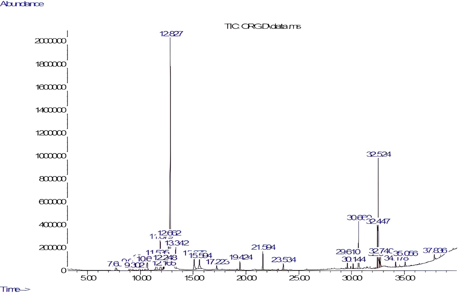
Fig. 1.

Gas chromatography–mass spectrometry (GC-MS) chromatogram of Origanum majorana (O. majorana) leaves extract.

Table 1.Phytoconstituents present in O. majorana leaves extract.
S. No. RT (min) Area % Compound name Molecular weight Molecular formula
1 7.655 0.95 Sabinene 136.23 C10H16
2 9.01 1.07 Alpha-thujene 136.23 C10H16
3 10.30 2.96 Alpha-terpinene 136.23 C10H16
4 10.65 2.77 Beta-Phellandrene 136.23 C10H16
5 11.53 3.56 Gamma-terpinene 136.23 C10H16
6 11.87 4.64 Beta-terpineol 154.25 C10H18O
7 12.66 4.19 Carane 138.25 C10H18
8 12.82 35.18 Alpha-terpineol 154.25 C10H18O
9 13.34 3.04 3-Formyl-1-methyl-2(1H)-pyridine-thione 153.20 C7H7NOS
10 15.07 3.05 Terpinen-4-ol 154.25 C10H18O
11 17.22 0.75 Linalyl acetate 196.29 C12H20O2
12 19.42 1.17 1,3,6-Heptatriene, 2,5,5-trimethyl 136.23 C10H16
13 21.59 3.06 Beta-caryophyllene 204.36 C15H24
14 23.53 1.30 Gamma-Pyronene 136.23 C10H16
15 29.61 1.24 Pinane 138.25 C10H18
16 30.66 5.68 Methyl palmitate 270.5 C17H34O2
17 32.44 3.92 Methyl linoleate 294.5 C19H34O2
18 32.52 13.30 Linoleic acid 280.4 C18H32O2
19 32.64 1.53 Phytol 296.5 C20H40O
20 32.74 1.48 Methyl isostearate 298.5 C19H38O2
21 35.05 1.42 Dehydroabeityl alcohol 290.5 C20H34O

S. No., Serial Number; RT, Retention Time.

3.3 Antidiarrheal Effect

Both the increasing doses of orally administered O. majorana extract in mice exhibited significant antidiarrheal effects in comparison to the control group (Table 2). When administered a dose of 200 mg/kg, two out of five mice exhibited protection, indicating a 40% protection rate. In contrast, a higher dose of 400 mg/kg resulted in 80% protection. The positive control drug, loperamide, showed 100% protection at 10 mg/kg, as detailed in Table 2.

Table 2.Antidiarrheal activity of the methanolic leaf extract of Origanum majorana (O. majorana) using mice.
Group Dose No. of mice with diarrhea % Protection
Castor oil 10 mL/kg 5/5 0
Castor oil + O. majorana 200 mg/kg 3*/5 40
Castor oil + O. majorana 400 mg/kg 1*/5 80
Castor oil + loperamide 10 mg/kg 0**/5 100

*p < 0.05 and **p < 0.01 vs. Saline + Castor oil treated group (χ2-test).

3.4 Ex Vivo Antispasmodic Effects

When tested against CCh and high K+-mediated spasm in rat-isolated ileal tissues, O. majorana resulted in complete inhibition with a resultant EC50 value of 1.38 mg/mL (1.06–1.82, 95% CI, n = 4) recorded against high K+ whereas the potency against CCh was found weak with a resultant EC50 of 7.14 mg/mL (6.85–7.84, 95% CI, n = 4), as shown Fig. 2A. Similarly, verapamil resulted in distinctly higher potency to inhibit high K+compared to CCh-evoked spasms with EC50 values of 0.24 µM (0.19–0.32, 95% CI, n = 4) and 2.28 µM (1.84–2.92, 95% CI, n = 4), respectively as shown in Fig. 2B.

Fig. 2.

The concentration-response curves compare the inhibitory effect of the methanolic Origanum majorana (O. majorana) leaf extract (A) and verapamil (B) against carbachol (CCh, 1 µM) and high K+ (80 mM)-induced contractions in isolated rat ileum preparations. The values presented are the mean ± Standard error of the mean (SEM), n = 4.

3.5 Calcium Channel Blocking (CCB)-Like Effect

To further confirm the Ca++ inhibitory effect, ileal tissues preincubated with O. majorana methanolic extract shifted the Ca++ CRCs curves at tested concentrations of 0.3 and 1.0 mg/mL (Fig. 3A), similar to that caused by verapamil at concentrations of 0.03 and 0.10 µM (Fig. 3B).

Fig. 3.

The concentration-response curves of Ca++ were measured in isolated rat ileum preparations in the absence and presence of increasing concentrations of (A) the methanolic Origanum majorana (O. majorana) leaf extract and (B) verapamil. The values presented are the mean ± SEM, n = 4.

4. Discussion

The therapeutic effect of O. majorana in digestive disorders [24, 25] raised our interest in evaluating this plant scientifically using rodents and employing GC-MS to determine its phytochemical analysis. In the castor oil-induced diarrhea model, the preincubated mice with plant extract resulted in a dose-dependent antidiarrheal effect by protecting the mice from typical diarrheal drops at 200 and 400 mg/kg when compared to a saline pretreated group where no protection was observed. The dose selection of the plant extract was based on a previously reported acute toxicity study that revealed the safety of O. majorana ethanolic extract up to the maximum tested dose of 5 g/kg [26, 27]. The induction of diarrhea in normal mice was achieved by administering castor oil, which, upon hydrolysis into ricinoleic acid, alters the transportation of electrolytes and water. This disruption leads to spasms in the gastrointestinal tract [28]. Thus, a potential antidiarrheal agent may exhibit its antidiarrheal effect by inhibiting bowel contraction. The antidiarrheal activity of the plant extract of O. majorana can be attributed to multiple inhibitory components present in the plant.

To identify the potential pharmacodynamics responsible for the observed antidiarrheal activity, the methanolic O. majorana extract was tested in increasing doses on isolated rat ileum, as previously documented [29]. Based on previous findings that antispasmodic medicines typically have inhibitory effects on the gut by blocking calcium channels [30], we conducted further experiments to assess the impact of O. majorana extract on induced contractions in rat ileum using CCh and high K+ as contractile agents [31]. The critical analysis of the pattern of the inhibitory CRCs of O. majorana against CCh and high K+ in the ileal tissues shows a verapamil-like effect by recording significantly higher potency (p < 0.05) against high K+ compared to CCh, which indicates the involvement of the predominant CCB-like components. Verapamil, a standard CCB, was run in parallel experiments [23, 32] for comparison.

Godfraind et al. [33] reported that high concentrations of K+ (>30 mM) lead to depolarization of certain smooth muscles due to the activation of voltage-dependent Ca++ channels (specifically, L-type channels), resulting in persistent contractions. A substance that reverses high K+ (˃30 mM)-mediated spasm depicted CCBs [34, 35]. Hence, to support and further confirm the CCB-like action of O. majorana, the ileal tissues were first made Ca++-free by replacing the bath solution with Ca++-free Tyrode’s solution having a chelating agent, ethylenediaminetetraacetic acid (EDTA) to chelate the Ca++ from tissue. Calcium-free ileal tissues were then preincubated with increasing concentrations of O. majorana. The contractile CRCs of exogenously added Ca++ were made in the absence and presence of preincubated tissues with O. majorana. O. majorana extract, at both preincubated concentrations, caused a rightward shift in Ca++-CRCs and suppressed the highest peak, comparable to verapamil, a standard CCB [32]. This confirms the verapamil-like CCB impact of O. majorana. The GC-MS analysis of O. majorana extract showed the presence of twenty-one constituents, where alpha-terpineol was found to be the major compound in terms of concentration. Alpha-terpineol has been reported to possess antidiarrheal effects in mice [36]. Hence, the plant’s antidiarrheal and antispasmodic effect might be due to the presence of this compound, whereas the involvement of additional compounds may have a parallel contribution.

5. Conclusions

The study’s findings show that the methanolic leaf extract of O. majorana inhibits with higher potency the contractions against high K+ compared to CCh-mediated contractions and thus depicts that voltage-gated Ca++ channels predominantly mediate the antidiarrheal and antispasmodic effects of the plant. However, additional mechanism(s) cannot be ignored.

Availability of Data and Materials

The datasets used and/or analyzed during the current study are available from the corresponding author on reasonable request.

Author Contributions

NUR and WA designed the research study. MNA, NUR, AA, and WA performed the research. NUR, MNA, and WA analyzed the data. MNA, NUR, AA, and WA wrote the manuscript. All authors have participated sufficiently in the work and agreed to be accountable for all aspects of the work. All authors read and approved the final manuscript.

Ethics Approval and Consent to Participate

The study has obtained approval from the Bio-Ethical Research Committee (BERC) at PSAU, with the reference number BERC-004-12-19.

Acknowledgment

The authors are thankful to the Department of Pharmacology and Toxicology, College of Pharmacy, PSAU, for providing the facility to perform the study.

Funding

The authors extend their appreciation to Prince Sattam Bin Abdulaziz University for funding this research work through the project number (PSAU/2023/03/25570).

Conflict of Interest

The authors declare no conflict of interest.

References
[1]
Özer Z, Gören AC, Kılıç T, Öncü M, Çarıkçı S, Dirmenci T. The phenolic contents, antioxidant and anticholinesterase activity of section Amaracus (Gled.) Vogel and Anatolicon Ietsw. of Origanum L. species. Arabian journal of chemistry. 2020; 13: 5027–5039.
[2]
Celep F, Dirmenci T. Systematic and biogeographic overview of Lamiaceae in Turkey. Natural Volatiles and Essential Oils. 2017; 4: 14–27.
[3]
Bellakhdar J. La pharmacopée marocaine traditionnelle. Médecine arabe ancienne et savoirs populaires (pp. 349). Ibiss press: Paris. 1997.
[4]
Makrane H, Aziz M, Mekhfi H, Ziyyat A, Legssyer A, Melhaoui A, et al. Origanum majorana L. extract exhibit positive cooperative effects on the main mechanisms involved in acute infectious diarrhea. Journal of Ethnopharmacology. 2019; 239: 111503.
[5]
Hossain MB, Barry-Ryan C, Martin-Diana AB, Brunton NP. Optimisation of accelerated solvent extraction of antioxidant compounds from rosemary (Rosmarinus officinalis L.), marjoram (Origanum majorana L.) and oregano (Origanum vulgare L.) using response surface methodology. Food Chemistry. 2011; 126: 339–346.
[6]
Hossain MB, Brunton NP, Patras A, Tiwari B, O’Donnell CP, Martin-Diana AB, et al. Optimization of ultrasound assisted extraction of antioxidant compounds from marjoram (Origanum majorana L.) using response surface methodology. Ultrasonics Sonochemistry. 2012; 19: 582–590.
[7]
Gök HN, Luca SV, Ay ST, Komsta Ł, Salmas RE, Orhan IE, et al. Profiling the annual change of the neurobiological and antioxidant effects of five Origanum species in correlation with their phytochemical composition. Food Chemistry. 2022; 368: 130775.
[8]
Fecka I, Turek S. Determination of polyphenolic compounds in commercial herbal drugs and spices from Lamiaceae: thyme, wild thyme and sweet marjoram by chromatographic techniques. Food Chemistry. 2008; 108: 1039–1053.
[9]
Lee CJ, Chen LG, Chang TL, Ke WM, Lo YF, Wang CC. The correlation between skin-care effects and phytochemical contents in Lamiaceae plants. Food Chemistry. 2011; 124: 833–841.
[10]
Komaitis ME, Ifanti-Papatragianni N, Melissari-Panagiotou E. Composition of the essential oil of marjoram (Origanum majorana L.). Food Chemistry. 1992; 45: 117–118.
[11]
Møller JKS, Catharino RR, Eberlin MN. Electrospray ionization mass spectrometry fingerprinting of essential oils: Spices from the labiatae family. Food Chemistry. 2007; 100: 1283–1288.
[12]
Novak J, Bitsch C, Langbehn J, Pank F, Skoula M, Gotsiou Y, et al. Ratios of cis- and trans-Sabinene Hydrate in Origanum majorana L. and Origanum microphyllum (Bentham) Vogel. Biochemical Systematics and Ecology. 2000; 28: 697–704.
[13]
Pedersen JA. Distribution and taxonomic implications of some phenolics in the family Lamiaceae determined by ESR spectroscopy. Biochemical Systematics and Ecology. 2000; 28: 229–253.
[14]
Kaliora AC, Kogiannou DAA, Kefalas P, Papassideri IS, Kalogeropoulos N. Phenolic profiles and antioxidant and anticarcinogenic activities of Greek herbal infusions; balancing delight and chemoprevention? Food Chemistry. 2014; 142: 233–241.
[15]
Kaiser A, Carle R, Kammerer DR. Effects of blanching on polyphenol stability of innovative paste-like parsley (Petroselinum crispum (Mill.) Nym ex A. W. Hill) and marjoram (Origanum majorana L.) products. Food Chemistry. 2013; 138: 1648–1656.
[16]
Almasi H, Azizi S, Amjadi S. Development and characterization of pectin films activated by nanoemulsion and Pickering emulsion stabilized marjoram (Origanum majorana L.) essential oil. Food Hydrocolloids. 2020; 99: 105338.
[17]
Ma B, Ma J, Li B, Tao Q, Gan J, Yan Z. Effects of different harvesting times and processing methods on the quality of cultivated Fritillaria cirrhosa D. Don. Food Science & Nutrition. 2021; 9: 2853–2861.
[18]
National Research Council. Guide for the Care and Use of Laboratory Animals (pp. 1–7). National Academy Press: Washington. 1996.
[19]
Khan W, Chester K, Anjum V, Ahmad W, Ahmad S, Narwaria A, et al. Chromatographic Profiling of Pancharishta at Different Stages of its Development Using HPTLC, HPLC, GC-MS and UPLC-MS. Phytochemistry Letters. 2017; 20: 391–400.
[20]
Ahmad W, Parveen R, Mujeeb M, Zaidi SMA. Comparative Fingerprint Profiling of Unani Polyherbomineral (Safoof-e-Pathar Phori) Formulation by HPTLC, HPLC, and GC-MS. Journal of AOAC International. 2020; 103: 659–668.
[21]
Jebunnessa Uddin SB, Mahbub-Uz-Zaman M, Akhtar R, Ahmed NU. Antidiarrheal activity of ethanolic bark extract of Mitragyna diversifolia. Bangladesh Journal of Pharmacology. 2009; 4: 144–146.
[22]
Rehman NU, Ansari MN, Samad A. In Silico, Ex Vivo and In Vivo Studies of Roflumilast as a Potential Antidiarrheal and Antispasmodic agent: Inhibition of the PDE-4 Enzyme and Voltage-gated Ca++ ion Channels. Molecules (Basel, Switzerland). 2020; 25: 1008.
[23]
Fleckenstein A. Specific pharmacology of calcium in myocardium, cardiac pacemakers, and vascular smooth muscle. Annual Review of Pharmacology and Toxicology. 1977; 17: 149–166.
[24]
Cinbilgel I, Kurt Y. Oregano and/or marjoram: Traditional oil production and ethnomedical utilization of Origanum species in southern Turkey. Journal of Herbal Medicine. 2019; 16: 100257.
[25]
Eddouks M, Ajebli M, Hebi M. Ethnopharmacological survey of medicinal plants used in Daraa-Tafilalet region (Province of Errachidia), Morocco. Journal of Ethnopharmacology. 2017; 198: 516–530.
[26]
Seoudi DM, Medhat AM, Hewedi IH, Osman SA, Mohamed MK, Arbid MS. Evaluation of the anti-inflammatory, analgesic, and antipyretic effects of Origanum majorana ethanolic extract in experimental animals. Journal of Radiation Research and Applied Sciences. 2009; 2: 513–534.
[27]
Selim SA, Aziz MA, Mashait MS, Warrad MF. Antibacterial activities, chemical constituents and acute toxicity of Egyptian Origanum majorana L., Peganum harmala L. and Salvia officinalis L. essential oils. African Journal of Pharmacy and Pharmacology. 2013; 7: 725–735.
[28]
Croci T, Landi M, Emonds-Alt X, Le Fur G, Maffrand JP, Manara L. Role of tachykinins in castor oil diarrhoea in rats. British Journal of Pharmacology. 1997; 121: 375–380.
[29]
IWAO I, TERADA Y. On the mechanism of diarrhea due to castor oil. Japanese Journal of Pharmacology. 1962; 12: 137–145.
[30]
Rehman NU, Gilani AH, Khan A, Nazneen M, El Gamal AA, Fawzy GA, et al. Antidiarrheal and Antispasmodic Activities of Buddleja polystachya are Mediated Through Dual Inhibition of Ca(++) Influx and Phosphodiesterase Enzyme. Phytotherapy Research: PTR. 2015; 29: 1211–1218.
[31]
Gilani AH, Rehman NU, Mehmood MH, Alkharfy KM. Species differences in the antidiarrheal and antispasmodic activities of Lepidium sativum and insight into underlying mechanisms. Phytotherapy Research: PTR. 2013; 27: 1086–1094.
[32]
Downie JW, Twiddy DA, Awad SA. Antimuscarinic and noncompetitive antagonist properties of dicyclomine hydrochloride in isolated human and rabbit bladder muscle. The Journal of Pharmacology and Experimental Therapeutics. 1977; 201: 662–668.
[33]
Godfraind T, Miller R, Wibo M. Calcium antagonism and calcium entry blockade. Pharmacological Reviews. 1986; 38: 321–416.
[34]
Hamilton TC, Weir SW, Weston AH. Comparison of the effects of BRL 34915 and verapamil on electrical and mechanical activity in rat portal vein. British Journal of Pharmacology. 1986; 88: 103–111.
[35]
Shah AJ, Bhulani NN, Khan SH, Ur Rehman N, Gilani AH. Calcium channel blocking activity of Mentha longifolia L. explains its medicinal use in diarrhoea and gut spasm. Phytotherapy Research: PTR. 2010; 24: 1392–1397.
[36]
Dos Santos Negreiros P, da Costa DS, da Silva VG, de Carvalho Lima IB, Nunes DB, de Melo Sousa FB, et al. Antidiarrheal activity of α-terpineol in mice. Biomedicine & Pharmacotherapy = Biomedecine & Pharmacotherapie. 2019; 110: 631–640.

Publisher’s Note: IMR Press stays neutral with regard to jurisdictional claims in published maps and institutional affiliations.

Share
Back to top