IMR Press / FBL / Volume 28 / Issue 8 / DOI: 10.31083/j.fbl2808192
Open Access Original Research
SUMOylation of SMAD4 by PIAS1 in Conjunction with Vimentin Upregulation Promotes Migration Potential in Non-Small Cell Lung Cancer
Show Less
1 Department of Respiratory, Ningbo No.2 Hospital, 315000 Ningbo, Zhejiang, China
2 Department of Radiology, Ningbo No.2 Hospital, 315000 Ningbo, Zhejiang, China
*Correspondence: dongzhaoxing1978@outlook.com (Zhaoxing Dong)
Front. Biosci. (Landmark Ed) 2023, 28(8), 192; https://doi.org/10.31083/j.fbl2808192
Submitted: 2 December 2022 | Revised: 13 April 2023 | Accepted: 23 April 2023 | Published: 31 August 2023
Copyright: © 2023 The Author(s). Published by IMR Press.
This is an open access article under the CC BY 4.0 license.
Abstract

Background: The expression of vimentin as a marker of epithelial-to-mesenchymal transition (EMT) has been speculated to be associated with tissue heterogeneity and metastases of non-small cell lung cancer (NSCLC). Methods: This study utilized in vitro co-immunoprecipitation with small interfering RNAs (siRNAs) against protein inhibitors of STAT system type 1 (PIAS1) or SMAD4 in transforming growth factor-beta (TGF-β) signaling pathway in combination with SUMOylation assay. Results: We successfully demonstrated that PIAS1 enhanced SUMOylation of SMAD4 by forming a complex PIAS1-SUMO1-SMAD4 protein complex. This, in accordance with subsequently increased production of vimentin microfilaments, led to enhanced migration ability of non-small cell lung cancer (NSCLC) A549 line, observed from wound healing assay. Conclusions: Our results further supported the positive correlation of SUMOylated SMAD4 mediated by PIAS1 and downstream overexpression of vimentin. In addition, the observation that overexpression of vimentin in this certain cell line was not necessarily linked with accelerated relative wound closure raised concerns that further exploration will be needed to confirm if the causal relationship exists between vimentin expression and the metastases of NSCLC, and if so, to what extent vimentin contributes to it.

Keywords
non-small cell lung cancer
SUMOylation
SMAD4
PIAS1
Vimentin
1. Introduction

Lung cancer is the most common cancer globally, with 2.21 million cases in 2020 and over 1.8 million deaths from lung cancer, making it the most common cause of cancer-related deaths [1]. In China, the estimated age-standardized incidence rate of lung cancer is as high as 34.8 per 100,000, ranking the second-highest worldwide [2], to a large extent due to the drastic increase of inhalant carcinogens (cigarette smoking, for example) in the past few decades. Non-small cell lung cancer (NSCLC) comprises the main histologic types of most lung cancer, with the most common ones being lung adenocarcinoma (LUAD) and lung squamous cell carcinoma (LUSC). When diagnosed, over half of NSCLC cases are already in stage IV with metastasis [3]. Recent advancement in targeted therapy transforms disease management into an actionable molecular alteration. Targeted therapy in combination with chemotherapeutic agents indeed elongated the median survival (OS) of the selected patients’ group [4, 5]. However, due to universally developed resistance or the nature of lacking identifiable targets, the OS of patients with a metastasized situation is still as low as no more than two years [6, 7].

The oncogenic process in around 20% of patients with LUAD type of NSCLC is thought to result from mutations of epithelial growth factor receptor (EGFR) [8], thus molecules targeting tyrosine kinase (TK) of EGFR like Osimertinib has become a recommended treatment for patients falling into this category [9]. Since intrinsic and acquired resistance against these TK inhibitors (TKI) seems to be inevitable nonetheless [10, 11], and acquisition of mesenchymal proteins has been associated with more advanced stage and poorer prognosis [12], the role of epithelial-mesenchymal transition (EMT) has thus been extensively discussed in the treatment as well as its function in NSCLC and other cancers [13, 14]. EMT is a complex program of trans-differentiation necessary during embryogenesis and important for organ development. During this process, epithelial cells lose their characteristic apical-to-basal polarity as well as dense adhesion between cells and approach to more mesenchymal-like cells, gaining motility and resistance to apoptosis instead [15, 16]. Under precisely controlled mechanical and physiological induction, appropriate EMT is essential for early development and tissue regeneration or healing in later life [17]. However, cells undergoing inappropriate EMT could acquire the enhanced capacity to migrate and invade, which could be disastrous in tumor development and metastasis [14, 18].

In accordance with this hypothesis, increased mesenchymal characteristics could be observed across multiple different studies involving lung cancer cells with resistance to first-line drugs such as Gefitinib and Erlotinib, with universally increased expression of vimentin, a typical mesenchymal marker of intermediate filament [13, 19, 20, 21]. Metabolism-inhibiting drugs with EMT-inhibiting features are not new in the cancer treatment regimen [1]. Several EMT transcription factors have been identified and targeted as therapeutic candidates in cancer management [2, 3]. Emerging evidence has gradually established the role of vimentin in the occurrence of metastasis in NSCLC, especially in poorly differentiated large cell endocrine, adenosquamous and sarcomatous [22, 23, 24, 25].

SMAD4 protein is a significant transducer in transforming the growth factor-beta (TGF-β) receptor signaling pathway; it also serves as a tumor suppressor [26, 27]. Current evidence diverges on the effect of SMAD4 in EMT, especially those using vimentin as an indicative characteristic of mesenchymal transition. Even though increased vimentin is almost always linked with progressed EMT, modifications of SMAD4 seem to have opposite effects on the expression level of vimentin itself [28, 29, 30, 31]. Notably, many of these studies exploited straightforward methods that either silence or upregulate the expression of SMAD4. Considering its complex role as both a transcription factor and a tumor suppressor, there is a possibility that upper stream regulations exist to modify the effect of SMAD4 on its downstream targets, such as vimentin.

An enzymatic cascade directed by small ubiquitin-like modifiers (SUMO) has been shown to be important in directing SMAD4 activity: SUMOylation of SMAD4 has been reported to stabilize SMAD4, thus enhancing/activating the TGF-βsignaling pathway and other target proteins associated with SMAD4 [32, 33], but suppression has also been documented [34]. It seems that depending on the target promoter analyzed, the effect of SUMOylation of SMAD4 could be either inhibitory or stimulatory.

A great portion of the target proteins of SUMOylation can be directly recognized by at least one member of the protein inhibitors of the STAT system (PIAS) family. Several members of this family, such as PIASy, and PIAS1, have already been shown to interact with members of the SMAD family to some extent; the interaction in some cases was minimal or weak, but in certain situations was influential enough on the activity of SMAD [34, 35, 36].

In this study, we uncovered the interactions of PIAS1 with SMAD4 protein and confirmed that SUMOylation of SMAD4 by PIAS1 favors the migration ability of A549 cells. We also speculate that the modification of SMAD4 further contributes to the metastasis of NSCLC partially through upregulating the expression of vimentin and promoting the process of EMT.

2. Materials and Methods
2.1 Cell Culture

NSCLC line A549 cells were maintained in an ATCC-formulated F-12K medium supplemented with 10% fetal bovine serum (FBS, 10099141C, Thermo Fisher, Waltham, Illiniwek, US) in a humidified atmosphere with 5% CO2 at 37 °C. A549 cell lines were identified prior to performing the following experiments, performed by GENEWIX. Inc (Suzhou, Nanjing, China). We were commissioned by Suzhou Jinwei Zhi Biotechnology Co., Ltd. for mycoplasma testing, and all cell lines were tested for mycoplasma every other week using the Promege geneprint10 mycoplasma test kit (Cat. No. B9510), and the mycoplasma test was negative.

2.2 Plasmids

Plasmids expressing hemagglutinin-tagged PIAS1 (HA-PIAS1), c-Myc-tagged SUMO1 (Myc-SUMO), and FLAG-SMAD4 were purchased from Sino Biological. Full-length cDNA clones of PIAS1 (1956 bp), SMAD4 (2388 bp), and SUMO1 (306 bp) were constructed between the KpnI and XbaI sites on mammalian expression vector pCMV3 separately. PIAS1 and SMAD4 siRNAs were synthesized by Thermo Fisher Scientific. General transfection was conducted using Lipofectamine 3000 transfection reagents (Invitrogen). Processes involving siRNAs were carried out using RiboJuice siRNA transfection reagent (Sigma Aldrich) following the instructions of the manufacturers.

2.3 Antibodies

The following antibodies were employed in this study:

Rabbit DYKDDDDK tag antibody (ProteinTech, Wuhan, Hubei, China, Cat#80010-1-RR), rabbit anti-Myc-Tag antibody (ProteinTech, Wuhan, Hubei, China, Cat#10828-1-AP), rabbit anti-HA antibody (ProteinTech, Wuhan, Hubei, China, Cat#51064-2-AP), rabbit anti-PIAS1 antibody (ProteinTech, Wuhan, Hubei, China, Cat#23395-1-AP), rabbit anti-SMAD4 antibody (ProteinTech, Wuhan, Hubei, China, Cat#10231-1-AP), rabbit anti-SUMO1 (ProteinTech, Wuhan, Hubei, China, Cat#10329-1-AP), rabbit anti-β-actin antibody (ProteinTech, Wuhan, Hubei, China, Cat#20536-1-AP), Mouse anti-Vimentin antibody (Abcam, Cambridge, UK, Cat#AB92547). Secondary antibodies for western blot were goat anti-mouse (Abcam, Cambridge, UK, Cat#AB96879) and goat anti-rabbit (Abcam, Cambridge, UK, Cat#AB7090).

2.4 In Vitro Immunoprecipitation

Cells were co-transfected with Myc-SUMO1, HA-PIAS1, or FLAG-SMAD4, in the presence or absence of PIAS1si or SMAD4si or both. At 48 hours post-transfection, cells were collected and lysed with 1 mL of Triton lysis buffer (150 mM NaCl, 0.2% Triton X-100, 1 mM EDTA, 10% Glycerol, 50 mM Tris-HCl pH 7.5) supplemented with protease inhibitor cocktail (Sigma Aldrich, Chengdu, Sichuan, China) and phosphatase inhibitor cocktail (Sigma Aldrich) at 4 °C for 15 min with gentle rocking. Cell extracts were centrifuged at 14,000 rpm for 10 min at 4 °C. The supernatants were immunoprecipitated with ANTI-c-MYC Affinity Gel, ANTI-FLAG M2 Affinity Gel, or Anti-HA Affinity Matrix (Millipore Sigma, Massachusetts, Burlington, US) with gentle rocking at 4 °C overnight. Immunoprecipitates were then washed three times with cold Triton lysis buffer and were analyzed by western blotting.

2.5 Western Blot

Cell lysates prepared in Triton lysis buffer were diluted in Laemmli buffer (10% (w/v) glycerol, 2% sodium dodecyl sulfate (SDS), 10% (v/v) 2-mercaptoethanol, and 62.5 mM Tris-HCl pH 6.8), boiled for 10 min, and separated on a 10% SDS- polyacrylamide gel electrophoresis (PDGE, Bio-Rad, Hercules, California, US) followed by transfer onto polyvinylidene fluoride (PVDF) membranes (Millipore Sigma). Before blocking the membrane for 1 h with 5% non-fat milk in Tris-buffered saline with Tween 20 (TBST), membranes were briefly stained with 0.1% Ponceau-S in 5% acetic acid to represent total protein content. Membranes were subsequently incubated overnight with a 1:1000 dilution of appropriate primary antibodies at 4 °C. Membranes were then incubated with peroxidase-conjugated with secondary antibodies for 1 hour at room temperature (RT) at a 1:250 dilution. Membranes were washed three times with TBST for 10 min each and revealed using ECL (GE Healthcare, Chicago, Illiniwek, US) as the manufacturer instructed.

2.6 Real-Time Polymerase Chain Reaction

Total RNA was extracted by TRIzol reagent (Invitrogen, Los Angeles, California, US). The cDNA was synthesized using the QuantiTect Reverse Transcription kit (Qiagen, Shenzhen, China). Quantitative RT-PCR experiments were performed with TaqMan Multiplex master mix (Thermo Fisher Scientific, Waltham, Illiniwek, US) using Applied Biosystems ViiA 7 Real-Time PCR System; the following primers were used: human vimentin F: AGGCAAAGCAGGAGTCCACTGA, human vimentin R: ATCTGGCGTTCCAGGGACTCAT. Human glyceraldehyde-3-phosphate dehydrogenase (GAPDH) was used as a control; the primer set was GAPDH F: GTCTCCTCTGACTTCAACAGCG, GAPDH R: ACCACCCTGTTGCTGTAGCCAA. Controls without reverse transcriptase were assessed for all samples.

2.7 Wound Healing Assay

Cell migration ability was evaluated with a wound-healing assay. 3 days post-transfection, cells were collected by brief trypsinization and were seeded in Ibidi wound-healing two-well Culture Inserts (Ibidi, Fitchburg, Wisconsin, US) into 24-well plates. Cells were grown to confluence in DMEM containing 10% FBS for another 12 h before the Ibidi wound-healing two-well Culture Inserts were removed. The cells were washed twice with phosphate-buffered saline (PBS) to remove the cell debris and grown in DMEM containing 1% FBS. The cell migration into the gap area was observed and photographed at time points 0 h, 24 h, and 48 h. The closure of the gap was measured using a phase-contrast microscope. Wound healing was analyzed using the “MRI Wound Healing Tool” plugin in ImageJ (version 1.52 LOCI, University of Wisconsin, Madison, WI, USA) and estimated as a percentage of the starting wound area.

2.8 Statistical Analyses

All experiments were performed in biological triplicates or quadruplicates. One-way, two-way analyses of variances (ANOVA) or unpaired t-test with two-tailed p value was applied accordingly. Data were presented as mean ± standard deviation (SD). Prism 9.0 (Dotmatics, Boston, MA, USA) was utilized to analyze the quantitative data. p < 0.05 was considered statistically significant and asterisks were interpreted as: *p < 0.05 and **p < 0.01.

3. Results
3.1 PIAS1 Enhances SMAD4 SUMOylation by Forming PIAS1-SUMO1-SMAD4 Multiprotein Complex

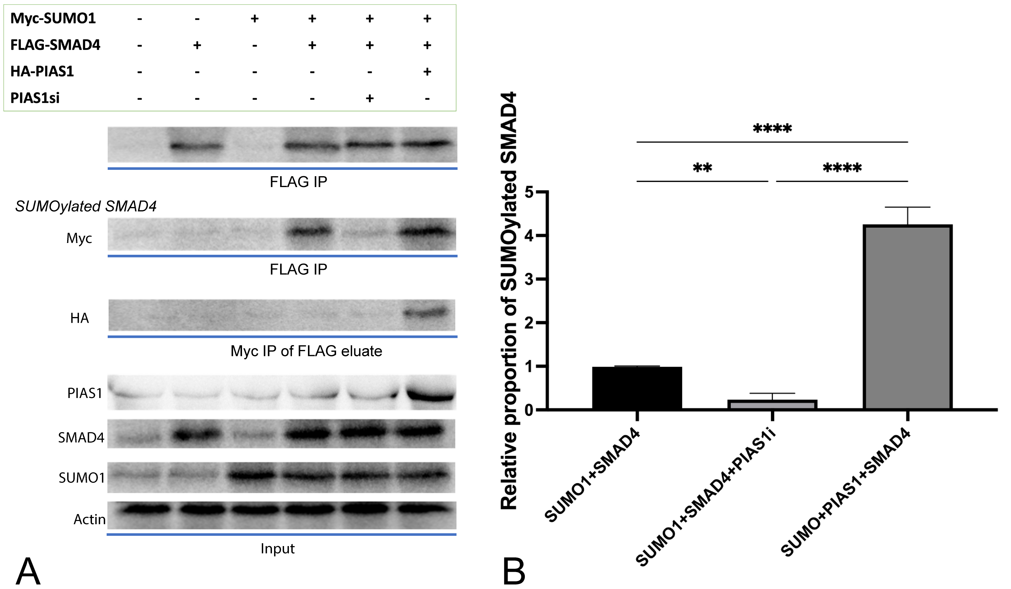
SMAD4 was indeed SUMOylated, confirmed by our in vitro SUMOylation assay conducted in NSCLC cell line A549 (Fig. 1B, column 1), and such modification was enhanced obviously (p < 0.0001) by overexpression of PIAS1 through the incorporation of HA-PIAS1 plasmids into this cell line. On the other hand, endogenous inhibition of PIAS1 by introducing small interfering RNA (siRNA) decreased the SUMOylated SMAD4 level significantly (p = 0053); this further confirmed that PIAS1 enhances the SUMOylation of SMAD4 at the post-translational level. This result raised the possibility that PIAS1 might have achieved SMAD4-SUMOylation by forming a multiprotein complex. To further test this, we performed sequential co-immunoprecipitation by subjecting lysates to SMAD4 immunoprecipitation (FLAG IP), followed by SUMO1 immunoprecipitation (Myc IP) of the eluates. Successful co-immunoprecipitated PIAS1, expressed as an HA-tagged protein, supported the existence of a PIAS1-SUMO1-SMAD4 complex (Fig. 1A).

Fig. 1.

PIAS1 enhances SMAD4 SUMOylation by forming PIAS1-SUMO1-SMAD4 multiprotein complex. (A) Cell lysates transfected with vector control or a plasmid containing cDNA encoding Myc-SUMO1, FLAG-SMAD4, and HA-PIAS1, alone or in combination, were subjected to SMAD4 immunoprecipitation (FLAG IP) followed by SMAD4 or Myc immunoblotting (IB). At the same time, 10% lysates were subjected to SMAD4, PIAS1, SUMO1 IB as the input, and β-actin as the input control. (B) Relative SUMOylated SMAD4 level with vector control, infused with PIAS1, or with endogenous inhibition of PIAS1. Experiments were performed with quadruplicates in each group, quantitative data analyses were conducted in Prism 9.0, the error bar showed standard error (SD), ANOVA was applied, two-tailed p values were indicated as **p 0.01, ****p 0.0001, and Bonferroni’s correction test was conducted for multiple comparisons.

3.2 PIAS1mediated SUMOylation of SMAD4 Boosted the Migration Ability of A549 Cells

To elucidate whether upregulated SUMOylation of SMAD4 is related to metastasis of NSCLC, we tested the effect of different combinations of plasmid transfection on cell migration ability through a wound-healing assay. The maximally enhanced migration ability (compared to control, p
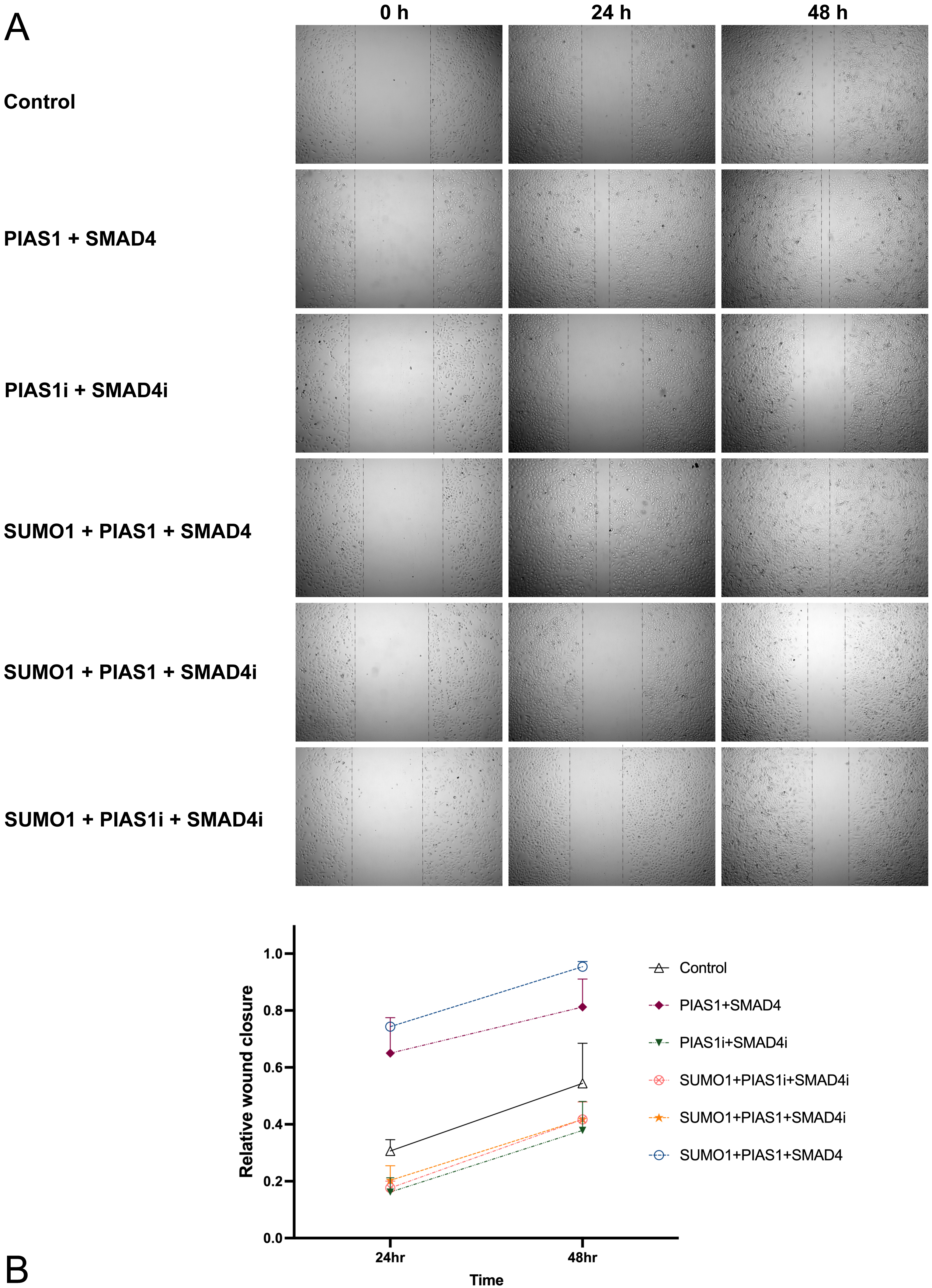
 < 0.0001) was achieved when there was overexpression of PIAS1, SMAD4, as well as SUMO1 (Fig. 2). Yet without exogenous addition of SUMO1, cell migration ability was less increased (p = 0.0342) but still apparently higher than control WT A549. When there was abundant SUMO1 in the system, knocking down SMAD4 significantly compromised the cell migration ability (p = 0.0413, 0.0166 with or without the presence of endogenous PIAS1, respectively), and minimal improvement was observed even overexpression of PIAS1 was introduced (Fig. 2). This, in accordance with previous results, suggested that even though PIAS1 turned up the SUMOylation of SMAD4 by forming an integrated protein complex, the subsequent effect on cell migration was probably mainly achieved by SMAD4 serving as the most important mediator.

Fig. 2.

PIAS1mediated SUMOylation of SMAD4 boosted the migration ability of A549 cells. (A) A549 cells were transfected with vector control or a plasmid containing cDNA encoding Myc-SUMO1, FLAG-SMAD4, HA-PIAS1, alone or in combination with siRNA against PIAS1 or SMAD4, and wound healing assay was performed. The photographs were taken at 0 h, 24 h, and 48 h. Cell migration was quantified by measuring the difference in area between the leading edge and the initiation edge of the experiment. The wound area was assessed by ImageJ software, quantitative data analyses were conducted in Prism 9.0, and the results were shown as (B).

3.3 Increased Migration Ability Induced by PIAS1-SUMO1-SMAD4 Complex was Accompanied by High Expression of Vimentin

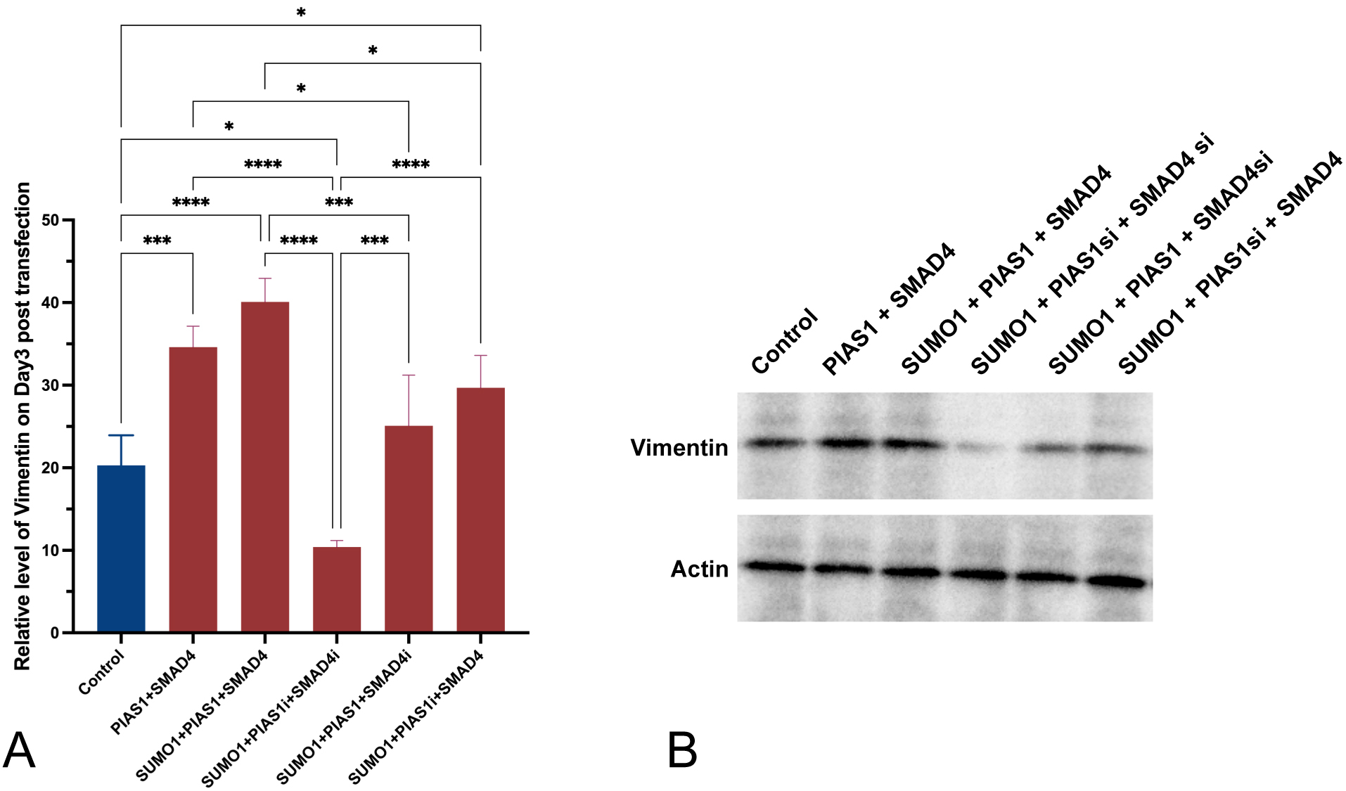
In a parallel experiment involving all groups of cell cultures generated above, we quantified both mRNA expression level and protein level of vimentin microfilament using RT-qPCR and WB, respectively.

Introducing excessive SUMO1 only, with endogenous inhibition of both SMAD4 and PIAS1, was accompanied by decreased vimentin expression and translation in A549 cells, in comparison to control (p = 0.0202, Fig. 3A). If excessive SMAD4 was introduced, vimentin mRNA expression and translation were enhanced (p = 0.0303 with SUMO1 and endogenous inhibition of PIAS1). PIAS1 added up to the effect generated by SUMO1 plus SMAD4 (p = 0.0129), and the upregulating effect was achieved to the maximum when all SUMO1, PIAS1, and SMAD4 were excessively produced. Interestingly, with the presence of upregulation of SUMO1 and inhibition of endogenous SMAD4, the level of vimentin was significantly (p = 0.0004) overcome by the introduction of PIAS1 and comparable to the level observed in control.

Fig. 3.

Increased production of vimentin was observed to be accompanied by the excessive PIAS1-SUMO1-SMAD4 complex. (A) Relative level of vimentin measured using RT-qPCR three days post-transfection. (B) Western blot showing vimentin protein amount across different conditions. Experiments were performed with quadruplicates in each group, quantitative data analyses were conducted in Prism 9.0, the error bar showed standard error (SD), ANOVA was applied, two-tailed p values were indicated as *p 0.05, ***p 0.001, ****p 0.0001, and Bonferroni’s correction test was conducted for multiple comparisons.

4. Discussion

The high incidence and mortality of lung cancer, especially NSCLC as one major component, makes it an important area to explore. Despite the advances that we have been making in terms of the combinative treatment regimen, such as target therapy plus chemotherapy on the foundation of surgical evaluation and interventions, eradication of NSCLC seems to be extremely hard, as indicated by the low survival rate and frequent reoccurrence, which is especially true for metastasized cases. Most NSCLC cases, notably those with increased intra-tumoral heterogeneity, are associated with an increased likelihood of relapse and a greater chance of metastases; this directly leads to poorer prognosis since patients are mostly in late stages when a diagnosis is confirmed. Even though the genomic landscapes vary depending on the different histological subtypes and also whether or not some predisposing factors exist (cigarette exposure, for example), some interesting common genetic mutations have been identified, such as KRAS from both LUAD and LUSC [37], which is later linked to EMT [38, 39], a process thought to be playing a crucial role in the metastasis of many malignancies including NSCLC [13, 14].

One group explored the function of SMAD4 in colorectal cancer and observed that direct silencing SMAD4 compromised the sensitivity of colorectal cancer cells to Cetuximab (a monoclonal antibody against EGFR) as the EMT is promoted and cells are losing epithelial characteristics [30]. Another group focused on prostate carcinoma and also found the inhibitory effect of suppressed SMAD4 on EMT, with a subsequential decrease in mesenchymal markers such as vimentin [31]. Lin et al. [32] revealed that the TGF-β signaling pathway is indeed SMAD4-dependent. Its activation can be achieved through post-translational SUMOylation of SMAD4, which does not affect the transcription activator complex, but stabilizes SMAD4 from being degraded [32]. The effect of SMAD4 on EMT has been further confirmed in the human pancreatic ductal epithelium and invasive growth hormone-secreting adenomas [28, 29]; however, the level of vimentin (whether transcriptional or translational level) was not affected, and so were proteins from canonical TGF-β signaling pathway [29]. Post-translational modification by SUMOylation has been speculated to be substantially important for a lot of mediators and regulators on TGF-β the signaling pathway [40, 41]. In the in vitro experiments we conducted on the A549 NSCLC line, our detection of Myc IB from FLAG eluate confirmed the SUMOylation of SMAD4 (Fig. 1), supported previous findings. In addition, we found out that introducing FLAG-SMAD4 while knocking down PIAS1 could not bring the level of SUMOylated SMAD4 to a comparable level with the SUMO1 + SMAD4 group (Fig. 1, p = 0.0053). This aligns with Liang et al. [36], who described close interactions between SMAD4 and E3 SUMO ligase PIAS1. We also utilized a wound healing assay and identified the positive effect of excessive SUMOylated SMAD4 on the migration/healing ability of 2D A549 cultures; this is established by the significantly increased relative wound closure compared to the wild-type culture (Fig. 2).

As described formerly, vimentin has been widely used as an indicator of EMT, and its expression is linked with the ability of NSCLC to metastasize. However, discrepancies exist when it comes to whether SUMOylation of SMAD4 necessarily means upregulation of vimentin. Contrary to what Kang et al. [29] have described, we observed higher production of vimentin protein accompanied by increased SMAD4 and PIAS1. Still, endogenous SUMO1 was enough to mediate this effect, as shown by the insignificant differences between cultures with excessive SUMO1 or not (p = 0.7569, Fig. 3). The differences between the cell lines tested might have contributed to the differences, and considering that it could take days for this screening marker for EMT to change even with ideal direct stimulation from TGF-β [42], we extended the timepoint to collect samples as long as possible to three days post-transfection. There is also evidence suggesting that vimentin by itself is an important one among SUMOylation targets of PIAS1, and this modification enhances its ability to actively assemble [43]. Direct TGF-β stimulation enhances the promoter of vimentin through AP1 biding sites [44], and non-promoter-mediated upregulation of vimentin also exists [42]. We speculate that the advanced level/functioning of vimentin could result from PIAS1-mediated SMAD4 SUMOylation both at the transcriptional level (promoter-dependent activation by SMAD4) and post-translationally (direct SUMOylation and stabilization of vimentin by PIAS1). However, when comparing the effect of infusion with SUMO1 and endogenous inhibition of SMAD4 plus excessive PIAS1 or further inhibition of PIAS1, PIAS1 could make up for the deficiency of SMAD4, and the net result was recovered vimentin level (Fig. 3), but it did not contribute to cell ability to migrate (Fig. 2). This suggests that even though vimentin is an important marker for EMT, it is not the only or major reason that has contributed to metastasis. As the immunochemistry characterization of the NSCLC patient population carried out by Dauphin et al. [24] pointed out, vimentin could be useful as an indicator for metastases but not necessarily for prognosis or recurrence, and the causal relationship requires further confirmation. In future studies, three-dimensional models might be applied since they have been shown to have a greater capability of simulating what happens in vivo in the NSCLC [45, 46].

In conclusion, this study uncovers novel interactions between PIAS1 and SMAD4 in NSCLC, revealing their role in regulating cell migration and potential metastasis mechanisms. The findings contribute to understanding NSCLC’s molecular mechanisms, potentially leading to innovative therapeutic strategies, prognostic markers, and drug targets for improved patient treatment and management. But in the same time, we acknowledge several limitations in our study. We did not explicitly investigate whether cell migration activities originated from TGFβ signaling. In future research, we should consider elucidating the role of the TGFβ signaling pathway in this process. Although vimentin plays a critical role in EMT, our next step should involve silencing vimentin in tumor cells overexpressing SUMO1, PIAS1, and SMAD4. By doing so, we can observe whether the EMT properties, cell migration activity, and cancer metastasis of these cancer cells are affected. To validate our conclusions, it would be essential to use animal models, which will serve as an important direction for our future research.

5. Conclusions

To conclude, our study further confirmed the interactions between PIAS1 and SMAD4 on the post-translational level. With the presence of high PIAS1, SUMO1-SMAD4 proportion was greatly increased in vitro; this in accordance with highly expressed cytoskeletal element vimentin, increased the ability of A549 NSCLC cells to migrate.

Availability of Data and Materials

The datasets used and/or analyzed during the current study are available from the corresponding author on reasonable request.

Author Contributions

CW and XZ conceptualised and conceived the main idea, designed the program and extracted the data, QD and ZC conducted the experimental study. SY provided help and advice on CW analysed of the data. CW and ZD wrote the manuscript. ZD curated the data. All authors read and approved the final version of the manuscript for publication. All authors contributed to editorial changes in the manuscript.

Ethics Approval and Consent to Participate

Not applicable.

Acknowledgment

We are grateful to the University of Chinese Academy of Sciences for their help with this research.

Funding

This work was supported by Key Reasearch Foundation of Hwa Mei Hospital, University of Chinese Academy of Sciences, China (Grant No. 2020HMZD02, No. 2022HMZD09); supported by Ningbo Health Branding Subject Fund (Grant No. PPXK2018-05); supported by Basic Public Welfare Research Program of Zhejiang Province, China (No. LTGY23H010001); supported by Medical Scientific Research Foundation of Zhejiang Province, China (No. 2022KY1125, No. 2023KY280); supported by Ningbo Clinical Research Center for Respiratory System Disease of Zhejiang Province, China (No. 2022L004).

Conflict of Interest

The authors declare no conflict of interest.

References
[1]
World Health Organization. Cancer Fact Sheet. 2021. Available at: https://www.who.int/news-room/fact-sheets/detail/cancer (Accessed: 23 February 2022).
[2]
Ferlay J, Ervik M, Lam F, Colombet M, Mery L, Piñeros M, et al. Global Cancer Observatory: Cancer Today. Lyon: International Agency for Research on Cancer. 2021. Available at: https://gco.iarc.fr/today (Accessed: 23 February 2022).
[3]
Schad F, Thronicke A, Steele ML, Merkle A, Matthes B, Grah C, et al. Overall survival of stage IV non-small cell lung cancer patients treated with Viscum album L. in addition to chemotherapy, a real-world observational multicenter analysis. PLoS ONE. 2018; 13: e0203058.
[4]
Paz-Ares L, Luft A, Vicente D, Tafreshi A, Gümüş M, Mazières J, et al. Pembrolizumab plus Chemotherapy for Squamous Non-Small-Cell Lung Cancer. The New England Journal of Medicine. 2018; 379: 2040–2051.
[5]
Fujimoto D, Miura S, Yoshimura K, Wakuda K, Oya Y, Yokoyama T, et al. Pembrolizumab plus chemotherapy-induced pneumonitis in chemo-naïve patients with non-squamous non-small cell lung cancer: A multicentre, retrospective cohort study. European Journal of Cancer. 2021; 150: 63–72.
[6]
Garon EB, Hellmann MD, Rizvi NA, Carcereny E, Leighl NB, Ahn MJ, et al. Five-Year Overall Survival for Patients With Advanced Non‒Small-Cell Lung Cancer Treated With Pembrolizumab: Results From the Phase I KEYNOTE-001 Study. Journal of Clinical Oncology. 2019; 37: 2518–2527.
[7]
Minna JD, Roth JA, Gazdar AF. Focus on lung cancer. Cancer Cell. 2002; 1: 49–52.
[8]
Majeed U, Manochakian R, Zhao Y, Lou Y. Targeted therapy in advanced non-small cell lung cancer: current advances and future trends. Journal of Hematology & Oncology. 2021; 14: 108.
[9]
Soria JC, Ohe Y, Vansteenkiste J, Reungwetwattana T, Chewaskulyong B, Lee KH, et al. Osimertinib in Untreated EGFR-Mutated Advanced Non-Small-Cell Lung Cancer. The New England Journal of Medicine. 2018; 378: 113–125.
[10]
Mitsudomi T, Yatabe Y. Mutations of the epidermal growth factor receptor gene and related genes as determinants of epidermal growth factor receptor tyrosine kinase inhibitors sensitivity in lung cancer. Cancer Science. 2007; 98: 1817–1824.
[11]
Yano S, Wang W, Li Q, Matsumoto K, Sakurama H, Nakamura T, et al. Hepatocyte growth factor induces gefitinib resistance of lung adenocarcinoma with epidermal growth factor receptor-activating mutations. Cancer Research. 2008; 68: 9479–9487.
[12]
Soltermann A, Tischler V, Arbogast S, Braun J, Probst-Hensch N, Weder W, et al. Prognostic significance of epithelial-mesenchymal and mesenchymal-epithelial transition protein expression in non-small cell lung cancer. Clinical Cancer Research. 2008; 14: 7430–7437.
[13]
Yauch RL, Januario T, Eberhard DA, Cavet G, Zhu W, Fu L, et al. Epithelial versus mesenchymal phenotype determines in vitro sensitivity and predicts clinical activity of erlotinib in lung cancer patients. Clinical Cancer Research. 2005; 11: 8686–8698.
[14]
Singh A, Settleman J. EMT, cancer stem cells and drug resistance: an emerging axis of evil in the war on cancer. Oncogene. 2010; 29: 4741–4751.
[15]
Thompson EW, Newgreen DF, Tarin D. Carcinoma invasion and metastasis: a role for epithelial-mesenchymal transition? Cancer Research. 2005; 65: 5991–5995; discussion 5995.
[16]
Polyak K, Weinberg RA. Transitions between epithelial and mesenchymal states: acquisition of malignant and stem cell traits. Nature Reviews. Cancer. 2009; 9: 265–273.
[17]
Kalluri R, Weinberg RA. The basics of epithelial-mesenchymal transition. The Journal of Clinical Investigation. 2009; 119: 1420–1428.
[18]
Das V, Bhattacharya S, Chikkaputtaiah C, Hazra S, Pal M. The basics of epithelial-mesenchymal transition (EMT): A study from a structure, dynamics, and functional perspective. Journal of Cellular Physiology. 2019; 234: 14535–14555.
[19]
Rho JK, Choi YJ, Lee JK, Ryoo BY, Na II, Yang SH, et al. Epithelial to mesenchymal transition derived from repeated exposure to gefitinib determines the sensitivity to EGFR inhibitors in A549, a non-small cell lung cancer cell line. Lung Cancer. 2009; 63: 219–226.
[20]
Yao Z, Fenoglio S, Gao DC, Camiolo M, Stiles B, Lindsted T, et al. TGF-beta IL-6 axis mediates selective and adaptive mechanisms of resistance to molecular targeted therapy in lung cancer. Proceedings of the National Academy of Sciences of the United States of America. 2010; 107: 15535–15540.
[21]
Zeisberg M, Neilson EG. Biomarkers for epithelial-mesenchymal transitions. The Journal of Clinical Investigation. 2009; 119: 1429–1437.
[22]
Cheng Z, Xin H, Han T. BECN1 promotes the migration of NSCLC cells through regulating the ubiquitination of Vimentin. Cell Adhesion & Migration. 2019; 13: 249–259.
[23]
Ye Z, Zhang X, Luo Y, Li S, Huang L, Li Z, et al. Prognostic Values of Vimentin Expression and Its Clinicopathological Significance in Non-Small Cell Lung Cancer: A Meta-Analysis of Observational Studies with 4118 Cases. PLoS ONE. 2016; 11: e0163162.
[24]
Dauphin M, Barbe C, Lemaire S, Nawrocki-Raby B, Lagonotte E, Delepine G, et al. Vimentin expression predicts the occurrence of metastases in non small cell lung carcinomas. Lung Cancer. 2013; 81: 117–122.
[25]
Kidd ME, Shumaker DK, Ridge KM. The role of vimentin intermediate filaments in the progression of lung cancer. American Journal of Respiratory Cell and Molecular Biology. 2014; 50: 1–6.
[26]
Miyaki M, Kuroki T. Role of Smad4 (DPC4) inactivation in human cancer. Biochemical and Biophysical Research Communications. 2003; 306: 799–804.
[27]
Schwarte-Waldhoff I, Volpert OV, Bouck NP, Sipos B, Hahn SA, Klein-Scory S, et al. Smad4/DPC4-mediated tumor suppression through suppression of angiogenesis. Proceedings of the National Academy of Sciences of the United States of America. 2000; 97: 9624–9629.
[28]
Shan X, Liu Q, Li Z, Li C, Gao H, Zhang Y. Epithelial–Mesenchymal Transition Induced by SMAD4 Activation in Invasive Growth Hormone-Secreting Adenomas. Open Chemistry. 2018; 16: 571–582.
[29]
Kang Y, Ling J, Suzuki R, Roife D, Chopin-Laly X, Truty MJ, et al. SMAD4 regulates cell motility through transcription of N-cadherin in human pancreatic ductal epithelium. PLoS ONE. 2014; 9: e107948.
[30]
Lin Z, Zhang L, Zhou J, Zheng J. Silencing Smad4 attenuates sensitivity of colorectal cancer cells to cetuximab by promoting epithelial mesenchymal transition. Molecular Medicine Reports. 2019; 20: 3735–3745.
[31]
Lan X, Lu G, Yuan C, Mao S, Jiang W, Chen Y, et al. Valproic acid (VPA) inhibits the epithelial-mesenchymal transition in prostate carcinoma via the dual suppression of SMAD4. Journal of Cancer Research and Clinical Oncology. 2016; 142: 177–185.
[32]
Lin X, Liang M, Liang YY, Brunicardi FC, Melchior F, Feng XH. Activation of transforming growth factor-beta signaling by SUMO-1 modification of tumor suppressor Smad4/DPC4. The Journal of Biological Chemistry. 2003; 278: 18714–18719.
[33]
Lee PSW, Chang C, Liu D, Derynck R. Sumoylation of Smad4, the common Smad mediator of transforming growth factor-beta family signaling. The Journal of Biological Chemistry. 2003; 278: 27853–27863.
[34]
Long J, Wang G, He D, Liu F. Repression of Smad4 transcriptional activity by SUMO modification. The Biochemical Journal. 2004; 379: 23–29.
[35]
Imoto S, Sugiyama K, Muromoto R, Sato N, Yamamoto T, Matsuda T. Regulation of transforming growth factor-beta signaling by protein inhibitor of activated STAT, PIASy through Smad3. The Journal of Biological Chemistry. 2003; 278: 34253–34258.
[36]
Liang M, Melchior F, Feng XH, Lin X. Regulation of Smad4 sumoylation and transforming growth factor-beta signaling by protein inhibitor of activated STAT1. The Journal of Biological Chemistry. 2004; 279: 22857–22865.
[37]
Herbst RS, Morgensztern D, Boshoff C. The biology and management of non-small cell lung cancer. Nature. 2018; 553: 446–454.
[38]
Kharbanda A, Rajabi H, Jin C, Alam M, Wong KK, Kufe D. MUC1-C confers EMT and KRAS independence in mutant KRAS lung cancer cells. Oncotarget. 2014; 5: 8893–8905.
[39]
Kitai H, Ebi H. Key roles of EMT for adaptive resistance to MEK inhibitor in KRAS mutant lung cancer. Small GTPases. 2017; 8: 172–176.
[40]
Hendriks IA, Vertegaal ACO. A comprehensive compilation of SUMO proteomics. Nature Reviews. Molecular Cell Biology. 2016; 17: 581–595.
[41]
Chanda A, Sarkar A, Bonni S. The SUMO System and TGFβ Signaling Interplay in Regulation of Epithelial-Mesenchymal Transition: Implications for Cancer Progression. Cancers. 2018; 10: 264.
[42]
Yates B, Zetterberg C, Rajeev V, Reiss M, Rittling SR. Promoter-independent regulation of vimentin expression in mammary epithelial cells by val(12)ras and TGFbeta. Experimental Cell Research. 2007; 313: 3718–3728.
[43]
Li C, McManus FP, Plutoni C, Pascariu CM, Nelson T, Alberici Delsin LE, et al. Quantitative SUMO proteomics identifies PIAS1 substrates involved in cell migration and motility. Nature Communications. 2020; 11: 834.
[44]
Wu Y, Zhang X, Salmon M, Lin X, Zehner ZE. TGFbeta1 regulation of vimentin gene expression during differentiation of the C2C12 skeletal myogenic cell line requires Smads, AP-1 and Sp1 family members. Biochimica et Biophysica Acta. 2007; 1773: 427–439.
[45]
Gamerith G, Rainer J, Huber JM, Hackl H, Trajanoski Z, Koeck S, et al. 3D-cultivation of NSCLC cell lines induce gene expression alterations of key cancer-associated pathways and mimic in-vivo conditions. Oncotarget. 2017; 8: 112647–112661.
[46]
Amann A, Zwierzina M, Gamerith G, Bitsche M, Huber JM, Vogel GF, et al. Development of an innovative 3D cell culture system to study tumour–stroma interactions in non-small cell lung cancer cells. PLoS ONE. 2014; 9: e92511.

Publisher’s Note: IMR Press stays neutral with regard to jurisdictional claims in published maps and institutional affiliations.

Share
Back to top