


 , Lianzhong Zhang 2,3,*
1 Department of Pharmacy, Henan Provincial People’s Hospital, People’s Hospital of Zhengzhou University, People’s Hospital of Henan University, 450003 Zhengzhou, Henan, China
2 Department of Ultrasound, Henan Provincial People’s Hospital, People's Hospital of Zhengzhou University, People's Hospital of Henan University, 450003 Zhengzhou, Henan, China
3 Henan Engineering Technology Research Centre of Ultrasonic Molecular Imaging and Nanotechnology, 450003 Zhengzhou, Henan, China
4 Department of Medical Imaging, Henan Provincial People’s Hospital & the People’s Hospital of Zhengzhou University, 450003 Zhengzhou, Henan, China
Abstract
Background: Ultrasound-responsive nanodroplets (NDs) targeting tumors
have shown great potential in ultrasound imaging and tumor therapy, but most of
these studies are based NDs with lipid shells that cannot overcome the uptake by
cells of the reticulo-endothelial system (RES). NDs with shells comprised of
polyethylene glycol (PEG)-based polymers could effectively suppressed the uptake
of RES, but the phase transition, contrast-enhanced imaging and drug release
about these NDs have not been well illuminated. Methods: Folate receptor
targeted NDs with shells of polymers and loaded with DOX (FA-NDs/DOX) were
prepared. The particle size distribution and morphology of NDs was characterized
with dynamic light scattering (DLS) and microscope. Phase transition and
contrast-enhanced ultrasound imaging under different mechanical indices (MIs) was
studied, and the intensity of contrast enhancement were quantitatively analyzed.
The targeting property of FA-NDs/DOX to MDA-MB-231 cells and cellular uptake were
observed using a fluorescence microscope. The anti-tumor effects of FA-NDs/DOX
combined with low-intensity focused ultrasound (LIFU) was studied through
cytotoxicity tests. Flow cytometry assays were used to detect cell apoptosis.
Results: The average particle size of the FA-NDs/DOX was 448.0
Keywords
- polymeric nanodroplets
- tumor targeting
- ultrasound imaging
- phase transition
- tumor therapy
Ultrasound is widely used in clinical diagnosis due to its high sensitivity, low cost and safety. Contrasted-enhanced ultrasound (CEUS) can further improve the accuracy of diagnosis by using ultrasound contrast agents (UCAs). Current UCAs used in clinical practice are microbubbles (MBs) composed of a gas core and a shell of lipid or protein, which usually has a diameter of 1–8 µm [1]. In the past two decades, many efforts have been undertaken to develop MBs targeted to vascular endothelial markers of diseases [2, 3]. However, the inefficiency of MBs in extravasating beyond the vasculaturedue to its micron-size has limited the advancement of molecular ultrasound imaging [4]. Nano-scaled UCAs provides an effective method for molecular ultrasound (US) imaging especially for tumors, because of their unique enhanced permeability and retention effects (EPR) at the tumor sites could facilitate the accumulation of nanoparticles in tumor tissues [5].
In recent years, ultrasound-responsive nanodroplet (NDs) with liquid core of perfluorocarbon (PFC) have attracted much attention in ultrasound imaging and drug delivery [6, 7, 8]. NDs could maintain their initial particle size when they are injected into the body, which allows NDs to extravasate beyond the vascular endothelium by taking advantage of the EPR effects of the tumor tissue. When exposed to ultrasound, NDs can be transformed from droplets into MBs and produce acoustic signal [9, 10]. Moreover, the ultrasound-triggered MBs destruction could act as a driving force to push drugs into the target cells [7]. A clinical trial in 2016 showed that ultrasound combined with MBs and gemcitabine improved patients’ tolerance to chemotherapy and significantly prolonged the survival time of patients without increasing side effects [11]. Based on the ultrasound-triggered drug release, various drug loaded NDs has been designed for tumor therapy [12, 13, 14]. In addition to diagnostic ultrasound, low-intensity focused ultrasound (LIFU) has been introduced to cavitation of MBs and drug release for NDs due to its ability of controlling the irradiation within a small area [15, 16, 17], which improved the targeting of tumors therapy.
Although EPR effects increase the passive accumulation of NDs at the tumor site, it has been reported that the that the drug concentration at the tumor site could only be improved to twofold of that in normal tissue through the EPR effect, resulting in an unsatisfactory therapeutic outcome [18]. To improve the targeting of the NDs, NDs have been modified to target the overexpressed receptors on the surface of tumor cells. Folate (FA) has been introduced to design of tumor targeted NDs [19, 20, 21] due to the highly expressed folate receptor (FR) on the surface of various tumor cells such as ovarian, breast, and cervical tumors [22, 23]. The study of Chen et al. [21] proved that folate-conjugated and drug-loaded NDs could successfully deliver the drugs to the targeted tumor site in vitro and in vivo. However, most of these studies are based NDs with lipid shells that cannot overcome the recognition and uptake by cells of the RES [24]. Suppressing the uptake by RES and prolonging the circulation time in vivo are required for NDs to achieve effective accumulation and targeting at tumor sites [25].
It has been reported that the PEG-based polymers can effectively reduce the uptake of RES of nano-scale constructs [9, 26] due to the minimal protein absorption on the PEG-coated surface. The study of Rapoport et al. [25] proved the stability and long circulation time of NDs with shells of PEG-based polymers in vivo. However, the in-depth study of the FR-targeted NDs with polymer shells about the threshold of phase transition, intensity of CEUS and enhancement of drug delivery are still lacking, which have limited their application in molecular imaging and targeted tumor therapy.
In the present study, FR-targeted NDs with polymer shells and loaded with DOX were constructed. The phase transition and in vitro CEUS of the NDs under different MIs were studied, and the intensity of CEUS with MIs and time were quantitatively analyzed. The targeting of the ND to breast cancer cell and enhanced anti-tumor effects mediated by LIFU were also evaluated. The insight into the CEUS and targeted therapy of NDs in this study provide basis for the development and application of tumor targeted NDs with polymer shells.
Poly(ethyleneoxide)-block-poly(caprolactone) (PEG-PCL, PEG: 2000 Da, PCL: 2000 Da) and FA modified PEG-PCL (FA-PEG-PCL) were obtained from the Ruixi Biological Technology Co., Ltd (Xi’an, China). Perfluoropentane (PFP) was purchased from Strem Chemicals, Inc. (Newburyport, USA). Doxorubicin (DOX) was purchased from Aladdin Biochemical Technology Co., Ltd. (Shanghai, China). DMEM medium, L-15 medium, fetal bovine serum (FBS) and penicillin/streptomycin were all provided by Life Technologies corporation (gibco®, Grand Island, NY, USA). Human breast cancer cells MDA-MB-231 were obtained from the Shanghai cell bank (Chinese Academy of Sciences, China). Fluorescent dyes DiI and Hoechst were obtained from Beyotime biotechnology Co., Ltd. (Shanghai, China).
The preparation of FA-NDs/DOX follows the process shown in Fig. 1. The FA-PEG-PCL copolymer and DOX were dissolved in Tetrahydrofuran (THF), and the solution was added to stirred deionized water. The ratio of THF and deionized water was 1:4. The concentration of FA-PEG-PCL and DOX were 3 mg/mL and 0.75 mg/mL respectively. Then the THF was removed by rotary evaporation, and the solution of FA linked micelle loaded with DOX (FA-micelle/DOX) was obtained. To remove the unloaded DOX, the micelle solution was dialyzed in deionized water using a dialysis bag (3000 Da, Viskase®, Chicago, IL, USA).
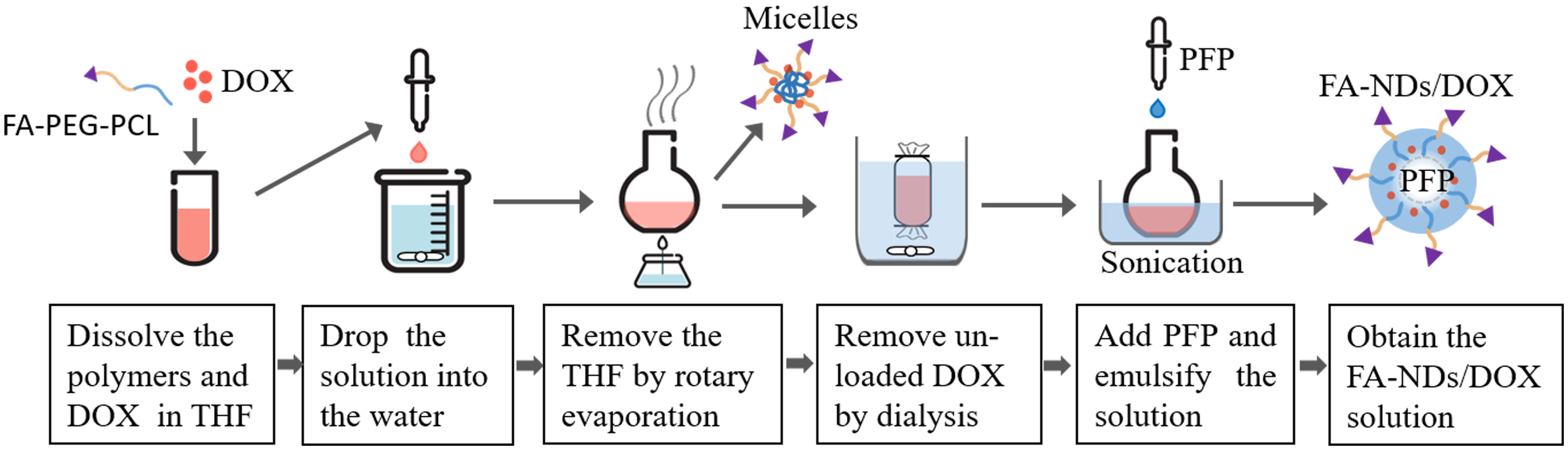 Fig. 1.
The schematic diagram of preparation process of FA-NDs/DOX.
As the liquid core of NDs, the PFP (2%, v/v) was added to the solution of FA-micelle/DOX, and the mixed solution was sonicated for 5 min by a Scientz08-Ⅱ non-contact ultrasonic cell crushing instrument (Scientz Biotechnology Co., Ltd, Ningbo, China). An ice water bath was used to maintain the temperature around the sample during sonication. The resulting emulsion was FA linked NDs loaded with DOX (FA-NDs/DOX). The non-targeted NDs was prepared by replacing the FA-PEG-PCL with PEG-PCL. Blank FA linked NDs (FA-NDs) were prepared without the addition of DOX. NDs with fluorescent dye DiI (10 µM) were prepared by adding DiI in the steps used for micelle preparation.
A Zetasizer Nano ZSE instrument (Malvern Instruments Ltd., Malvern, UK) was used to measure the particle size of the FA-micelle/DOX and FA-NDs/DOX. Transmission electron microscopy (TEM, HT7700, HITACHI, Tokyo, Japan) was used to observe the morphology of the NDs. Prior to observation, NDs dispersions were deposited onto copper grids and then stained with uranyl acetate and dried. The fluorescence of DOX loaded by FA-NDs and the morphology of the FA-NDs/DOX after incubated at 37 °C was observed through an Olympus IX73 inverted fluorescence microscope (Tokyo, Japan).
To determine the drug loading of FA-NDs/DOX, standard curves of DOX concentration and absorbance at 480 nm were plotted using an UV-1800S spectrophotometer (Macy, Shanghai, China). The DOX concentrations of FA-micelle/DOX solution and supernatant of the FA-NDs/DOX solution after centrifugation were determined, and the drug-loading content of the FA-NDs/DOX was calculated as the difference between the two concentrations.
It is assumed that the loss of very small amount of polymer and PFP during preparation is negligible, the mass concentration of the NDs could be calculated according to the total mass of the component (the mass of FA-PEG-PCL, PFP and DOX loaded) and the volume of the NDs solution. The concentration of NDs after dilution can be calculated based on the initial concentration and dilution factor.
The in vitro ultrasound imaging of FA-NDs/DOX was conducted in a glass container filled with degassed water to prevent the impact of air on ultrasound penetration. A sound-absorbing sponge was used to reduce the interference of echo reflection. The water in the glass container was heated and monitored by a water bath (Yushen Instruments Co. Ltd., Shanghai, China). The FA-NDs/DOX solution was diluted to the concentration of 0.75 mg/mL and 1.5 mg/mL with degassed water. Then plastic pipettes (Xinkang Medical Devices Co., Ltd., Taizhou, China) filled with FA-NDs/DOX solution (1.5 mL) was sealed and immersed into the beaker. A control group was developed using plastic pipettes with degassed water. About 10 min later, a linear array transducer (L74M, 5–13 MHz, HITACHI) was immersed into the water to radiate the pipettes with NDs, and the contrast enhancement under contrast mode was monitored and recorded by a ultrasound diagnostic system (HI VISION Ascendus, HITACHI) [10]. The frequency of the transducer was set at 13 MHz during experiments, and the single focus was placed at 2 cm. The imaging acquisition type under contrast enhanced ultrasound mode was pulse inversion. The ultrasound imaging was conducted at 25 °C and 37 °C respectively, and MIs of ultrasound transducer ranged from 0.08 to 1.0 at each temperature.
MI is a parameter directly given by the ultrasound diagnostic system in the present study, which is a measure of the power of an ultrasound transducer and can be defined as [27]:
where P was the peak negative pressure of the ultrasound wave (MPa); f is the frequency of the ultrasound wave (MHz). When the frequency is fixed, the MI is proportional to the peak negative pressure. The peak negative pressure of the ultrasound could be measured by a hydrophone [28].
For the ultrasound diagnostic system used in this study, the MI displayed by the device is the MI at the transducer. Considering the attenuation of ultrasound in water and pipette wall (made of polyethylene), as well as the ultrasound reflection at the interface between water and pipette wall, a compensation factor for the MIs need to be calculated.
For a sound wave with initial acoustic pressure of P
where a is the attenuation coefficient of the medium (Neper, 1 Neper = 8.686 dB/cm); x represents the distance traveled (cm); e is the base of the natural log.
When the sound wave vertically transmit from medium Ⅰ to medium Ⅱ, the
reflection coefficient r
where Z
The attenuation coefficient and acoustic impedance of water is 0.002
dB/cm
The contrast enhancement intensity was analyzed using EZU-CH9 software version
Q1C-EZ1249-5 (HITACHI,) installed on the HI VISION Ascendus diagnostic system.
The dynamic ultrasound imaging was loaded into the software, and ellipse regions
of 0.8 cm
MDA-MB-231 cells (10
MDA-MB-231 cells (10
MDA-MB-231 cells (2
The flow cytometry assay was carried out to assess cell apoptosis. MCF-7 cells or MDA-MB-231 cells were seeded into 6-well plates with L-15 medium (10% FBS, 1% penicillin/streptomycin) at 37 °C for 24 h. Then, the two types of cells were each divided into five groups: control (PBS), FA-NDs without DOX (FA-NDs), LIFU, FA-NDs/DOX and FA-NDs/DOX combined LIFU (FA-NDs/DOX-LIFU). Three parallel holes were set for each group. The specific treatment method of the cells was similar to that previously described, and the concentration of NDs was 250 µg/mL for all the groups. After addition of FA-NDs for 2 h, the group of FA-NDs/DOX-LIFU was irradiated with LIFU for 3 min per well (650 KHz, 3.5 W, 50% duty cycle). After 72 h of interaction with FA-NDs, the cell apoptosis was measured by applying the Annexin V-FITC Apoptosis Detection Kit (Solarbio, Beijing, China) and PI Flow Cytometry Kit (Solarbio) to treat each group of cells in line with the specifications. The apoptotic rates were recorded under flow cytometry. The experimental procedure to obtain the cytotoxicity is shown in Fig. 2.
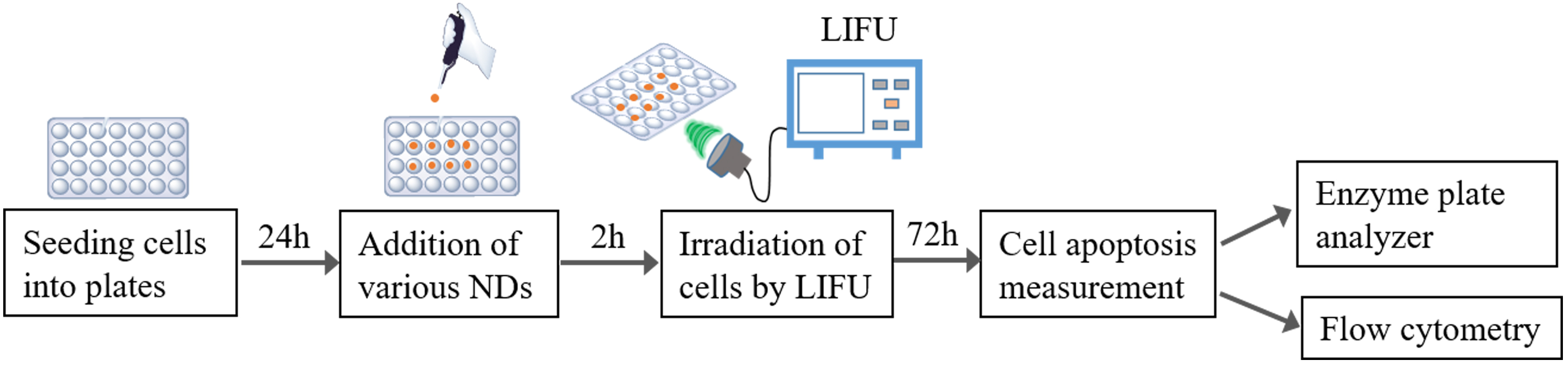 Fig. 2.
The flow chart of cell apoptosis measurement of FA-NDs/DOX combined LIFU on breast cancer cells.
To evaluate the cavitation effect of LIFU on MBs, CEUS experiments of the FA-NDs/DOX irradiated by LIFU was performed. The FA-NDs/DOX solution (3 mg/mL) was added to the 96-well plate and incubated at 37 °C for 2 h. Then the FA-NDs/DOX were divided into 4 groups, each group was irradiated with LIFU (650 KHz, 3.5 W, 50% duty cycle) for 0 s (i.e., without LIFU irradiation), 30 s, 1 min and 2 min, respectively. The CEUS and intensity analysis for each group of NDs solution were conducted using the method and parameters in section 2.4.
The mean
The particle size distribution of FA-micelle/DOX (Fig. 3A) and FA-NDs/DOX (Fig. 3B) measured through dynamic light scattering (DLS) is shown in Fig. 3. The
particle size distribution were obtained in intensity, volume and number. The
particle size of FA-micelle/DOX in intensity, volume and number were 139.9
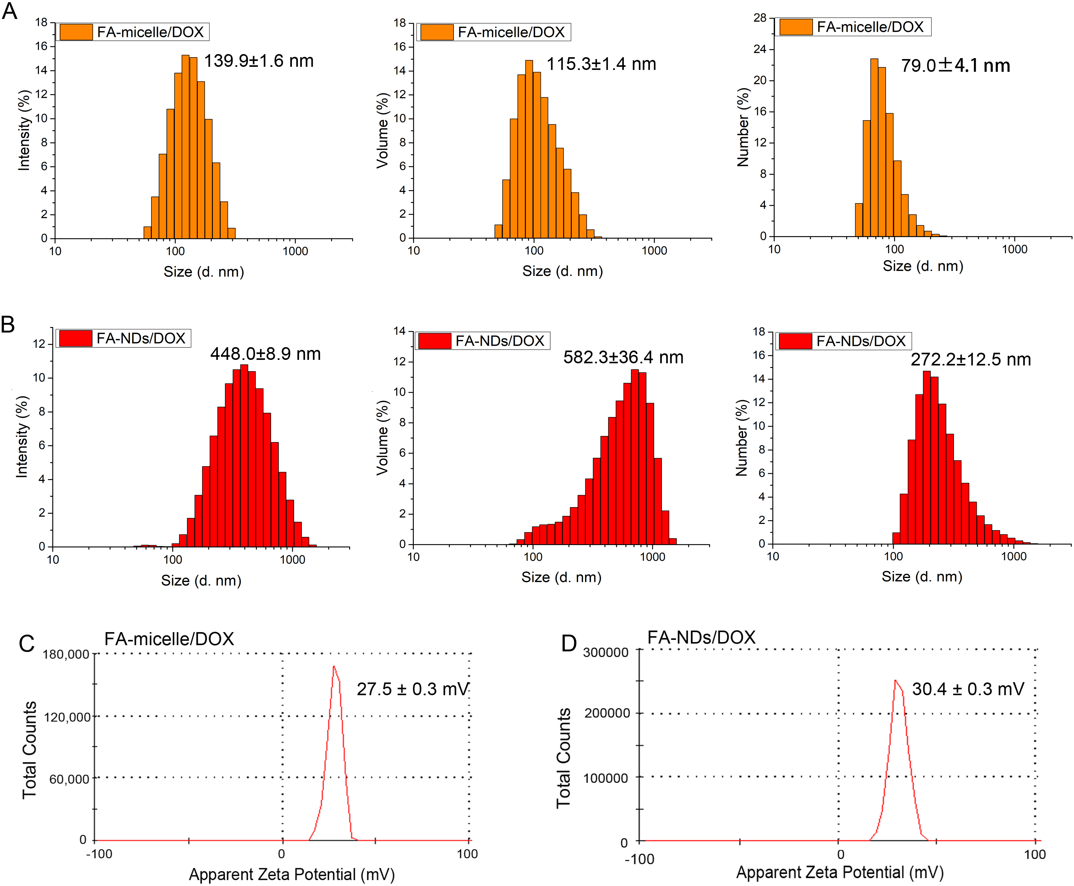 Fig. 3.
Particle size distribution and zeta potential of FA-micelles/DOX and FA-NDs/DOX. Particle size distribution of FA-micelles/DOX (A) and FA-NDs/DOX (B) presented in intensity, volume and number. Zeta potential of FA-micelle/DOX (C) and FA-NDs/DOX (D).
The larger particle size of NDs compared with that of the micelle indicates the
successful encapsulation of PFP. The differences among the particle size in
intensity, volume and number may be due to the fact that the uniformity of the
particles is not very high. For a single particle, the volume and intensity of
large particle is much larger than that of small particle, so the ratio of volume
to intensity could not directly represent the number of particles. The particle
size distribution in number indicated that the number of particles with particle
size below 300 nm accounts for the majority, which is beneficial for the NDs to
penetrate blood vessels at the tumor site. The zeta potential of FA-micelle/DOX
and FA-NDs/DOX were 27.5
The morphology of FA-micelle/DOX and FA-NDs/DOX were observed by TEM. Both micelle (Fig. 4A) and NDs (Fig. 4B) appeared spherical under TEM, but NDs showed a clear shell-core structure due to the introduction of PFP. The particle size of micelle and NDs observed by TEM were about 100 nm and 500 nm respectively, which were in good agreement with that measured by DLS. The red fluorescence of DOX, shown in Fig. 4C, indicates that the DOX is successfully encapsulated, and the drug loading of FA-NDs was 15.6 µg/mg.
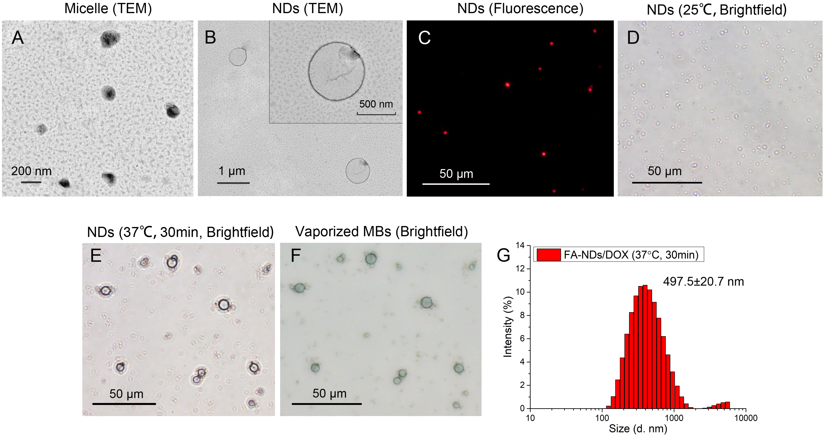 Fig. 4.
Morphology of FA-micelle/DOX and FA-NDs/DOX. Morphology of FA-micelle/DOX (A) and FA-NDs/DOX (B) observed by TEM. (C) Image of FA-NDs/DOX observed by the fluorescence microscope. Optical image of FA-NDs/DOX at 25 °C (D) and heated at 37 °C for 30 min (E). (F) Optical image of vaporized MBs in the upper layer of FA-NDs/DOX solution after heated at 37 °C for 30 min. (G) Particle size distribution (in intensity) of FA-NDs/DOX heated at 37 °C for 30 min.
The morphology of the FA-NDs/DOX heated at 37 °C was further observed. Before
heating, the NDs were uniformly spherical particles under the microscope (Fig. 4D). A small amount of particles with larger size (5–10 µm in diameter)
appeared after heating the NDs at 37 °C for 30 min (Fig. 4E). These larger
particles and NDs distributed in different layers of the solution, because the
shape of the particles was not clear in Fig. 4E. However, these larger particles
can be clearly seen when the microscope was focused on the upper layer of the
solution (Fig. 4F), and the smaller NDs in Fig. 4E almost disappeared from the
image at this time, indicating that the larger particles should be vaporized MBs
rather than large NDs formed by coalescence/fusion of small NDs. Though the
vaporized MBs represent the phased transition induced by heating, the large
amount of NDs that did not vaporize. Fig. 4G showed the particle size
distribution of FA-NDs/DOX (37 °C, 30 min) in intensity. Compared with the average
particle size of FA-NDs/DOX before heating (Fig. 4B, 448.0
The results of the CEUS of FA-NDs/DOX in vitro is shown in Fig. 5. The ultrasound imaging of FA-NDs/DOX was conducted under MIs ranging from 0.04 to 0.48. For degassed water, no acoustic signal was observed at all of the MIs under 25 °C and 37 °C. For NDs (25 °C, 0.75 mg/mL), no acoustic signal was observed until the MI reached 0.48 at 25 °C, and the contrast enhancement at MI of 0.48 was very weak. At 37 °C, the contrast enhancement of NDs with concentration of 0.75 mg/mL and 1.5 mg/mL both began to appear when MI reached 0.19, and the contrast enhancement was stronger for NDs under ultrasound with higher MIs. The duration of ultrasound imaging was also monitored. The contrast enhancement of FA-NDs/DOX decayed with time, but an obvious acoustic signal could still be observed at 30 min.
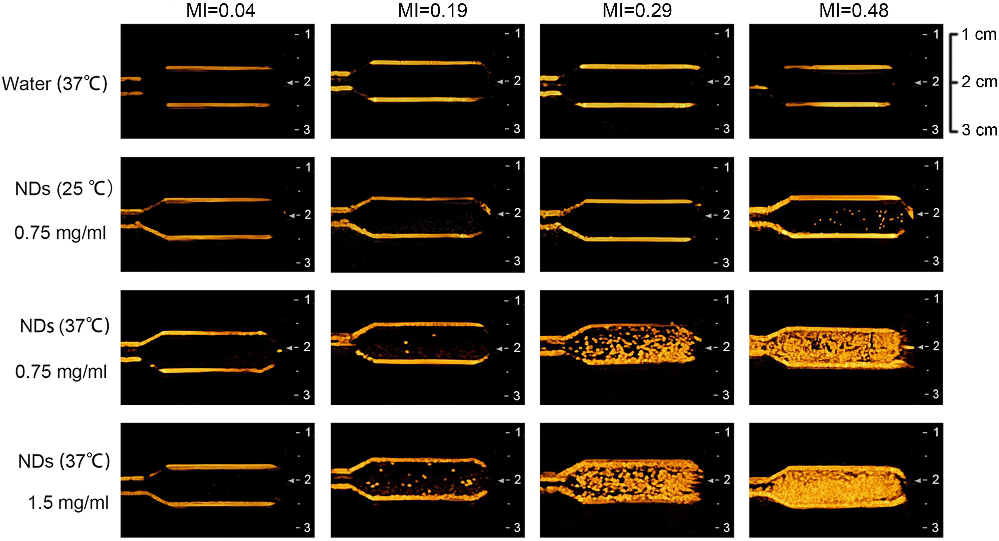 Fig. 5.
Contrast-enhanced ultrasound imaging of water at 37 °C, FA-NDs/DOX (25 °C, 0.75 mg/mL), FA-NDs/DOX (37 °C, 0.75 mg/mL) and FA-NDs/DOX (37 °C, 1.5 mg/mL) under MIs of 0.04, 0.19, 0.29 and 0.48.
Signal intensity of contrast-enhanced ultrasound under different MIs was
analyzed to describe the ultrasound imaging of NDs (Fig. 6). The ROIs selected
within the pipettes is shown in Fig. 6A. The results in Fig. 6B indicated that
the signal intensity of NDs (37 °C, 0.75 mg/mL) were stronger than NDs (25 °C, 0.75
mg/mL) at MI of 0.19, 0.29 and 0.48 (p
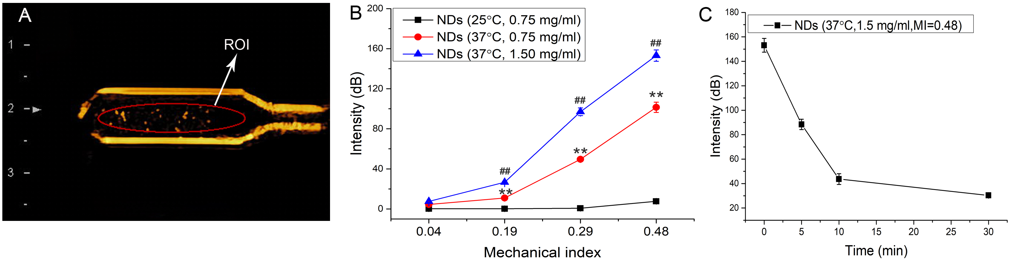 Fig. 6.
Signal intensity of contrast-enhanced ultrasound for FA-NDs/DOX.
(A) Schematic diagram of ROI with area of 0.8 cm
In order to improve the tumor targeting of NDs, FA was used to modify the NDs.
MDA-MB-231 cells with high expression of FR were selected to conduct the
targeting experiment. In Fig. 5, the NDs and FA-NDs were stained by DiI (red),
and the nucleus of MDA-MB-231 cells were stained by Hoechst (blue). The blue in
Fig. 7 are the nucleus, and the red around the nucleus are mainly FA-NDs that
bind to the cells after 45 min of co-incubation. The unbound NDs have been
removed during the experimental operation. In Fig. 7A, there was nearly no red
fluorescence around the nucleus for the non-targeted NDs group, implying that the
NDs without modification of FA could not recognize and bind to cells. In Fig. 7B,C, the red around the nucleus should be FA-NDs binding to the membrane or
entering the cytoplasm, indicating the targeted recognition of FA-NDs to cells.
The intensity ratio of red fluorescence (DOX) to blue fluorescence (nucleus) was
used to estimate the tumor targeting of NDs (Fig. 7D). The higher ratio of
FA-NDs/DiI than NDs/DiI (p
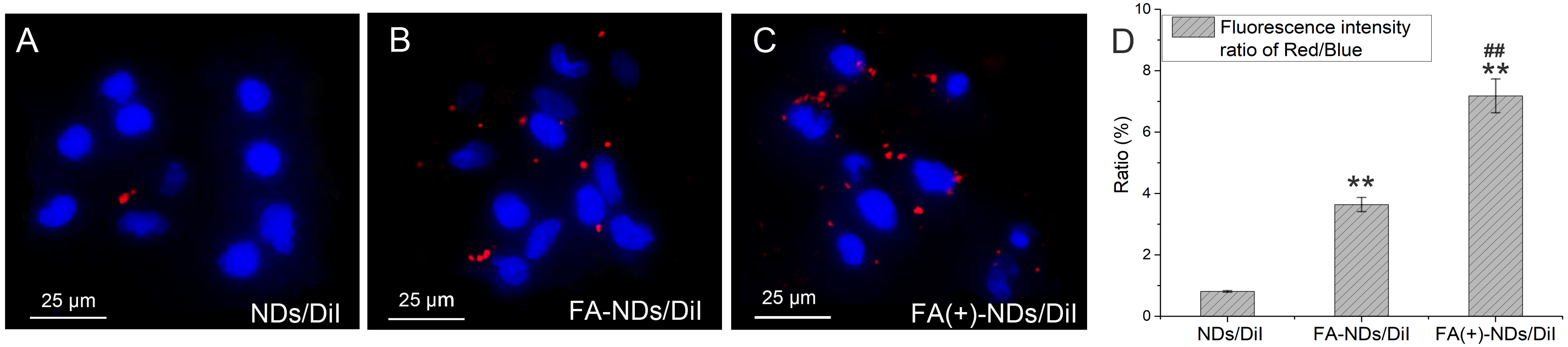 Fig. 7.
Targeted recognition of NDs by MDA-MB-231 cells observed by
fluorescence microscope. (A) NDs/DiI. (B) FA-NDs/DiI. (C) FA (+)-NDs/DiI, i.e.,
FA-NDs/DiI and cells cultured without FA. The nucleus of MDA-MB-231 cell were
stained with Hoechst and shown in blue, whereas NDs or FA-NDs were stained with
DiI and shown in red. (D) Fluorescence intensity ratio of red to blue in (A–C)
(mean
The cellular uptake of FA-NDs/DOX was compared with that of NDs/DOX (without modification of FA) by fluorescent microscopy, and the results was shown in Fig. 8. Both the NDs (Fig. 8A) and the FA-NDs (Fig. 8B) entered cells after 4 h of co-incubation. However, the red fluorescence of DOX appeared in cells was significant higher for FA-NDs/DOX than that of NDs/DOX, and most of the DOX had been entered cell nucleus for FA-NDs/DOX. The higher ratio of red fluorescence (DOX) to the blue fluorescence (nucleus) for FA-NDs/DOX (Fig. 8C) indicated that modification of FA could promote tumor cells to endocytosis of FA-NDs/DOX.
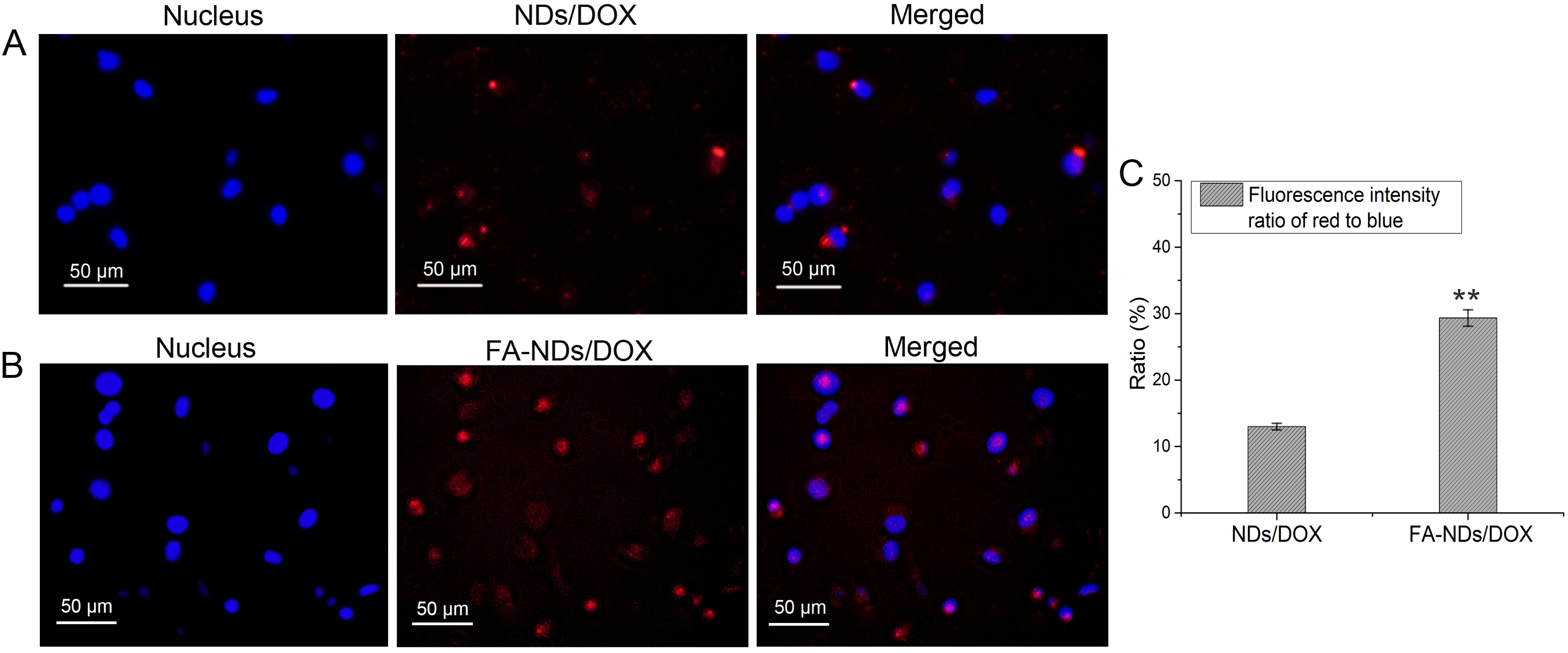 Fig. 8.
Cellular uptake of NDs by MDA-MB-231cells observed by
fluorescent microscopy. (A) NDs/DOX without modification of FA. (B) FA-NDs/DOX.
The nucleus of MDA-MB-231 cell were stained with Hoechst and shown in blue,
whereas NDs/DOX or FA-NDs/DOX were shown in red. (C) Fluorescence intensity ratio
of red to blue in (A,B) (mean
As LIFU was introduced to the anti-tumor experiments, the cavitation effect of LIFU on FA-NDs/DOX was estimated by CEUS. The contrast-enhanced ultrasound imaging and signal intensity of FA-NDs/DOX irradiated by LIFU (3.5 W, 50% duty cycle) for 0 s, 30 s, 1 min and 2 min are shown in Fig. 9A,B. The enhancement intensity of FA-NDs/DOX irradiated by LIFU for 30 s, 1 min and 2 min was about 42%, 26% and 16% of that without irradiation by LIFU, respectively. The results proved that the irradiation of LIFU could induced effective cavitation and destruction of MBs, and the number of MBs decreased with the extension of irradiation time.
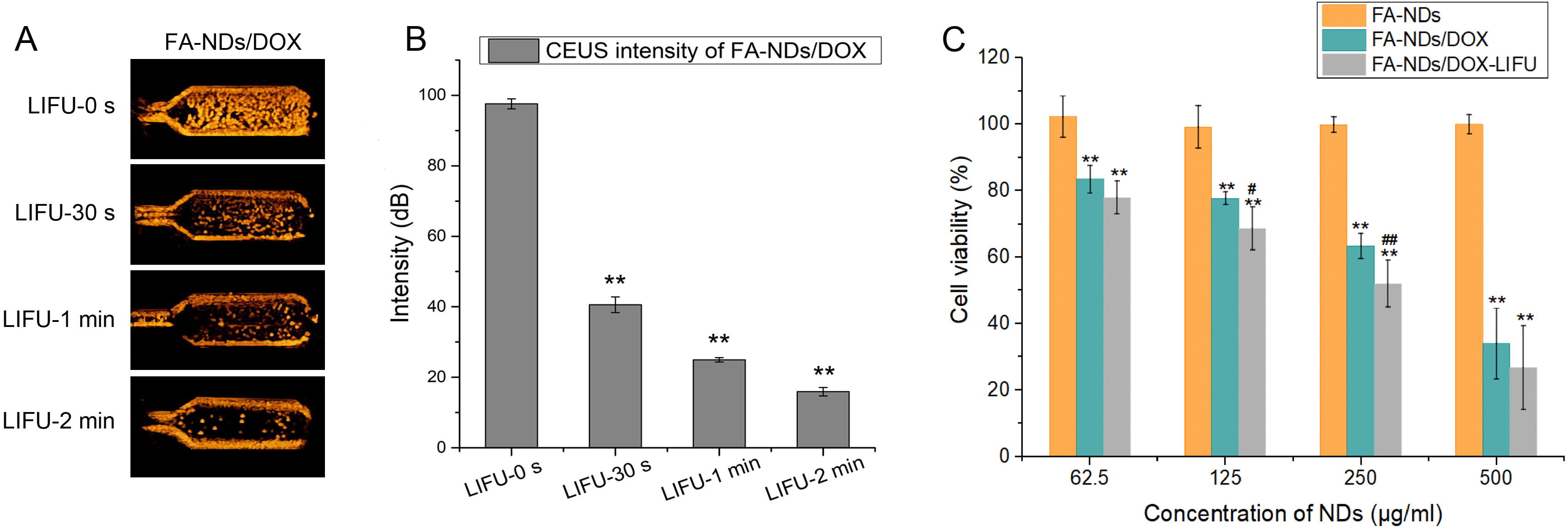 Fig. 9.
The CEUS of FA-NDs/DOX irradiated by LIFU and viability of
MDA-MB-231 cells cultured with different NDs. (A) CEUS of FA-NDs/DOX irradiated
by LIFU for 0 s, 30 s, 1 min and 2 min (37 °C, 0.75 mg/mL, MI = 0.48). (B) The
intensity of contrast enhancement of FA-NDs/DOX irradiated by LIFU for 0 s, 30 s,
1 min and 2 min (mean
The cytotoxicity of FA-NDs/DOX with different concentrations to MDA-MB-231 cells
were investigated. In Fig. 9C, the cell viability was above 90% at all
concentrations for blank NDs, which indicated the well biocompatibility of FA-NDs
as UCA. For FA-NDs/DOX, the cell viability decreased with the increase of
concentrations (p
The apoptosis of MDA-MB-231 cells and MCF-7 cells were also detected by flow
cytometry after treatment with FA-NDs, LIFU, FA-NDs/DOX and FA-NDs/DOX combined
with LIFU (FA-NDs/DOX-LIFU). In Fig. 10, we found that FA-NDs treatment had no
effect on apoptosis of MDA-MB-231 cells (Fig. 10A) and MCF-7 cells (Fig. 10B).
The cell apoptosis MCF-7 cell and MDA-MB-231 were significant after irradiation
by LIFU alone or treated with FA-NDs/DOX alone (p
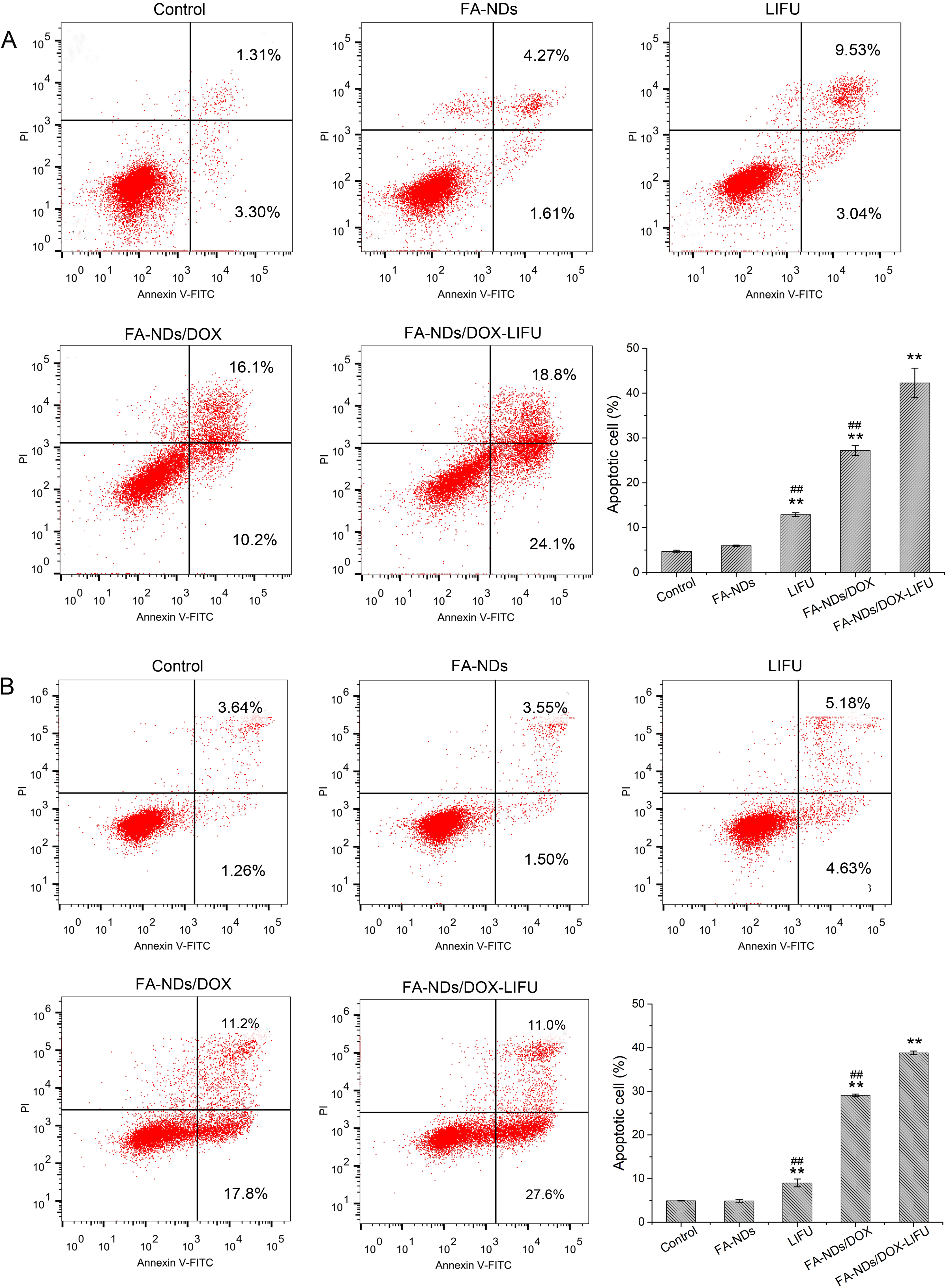 Fig. 10.
 The apoptotic rate of MDA-MB-231 cells (A) and MCF-7 cells (B)
detected by flow cytometry after treated with FA-NDs, LIFU, FA-NDs/DOX and
FA-NDs/DOX combined with LIFU (FA-NDs/DOX-LIFU). Representative images of cell
apoptosis. Data are presented as the mean
The FA-NDs/DOX prepared in this study provides a novel UCA for extravascular CEUS. It has been reported that the particle size of nanocarriers between 380 nm and 780 nm can be passively targeted to tumor sites through large inter-endothelial gaps [15, 33]. The average particle size of FA-NDs/DOX was about 500 nm, which was suitable for extravasation beyond the vascular endothelium of tumor sites. Furthermore, the shell of NDs comprised of PEG could reduce the recognition and non-specific uptake by the reticuloendothelial system [34], prolonging the half-life of the drug carrier system in circulation and providing the basis for tumor targeted imaging and therapy.
Although FA-NDs/DOX has the particle size of a nano scale, the strong ultrasound signal could still be retained due to the phase transition of FA-NDs/DOX under ultrasound. The phase transition property of FA-NDs/DOX was associated with the low boiling liquid core of PFP, which has a boiling point of 29 °C. Most of the NDs could maintain the state of liquid at 37 °C due to the Laplace pressure, which is the pressure difference between the internal and external environment of the NDs resulting from the shells [10, 35]. The Laplace pressure provided by the shells comprised of block polymers may significantly increase the boiling point of the PFP, so that vaporization could not occur even if the temperature around the NDs is above 29 °C [35]. The Laplace pressure is inversely proportional to the radius of NDs, so that NDs with smaller size have a higher vaporization threshold, and NDs with larger size can more easily undergo phase transition.
The vaporization of NDs activated by the ultrasound is called acoustic droplet vaporization (ADV) [36, 37]. The relation between Laplace pressure and radius of NDs determines the difference in ultrasound energy required for vaporization of NDs with different particle sizes [38]. According to the theory of ultrasound imaging, contrast enhancement is mainly derived from the MBs in solution, therefore the acoustic signals can be used to predict whether the NDs have undergone phase transition. The results in Fig. 3 revealed that the phase transition was influenced by temperature and ultrasound. The core of PFP has a boiling point of 29 °C, but phase transition was observed at 25 °C under ultrasound with MI of 0.48; while at 37 °C, contrast enhancement was not observed until the MI was higher than 0.19. As the ultrasound energy is proportional to the MI, the weak acoustic signal under an MI of 0.19 at 37 °C should result from the vaporization of a small amount of NDs with a larger particle size (Fig. 3). With the increase of MI, more NDs was activated by ultrasound and produced stronger acoustic signals. The long duration of the acoustic signal of FA-NDs/DOX, even under higher MIs, may be due to the polymer shells. The strong pressure resistance of NDs resulting from the stiffness of the polymers shells leads to high cavitation thresholds [15, 39], which protect the vaporized MBs from destruction when exposed to ultrasound even under high MIs.
As a tumor targeted UCA, FA-NDs/DOX could not only achieve passive targeting by
taking advantage of particle size, but also achieve active targeting through
modifying the NDs with FA. It has been suggested that the FR is up-regulated in
approximately 40% of all cancers and this overexpression can be seen in
malignant tumor cells [40, 41], therefore the FR could act as an effective
biomarker for tumor targeted imaging. NDs modified by FA showed obvious tumor
targeting, which provided the basis for tumor targeted ultrasound imaging and
drug delivery. In addition, the FR highly expressed on the surface of MDA-MB-231
cells has the potential to facilitate the cellular uptake of FA-NDs/DOX through
receptor-mediated endocytosis [40]. The combination of FR on the surface of tumor
cells and FA-NDs/DOX leads to cell membrane invagination and formation of
intracellular vesicles, which further forms endosomes with an acidic environment
(pH = 5) [42]. Then the FR dissociates from the FA-NDs/DOX and returns to the
cell surface, and the FA-NDs/DOX is destroyed or degraded, resulting in the
release and diffusion of DOX into the cytoplasm and nucleus [43]. Therefore, more
FA-NDs/DOX entered MDA-MB-231 cells than NDs/DOX without modification of FA in
Fig. 6 (p
For anti-tumor experiments, the results of the flow cytometry assay implied that LIFU irradiation alone could induce apoptosis in about 10% of cells. Feng et al. [44] suggested that several proteins, e.g., cellular tumor antigen protein 53, BH3-interacting domain death agonist, apoptosis regulator Bcl-2 and hemeoxygenase 1 were identified as responding to ultrasound irradiation on a molecular level, indicating that mitochondrial dysfunction and oxidative stresses were involved in ultrasound-induced apoptosis. The cells apoptosis induced by FA-NDs/DOX alone should be mainly due to the endocytosis of NDs and release of DOX. When FA-NDs/DOX was combined with LIFU, the cells apoptosis were significantly enhanced. The increase of cells apoptosis resulted from the enhanced drug release and penetration induced by LIFU-triggered MBs cavitation [45]. The mechanism could be interpreted as follows. First, the cavitation and destruction of MBs resulted in maximized release of the loaded drug. Second, the microstreams, shock waves and liquid jets created by the MB cavitation resulted in widening of the intercellular space of the surrounding target cells, which is known as sonoporation [46]. The sonoporation increased cellular permeability and facilitated the entry of the drug into the cells [47, 48]. Finally, the mechanical energy of MB cavitation induced by ultrasound acts as a driving force to push drugs into the deep tumor region. Therefore, the therapeutic effectiveness of FA-NDs/DOX could be significantly enhanced by combing LIFU, providing a more effective approach for anti-tumor therapy.
The results of imaging and anti-tumor experiment provide basis for clinical application of FA-NDs/DOX in diagnosis and treatment. When applied in medical field, the FA-NDs/DOX could accumulate at the tumor site after injected by ERP effects and tumor targeting of FA. Then, molecular imaging at the tumor site could be performed by contrast-enhanced ultrasound, and targeted therapy could be achieved with ultrasound guidance and LIFU assistance. As a small amount of the drug may be released before irradiation of LIFU, the imaging process may be accompanied by a small release of the drug, which is a limitation for application of FA-NDs/DOX to undiagnosed patients. To be safe, drug loaded drops should be used to image and treat patients who are initially diagnosed with tumors, while drug-free NDs may be more suitable for imaging undiagnosed patients.
In this study, the FR-targeted and drug loading NDs with shells of PEG-PCL was developed. The ability of phase transition and CEUS under diagnostic ultrasound of FA-NDs/DOX was proved, and the effect of MIs on intensity of CEUS was elucidated by quantitative analysis. The remarkable targeting ability of FA-NDs to MDA-Mb-231 cells indicated their potential in tumor molecular imaging. The combination of FA-NDs/DOX with LIFU significantly enhanced apoptosis of different breast cancer cells. These results provide evidence for the application of FR-targeted NDs with polymer shells in tumor targeted ultrasound imaging and enhanced chemotherapy.
Data contained within the article.
LZ, YL and JS conceived the study and wrote the manuscript. YL and YC performed the experiments. RL and SD performed statistical analysis and interpretation of data. YL and LC revised the manuscript. All authors contributed to editorial changes in the manuscript. All authors read and approved the final manuscript. All authors have participated sufficiently in the work and agreed to be accountable for all aspects of the work.
Not applicable.
The authors would like to thank Na Li and Linlin Zhang for technical assistance with the cell culture work. The authors would like to thank Ye Zhang for technical support with the ultrasound diagnostic system. We acknowledge Transmission Electron Microscopy at Henan University of Technology.
This work was supported by the Medical Science and Technology Breakthrough Project of Henan Province (Grant No. LHGJ20210031), the Key Research and Development Project of Henan Province (Grant No. 221111310400), the Science and Technology Project of Henan Province (Grant No. 222102310126), the Natural Science Foundation of He’nan Province (Grant No. 212300410240) and the Science and Technology Project of Henan Province (Grant No. 232102311169).
The authors declare no conflict of interest.
References
Publisher’s Note: IMR Press stays neutral with regard to jurisdictional claims in published maps and institutional affiliations.