IMR Press / FBL / Volume 28 / Issue 5 / DOI: 10.31083/j.fbl2805104
Open Access Original Research
TRPM7 is Involved in the Regulation of Proliferation, Migration and Osteogenic Differentiation of Human Dental Follicle Cells
Dongchuan Zuo1,2,†Jiali Li3,4,†Yueyue Huang3,4Jiantao Li3,4Shunzhi Yao3,4Lei Xiong3,4Jin Zeng3,4,*
Show Less
1 The Key Laboratory of Medical Electrophysiology, Ministry of Education, Collaborative Innovation Center for Prevention and Treatment of Cardiovascular Disease, Institute of Cardiovascular Research, Southwest Medical University, 646000 Luzhou, Sichuan, China
2 Department of Cardiology, The Affiliated Hospital of Southwest Medical University, 646000 Luzhou, Sichuan, China
3 Department of Orthodontics, The Affiliated Stomatology Hospital of Southwest Medical University, 646000 Luzhou, Sichuan, China
4 Luzhou Key Laboratory of Oral & Maxillofacial Reconstruction and Regeneration, The Affiliated Stomatological Hospital of Southwest Medical University, 646000 Luzhou, Sichuan, China
*Correspondence: zengjin@swmu.edu.cn (Jin Zeng)
These authors contributed equally.
Front. Biosci. (Landmark Ed) 2023, 28(5), 104; https://doi.org/10.31083/j.fbl2805104
Submitted: 1 September 2022 | Revised: 24 November 2022 | Accepted: 29 November 2022 | Published: 25 May 2023
Copyright: © 2023 The Author(s). Published by IMR Press.
This is an open access article under the CC BY 4.0 license.
Abstract

Background: Dental follicle cells (DFCs) are promising candidates for tissue engineering. However, the molecular mechanisms that regulate the biological characteristics of DFCs are still unclear. Transient receptor potential melastatin 7 (TRPM7) is a Ca2+- and Mg2+-permeable cation channel. The aim of this study was to determine the impact of TRPM7 on the proliferation, migration and osteogenic differentiation of DFCs. Methods: PCR, Western blotting, Immunocytochemical staining and Patch clamp methods were used to identify the gene and protein expression of TRPM7 in DFCs. DFCs were infected with lentiviruses that expressed either TRPM7 specific shRNA or scrambled non-effective shRNA to investigate its functional role. Cell proliferation and migration were assessed using Cell Counting Kit-8 assays and transwell cell culture chambers separately. Cell osteogenic differentiation were determined by ALP assay kit and Alizarin Red staining. Results: Gene and protein expression of TRPM7 were detected in DFCs, but not of TRPM6, which is a closely related channel with similar function. In the absence of Mg2+, typical whole cell TRPM7-like currents were recorded by patch clamp. These were inhibited by low concentrations of 2-APB, but activated by high concentrations of 2-APB. Functional studies demonstrated that suppression of TRPM7 expression inhibited the proliferation and migration of DFCs, and promoted their osteogenic differentiation. Furthermore, Mg2+ deficiency mimicked the effects of TRPM7 knockdown in terms of osteogenic differentiation of DFCs. Conclusions: These results demonstrate that TRPM7 is involved in regulating the proliferation, migration and osteogenic differentiation of DFCs.

Keywords
DFCs
proliferation
migration
osteogenic differentiation
TRPM7
1. Introduction

Dental follicle cells (DFCs) are believed to contain precursor cells for periodontal ligament cells, cementoblasts and osteoblasts. These precursor cells are responsible for periodontal ligament, cementum and alveolar bone formation, respectively, in the process of tooth development [1]. Wise et al. [2] first isolated DFCs from rat molars in 1992. In 2005, Morsczeck et al. [3, 4] successfully isolated DFCs from the human impacted third molars and confirmed they were mesenchymal derived stem cells with multi-differentiation capability in vitro. Over the past few decades, DFCs have received a lot of attention due to their use in regenerative medicine research, as well as their use in basic research for studying molecular processes during in vitro differentiation conditions [5]. The molecular mechanisms that underlie the osteogenic differentiation of DFCs are not yet fully elucidated. However, the Bone Morphogenetic Protein signaling pathway, the canonical Wingless Type Mouse mammary tumor virus (MMTV) Integration Site Family Member (WNT) signaling pathway, the Mitogen-Activated Protein Kinase (MAPK) signaling pathway, and the Notch signaling pathway have all been implicated in the osteogenic differentiation of DFCs [6].

Transient receptor potential melastatin 7 (TRPM7) is a member of the transient receptor potential (TRP) family, which is widely expressed in both electrically excitable and non-excitable cells. TRPM7 is a bi-functional protein containing a kinase domain fused to an ion channel [7]. As an ion channel, TRPM7 is a key regulator for Mg2+ homeostasis, which is essential for cell survival, proliferation, migration and differentiation of various cell types [8, 9]. In addition, TRPM7-mediated Ca2+ influx under extracellular stimulation (such as mechanical stress) also plays a vital role in fundamental cellular processes. For example, TRPM7 can sense mechanical stimulation and induce osteogenesis in human bone marrow mesenchymal stem cells (MSCs) by mediating Ca2+ influx [10]. Moreover, TRPM7 contains an atypical kinase domain at its C-terminus which has been reported to have several substrates in vitro, including annexin-A1, myosin IIA, eukaryotic elongation factor 2 cognate kinase, Smad2, and phospholipase Cγ2 [11, 12, 13, 14, 15]. Therefore, TRPM7 can exert its physiological function through diverse downstream signaling pathways. Of all the TRP channels described to date, only TRPM4 has been reported in DFCs. Nelson et al. [16] found that TRPM4 had an inhibitory effect on the osteogenic differentiation of mouse DFCs, thus potentially implicating TRP channels in DFCs. The aims of the present study were to assess the expression of TRPM7 in DFCs and to determine its impact on the proliferation, migration and osteogenic differentiation of these cells.

2. Materials and Methods
2.1 Constructs and Lentiviruses

The human sequences encoding TRPM6 or TRPM7 were synthesized and cloned into pcDNA3.0 vector to generate pcDNA-TRPM6 and pcDNA-TRPM7 plasmids. These were subcloned into pEGFP-N1 vector with forward primer (5-ACTAAGCTTAATGGGCGACGCGGCCG-CAG-3) containing a HindIII site, and reverse primer (5-CATGGATCCGAAAGGTGCTGGTGGAGGTTG-3) containing a BamHI site. All plasmids were sequenced to confirm their identities. Oligos encoding human TRPM7-specific shRNA (GCAGATCTGCTAGCGTATATTCTCGAG), or scrambled non-effective shRNA, were cloned into the LV-U6-mPGK-EGFP-T2A-Puro vector (Cyagen, Guangzhou, China) to produce recombinant lentiviral vectors in which green fluorescence protein (GFP) functions as an expression reporter.

2.2 Cell Culture, Transduction, and Transfection

Chinese hamster ovary (CHO) cells were cultured with Dulbecco’s Modified Eagle’s Medium (DMEM; catalog #SH3002302, HyClone, Logan, UT, USA) containing 10% fetal bovine serum (FBS; catalog #FB12999102, Gibco, Carlsbad, CA, USA), 100 units/mL penicillin, and 100 mg/mL streptomycin (catalog #15140148, Gibco, Carlsbad, CA, USA) in a 5% CO2 incubator. CHO cells were passaged into 35 mm dishes 24 hours before transfection. Cells at 60 to 80% confluence were transfected with TRPM6 or TRPM7 plasmids using Lipofectamine 3000 reagent (catalog #L3000001, Invitrogen, Shanghai, China) according to the manufacturer’s protocol.

The ethics committee of Southwest Medical University approved all human cell culture experiments. Informed consent was obtained from donors who provided teeth for subsequent research. DFCs were obtained from embedded third molars collected from 20 individuals (aged 12–20 years). The dental follicle was gently separated from the tooth and then carefully washed with PBS containing 100 units/mL penicillin and 100 mg/mL streptomycin. The tissue blocks were minced and digested in a solution of 1% collagenase and 1% dispase (catalog #17105041, Invitrogen, Shanghai, China) for 40 minutes at 37 °C. Single cells and digested tissues were incubated in DMEM supplemented with 10% FBS, 100 units/mL penicillin and 100 mg/mL streptomycin at 37 °C and with 5% CO2. The culture media was changed every 2 days before cell passage. Cells from passages 2 or 3 were used in the experiments. DFCs were infected with lentiviruses that expressed either TRPM7 specific shRNA or scrambled non-effective shRNA at a concentration of 1010 plaque-forming units/mL. GFP allowed the identification of transduced cells.

2.3 Immunocytochemical Staining

DFCs cultured in 6-well plates (2 × 10⁴ cells/well) were fixed with 4% paraformaldehyde for 10 min, permeabilized with 0.5% triton for 15 min, and blocked with 1% bovine serum albumin in PBS for 30 minutes. Cells were then incubated overnight with primary antibodies at 4 °C. The next day, the cells were incubated with secondary antibody for 1 h. After washing with PBS, the nuclei were stained with 100 ng/mL of 4’,6-Diamidino-2-Phenylindole, Dihydrochloride (DAPI , catalog #C1005, Beyotime Biotechnology, Shanghai, China) for 5 min. Primary antibody for TRPM7 was purchased from Abcam (catalog #ab245408, Cambridge, MA, USA) and diluted at a 1:100 ratio. Images were captured with a fluorescence microscope.

2.4 Polymerase Chain Reaction (PCR)

Total RNA was isolated from DFCs and CHO cells with ectopic expression of TRPM6 or TRPM7 using Trizol reagent (catalog #15596026, Invitrogen, Shanghai, China) according to the manufacturer’s instructions. The isolated RNA was treated with DNase 1 and reverse transcription PCR was performed with PrimeScriptTM RT reagent Kit (catalog #RR037B, TaKaRa, Tokyo, Japan) according to the manufacturer’s protocol. The expression of target mRNA was visualized by ethidium bromide staining under UV light after agarose gel electrophoresis (1.5%).

Quantitative real-time PCR (qRT-PCR) was used to quantify TRPM7 and osteogenic-related gene expression levels in knockdown cells during osteogenic differentiation. Briefly, total RNA was isolated from DFCs and reverse-transcribed to cDNA using the PrimeScriptTM RT reagent Kit (catalog #RR037B, TaKaRa, Tokyo, Japan). qRT-PCR was performed using the ABI Prism 7300 Real-Time PCR System (Thermo Fisher, MA, USA) with PrimeScript RT-PCR Kit (catalog #RR014B, TaKaRa, Tokyo, Japan). Specific primers used for detection of the target mRNA were (5′-3′): TRPM7 forward CTGGTTCCTCTTCTGGTGCCTTATTC, reverse TGCAACTTGGCTGAGATGGTGTAC; TRPM6: forward AGGGCCTGCAAATCAAAGATGA, reverse: TCTCCAATCAGTCGGCCACA; ALP: forward ACTCTCCGAGATGGTGGTGGTG, reverse CGTGGTCAATTCTGCCTCCTTCC; RUNX2: forward GCAGCAGCAGCAGCAGGAG, reverse: GCACCGAGCACAGGAAGTTGG; BSP: forward TGAAGAAGAAGAGGAGGAAGAGGAAGG, reverse CTCCGCTGCTGCCGTTGC; Human glyceraldehyde-3-phosphate dehydrogenase (GAPDH) was used as the internal control to normalize the amount of RNA in each sample. Quantitative data for mRNA expression was calibrated to the results of control cells.

2.5 Osteogenic Differentiation of DFCs

DFCs were cultured in DMEM supplemented with 10% FBS, 10 mM β-glycerophosphate (catalog # G9422, Sigma, Darmstadt, Germany), 10-8 M dexamethasone (catalog #D4902, Sigma, Darmstadt, Germany), 50 μg/mL ascorbic acid (catalog # A92902, Sigma, Darmstadt, Germany), and 10 nM vitamin D3 (catalog #731285, Sigma, Darmstadt, Germany). The medium was changed every 3 days. After 14 days of osteogenic differentiation, the cells were fixed with 4% polyoxymethylene for 10 minutes followed by Alizarin Red staining. After washing with PBS, stained cells were visualized using Leica DM2500 optical microscope (Leica Optical, Wetzlar, Germany).

2.6 Cell Proliferation Assay

Cell proliferation of DFCs was determined using Cell Counting Kit-8 assays (CCK-8, catalog #CK0411, Dojindo, Japan). DFCs were seeded into 96-well plates (5 × 10³ cells/well) in 100 μL of DMEM. At different time points (day 2, day 4 and day 6), the culture medium was replaced with 110 μL of fresh medium containing 10 μL of CCK-8. Absorbance at 490 nm was measured in each well with a microplate reader.

2.7 Transwell Chamber Migration Assay

Cell migration of DFCs was assayed usingtranswell cell culture chambers (catalog #3384, Corning, NY, USA). The migration of cells was stimulated by DMEM containing 10% FBS in the lower chamber. After 48 hours culture, cells that had migrated through the membrane were stained by 100 ng/mL DAPI for 5 min. To quantify the migrated cells, cells were counted in 5 random fields with a Leica DM2500 optical microscope (Leica Optical, Wetzlar, Germany) and expressed as the average number of cells per field.

2.8 Electrophysiological Recordings

Whole cell recordings were performed using an EPC-10 USB amplifier with PatchMaster software version 2x91 (HEKA Elektronik, Reutlingen, Germany). Patch pipettes with resistances of 2.0–3.0 M were used. Series resistance was smaller than 10 M and compensated at least 80% to minimize voltage errors. To elicit whole cell TRPM7 currents, a standard 2.2 s voltage ramp from –100 mV to +100 mV was applied. The standard extracellular solution contained (in mM): 140 NaCl, 2 CaCl2, 2.8 KCl, 2 MgCl2, 4 glucose, and 10 HEPES-NaOH, with pH adjusted to 7.2 with NaOH. Mg2+-free intracellular solution contained (in mM): 120 Cs-glutamate, 8 NaCl, 10 Cs-BAPTA, 10 HEPES-CsOH, with pH adjusted to 7.2 with CsOH.

2.9 Measurement of ALP Activities

DFCs (2 × 10⁴ cells/well) were first cultured in osteogenic differentiation media for 14 days. Alkaline phosphatase (ALP) activities were then determined by the p-nitrophenol phosphate conversion method using the ALP assay kit (catalog #C3206, Beyotime, Shanghai, China) according to the manufacturer’s instructions. Absorbance at 405 nm was measured using a microplate reader. Quantitative data for ALP activity was calibrated against the results of control cells at day 14 of osteogenic differentiation.

2.10 Western Blotting

DFCs were washed with PBS and lysed on ice for 30 minutes with Radio Immunoprecipitation Assay (RIPA, catalog #89900, Thermo Fisher, MA, USA) buffer containing protease inhibitor (50 mM Tris-HCl, 0.1% Sodium dodecyl sulfate (SDS), 150 mM NaCl, 1% Triton X-100, 1% sodium deoxycholate and 1 mM EGTA). The lysed cells were centrifuged at 12,000 g at 4 °C for 20 minutes, the supernatant was collected, and the protein quantified with the BCA protein assay kit (catalog #23227, Pierce, Rockford, IL, USA). After normalization, cell lysates were resuspended in SDS sample buffer and denatured at 95 °C for 5 minutes. Twenty μg of total target protein was separated by 10% SDS-PAGE gel and then transferred to PVDF membranes. PVDF membranes were blocked at room temperature with 5% non-fat milk in Tween-Tris-buffered saline (TTBS, 0.1% Tween 20, 50 mM Tris, 500 mM NaCl) for 1 h, and then incubated overnight at 4 °C with primary antibodies directed against GAPDH or TRPM7 (dilution 1:1000; Santa Cruz Biotechnology, Santa Cruz, CA, USA). The membranes were then incubated with secondary antibody for 1 hour. Protein bands were visualized with a Bio-Rad Universal Hood II System (Bio-Rad, Hercules, CA, USA).

2.11 Data Analysis

All data are presented as the mean ± SD from at least three independent experiments, each performed in triplicate. The Student’s t-test was used to calculate p-values with GraphPad Prism version 9.2.0 (GraphPad Software, San Diego, CA, USA). p values of <0.05 were considered statistically significant.

3. Results
3.1 Confirmation of TRPM7 Expression in DFCs

The expression of TRPM7 mRNA and protein in DFCs was confirmed using PCR, western blot and immunofluorescence staining (Fig. 1a–d). Electrophysiological recordings were then made to determine whether TRPM7 currents were active in DFCs. In the absence of Mg
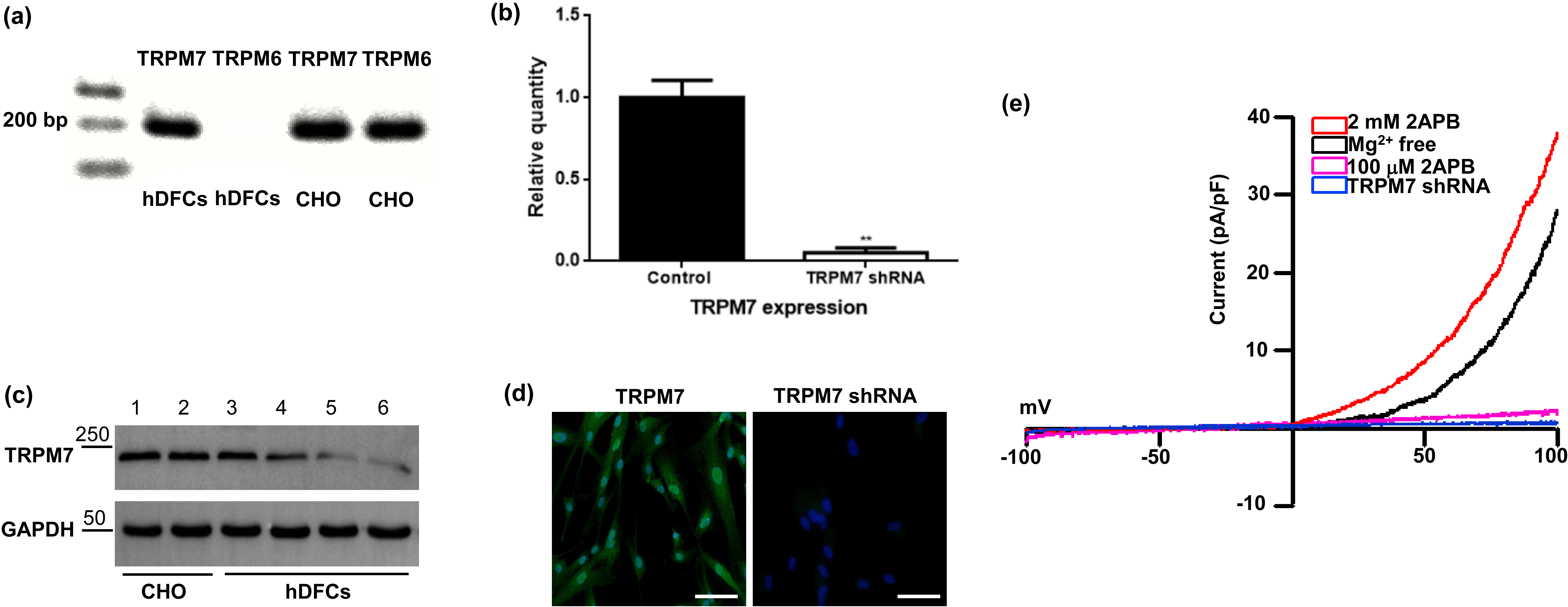
2+, typical whole cell TRPM7-like currents with small inward currents were recorded at negative potential, and strong outward currents at positive potential. To identify the native TRPM7-like currents present in DFCs, two concentrations of 2-APB (100 μM and 2 mM) were used to examine the currents in response to a voltage ramp. The results showed that TRPM7-like currents increased after exposure to 2 mM 2-APB, whereas they decreased after exposure to 100 μM 2-APB (Fig. 1e). These biophysical properties are similar to those reported for recombinant TRPM7 [17, 18]. Together, the results demonstrate that TRPM7 is functionally expressed in DFCs.

Fig. 1.

Knockdown of TRPM7 in DFCs using lentiviruses that express TRPM7-specific shRNA. (a) TRPM7 mRNA, but not TRPM6 mRNA, was detected in DFCs by reverse transcription PCR. CHO cells with ectopic expression of TRPM6 or TRPM7 were used as positive controls. (b) qRT-PCR showed that TRPM7 gene expression was significantly reduced in TRPM7 knockdown cells. Quantitative data from three independent experiments is shown. **, p < 0.01. (c) Western blot was performed to detect the TRPM7 protein expression. Proteins isolated from CHO cells transduced with TRPM7 served as positive controls. lanes 1 and 2: CHO cells transduced with TRPM7. lanes 3 and 4: DFCs transduced with scrambled shRNA. lanes 5 and 6: DFCs transduced with TRPM7 shRNA. (d) Immunocytochemistry showed that TRPM7 was highly expressed in DFCs, but barely expressed in TRPM7 knockdown cells. Scale bar: 100 μM. (e) Typical whole cell TRPM7-like currents recorded in DFCs in the absence of Mg2+ (black color), in the presence of 2 mM 2APB (red color), of 100 μM 2APB (pink color), or infected with TRPM7-specific shRNA (blue color).

3.2 Knockdown of TRPM7 Inhibits the Proliferation and Migration of DFCs

We next evaluated whether TRPM7 has a role in the proliferation and migration of DFCs. For this purpose, shRNA-expressing lentiviruses were used to knockdown TRPM7 expression in DFCs. The effectiveness of shRNA was confirmed by performing qRT-PCR, immunofluorescence, western blot and electrophysiological recordings. The results showed significant reductions in TRPM7 mRNA and protein levels, as well as in whole cell TRPM7-like currents in the TRPM7 knockdown cells compared to control cells infected with scrambled shRNA (Fig. 1b–e).

CCK-8 and transwell chamber assays were used to assess the effect of TRPM7 knockdown on the proliferation and migration of DFCs. CCK-8 assay results showed that at all time points (2, 4 and 6 days), the proliferation of TRPM7 knockdown cells was significantly reduced compared to control cells infected with scrambled shRNA (Fig. 2a). Transwell chamber assay results showed that fewer cells with TRPM7 knockdown migrated after 48 hours culture compared to control cells infected with scrambled shRNA (Fig. 2b). Quantitative data showed that TRPM7 knockdown reduced the number of migrated cells by almost half (52.17%) compared to the control cells (Fig. 2c).
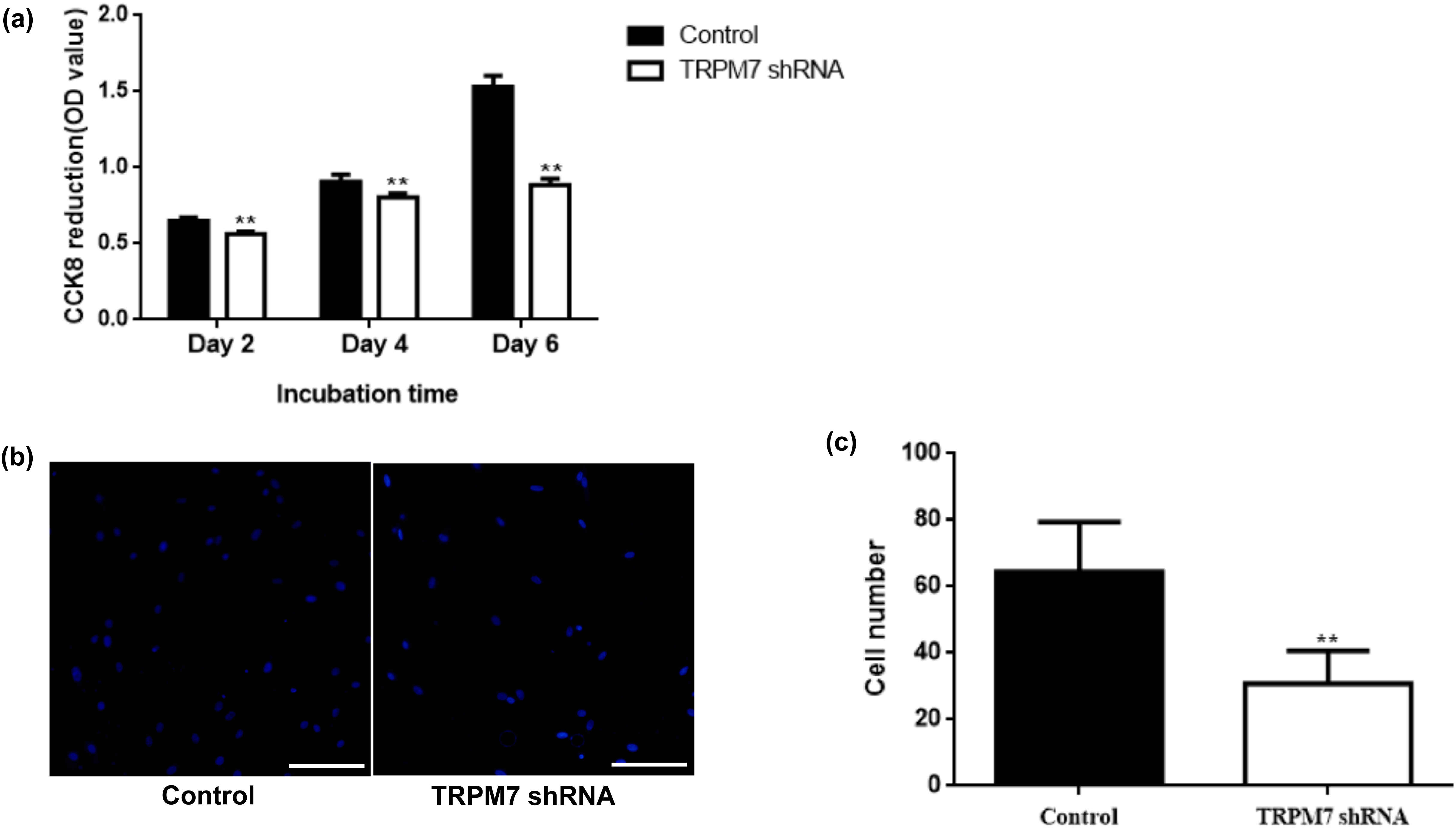
Fig. 2.

TRPM7 knockdown inhibited the proliferation and migration of DFCs. (a) CCK-8 assay showed that TRPM7 knockdown inhibited cell proliferation of DFCs. Quantitative data from five independent experiments are shown. **, p < 0.01. (b) Transwell chamber assays showed less migration of TRPM7 knockdown cells. Scale bar: 100 μM. (c) Quantitative data from five independent experiments for (b) are shown. **, p < 0.01.

3.3 Knockdown of TRPM7 Promotes Osteogenic Differentiation of DFCs

To evaluate the effect of TRPM7 on the osteogenic differentiation of DFCs, we cultured these cells in osteogenic differentiation media for 14 days and then determined ALP activity, expression of osteogenic related genes, and formation of mineral deposits. Inhibition of TRPM7 with specific shRNA was found to enhance mineralization (Fig. 3a,b), increase ALP activities (Fig. 3c), and upregulate the expression of ALP, RUNX2 and BSP (Fig. 3d–f) compared to control cells infected with scrambled shRNA.
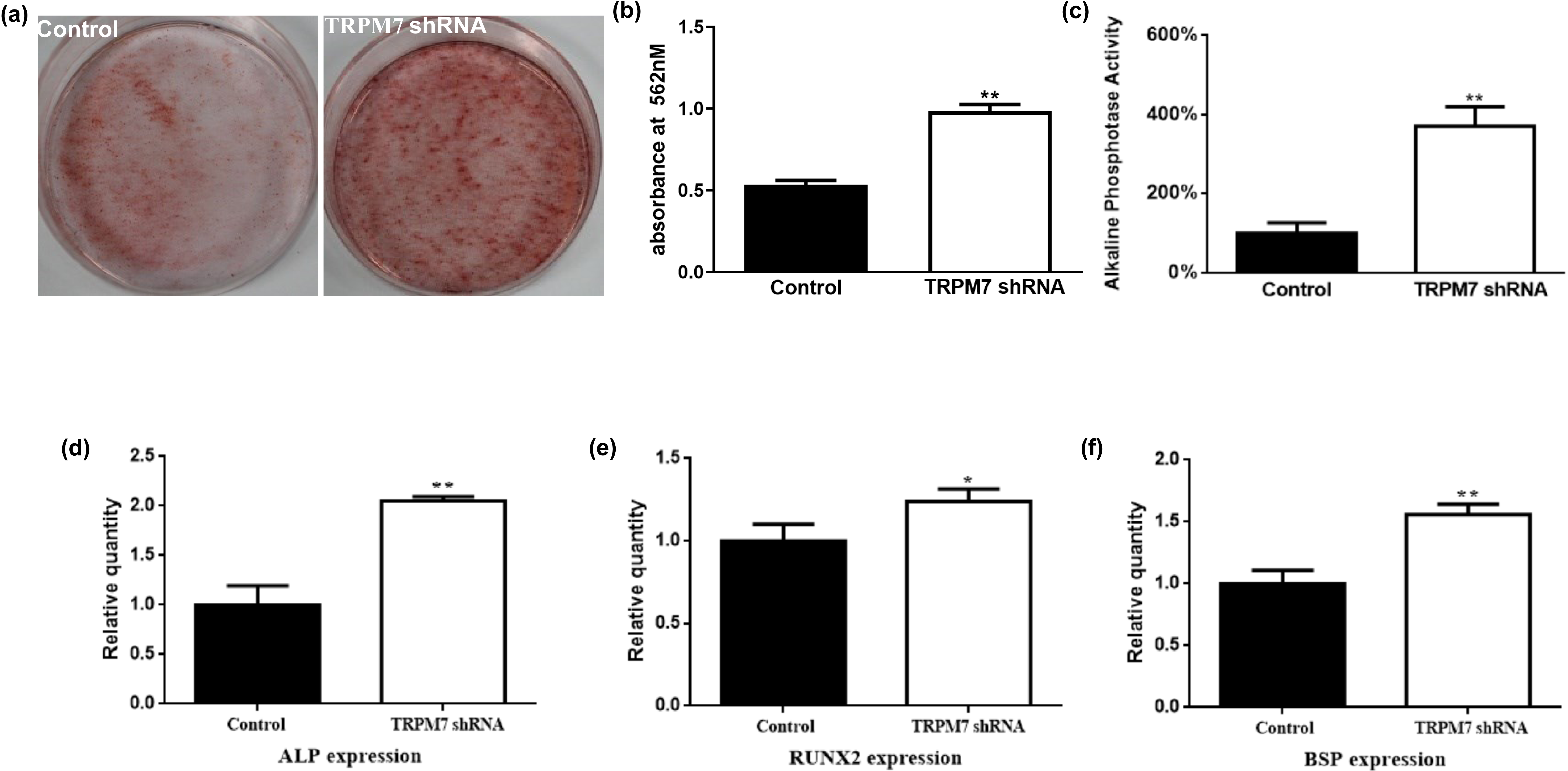
Fig. 3.

TRPM7 knockdown promoted the osteogenic differentiation of DFCs. (a,b) Alizarin red staining showed that TRPM7 knockdown enhanced mineralization. (c) TRPM7 knockdown increased the ALP activities of DFCs. Quantitative data from five independent experiments are shown. **, p < 0.01. (d–f) TRPM7 knockdown upregulated the expression of ALP, RUNX2 and BSP genes. Quantitative data from three independent experiments are shown. **, p < 0.01; *, p < 0.05.

3.4 Low Extracellular Mg2+ Promotes Osteogenic Differentiation of DFCs

We next examined whether Mg
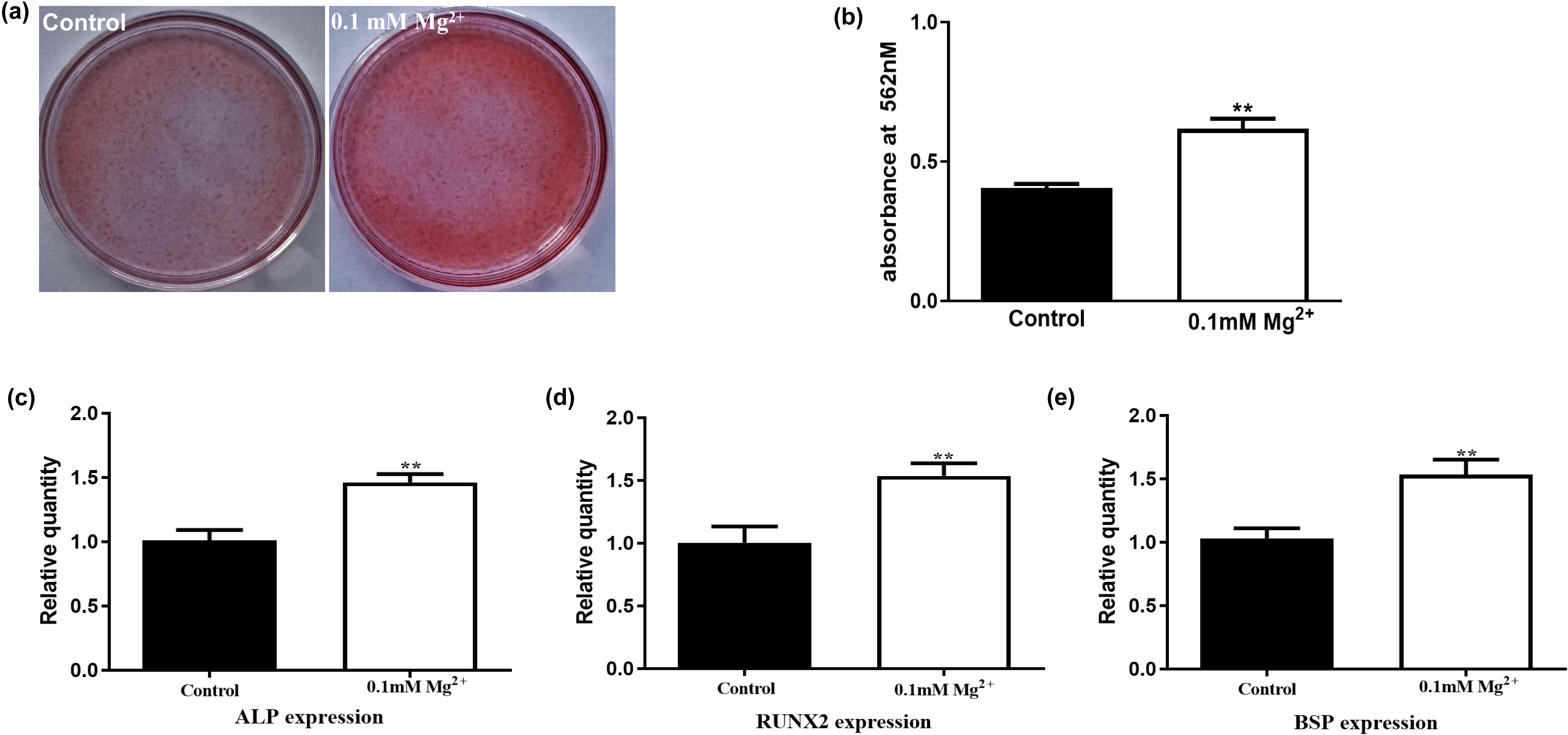
2+ deficiency had an effect on the osteogenic differentiation of DFCs. DFCs cultured for 14 days in osteogenic differentiation media containing 0.1 mM Mg2+showed increased formation of mineral deposits, as detected by Alizarin Red S staining (Fig. 4a,b). The expression of ALP, RUNX2 and BSP was also higher in Mg2+-deficient cells compared to control cells (Fig. 4c–e).

Fig. 4.

Mg2+ deficiency promotes osteogenic differentiation of DFCs. (a,b) Alizarin red staining showed that 0.1 mM Mg2+ enhanced mineralization. (c–e) 0.1 mM Mg2+ upregulated the expression of ALP, RUNX2 and BSP genes. Quantitative data from five independent experiments are shown. **, p < 0.01.

4. Discussion

TRPM7 expression was detected in the ameloblast, odontoblast and osteoblast cells of dental tissue [19, 20]. Pharmacological block of TRPM7 inhibits mechanically-evoked Ca2+ responses in odontoblasts, suggesting that TRPM7 might mediate mechanical sensitivity in odontoblasts [21]. Furthermore, TRPM7 kinase-inactive, knock-in mutant mice exhibited small enamel volume and hypo-mineralized craniofacial structures, including the incisors, molars, and cranial bones [19, 20]. Although these results demonstrate that TRPM7 has vital roles in dental development, the physiological functions of this protein in DFCs remain largely unknown.

To investigate this, we first confirmed the presence of TRPM7 transcript and protein in DFCs. TRPM6, a closely related channel with similar function, was not detected. This agrees with a previous report showing that TRPM7 was more widely distributed than TRPM6 [22]. In addition, we performed electrophysiological recordings to determine whether TRPM7 was functionally active in DFCs. In the absence of intracellular Mg2+, we recorded typical TRPM7-like currents characterized by a large outward current at positive potentials and a small inward current at negative potentials, consistent with the expression of TRPM7 [23]. We also used two concentrations of 2-APB to identify the native TRPM7-like currents present in DFCs. The TRPM7-like currents were inhibited by 100 μM 2-APB, but activated by 2 mM 2-APB. These results concur with previous studies showing that low concentrations of 2-APB block TRPM7 channels, whereas high concentrations activate TRPM7 currents [18, 24]. Based on these results, we confirm that active TRPM7 is expressed in DFCs.

To study the function of TRPM7 in DFCs, we generated stable knockdown cells with a lentiviral vector expressing specific TRPM7 shRNA. The knockdown efficiency of TRPM7 was confirmed by qRT-PCR, western blotting, immunofluorescence and electrophysiological recordings. Our results showed that knockdown of TRPM7 significantly inhibited the proliferation and migration of DFCs. These results were expected, since TRPM7 is thought to be the main pathway for Mg2+ entry into MSCs [22]. Mg2+ is critical for cell proliferation due to its involvement in DNA and protein synthesis, as well as for the proper functioning of numerous enzymes required for cell proliferation [25]. Recently, suppression of TRPM7 was shown to inhibit the proliferation and migration of MSCs derived from bone marrow and dental pulp tissues [10, 26]. Based on these previous studies, we speculate that TRPM7-mediated Mg2+ entry plays an important role in the proliferation and migration of DFCs.

An important observation in the present study was that TRPM7 had an inhibitory effect on the osteogenic differentiation of DFCs. We observed that ALP activities, osteogenic-related gene expression, and the number of mineralized matrix nodules all increased in TRPM7 knockdown cells after osteogenic induction compared to control cells. Castiglioni et al. [27] previously reported that silencing of TRPM7 accelerated the osteogenic differentiation of human MSCs. Interestingly, similar results were obtained when the cells were cultured in Mg2+-deficient conditions. In the present study, we also observed that Mg2+ deficiency promoted the osteogenic differentiation of DFCs, thus highlighting the contribution of Mg2+. Based on previous studies and on the present results, we hypothesize that TRPM7 and Mg2+ act together to coordinate the human marrow mesenchymal stem cells(hMSC) response to osteogenic stimuli by preventing excessive osteogenesis. However, TRPM7 contains an atypical kinase domain at its C-terminus. We cannot rule out the possibility that silencing of TRPM7 might affect the phosphorylation of its specific substrates. For example, one of the substrates phosphorylated by TRPM7, annexin-1, has been implicated in osteogenic differentiation [28]. Furthermore, the ameloblast, odontoblast and osteoblast cells isolated from TRPM7 kinase-inactive, knock-in mutant mice show decreased osteogenic capacity [19, 20]. Therefore, specific TRPM7 channel inhibitors or TRPM7 kinase inhibitors are needed to clarify the role of each TRPM7 domain in the osteogenic differentiation of DFCs.

5. Conclusions

In conclusion, this study has demonstrated for the first time that TRPM7 channels are functionally expressed in DFCs. Functional studies showed that suppression of TRPM7 inhibited the proliferation and migration of DFCs, while promoting their osteogenic differentiation. These results suggest that TRPM7 has an important role in regulating the biological behavior of DFCs.

Availability of Data and Materials

The datasets used and/or analyzed during the current study are available from the corresponding author on reasonable request.

Author Contributions

JZ, JialiL, YH, JiantaoL, SY, LX performed the experiments. JZ and DZ participated in the design of the experiments, analysis of data, and writing of the paper. All authors contributed to editorial changes in the manuscript. All authors read and approved the final manuscript. All authors have participated sufficiently in the work and agreed to be accountable for all aspects of the work.

Ethics Approval and Consent to Participate

The ethics committee of Southwest Medical University approved all human cell culture experiments (20221107004). Informed consent was obtained from donors who provided teeth for subsequent research.

Acknowledgment

We thank Dr. Weidong Tian, Dr. Weihua Guo and Dr. Li Xie (West China Hospital of Stomatology, Sichuan University, Sichuan, China) for technical assistance.

Funding

This work was supported by Sichuan Science and Technology Program (2023NSFSC1522 and 2022YFS0634); Luzhou-Southwest Medical University Joint Project (2020LZXNYDJ05, 2019LZXNYDJ01).

Conflict of Interest

The authors declare no conflict of interest.

References
[1]
Zhou T, Pan J, Wu P, Huang R, Du W, Zhou Y, et al. Dental Follicle Cells: Roles in Development and Beyond. Stem Cells International. 2019; 2019: 9159605.
[2]
Wise GE, Lin F, Fan W. Culture and characterization of dental follicle cells from rat molars. Cell and Tissue Research. 1992; 267: 483–492.
[3]
Morsczeck C, Götz W, Schierholz J, Zeilhofer F, Kühn U, Möhl C, et al. Isolation of precursor cells (PCs) from human dental follicle of wisdom teeth. Matrix Biology. 2005; 24: 155–165.
[4]
Morsczeck C, Moehl C, Götz W, Heredia A, Schäffer TE, Eckstein N, et al. In vitro differentiation of human dental follicle cells with dexamethasone and insulin. Cell Biology International. 2005; 29: 567–575.
[5]
Morsczeck C, Reichert TE. Dental stem cells in tooth regeneration and repair in the future. Expert Opinion on Biological Therapy. 2018; 18: 187–196.
[6]
Morsczeck C. Mechanisms during Osteogenic Differentiation in Human Dental Follicle Cells. International Journal of Molecular Sciences. 2022; 23: 5945.
[7]
Runnels LW, Yue L, Clapham DE. TRP-PLIK, a bifunctional protein with kinase and ion channel activities. Science. 2001; 291: 1043–1047.
[8]
Nadler MJ, Hermosura MC, Inabe K, Perraud AL, Zhu Q, Stokes AJ, et al. LTRPC7 is a Mg.ATP-regulated divalent cation channel required for cell viability. Nature. 2001; 411: 590–595.
[9]
Schmitz C, Perraud A, Johnson CO, Inabe K, Smith MK, Penner R, et al. Regulation of vertebrate cellular Mg2+ homeostasis by TRPM7. Cell. 2003; 114: 191–200.
[10]
Xiao E, Yang HQ, Gan Y, Duan D, He L, Guo Y, et al. Brief reports: TRPM7 Senses mechanical stimulation inducing osteogenesis in human bone marrow mesenchymal stem cells. Stem Cells. 2015; 33: 615–621.
[11]
Ryazanova LV, Dorovkov MV, Ansari A, Ryazanov AG. Characterization of the protein kinase activity of TRPM7/ChaK1, a protein kinase fused to the transient receptor potential ion channel. The Journal of Biological Chemistry. 2004; 279: 3708–3716.
[12]
Clark K, Middelbeek J, Lasonder E, Dulyaninova NG, Morrice NA, Ryazanov AG, et al. TRPM7 regulates myosin IIA filament stability and protein localization by heavy chain phosphorylation. Journal of Molecular Biology. 2008; 378: 790–803.
[13]
Perraud A, Zhao X, Ryazanov AG, Schmitz C. The channel-kinase TRPM7 regulates phosphorylation of the translational factor eEF2 via eEF2-k. Cellular Signalling. 2011; 23: 586–593.
[14]
Romagnani A, Vettore V, Rezzonico-Jost T, Hampe S, Rottoli E, Nadolni W, et al. TRPM7 kinase activity is essential for T cell colonization and alloreactivity in the gut. Nature Communications. 2017; 8: 1917.
[15]
Deason-Towne F, Perraud A, Schmitz C. Identification of Ser/Thr phosphorylation sites in the C2-domain of phospholipase C γ2 (PLCγ2) using TRPM7-kinase. Cellular Signalling. 2012; 24: 2070–2075.
[16]
Nelson P, Ngoc Tran TD, Zhang H, Zolochevska O, Figueiredo M, Feng J, et al. Transient receptor potential melastatin 4 channel controls calcium signals and dental follicle stem cell differentiation. Stem Cells. 2013; 31: 167–177.
[17]
Takezawa R, Schmitz C, Demeuse P, Scharenberg AM, Penner R, Fleig A. Receptor-mediated regulation of the TRPM7 channel through its endogenous protein kinase domain. Proceedings of the National Academy of Sciences of the United States of America. 2004; 101: 6009–6014.
[18]
Carvacho I, Ardestani G, Lee HC, McGarvey K, Fissore RA, Lykke-Hartmann K. TRPM7-like channels are functionally expressed in oocytes and modulate post-fertilization embryo development in mouse. Scientific Reports. 2016; 6: 34236.
[19]
Nakano Y, Le MH, Abduweli D, Ho SP, Ryazanova LV, Hu Z, et al. A Critical Role of TRPM7 As an Ion Channel Protein in Mediating the Mineralization of the Craniofacial Hard Tissues. Frontiers in Physiology. 2016; 7: e00258.
[20]
Ogata K, Tsumuraya T, Oka K, Shin M, Okamoto F, Kajiya H, et al. The crucial role of the TRPM7 kinase domain in the early stage of amelogenesis. Scientific Reports. 2017; 7: 18099.
[21]
Won J, Vang H, Kim JH, Lee PR, Kang Y, Oh SB. TRPM7 Mediates Mechanosensitivity in Adult Rat Odontoblasts. Journal of Dental Research. 2018; 97: 1039–1046.
[22]
Cheng H, Feng J, Figueiredo ML, Zhang H, Nelson PL, Marigo V, et al. Transient receptor potential melastatin type 7 channel is critical for the survival of bone marrow derived mesenchymal stem cells. Stem Cells and Development. 2010; 19: 1393–1403.
[23]
Bates-Withers C, Sah R, Clapham DE. TRPM7, the Mg(2+) inhibited channel and kinase. Advances in Experimental Medicine and Biology. 2011; 704: 173–183.
[24]
Sah R, Mesirca P, Mason X, Gibson W, Bates-Withers C, Van den Boogert M, et al. Timing of myocardial trpm7 deletion during cardiogenesis variably disrupts adult ventricular function, conduction, and repolarization. Circulation. 2013; 128: 101–114.
[25]
de Baaij JHF, Hoenderop JGJ, Bindels RJM. Magnesium in man: implications for health and disease. Physiological Reviews. 2015; 95: 1–46.
[26]
Cui L, Xu SM, Ma DD, Wu BL. The effect of TRPM7 suppression on the proliferation, migration and osteogenic differentiation of human dental pulp stem cells. International Endodontic Journal. 2014; 47: 583–593.
[27]
Castiglioni S, Romeo V, Locatelli L, Zocchi M, Zecchini S, Maier JAM. The simultaneous downregulation of TRPM7 and MagT1 in human mesenchymal stem cells in vitro: Effects on growth and osteogenic differentiation. Biochemical and Biophysical Research Communications. 2019; 513: 159–165.
[28]
Pan X, Peng L, Yin G. Downregulation of Annexin A1 by short hairpin RNA inhibits the osteogenic differentiation of rat bone marrow-derived mesenchymal stem cells. International Journal of Molecular Medicine. 2015; 36: 406–414.

Publisher’s Note: IMR Press stays neutral with regard to jurisdictional claims in published maps and institutional affiliations.

Share
Back to top