IMR Press / FBL / Volume 28 / Issue 3 / DOI: 10.31083/j.fbl2803062
Open Access Review
Reducing Oxygen Demand to Alleviate Acute Kidney Injury
Show Less
1 Department of Medicine, Uniformed Services University of the Health Sciences, Bethesda, MD 20814, USA
*Correspondence: xiaoming.zhou@usuhs.edu (Xiaoming Zhou)
Front. Biosci. (Landmark Ed) 2023, 28(3), 62; https://doi.org/10.31083/j.fbl2803062
Submitted: 13 November 2022 | Revised: 8 March 2023 | Accepted: 15 March 2023 | Published: 28 March 2023
(This article belongs to the Special Issue Mitochondrial Biology in Health and Disease)
Copyright: © 2023 The Author(s). Published by IMR Press.
This is an open access article under the CC BY 4.0 license.
Abstract

Maintaining a balance between the supply and demand of oxygen is vital for proper organ function. Most types of acute kidney injury (AKI) are characterized by hypoxia, a state where the supply of oxygen cannot match the demand for normal cellular activities. Hypoxia results from hypo perfusion and impaired microcirculation in the kidney. It inhibits mitochondrial oxidative phosphorylation, resulting in a decrease in production of adenosine triphosphate (ATP), which is essential to power tubular transport activities, especially reabsorption of Na+, and other vital cellular activities. To ameliorate AKI, the majority of studies have focused on increasing renal oxygen delivery by restoring renal blood flow and altering intra-renal hemodynamics. However, to date these approaches remain inadequate. In addition to augmenting oxygen supply, increasing renal blood flow also increases glomerular filtration rate, leading to increased solute deliver and workload for the renal tubules, causing an increase in oxygen consumption. The relationship between Na+ reabsorption and oxygen expenditure in the kidney is linear. Experimental models have demonstrated that inhibition of Na+ reabsorption can alleviate AKI. Since the proximal tubules reabsorb approximately 65% of filtered Na+, consuming the largest portion of oxygen, many studies focus on examining the effects of inhibiting Na+ reabsorption in this segment. Potential therapeutics that have been examined include acetazolamide, dopamine and its analog, inhibitors of the renin-angiotensin II system, atrial natriuretic peptide, and empagliflozin. The effectiveness of inhibition of Na+ reabsorption in the thick ascending limb of the Loop of Henle by furosemide has been also examined. While these approaches produced impressive results in animal models, their clinical benefits remain mixed. This review summarizes the progress in this area and argues that the combination of increasing oxygen supply with decreasing oxygen consumption or different approaches to reducing oxygen demand will be more efficacious.

Keywords
mitochondria
oxygenation
hypoxia
Na+-H+ exchanger 3
Na+-dependent glucose transporter 2
Na+ reabsorption
1. Introduction

Acute kidney injury (AKI) is defined by a sudden decrease in glomerular filtration rate (GFR), occurring within hours to weeks, along with the retention of nitrogenous waste products and renal parenchymal damage. Examples of AKI include acute tubular necrosis, acute interstitial nephritis, and glomerulonephritis. Based on the Kidney Disease: Improving Global Outcomes criteria, AKI is diagnosed by an absolute increase in serum creatinine levels of at least 0.3 mg/dL (26.5 μmol/L) within 48 hours, or a 50% increase in serum creatinine from baseline within 7 days, or a urine volume of less than 0.5 mL/kg/h for at least 6 hours [1]. Acute kidney injury occurs in a variety of settings, including major surgeries, transplantation, hemorrhage, burns, sepsis, lower limb ischemia/reperfusion, and the administration of nephrotoxic medications [2, 3, 4, 5, 6]. Conversely, chronic kidney disease (CKD) is the gradual loss of nephrons in both number and function. Chronic kidney disease is diagnosed by a persistent abnormality in kidney structure or function such as GFR <60 mL/min/1.73 m2 or albuminuria 30 mg per 24 hours for more than 3 months. Arteriolosclerosis, glomerulosclerosis and tubulointerstitial fibrosis are the common pathologic themes of CKD. Important risk factors for developing CKD include hypertension, diabetes, polycystic kidney disease, and sickle cell disease [7, 8, 9]. Chronic kidney disease can eventually result in end-stage kidney disease. Traditionally, AKI and CKD were considered as separate entities. However, newer paradigms recognize continuity between these two diseases, with AKI resulting in CKD and CKD being a recognized risk factor for AKI [7, 9].

Acute kidney injury is associated with poor outcomes. Acute kidney injury is independently associated with both short and long-term morbidity and mortality [6, 10, 11, 12, 13]. For example, a recent study of 1286 COVID-19 patients in Belgium demonstrated that all stages of AKI were associated with increased ICU mortality rates with 9.3% at stage 1, 40.1% at stage 2 and 47.0% at stage 3 compared to 3.6% with no AKI [14]. Furthermore, the incidence of AKI is steadily rising due to an aging population, increased prevalence of CKD, and improved recognition by physicians [15]. In addition to the high human cost, AKI imposes a heavy financial burden on society. In the United States, the in-hospital costs for AKI ranged from $5.4 to $24.0 billion annually [16]. In Queensland, Australia, a study found that the mean total hospital cost in patients with AKI was more than triple that of patients without AKI ($93,042 vs $30,778) [17].

In order to achieve clearance, the human kidney filters approximately 180 liters of blood each day. This filtrate is composed of both harmful metabolites and essential elements for life, such as NaCl, amino acids, and glucose. Approximately 99% of water and ions, and 100% of amino acids and glucose are reabsorbed by the renal tubules through the transcellular and paracellular pathways. The Na+,K+-ATPase is fundamental for the absorptions. In the transcellular pathway, it keeps the intracellular Na+ electrochemical potential lower than the extracellular one, which allows Na+ and Na+-coupled reabsorption [18, 19, 20, 21]. In the paracellular pathway, the Na+,K+-ATPase-dependent ion transport generates the transepithelial voltage favoring certain ion absorption [21]. In aerobic states ATP is generated by mitochondria predominantly through oxidative phosphorylation. To meet the demand of mitochondria for oxygen, the kidney has the largest blood supply (approximately 25% of cardiac output) only next to the heart under a resting state [22]. There is an almost perfect linear relationship between Na+reabsorption and oxygen consumption by the kidney [23]. In addition to supporting aerobic metabolism, oxygen is also needed to generate reactive oxygen species which, at a low level, signal many fundamental cellular activities [24].

Despite diverse etiologies, the majority of AKI sub types share common pathophysiologic mechanisms, including microvascular dysfunction and inflammation [3]. Microvascular dysfunction leads to hypoxia. Hypoxia is a state where oxygen supply cannot meet oxygen demand, which has a profound impact on kidney function (Fig. 1). Artificially induced hypoxia by increasing oxygen consumption with triiodothyronine without other compounding factors induces kidney injury in rats [25]. Similarly, treating rats with normobaric hyperoxia per se helps renal recovery from warm ischemia-induced injury [26]. Hypoxia compromises mitochondrial aerobic metabolism by diminishing nicotinamide adenine dinucleotide hydrogen (NADH) supply and electron transport [27]. Hypoxia leads to the accumulation of non-esterified fatty acids, which contributes to activation of pro-inflammatory pathways and produces reactive oxygen species, resulting in apoptosis and necrosis [28, 29]. Furthermore, inflammation directly inhibits mitochondrial respiration. As a result, the ability of mitochondria to generate ATP is compromised and the cellular ATP levels are reduced in AKI [30]. Therefore, AKI has been broadly characterized as a state of tubular ATP depletion [31].
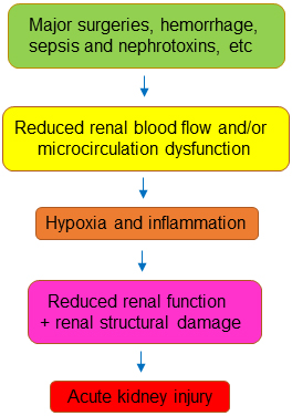
Fig. 1.

A common mechanism for acute kidney injury.

To make matters worse, the efficiency of the kidney in using oxygen to reabsorb Na+ is reduced in AKI. Redfors et al. [32] measured renal blood flow, oxygen extraction, Na+ filtration and excretion in 12 patients with AKI and 37 patients without AKI in a cardiothoracic intensive care unit. They found that renal blood flow was reduced by 40% and renal Na+ reabsorption was reduced by 59% in subjects with AKI. However, renal oxygen extraction was increased by 68% in patients with AKI compared to those without AKI. This resulted in an approximately 2.4-fold increase in the amount of oxygen required to absorb the same amount of Na+ [32]. Similar results were also found for sepsis-induced AKI in humans, rats, and mice [30, 33, 34]. A potential reason for this decrease in efficiency is the loss of epithelial polarization and tight junction integrity, which cause back leak and make Na+ transport less efficient [32, 34, 35, 36]. This has been observed in the mouse kidney after ischemia/reperfusion- and endotoxemia-induced injury [37, 38]. Inhibition of nitric oxide synthase more than doubles renal oxygen extraction/Na+ reabsorption in dogs and rats [39, 40]. It is also possible that AKI damages endothelial cells and inhibits endothelial nitric oxide synthase, leading to inefficient use of oxygen for Na+ reabsorption [32, 34, 35, 36]. Regardless of the cause, if more ATP is consumed for reabsorption of Na+, less ATP will be left for maintenance of cell integrity, resulting in cellular injury.

To restore the balance between oxygen supply and demand, many studies have examined increasing oxygen supply. Examples include fluid resuscitation, vasoconstrictors and removing the underlying cause of AKI (e.g., antibiotics and nephrotoxic medications). These approaches have been reviewed previously [41, 42, 43]. While these approaches unarguably improve patient outcomes, they are clearly insufficient. In most tissues, increasing blood flow will increase tissue oxygenation, however, the kidney is an exception to this rule. The variation in plasma Na+ concentrations is minimal in both healthy and disease states. Therefore, the delivery of Na+ into tubules is directly correlated to GFR. An increase of renal blood flow increases GFR, resulting in increased Na+ delivery and subsequent Na+ reabsorption and oxygen consumption [35, 36]. For example, infusion of a vasodilator atrial natriuretic peptide (ANP) to postoperative patients with normal renal function increases GFR by 15%, Na+ reabsorption by 9%, and oxygen consumption by 25% [44]. Although direct evidence is lacking, since approaches that increase renal blood flow inherently raise oxygen demand, it is unlikely that these treatments alone will be able to restore the supply-demand balance [35, 36].

Reducing oxygen demand is another way to maintain the supply-demand balance of oxygen when supply is low. Western painted turtles (Chrysemys picta) can survive under water with little oxygen for as long as 30 hours. They tolerate anoxia in part by reducing Na+ channels and metabolism in the hepatocytes and brain (channel arrest), therefore, reducing oxygen consumption [45, 46]. A similar defensive mechanism may occur in AKI. In one study, mitochondrial oxygen consumption in the proximal tubules is reduced 4 hours after septic AKI despite increased mitochondrial content and biogenesis [30]. This reduction in oxygen consumption is most likely due to reduced demand for ATP synthesis [30]. The Na+-H+ exchanger 3 (NHE3) reabsorbs approximate 65% of Na+ in the proximal tubules [47]. The NHE3-dependent pathway is one of the large oxygen consumers in the renal cortex, if not the largest. Acute kidney injury induced by ischemia/reperfusion, injection of lipopolysaccharides or mercury chloride decreases mRNA and protein levels of NHE3 in murine models [48, 49, 50, 51]. In human AKI, there is a loss of the brush border in the proximal tubules where NHE3 is localized [52]. This down-regulation of NHE3 has been interpreted as a defense mechanism against AKI [53]. The hypoxia-inducible factor (HIF) pathway, a key mediator of cellular adaptation in low oxygen tension states, may be another defense mechanism. Epithelial Na+ channels (ENaC) in the distal nephron participates in Na+ reabsorption. HIF-1α activation by hypoxia reduces expression of the γ-subunit of ENaC in mice and activity of ENaC in cultured principal cells [54]. Despite marked reduction in renal reabsorption and function, structural injury such as necrosis appears to be relatively mild in AKI [30, 55, 56, 57]. These observations have led to the hypothesis that early organ dysfunction is a defense mechanism that preserves energy to reduce cell injury and death [53].

How can reducing tubular cell injury and death help preserve and/or delay deterioration of GFR? The renal tubular cells are the primary site of injury in AKI. When the tubular cells undergo necrosis or apoptosis, they detach from the supporting basement membrane and obstruct the tubular lumen, causing back leakage of fluid with resultant decreased clearance. Even a sub-lethal injury can disrupt tight junctions, causing a loss of epithelial integrity and back leakage of fluid. The fluid back leakage further diminishes already impaired glomerular filtration induced by hypoperfusion of the kidney, eventually contributing to the filtration failure [58, 59, 60, 61]. Therefore, reducing oxygen demand through inhibition of Na+ transport in the renal epithelial cells may prevent injury and maintain clearance.

The present review summarizes progress in balancing supply and demand to prevent or treat AKI in patients (Table 1, Ref. [62, 63, 64, 65, 66, 67, 68, 69, 70, 71, 72, 73, 74, 75, 76, 77, 78, 79, 80, 81, 82, 83, 84, 85, 86, 87]) and animal models. Acute kidney injury is also associated with oxidative stress, which in turn increases oxygen consumption resulting in kidney tissue hypoxia [88, 89]. While various antioxidants have been shown to improve renal oxygenation and ameliorate AKI, reviewing the effect of antioxidants is beyond the scope of this article [90, 91, 92, 93].

Table 1.List of the clinical results in treatments of AKI.
Medications Mechanisms Inducers of AKI Timing therapy Patient # Results References
Prevention Treatment Control Medications
A. Proximal tubules
1. Inhibition of NHE3
Acetazolamide Reducing production of H+ Contrast X 46 received HCO3- 50 received acetazolamide Scr decreased [62]
Contrast X 96 received HCO3- 96 received acetazolamide Scr decreased [64]
eGFR increased
Cisplatin X 20 received mannitol 15 received acetazolamide AKI risk decreased from 30% to 8.9% [63]
Coronary artery bypass surgery X 65 received placebo 65 received acetazolamide ineffective [65]
ICU setting X 868 AKI 26.8% vs vancomycin 16% and gentamicin 20% [66]
Dopamine Inhibiting NHE3 translation and surface expression and promoting NHE3 degradation Operation for cardiac diseases X 40 received furosemide, bumetanide or ethacrynic acid 60 received furosemide, mannitol and dopamine Dialysis decreased from 90% to 6.7% [82]
Cardiac surgery X 40 received saline 42 received dopamine ineffective [84]
Fenoldopam Dopamine receptor agonist Post operation X 87 received furosemide 39 received furosemide and fenoldopam Tend to increase urine output (p = 0.06) [68]
Fenoldopam Dopamine receptor agonist X Total patient # [67]
Post operation 958 Reduced onset and incidence of AKI
917 ineffective
Contrast-induced 501 Reduced onset and incidence of AKI
714 ineffective
ICU setting 417 Reduced onset and incidence of AKI
155 ineffective
Human atrial natriuretic peptide (hANP) Possibly modulating dopamine system ICU setting X 30 received placebo 29 received hANP Improved creatinine clearance [83]
hANP ischemic or nephrotoxic insults X 114 received placebo 108 received hANP Increased morbidity and mortality [85]
ARBs and ACEIs Inhibiting the stimulatory effect of angiotensin II on NHE3 Major operation X 18,871 received no ARBs or ACEIs 268 received ARBs and 1137 received ACEIs Reduced incidence of AKI [72]
ARBs and ACEIs ICU setting X 3179 received no ARBs or ACEIs 3179 received ARBs or ACEIs Reduced all-cause mortality [73]
ARBs and ACEIs Multiple inducers X 40,000 received no ARBs or ACEIs 30,801 received ARBs or ACEIs Reduced all-cause mortality and recurrent AKI [74]
ARBs and ACEIs COVID-19 X X 279 received no ARBs or ACEIs 164 received ARBs or ACEIs Reduced all-cause mortality [69]
ARBs and ACEIs COVID-19 X 220 received no ARBs or ACEIs 80 received ARBs or ACEIs Ineffective [70]
ARBs and ACEIs COVID-19 X 100 received no ARBs or ACEIs 30 received ARBs or ACEIs Increased mortality and risk of AKI [71]
Spironolactone Inhibiting aldosterone-dependent Na+ reabsorption Cardiac surgery X 118 received placebo 115 received spironolactone Ineffective [75]
Clonidine Inhibiting sympathetic nerve-mediated activation of NHE3 Non-cardiac surgery X 3452 received placebo 3453 received clonidine Ineffective [76]
Dexmedetomidine Inhibiting sympathetic nerve-mediated activation of NHE3 Aortic surgery X 54 received placebo 54 received dexmedetomidine Reduced incidence of AKI [78]
Dexmedetomidine Laparoscopic prostatic surgery X 44 received placebo 45 received dexmedetomidine Ineffective [77]
Dexmedetomidine Sepsis X 719 received placebo 719 received dexmedetomidine Improved renal recovery rate and decreased in-hospital mortality [79]
2. Sglt2 inhibitors
Empagliflozin Inhibiting Sglt2 Decompensated heart failure with and without diabetes X 9 received placebo 10 received empagliflozin Reduced tubular injury [80]
Empagliflozin Decompensated heart failure with and without diabetes X 30 received standard medical care 10 received standard medical care plus empagliflozin Increased urine output [86]
Dapagliflozin Inhibiting Sglt2 COVID-19 X 606 received placebo 613 received dapagliflozin Ineffective [81]
B. Thick ascending limb of the Loop of Henle
Furosemide Inhibiting NKCC2 Cardiac surgery X 283 received placebo 283 received furosemide Ineffective [87]

Scr, serum creatinine concentrations; NHE3, Na+-H+ exchanger 3; ARBs, angiotensin receptor blockers; ACEIs, angiotensin converting enzyme inhibitors; AKI, acute kidney injury; Sglt2, Na+-dependent glucose transporter 2; NKCC2, Na+- K+-2Cl− cotransporter.

2. Approaches to Reduce Oxygen Demand in the Proximal Tubules
2.1 NHE3 Inhibition
2.1.1 Chemical Inhibitors

Inhibition of NHE3 with the NHE3 inhibitor #4167 protects against acute rejection of renal grafts in a rat model, an effect associated with preservation of the renal ATP levels [94]. Inhibitors of NHE3, 5-(N-ethyl-N-isopropyl) amiloride and S3226 protect against ischemia/reperfusion-induced AKI in rodents [95, 96]. Hypoxia induces intracellular acidosis. Activation of Na+-H+ exchanger 3 in the proximal tubules is critical to restore physiologic pH by extruding H+, but also increases intracellular Na+ levels, which lead to increased intracellular Ca2+ concentrations through the reverse mode of the Na+/Ca2+ exchanger. This increase in intracellular Ca2+ above the normal physiological levels (Ca2+ overload) contributes to AKI [97, 98]. Inhibition of NHE3 by S3226 induces acidosis [95]. Whether inhibition of NHE3 reduces the intracellular Ca2+ concentrations in the proximal tubules remains unknown, however multiple studies have demonstrated that inhibition of NHE1 reduces Na+-dependent Ca2+ overload in the heart [99]. Therefore, prevention of Ca2+ overload could be another mechanism by which NHE3 inhibitors in alleviate AKI.

2.1.2 Acetazolamide

Acetazolamide is an inhibitor of carbonic anhydrase. It is used clinically to treat glaucoma, epilepsy, high altitude sickness, and congestive heart failure [100]. It is also a diuretic, because inhibition of carbonic anhydrase indirectly leads to inhibition of NHE3 due to reduced production of H+ [101]. In animal models under healthy conditions, acetazolamide reduces cellular demand for oxygen by inhibiting Na+ reabsorption and increases oxygen delivery by stimulating blood flow (likely as a result of CO2 retention) [102, 103, 104, 105]. Acetazolamide has been shown to attenuate lower limb ischemia/reperfusion-induced AKI in mice [106]. However, the results for acetazolamide on renal artery-clamp-induced AKI are inconsistent. An et al. [107] pre-treated mice with 60 mg/kg/day in drinking water for 48 hours before they clamped the renal artery unilaterally for 30 minutes then released clamps for 48 hours. They found that acetazolamide reduced AKI by restoring renal blood flow, an effect that was associated with increased endothelial nitric oxide synthase (eNOS) activity, nitric oxide (NO) production, hypoxia inducible factor 1 subunit alpha (HIF-1α) expression, and decreased vascular permeability [107]. Conversely, Nensen et al. [108] intravenously injected 50 mg/kg of acetazolamide into rats 45 minutes prior to clamping the renal pedicle for 45 minutes unilaterally and then allowed reperfusion for 2 hours. Rats were kept under general anesthesia throughout the experiment. Under this condition, they found that acetazolamide impaired renal oxygenation, increased oxygen consumption, and decreased GFR compared to vehicle-treated rats [108]. In small human trials, there is some evidence to suggest that acetazolamide ameliorates contrast-, cisplatin-, cardiopulmonary bypass- and rhabdomyolysis-induced AKI (Table 1) [62, 63, 64, 109, 110]. However, other clinical studies have demonstrated no beneficial effect or even induction of AKI with acetazolamide (Table 1) [65, 66, 111]. Acetazolamide-induced AKI was attributed to intra-tubular obstruction by acetazolamide-induced crystalluria in some of these populations [111].

2.1.3 Dopamine

Dopamine inhibits NHE3 activity by inhibiting NHE3 translation [112], promoting ubiquitin-dependent NHE3 degradation [113], decreasing NHE3 exocytosis and cell membrane recycling [114, 115]. Dopamine has been shown to increase renal oxygenation in post-cardiac surgery patients. This effect was associated with profound pre- and post-glomerular vasodilation [116]. Fenoldopam is a dopamine receptor agonist, which has been used clinically to treat hypertension in part because of its inhibition of NHE3 [117]. Fenoldopam was effective in reducing the onset of postoperative AKI in adult patients, when used prophylactically (Table 1) [67, 68, 118]. However, other studies have failed to demonstrate a beneficial effect for fenoldopam (Table 1). Furthermore, its potential benefit to prevent AKI after cardiac surgery in pediatrics remains uncertain [119].

2.1.4 Natriuretic Peptides

Atrial natriuretic peptide is a peptide hormone that is produced by the walls of the heart in response to an increase in blood volume and pressure. It reduces blood volume and pressure in part through inhibition of NHE3 in the proximal tubules [120, 121]. Its analogs brain-type and c-type natriuretic peptides have similar functions [122]. How ANP inhibits NHE3 remains incompletely understood, but evidence indicates that it may be mediated in part by modulating the renal dopaminergic system [123]. It appears that while infusion of ANP raises oxygen supply by increasing renal blood flow, it also increases GFR and the subsequent burden for tubular Na reabsorption [44]. However, one small study in high risk surgery patients demonstrated a potential protective effect for ANP on kidney function (Table 1) [122]. Atrial natriuretic peptide is degraded by the endopeptidase neprilysin. Strategies that inhibit neprilysin offer an alternative means by which to raise the ANP levels. The challenge of this approach is that neprilysin also degrades endothelin, bradykinin, vasopressin and angiotensin II, which are critical for many physiologic activities. To circumvent this potential complication, Novartis Pharmaceuticals manufactures a drug named LCZ696, a mixture of the neprilysin inhibitor sacubitril and the angiotensin II receptor 1 blocker valsartan and markets it as Entresto®. Entresto protected the kidney from ischemic AKI in a porcine model of partial nephrectomy [124]. Multiple clinical trials have shown that Entresto does not increase the risk of AKI in patients with heart failure [122, 125]. However, whether Entresto has protective and/or therapeutic benefits against AKI in patients remains unknown.

2.1.5 Angiotensin II inhibitors

It has been well described that angiotensin II stimulates NHE3, which contributes to angiotensin II-induced hypertension [126, 127, 128]. Angiotensin II increases the levels of NHE3 mRNA and protein [129], stimulates cellular trafficking and exocytotic membrane insertion [130, 131], counteracts inhibitory phosphorylation by cAMP/PKA [132], and increases transport activity [133]. Not surprisingly, angiotensin II increases oxygen consumption by renal mitochondria [134]. While angiotensin receptor blockers (ARBs) have repeatedly shown benefits in animal models of AKI [135, 136, 137, 138] and CKD in humans [139, 140, 141], the results from clinical trials of AKI with ARBs and angiotensin converting enzyme inhibitors (ACEIs) have had mixed results (Table 1). One potential adverse impact of ACEIs and ARBs in the setting of AKI is that they could worsen renal hypoperfusion. In the cases where ACEIs and ARBs have shown potential benefit, namely hypertensive COVID-19 and sepsis, it remains unclear whether the effect was mainly due to control of blood pressure or reducing oxygen consumption in the proximal tubules [69, 142, 143]. Conversely, other studies have shown either no difference or increased risk of AKI and mortality in the hospitalized COVID-19 patients treated with ACEIs or ARBs [70, 71, 144, 145].

The results from trials examining patients undergoing major surgeries are also mixed. One study showed that ARBs and ACEIs were associated with a reduced risk of AKI after major surgery [72]. However, Zhou et al. [146] reported no significant association between perioperative use of renin-angiotensin system inhibitors and postoperative AKI in patients undergoing cardiac surgery. Other studies generally agree that ACEIs or ARBs increase survival, but also increase acute kidney disease, which is defined as persistent AKI from 7–90 days [73, 74, 147].

2.1.6 Aldosterone Inhibitors

Aldosterone binds to the mineralocorticoid receptors to increase sodium reabsorption and potassium secretion in the distal nephron. The expression of mineralocorticoid receptors has been detected in other tissues as well, where its activation may be pathophysiologic. Angiotensin II stimulates the biosynthesis and release of aldosterone from the adrenal cortex zona glomerulosa [148, 149]. Aldosterone stimulates NHE3 and Na+,K+-ATPase activities in cultured human renal proximal tubule cells [150, 151]. Whether aldosterone has a similar effect on NHE3 and Na+,K+-ATPase in the native proximal tubules remains unknown, but aldosterone appears to inhibit NHE3 in the microperfused medullary thick ascending limb [152]. Inhibition of angiotensin II (as reviewed above) should inhibit the effect of aldosterone. Blockade of aldosterone from binding its mineralocorticoid receptors with receptor antagonists, for example spironolactone, have been shown to prophylactically and therapeutically attenuate AKI in rats and pigs [138, 153, 154, 155]. However, the benefits of spironolactone seen in these animal models have not been reproduced clinically. A clinical trial with 115 patients on spironolactone and 118 patients on placebo demonstrated that spironolactone was not protective against AKI after cardiac surgery and there may be even a trend towards harm [75]. Indeed, a survey of the MEDLINE and EMBASE databases found that spironolactone had the highest odd ratio of inducing AKI among the reported medicine-induced AKI cases [156].

2.1.7 Angiotensin 1-7

Angiotensin 1-7 is generated mainly by angiotensin-converting enzyme 2 and exerts its actions via activation of its receptor Mas. Its functions frequently oppose angiotensin II [157]. Angiotensin 1-7 has been shown to inhibit NHE3 activity in the proximal tubules of normotensive and hypertensive rats [158, 159]. Angiotensin 1-7 also inhibits NHE3 activity by modulating the renal dopamine system [160]. Angiotensin 1-7 decreases oxygen consumption in the thick ascending limb of the loop of Henle and presumably in the proximal tubules as well [161]. Angiotensin 1-7 alleviates AKI in a variety of animal models [162, 163, 164, 165]. However, whether it has a similar effect in humans remains unknown.

2.1.8 Adenosine Receptors

Adenosine is present at low concentrations in the extracellular space, but its levels are greatly increased in conditions of metabolic stress, such as hypoxia as a result of enzymatic cleavage of the nucleotide adenosine 5-monophosphate (AMP) by 5-nucleotidase. Four types of adenosine receptors have been identified along the nephron. They are A1, A2A, A2B and A3. Although the exact molecular mechanisms are unclear, activation of A1 and A3 inhibits NHE3, whereas activation of A2A inhibits the action of A1 [166, 167]. Inhibition of A1 receptors by its knockout or its selective antagonist 1,3-dipropyl-8-cyclopentylxanthine worsens sepsis- and ischemia/reperfusion-induced AKI [168, 169]. On the contrary, activation of A1 receptor by its selective agonist 2-chlorocyclopentyladenosine, inhibits ischemia/reperfusion-induced AKI in mice [168].

2.1.9 Sympatholytics

The kidney is densely innervated by sympathetic nerves. Activation of these nerves increases renal vascular resistance and reduces renal blood flow by releasing norepinephrine and renin [170, 171]. Moreover, activation of these nerves increases NHE3 and Na+,K+-ATPase activity [172, 173, 174]. Acute kidney injury is frequently accompanied by activation of the sympathetic nerves. Inhibition of the sympathetic nervous system has been repeatedly demonstrated to alleviate AKI in various animal models [170, 171]. However, the clinical benefit of this approach is inconsistent. The sympatholytic effect of clonidine and dexmedetomidine is mediated by stimulating the pre-synaptic α2 adrenoceptors, thereby decreasing norepinephrine release from both central and peripheral sympathetic nerve terminals. A multi-center clinical trial of 6905 non-cardiac surgery patients with 3453 patients on clonidine and 3452 patients on placebo found that perioperative clonidine administered did not reduce the risk of AKI [76]. Similarly, dexmedetomidine has not shown a benefit for preventing AKI in patients undergoing laparoscopic prostatic surgery (Table 1) [77]. On the other hand, a single center trial with 54 patients on dexmedetomidine and 54 patients on placebo showed a beneficial effect for dexmedetomidine in patients undergoing aortic surgery [78]. A recent updated systematic review and meta-analysis of 16 studies involving 2148 patients revealed that dexmedetomidine administration may prevent AKI and postoperative delirium after cardiac surgery. This meta-analysis also suggests that dexmedetomidine may reduce the length of stay in the intensive care unit [175]. Likewise, dexmedetomidine administration was associated with improvements in renal function recovery and in-hospital survival in critically ill patients with septic AKI [79]. Whether the way of administration and types of AKI causes the different clinical outcomes of the sympatholytics on AKI remains unclear.

It is important to emphasize that the renoprotective effects of fenoldopam, ANP, activation of adenosine receptor A1, inhibition of angiotensin II system and sympatholytics are not entirely mediated by inhibition of NHE3. The well-known hemodynamic effects of these approaches also contribute significantly to their reno-protection [67, 118, 122, 157, 176, 177].

2.2 Na+-Dependent Glucose Transporter 2 (Sglt2) Inhibitors

Na+-dependent glucose transporter 2 has a high capacity and is responsible for reabsorption of almost all filtered glucose. By reducing glucose re-absorption from the proximal tubules, Sglt2 inhibitors are a class of anti-diabetic medications, which include empagliflozin, canagliflozin and dapagliflozin [178]. Hyperglycemia increases the burden of reabsorption of glucose and energy expenditure in the proximal tubules and induces hypoxia in the diabetic rat cortex. Inhibition of Sglt2 normalizes oxygen tension in the renal cortex of the diabetic rats [179, 180]. However, a lack of effect of dapagliflozin on renal microvascular oxygen tension in diabetic rats was recently reported [181]. Multiple systemic reviews of clinical studies and trials have revealed that diabetic patients who took Sglt2 inhibitors had lower odds of AKI compared to those who did not [178, 182, 183, 184, 185, 186]. However, this beneficial effect of Sglt2 inhibitors on AKI risk has not been universally observed [178, 187, 188]. The potential nephro-protective mechanisms of Sglt2 inhibitors include lowering blood glucose and body weight, inhibiting inflammation, improving cardiovascular function, increasing tubuloglomerular feedback, and increasing tubular oxygenation [178, 182, 183, 184, 185, 189]. The effects of Sglt2 inhibitors on AKI in non-diabetic patients have not been widely reported. Empagliflozin significantly reduced AKI parameters in patients with acute decompensated heart failure [80]. Dapagliflozin had no significant effect on the risk of AKI in the hospitalized COVID-19 patients (Table 1) [81]. In these two studies, both diabetic and non-diabetic patients were included. In contrast, the beneficial effects of Sglt2 inhibitors on AKI in non-diabetic animals has been repeatedly demonstrated. Na+-dependent glucose transporter 2 inhibitors protect the kidney against sepsis-, ischemia/reperfusion-, contrast-, and myocardial infarction-induced AKI in rodents [179, 190, 191, 192, 193]. However, knockout of Sglt2 offers no protection against renal artery clamping-induced AKI, implying that the renoprotective effects of Sglt2 inhibitors may not be mediated through Sglt2 in rodents [194]. Na+-dependent glucose transporter 2 is co-localized with NHE3 [195]. Studies have shown that empagliflozin inhibits NHE3 in the kidney and NHE1 in the heart [196, 197]. The cardioprotective effect of empagliflozin against ischemia is thought to be due to its inhibition of NHE1 [197]. Therefore, it is possible that the reno-protective effect of Sglt2 inhibitors against AKI may also involve inhibition of NHE3.

3. Approaches to Reduce Oxygen Demand in the Thick Ascending Limb of the Loop of Henle

Na+-K+-2Cl- cotransporter (NKCC2) is located in the apical membrane of the epithelial cells in the thick ascending limb of the loop of Henle. It facilitates approximately 20–25% of the reuptake of filtered NaCl. Therefore, oxygen consumption by this segment of nephron is second only to the proximal tubule. Furosemide, a diuretic that inhibits NKCC2, was shown to improve renal oxygenation in patients [44, 198] and reduces ischemia/reperfusion-induced AKI in animal models [199, 200]. However, furosemide has had disappointing results in clinical studies (Table 1). A meta- analysis of 2084 patients in 9 studies found that furosemide combined with intravenous fluids had no significant impact on the incidence of contrast-induced AKI in patients after coronary intervention, but could reduce major adverse cardiovascular events and mortality [201]. Combination of infusing furosemide with dopamine or ANP in patients with AKI after cardiac surgery decreased the need for dialysis and improved dialysis-free survival rates in two studies [82, 83]. However, a systemic review and meta-analysis of randomized clinical trials found that the quantity and quality of evidence for using furosemide to treat AKI in adult post-operative patients were very low with no firm evidence for benefit or harm [202]. Abraham et al. [203] also found that furosemide infusion in early-onset AKI did not reduce the progression to a higher stage of AKI in critically ill children.

4. Na+,K+-ATPase

Na+,K+-ATPase is localized to the basolateral membrane of the entire renal tubules, from the proximal tubules to the inner medullary collecting ducts. The basolaterally located Na+,K+-ATPase is the only pathway to pump out intracellular Na+ that enters the renal epithelial cells from the apical NHE3, Sglt2, NKCC2 and other Na+-dependent transporters. Inhibition of the apical Na+ entry will reduce the availability of Na+ to Na+,K+-ATPase, thus reducing the activity of the enzyme and oxygen expenditure, whereas stimulation of apical Na+ entry does the opposite. Dopamine inhibits Na+,K+-ATPase activity [204, 205]. Dopamine stimulates cAMP-dependent inhibitory phosphorylation α-subunit of the enzyme [206]. The renal dopaminergic system and ANP acted synergistically to produce a potent inhibition of Na+,K+-ATPase [123]. Dopamine and hypoxia induces endocytosis of Na+,K+-ATPase through protein kinase C-dependent phosphorylation of the α-subunit [207]. Angiotensin II stimulates Na+,K+-ATPase activity [208], whereas angiotensin 1-7 does the opposite [209]. However, it remains unknown whether these hormones directly regulate Na+,K+-ATPase or indirectly regulate the pump by affecting Na+ entry from the apical membrane [210].

4.1 Improving the Na+,K+-ATPase Efficiency

Chen et al. [211] recently reported the development of the third generation of a synchronization modulation electric field device. This device consists of three phases: synchronization, modulation and maintenance. In the synchronization phase, the device uses electric field to synchronize the Na+,K+-ATPase molecules by using ATPs to actively transport Na+ and K+. At the same time, the device applies electric energy to the Na+,K+-ATPase molecules so that the pumps could synthesize one ATP at the end of each pumping cycle. Thus, ATP consumption is markedly reduced. In the modulation phase, the Na+,K+-ATPase transporting rates are gradually modulated (increased or decreased) to the desired values, and the transporting rates are sustained at the target value throughout the maintenance phase. Application of this technique to a renal ischemia-reperfusion injury mouse model preserved Na+,K+-ATPase activity, thereby reducing kidney injury, as reflected by 40% lower plasma creatinine in the treated group as compared to the untreated control group. In a mouse kidney transplantation model, renal graft function was improved by more than 50% with the application of this technique based on GFR measurement compared with the untreated group.

4.2 Endothelin Inhibition

Endothelin-1 is an endogenous 21 amino acid peptide that has powerful vasoactive properties. In the kidney, endothelin-1 is produced by endothelial, epithelial and mesangial cells. Endothelin-1 acts through binding to its type A and type B endothelin receptors to modulate renal blood flow, GFR, reabsorption of sodium and water, and acid-base balance [212, 213, 214]. Ischemia/reperfusion of the kidney increases endothelin concentration in the renal cortex and decreases renal blood flow [215, 216]. Endothelin receptor antagonists such as ambrisentan and bosentan attenuated ischemia/reperfusion-induced experimental AKI [217, 218]. However, the reno-protective effect is due to an increase in oxygen supply from increased renal blood flow rather than a reduction of oxygen consumption [216, 217, 218, 219], because endothelin-1 actually inhibits Na+,K+-ATPase [220, 221] and NKCC2 [222]. The clinical benefits of endothelin receptor antagonists on AKI have not been reported.

5. Perspectives

The pathophysiology of AKI is complex, involving hypoperfusion, impaired microcirculation, hypoxia, oxidative stress, abnormal coagulation, and inflammation. There is no single approach that can address all aspects of this complicated pathophysiology. Further, addressing one aspect may exacerbate other aspects, even within the concept of reducing oxygen demand. For example, although furosemide reduces oxygen consumption by inhibiting NKCC2 in the thick ascending limb of the Loop of Henle, it also activates the renin-angiotensin-aldosterone system. Combining different treatments that increase oxygen supply and decrease oxygen consumption or different approaches to reduce oxygen consumption may be a better approach. A good example is the synergistic effect observed with using fenoldopam in combination with furosemide in treatment of AKI in critically ill surgical patients [68].

Abbreviations

ACEIs, angiotensin converting enzyme inhibitors; AKI, acute kidney injury; ANP, atrial natriuretic peptide; ARBs, angiotensin receptor blockers; CKD, chronic kidney disease; NHE3, Na+-H+ exchanger 3; NKCC2, Na+-K+-2Cl- cotransporter; Scr, serum creatinine concentration; Sglt2, Na+-dependent glucose transporter 2.

Disclaimer

The content and views expressed in this article are the sole responsibility of the author and do not necessarily reflect the views or policies of the Uniformed Services University or Department of Defense of USA. Mention of trade names, commercial products, or organizations does not imply endorsement by the Uniformed Services University or Department of Defense of USA.

Author Contributions

XZ conceived ideas, searched for references, drafted, edited and approved the manuscript.

Ethics Approval and Consent to Participate

Not applicable.

Acknowledgment

The author would like to thank Lieutenant Colonel Ian J. Stewart for his help with editing this manuscript.

Funding

This work was supported in part by the United States of America Department of Defense grants W81XWH-22-2-0068 and MED-83-12715.

Conflict of Interest

The author declares no conflict of interest.

References
[1]
Makris K, Spanou L. Acute Kidney Injury: Definition, Pathophysiology and Clinical Phenotypes. The Clinical Biochemist. Reviews. 2016; 37: 85–98.
[2]
Chopra TA, Brooks CH, Okusa MD. Acute Kidney Injury Prevention. Contributions to Nephrology. 2016; 187: 9–23.
[3]
Packialakshmi B, Stewart IJ, Burmeister DM, Chung KK, Zhou X. Large animal models for translational research in acute kidney injury. Renal Failure. 2020; 42: 1042–1058.
[4]
Packialakshmi B, Stewart IJ, Burmeister DM, Feng Y, McDaniel DP, Chung KK, et al. Tourniquet-induced lower limb ischemia/reperfusion reduces mitochondrial function by decreasing mitochondrial biogenesis in acute kidney injury in mice. Physiological Reports. 2022; 10: e15181.
[5]
Burmeister DM, Gómez BI, Dubick MA. Molecular mechanisms of trauma-induced acute kidney injury: Inflammatory and metabolic insights from animal models. Biochimica et Biophysica Acta. Molecular Basis of Disease. 2017; 1863: 2661–2671.
[6]
Harrois A, Soyer B, Gauss T, Hamada S, Raux M, Duranteau J, et al. Prevalence and risk factors for acute kidney injury among trauma patients: a multicenter cohort study. Critical Care. 2018; 22: 344.
[7]
Chawla LS, Eggers PW, Star RA, Kimmel PL. Acute kidney injury and chronic kidney disease as interconnected syndromes. The New England Journal of Medicine. 2014; 371: 58–66.
[8]
Wang H, Morris RG, Knepper MA, Zhou X. Sickle cell disease up-regulates vasopressin, aquaporin 2, urea transporter A1, Na-K-Cl cotransporter 2, and epithelial Na channels in the mouse kidney medulla despite compromising urinary concentration ability. Physiological Reports. 2019; 7: e14066.
[9]
Ferenbach DA, Bonventre JV. Acute kidney injury and chronic kidney disease: From the laboratory to the clinic. Nephrologie & Therapeutique. 2016; 12: S41–S48.
[10]
Wohlauer MV, Sauaia A, Moore EE, Burlew CC, Banerjee A, Johnson J. Acute kidney injury and posttrauma multiple organ failure: the canary in the coal mine. The Journal of Trauma and Acute Care Surgery. 2012; 72: 373–380.
[11]
Stewart IJ, Sosnov JA, Howard JT, Chung KK. Acute Kidney Injury in Critically Injured Combat Veterans: A Retrospective Cohort Study. American Journal of Kidney Diseases. 2016; 68: 564–570.
[12]
Heegard KD, Stewart IJ, Cap AP, Sosnov JA, Kwan HK, Glass KR, et al. Early acute kidney injury in military casualties. The Journal of Trauma and Acute Care Surgery. 2015; 78: 988–993.
[13]
Lafrance JP, Miller DR. Acute kidney injury associates with increased long-term mortality. Journal of the American Society of Nephrology. 2010; 21: 345–352.
[14]
Schaubroeck H, Vandenberghe W, Boer W, Boonen E, Dewulf B, Bourgeois C, et al. Acute kidney injury in critical COVID-19: a multicenter cohort analysis in seven large hospitals in Belgium. Critical Care. 2022; 26: 225.
[15]
Siew ED, Davenport A. The growth of acute kidney injury: a rising tide or just closer attention to detail? Kidney International. 2015; 87: 46–61.
[16]
Silver SA, Chertow GM. The Economic Consequences of Acute Kidney Injury. Nephron. 2017; 137: 297–301.
[17]
Zhang J, Healy HG, Baboolal K, Wang Z, Venuthurupalli SK, Tan KS, et al. Frequency and Consequences of Acute Kidney Injury in Patients with CKD: A Registry Study in Queensland Australia. Kidney Medicine. 2019; 1: 180–190.
[18]
Larsen EH, Sørensen JN. Ion and Water Absorption by the Kidney Proximal Tubule: Computational Analysis of Isosmotic Transport. Function. 2020; 1: zqaa014.
[19]
Mather A, Pollock C. Glucose handling by the kidney. Kidney International. Supplement. 2011; S1–6.
[20]
Bröer S. Amino acid transport across mammalian intestinal and renal epithelia. Physiological Reviews. 2008; 88: 249–286.
[21]
Féraille E, Doucet A. Sodium-potassium-adenosinetriphosphatase-dependent sodium transport in the kidney: hormonal control. Physiological Reviews. 2001; 81: 345–418.
[22]
Palm F, Nordquist L. Renal tubulointerstitial hypoxia: cause and consequence of kidney dysfunction. Clinical and Experimental Pharmacology & Physiology. 2011; 38: 474–480.
[23]
Blantz RC, Deng A, Miracle CM, Thomson SC. Regulation of kidney function and metabolism: a question of supply and demand. Transactions of the American Clinical and Climatological Association. 2007; 118: 23–43.
[24]
Trayhurn P. Oxygen-A Critical, but Overlooked, Nutrient. Frontiers in Nutrition. 2019; 6: 10.
[25]
Sivertsson E, Friederich-Persson M, Persson P, Nangaku M, Hansell P, Palm F. Thyroid hormone increases oxygen metabolism causing intrarenal tissue hypoxia; a pathway to kidney disease. PLoS ONE. 2022; 17: e0264524.
[26]
Nensén O, Hansell P, Palm F. Intrarenal oxygenation determines kidney function during the recovery from an ischemic insult. American Journal of Physiology. Renal Physiology. 2020; 319: F1067–F1072.
[27]
Wheaton WW, Chandel NS. Hypoxia. 2. Hypoxia regulates cellular metabolism. American Journal of Physiology. Cell Physiology. 2011; 300: C385–C393.
[28]
Emma F, Montini G, Parikh SM, Salviati L. Mitochondrial dysfunction in inherited renal disease and acute kidney injury. Nature Reviews. Nephrology. 2016; 12: 267–280.
[29]
Li Z, Lu S, Li X. The role of metabolic reprogramming in tubular epithelial cells during the progression of acute kidney injury. Cellular and Molecular Life Sciences. 2021; 78: 5731–5741.
[30]
Li Y, Nourbakhsh N, Pham H, Tham R, Zuckerman JE, Singh P. Evolution of altered tubular metabolism and mitochondrial function in sepsis-associated acute kidney injury. American Journal of Physiology. Renal Physiology. 2020; 319: F229–F244.
[31]
Thadhani R, Pascual M, Bonventre JV. Acute renal failure. The New England Journal of Medicine. 1996; 334: 1448–1460.
[32]
Redfors B, Bragadottir G, Sellgren J, Swärd K, Ricksten SE. Acute renal failure is NOT an “acute renal success”–a clinical study on the renal oxygen supply/demand relationship in acute kidney injury. Critical Care Medicine. 2010; 38: 1695–1701.
[33]
Heemskerk AE, Huisman E, van Lambalgen AA, van den Bos GC, Hennekes M, Thijs LG, et al. Renal function and oxygen consumption during bacteraemia and endotoxaemia in rats. Nephrology, Dialysis, Transplantation. 1997; 12: 1586–1594.
[34]
Skytte Larsson J, Krumbholz V, Enskog A, Bragadottir G, Redfors B, Ricksten SE. Renal Blood Flow, Glomerular Filtration Rate, and Renal Oxygenation in Early Clinical Septic Shock. Critical Care Medicine. 2018; 46: e560–e566.
[35]
Singh P, Ricksten SE, Bragadottir G, Redfors B, Nordquist L. Renal oxygenation and haemodynamics in acute kidney injury and chronic kidney disease. Clinical and Experimental Pharmacology & Physiology. 2013; 40: 138–147.
[36]
Ricksten SE, Bragadottir G, Lannemyr L, Redfors B, Skytte J. Renal Hemodynamics, Function, and Oxygenation in Critically Ill Patients and after Major Surgery. Kidney360. 2021; 2: 894–904.
[37]
Hurtado KA, Janda J, Schnellmann RG. Lasmiditan promotes recovery from acute kidney injury through induction of mitochondrial biogenesis. American Journal of Physiology. Renal Physiology. 2023; 324: F56–F63.
[38]
Nakano D, Kitada K, Wan N, Zhang Y, Wiig H, Wararat K, et al. Lipopolysaccharide induces filtrate leakage from renal tubular lumina into the interstitial space via a proximal tubular Toll-like receptor 4-dependent pathway and limits sensitivity to fluid therapy in mice. Kidney International. 2020; 97: 904–912.
[39]
Laycock SK, Vogel T, Forfia PR, Tuzman J, Xu X, Ochoa M, et al. Role of nitric oxide in the control of renal oxygen consumption and the regulation of chemical work in the kidney. Circulation Research. 1998; 82: 1263–1271.
[40]
Deng A, Miracle CM, Suarez JM, Lortie M, Satriano J, Thomson SC, et al. Oxygen consumption in the kidney: effects of nitric oxide synthase isoforms and angiotensin II. Kidney International. 2005; 68: 723–730.
[41]
McIlroy DR, Lopez MG, Billings FT, 4th. Perioperative Clinical Trials in AKI. Seminars in Nephrology. 2020; 40: 173–187.
[42]
Kellum JA, Romagnani P, Ashuntantang G, Ronco C, Zarbock A, Anders HJ. Acute kidney injury. Nature Reviews. Disease Primers. 2021; 7: 52.
[43]
Zarbock A, Nadim MK, Pickkers P, Gomez H, Bell S, Joannidis M, et al. Sepsis-associated acute kidney injury: consensus report of the 28th Acute Disease Quality Initiative workgroup. Nature Reviews. Nephrology. 2023. (online ahead of print)
[44]
Swärd K, Valsson F, Sellgren J, Ricksten SE. Differential effects of human atrial natriuretic peptide and furosemide on glomerular filtration rate and renal oxygen consumption in humans. Intensive Care Medicine. 2005; 31: 79–85.
[45]
Hochachka PW, Lutz PL. Mechanism, origin, and evolution of anoxia tolerance in animals. Comparative Biochemistry and Physiology. Part B, Biochemistry & Molecular Biology. 2001; 130: 435–459.
[46]
Jackson DC. Hibernating without oxygen: physiological adaptations of the painted turtle. The Journal of Physiology. 2002; 543: 731–737.
[47]
Zhuo JL, Li XC. Proximal nephron. Comprehensive Physiology. 2013; 3: 1079–1123.
[48]
Wang Z, Rabb H, Haq M, Shull GE, Soleimani M. A possible molecular basis of natriuresis during ischemic-reperfusion injury in the kidney. Journal of the American Society of Nephrology. 1998; 9: 605–613.
[49]
Kwon TH, Frøkiaer J, Han JS, Knepper MA, Nielsen S. Decreased abundance of major Na(+) transporters in kidneys of rats with ischemia-induced acute renal failure. American Journal of Physiology. Renal Physiology. 2000; 278: F925–F939.
[50]
Schmidt C, Höcherl K, Schweda F, Kurtz A, Bucher M. Regulation of renal sodium transporters during severe inflammation. Journal of the American Society of Nephrology. 2007; 18: 1072–1083.
[51]
Vieira JVDA, Marques VB, Vieira LV, Crajoinas RDO, Shimizu MHM, Seguro AC, et al. Changes in the renal function after acute mercuric chloride exposure in the rat are associated with renal vascular endothelial dysfunction and proximal tubule NHE3 inhibition. Toxicology Letters. 2021; 341: 23–32.
[52]
Solez K, Morel-Maroger L, Sraer JD. The morphology of ”acute tubular necrosis” in man: analysis of 57 renal biopsies and a comparison with the glycerol model. Medicine. 1979; 58: 362–376.
[53]
Girardi ACC, Di Sole F. Deciphering the mechanisms of the Na+/H+ exchanger-3 regulation in organ dysfunction. American Journal of Physiology. Cell Physiology. 2012; 302: C1569–87.
[54]
Dizin E, Olivier V, Roth I, Sassi A, Arnoux G, Ramakrishnan S, et al. Activation of the Hypoxia-Inducible Factor Pathway Inhibits Epithelial Sodium Channel-Mediated Sodium Transport in Collecting Duct Principal Cells. Journal of the American Society of Nephrology. 2021; 32: 3130–3145.
[55]
Rosen S, Heyman SN. Difficulties in understanding human “acute tubular necrosis”: limited data and flawed animal models. Kidney International. 2001; 60: 1220–1224.
[56]
Hotchkiss RS, Swanson PE, Freeman BD, Tinsley KW, Cobb JP, Matuschak GM, et al. Apoptotic cell death in patients with sepsis, shock, and multiple organ dysfunction. Critical Care Medicine. 1999; 27: 1230–1251.
[57]
Langenberg C, Bagshaw SM, May CN, Bellomo R. The histopathology of septic acute kidney injury: a systematic review. Critical Care. 2008; 12: R38.
[58]
Donohoe JF, Venkatachalam MA, Bernard DB, Levinsky NG. Tubular leakage and obstruction after renal ischemia: structural-functional correlations. Kidney International. 1978; 13: 208–222.
[59]
Parekh N, Esslinger HU, Steinhausen M. Glomerular filtration and tubular reabsorption during anuria in postischemic acute renal failure. Kidney International. 1984; 25: 33–41.
[60]
Neugarten J, Aynedjian HS, Bank N. Role of tubular obstruction in acute renal failure due to gentamicin. Kidney International. 1983; 24: 330–335.
[61]
Shin NS, Marlier A, Xu L, Lam T, Cantley LG, Guo JK. Characterization of temporospatial distribution of renal tubular casts by nephron tracking after ischemia-reperfusion injury. American Journal of Physiology. Renal Physiology. 2022; 322: F322–F334.
[62]
Assadi F. Acetazolamide for prevention of contrast-induced nephropathy: a new use for an old drug. Pediatric Cardiology. 2006; 27: 238–242.
[63]
El Hamamsy M, Kamal N, Bazan NS, El Haddad M. Evaluation of the effect of acetazolamide versus mannitol on cisplatin-induced nephrotoxicity, a pilot study. International Journal of Clinical Pharmacy. 2018; 40: 1539–1547.
[64]
Pakfetrat M, Nikoo MH, Malekmakan L, Tabandeh M, Roozbeh J, Nasab MH, et al. A comparison of sodium bicarbonate infusion versus normal saline infusion and its combination with oral acetazolamide for prevention of contrast-induced nephropathy: a randomized, double-blind trial. International Urology and Nephrology. 2009; 41: 629–634.
[65]
Afzal G, Ansari Aval Z, Beheshti Monfared M, Khesali H, Ziaie S, Barati S, et al. Evaluating the Effect of Acetazolamide on the Prevention of Post-operative Acute Kidney Injury after Coronary Artery Bypass Grafting Surgery: A Randomized, Open-labeled Clinical Trial. Iranian Journal of Pharmaceutical Research. 2021; 20: 71–79.
[66]
Moffett BS, Kulik K, Khichi M, Arikan A. Acetazolamide-Associated Acute Kidney Injury in Critically Ill Pediatric Patients. The Journal of Pediatric Pharmacology and Therapeutics. 2021; 26: 467–471.
[67]
Noce A, Marrone G, Rovella V, Busca A, Gola C, Ferrannini M, et al. Fenoldopam Mesylate: A Narrative Review of Its Use in Acute Kidney Injury. Current Pharmaceutical Biotechnology. 2019; 20: 366–375.
[68]
Cagliani JA, Marinelli L, Cho Y, Miyara SJ, Ruhemann A, Loyola A, et al. Fenoldopam Increases Urine Output in Oliguric Critically Ill Surgical Patients. Cureus. 2021; 13: e20445.
[69]
Lam KW, Chow KW, Vo J, Hou W, Li H, Richman PS, et al. Continued In-Hospital Angiotensin-Converting Enzyme Inhibitor and Angiotensin II Receptor Blocker Use in Hypertensive COVID-19 Patients Is Associated with Positive Clinical Outcome. The Journal of Infectious Diseases. 2020; 222: 1256–1264.
[70]
Chaudhri I, Koraishy FM, Bolotova O, Yoo J, Marcos LA, Taub E, et al. Outcomes Associated with the Use of Renin-Angiotensin-Aldosterone System Blockade in Hospitalized Patients with SARS-CoV-2 Infection. Kidney360. 2020; 1: 801–809.
[71]
Lim JH, Cho JH, Jeon Y, Kim JH, Lee GY, Jeon S, et al. Adverse impact of renin-angiotensin system blockade on the clinical course in hospitalized patients with severe COVID-19: a retrospective cohort study. Scientific Reports. 2020; 10: 20250.
[72]
Yang SY, Huang TM, Lai TS, Chou NK, Tsao CH, Huang YP, et al. Angiotensin II Receptor Blockers but Not Angiotensin-Converting Enzyme Inhibitors Are Associated with a Reduced Risk of Acute Kidney Injury After Major Surgery. Frontiers in Pharmacology. 2021; 12: 662301.
[73]
Zhu X, Xue J, Liu Z, Dai W, Xiang J, Xu H, et al. The effects of angiotensin-converting enzyme inhibitors and angiotensin II receptor blockers in critically ill patients with acute kidney injury: An observational study using the MIMIC database. Frontiers in Pharmacology. 2022; 13: 918385.
[74]
Chen JY, Tsai IJ, Pan HC, Liao HW, Neyra JA, Wu VC, et al. The Impact of Angiotensin-Converting Enzyme Inhibitors or Angiotensin II Receptor Blockers on Clinical Outcomes of Acute Kidney Disease Patients: A Systematic Review and Meta-Analysis. Frontiers in Pharmacology. 2021; 12: 665250.
[75]
Barba-Navarro R, Tapia-Silva M, Garza-Garcia C, López-Giacoman S, Melgoza-Toral I, Vázquez-Rangel A, et al. The Effect of Spironolactone on Acute Kidney Injury After Cardiac Surgery: A Randomized, Placebo-Controlled Trial. American Journal of Kidney Diseases. 2017; 69: 192–199.
[76]
Garg AX, Kurz A, Sessler DI, Cuerden M, Robinson A, Mrkobrada M, et al. Perioperative aspirin and clonidine and risk of acute kidney injury: a randomized clinical trial. The Journal of the American Medical Association. 2014; 312: 2254–2264.
[77]
Wu S, Yao H, Cheng N, Guo N, Chen J, Ge M, et al. Determining whether dexmedetomidine provides a reno-protective effect in patients receiving laparoscopic radical prostatectomy: a pilot study. International Urology and Nephrology. 2019; 51: 1553–1561.
[78]
Soh S, Shim JK, Song JW, Bae JC, Kwak YL. Effect of dexmedetomidine on acute kidney injury after aortic surgery: a single-centre, placebo-controlled, randomised controlled trial. British Journal of Anaesthesia. 2020. (online ahead of print)
[79]
Hu H, An S, Sha T, Wu F, Jin Y, Li L, et al. Association between dexmedetomidine administration and outcomes in critically ill patients with sepsis-associated acute kidney injury. Journal of Clinical Anesthesia. 2022; 83: 110960.
[80]
Thiele K, Rau M, Hartmann NUK, Möller M, Möllmann J, Jankowski J, et al. Empagliflozin reduces markers of acute kidney injury in patients with acute decompensated heart failure. ESC Heart Failure. 2022; 9: 2233–2238.
[81]
Heerspink HJL, Furtado RHM, Berwanger O, Koch GG, Martinez F, Mukhtar O, et al. Dapagliflozin and Kidney Outcomes in Hospitalized Patients with COVID-19 Infection: An Analysis of the DARE-19 Randomized Controlled Trial. Clinical Journal of the American Society of Nephrology. 2022; 17: 643–654.
[82]
Sirivella S, Gielchinsky I, Parsonnet V. Mannitol, furosemide, and dopamine infusion in postoperative renal failure complicating cardiac surgery. The Annals of Thoracic Surgery. 2000; 69: 501–506.
[83]
Swärd K, Valsson F, Odencrants P, Samuelsson O, Ricksten SE. Recombinant human atrial natriuretic peptide in ischemic acute renal failure: a randomized placebo-controlled trial. Critical Care Medicine. 2004; 32: 1310–1315.
[84]
Lassnigg A, Donner E, Grubhofer G, Presterl E, Druml W, Hiesmayr M. Lack of renoprotective effects of dopamine and furosemide during cardiac surgery. Journal of the American Society of Nephrology. 2000; 11: 97–104.
[85]
Lewis J, Salem MM, Chertow GM, Weisberg LS, McGrew F, Marbury TC, et al. Atrial natriuretic factor in oliguric acute renal failure. Anaritide Acute Renal Failure Study Group. American Journal of Kidney Diseases. 2000; 36: 767–774.
[86]
Schulze PC, Bogoviku J, Westphal J, Aftanski P, Haertel F, Grund S, et al. Effects of Early Empagliflozin Initiation on Diuresis and Kidney Function in Patients with Acute Decompensated Heart Failure (EMPAG-HF). Circulation. 2022; 146: 289–298.
[87]
Xie CM, Yao YT, Yang K, Shen MQ, He LX, Dai Z, et al. Furosemide does not reduce the incidence of postoperative acute kidney injury in adult patients undergoing cardiac surgery: A PRISMA-compliant systematic review and meta-analysis. Journal of Cardiac Surgery. 2022; 37: 4850–4860.
[88]
Palm F, Cederberg J, Hansell P, Liss P, Carlsson PO. Reactive oxygen species cause diabetes-induced decrease in renal oxygen tension. Diabetologia. 2003; 46: 1153–1160.
[89]
Honda T, Hirakawa Y, Nangaku M. The role of oxidative stress and hypoxia in renal disease. Kidney Research and Clinical Practice. 2019; 38: 414–426.
[90]
Lan TY, Dun RL, Yao DS, Wu F, Qian YL, Zhou Y, et al. Effects of resveratrol on renal ischemia-reperfusion injury: A systematic review and meta-analysis. Frontiers in Nutrition. 2023; 9: 1064507.
[91]
Ergin B, Guerci P, Zafrani L, Nocken F, Kandil A, Gurel-Gurevin E, et al. Effects of N-acetylcysteine (NAC) supplementation in resuscitation fluids on renal microcirculatory oxygenation, inflammation, and function in a rat model of endotoxemia. Intensive Care Medicine Experimental. 2016; 4: 29.
[92]
El-Ashmawy NE, Khedr EG, Doghish AS, Elballal MS. Carnosine and crocin ameliorate oxidative stress in rats with rhabdomyolysis-induced acute kidney injury through upregulating HO-1 gene expression. Food Bioscience. 2022; 49: 101972.
[93]
Salman TM, Omran GA, El-Naa MM, Doghish AS, Youns S, Awad MM. Protective effect of proanthocyanidins on nephrotoxicity induced by antitumor dose of cisplatin in Ehrlich solid tumor-bearing mice‏. Arab Journal of Laboratory Medicine. 2014; 40: 953–965.
[94]
Reuter S, Velic A, Edemir B, Schröter R, Pavenstädt H, Gabriëls G, et al. Protective role of NHE-3 inhibition in rat renal transplantation undergoing acute rejection. Pflugers Archiv: European Journal of Physiology. 2008; 456: 1075–1084.
[95]
Hropot M, Juretschke HP, Langer KH, Schwark JR. S3226, a novel NHE3 inhibitor, attenuates ischemia-induced acute renal failure in rats. Kidney International. 2001; 60: 2283–2289.
[96]
Yamashita J, Ohkita M, Takaoka M, Kaneshiro Y, Matsuo T, Kaneko K, et al. Role of Na+/H+ exchanger in the pathogenesis of ischemic acute renal failure in mice. Journal of Cardiovascular Pharmacology. 2007; 49: 154–160.
[97]
Iwamoto T, Kita S. Development and application of Na+/Ca2+ exchange inhibitors. Molecular and Cellular Biochemistry. 2004; 259: 157–161.
[98]
Yang D, Yang D, Jia R, Ding G. Selective inhibition of the reverse mode of Na(+)/Ca(2+) exchanger attenuates contrast-induced cell injury. American Journal of Nephrology. 2013; 37: 264–273.
[99]
Deschaine B, Verma S, Rayatzadeh H. Clinical Evidence and Proposed Mechanisms of Sodium-Glucose Cotransporter 2 Inhibitors in Heart Failure with Preserved Ejection Fraction: A Class Effect? Cardiac Failure Review. 2022; 8: e23.
[100]
Farzam K, Abdullah M. Acetazolamide. In StatPearls. StatPearls Publishing LLC., Treasure Island (FL). 2020.
[101]
Krishnan D, Liu L, Wiebe SA, Casey JR, Cordat E, Alexander RT. Carbonic anhydrase II binds to and increases the activity of the epithelial sodium-proton exchanger, NHE3. American Journal of Physiology. Renal Physiology. 2015; 309: F383–F392.
[102]
Brezis M, Agmon Y, Epstein FH. Determinants of intrarenal oxygenation. I. Effects of diuretics. The American Journal of Physiology. 1994; 267: F1059–F1062.
[103]
Taki K, Oogushi K, Hirahara K, Gai X, Nagashima F, Tozuka K. Preferential acetazolamide-induced vasodilation based on vessel size and organ: confirmation of peripheral vasodilation with use of colored microspheres. Angiology. 2001; 52: 483–488.
[104]
Taki K, Kato H, Endo S, Inada K, Totsuka K. Cascade of acetazolamide-induced vasodilatation. Research Communications in Molecular Pathology and Pharmacology. 1999; 103: 240–248.
[105]
Horita Y, Yakabe K, Tadokoro M, Suyama N, Hayashida K, Kawano Y, et al. Renal circulatory effects of acetazolamide in patients with essential hypertension. American Journal of Hypertension. 2006; 19: 282–285.
[106]
Packialakshmi B, Stewart IJ, Burmeister DM, Li XC, Zhuo JL, Soleimani M, et al. Inhibition of Na-H exchanger 3 ameliorates lower limb ischemia/reperfusion-induced acute kidney injury through preservation of mitochondrial biogenesis in mice. The FASEB Journal. 2022; 36: R2760.
[107]
An Y, Zhang JZ, Han J, Yang HP, Tie L, Yang XY, et al. Hypoxia-inducible factor-1α dependent pathways mediate the renoprotective role of acetazolamide against renal ischemia-reperfusion injury. Cellular Physiology and Biochemistry. 2013; 32: 1151–1166.
[108]
Nensén O, Hansell P, Palm F. Role of carbonic anhydrase in acute recovery following renal ischemia reperfusion injury. PLoS ONE. 2019; 14: e0220185.
[109]
Davidov T, Hong JJ, Malcynski JT. Novel use of acetazolamide in the treatment of rhabdomyolysis-induced myoglobinuric renal failure. The Journal of Trauma. 2006; 61: 213–215.
[110]
Foote HP, Hornik CP, Hill KD, Rotta AT, Chamberlain R, Thompson EJ. A systematic review of the evidence supporting post-operative diuretic use following cardiopulmonary bypass in children with Congenital Heart Disease. Cardiology in the Young. 2021; 31: 699–706.
[111]
Abou-Mrad RM, Ibrahim M, Osman N, Babu RS. Anuric Acute Kidney Injury Requiring Dialysis Following Acetazolamide Use for Cataract Surgery. The American Journal of Case Reports. 2021; 22: e931319.
[112]
Hu MC, Di Sole F, Zhang J, McLeroy P, Moe OW. Chronic regulation of the renal Na(+)/H(+) exchanger NHE3 by dopamine: translational and posttranslational mechanisms. American Journal of Physiology. Renal Physiology. 2013; 304: F1169–F1180.
[113]
Armando I, Villar VAM, Jones JE, Lee H, Wang X, Asico LD, et al. Dopamine D3 receptor inhibits the ubiquitin-specific peptidase 48 to promote NHE3 degradation. FASEB Journal. 2014; 28: 1422–1434.
[114]
Hu MC, Bobulescu IA, Quiñones H, Gisler SM, Moe OW. Dopamine reduces cell surface Na+/H+ exchanger-3 protein by decreasing NHE3 exocytosis and cell membrane recycling. American Journal of Physiology. Renal Physiology. 2017; 313: F1018–F1025.
[115]
Bacic D, Kaissling B, McLeroy P, Zou L, Baum M, Moe OW. Dopamine acutely decreases apical membrane Na/H exchanger NHE3 protein in mouse renal proximal tubule. Kidney International. 2003; 64: 2133–2141.
[116]
Redfors B, Bragadottir G, Sellgren J, Swärd K, Ricksten SE. Dopamine increases renal oxygenation: a clinical study in post-cardiac surgery patients. Acta Anaesthesiologica Scandinavica. 2010; 54: 183–190.
[117]
Alshami A, Romero C, Avila A, Varon J. Management of hypertensive crises in the elderly. Journal of Geriatric Cardiology: JGC. 2018; 15: 504–512.
[118]
Chen X, Huang T, Cao X, Xu G. Comparative Efficacy of Drugs for Preventing Acute Kidney Injury after Cardiac Surgery: A Network Meta-Analysis. American Journal of Cardiovascular Drugs: Drugs, Devices, and other Interventions. 2018; 18: 49–58.
[119]
Van den Eynde J, Cloet N, Van Lerberghe R, Sá MPBO, Vlasselaers D, Toelen J, et al. Strategies to Prevent Acute Kidney Injury after Pediatric Cardiac Surgery: A Network Meta-Analysis. Clinical Journal of the American Society of Nephrology. 2021; 16: 1480–1490.
[120]
Winaver J, Burnett JC, Tyce GM, Dousa TP. ANP inhibits Na(+)-H+ antiport in proximal tubular brush border membrane: role of dopamine. Kidney International. 1990; 38: 1133–1140.
[121]
Hammond TG, Yusufi AN, Knox FG, Dousa TP. Administration of atrial natriuretic factor inhibits sodium-coupled transport in proximal tubules. The Journal of Clinical Investigation. 1985; 75: 1983–1989.
[122]
Brignone J, Assersen KB, Jensen M, Jensen BL, Kloster B, Jønler M, et al. Protection of kidney function and tissue integrity by pharmacologic use of natriuretic peptides and neprilysin inhibitors. Pflugers Archiv: European Journal of Physiology. 2021; 473: 595–610.
[123]
Choi MR, Rukavina Mikusic NL, Kouyoumdzian NM, Kravetz MC, Fernández BE. Atrial natriuretic peptide and renal dopaminergic system: a positive friendly relationship? BioMed Research International. 2014; 2014: 710781.
[124]
Brignone J, Jensen M, Jensen BL, Assersen KB, Goetze JP, Jødal L, et al. Protective effect of sacubitril/valsartan (Entresto) on kidney function and filtration barrier injury in a porcine model of partial nephrectomy. Nephrology, Dialysis, Transplantation. 2023; 38: 80–92.
[125]
Bhatt AS, Vaduganathan M, Zhuo M, Fu EL, Solomon SD, Desai RJ. Risk of Acute Kidney Injury Among Older Adults with Heart Failure and With Reduced Ejection Fraction Treated With Angiotensin-Neprilysin Inhibitor vs Renin-Angiotensin System Inhibitor in Routine Clinical Care. Journal of Cardiac Failure. 2023; 29: 138–146.
[126]
Nwia SM, Li XC, Leite APDO, Hassan R, Zhuo JL. The Na+/H+ Exchanger 3 in the Intestines and the Proximal Tubule of the Kidney: Localization, Physiological Function, and Key Roles in Angiotensin II-Induced Hypertension. Frontiers in Physiology. 2022; 13: 861659.
[127]
Azzam O, Kiuchi MG, Ho JK, Matthews VB, Gavidia LML, Nolde JM, et al. New Molecules for Treating Resistant Hypertension: a Clinical Perspective. Current Hypertension Reports. 2019; 21: 80.
[128]
Leite APDO, Li XC, Nwia SM, Hassan R, Zhuo JL. Angiotensin II and AT1a Receptors in the Proximal Tubules of the Kidney: New Roles in Blood Pressure Control and Hypertension. International Journal of Molecular Sciences. 2022; 23: 2402.
[129]
Dixit MP, Xu L, Xu H, Bai L, Collins JF, Ghishan FK. Effect of angiotensin-II on renal Na+/H+ exchanger-NHE3 and NHE2. Biochimica et Biophysica Acta. 2004; 1664: 38–44.
[130]
du Cheyron D, Chalumeau C, Defontaine N, Klein C, Kellermann O, Paillard M, et al. Angiotensin II stimulates NHE3 activity by exocytic insertion of the transporter: role of PI 3-kinase. Kidney International. 2003; 64: 939–949.
[131]
Riquier-Brison ADM, Leong PKK, Pihakaski-Maunsbach K, McDonough AA. Angiotensin II stimulates trafficking of NHE3, NaPi2, and associated proteins into the proximal tubule microvilli. American Journal of Physiology. Renal Physiology. 2010; 298: F177–F186.
[132]
Crajoinas RO, Polidoro JZ, Carneiro de Morais CPA, Castelo-Branco RC, Girardi ACC. Angiotensin II counteracts the effects of cAMP/PKA on NHE3 activity and phosphorylation in proximal tubule cells. American Journal of Physiology. Cell Physiology. 2016; 311: C768–C776.
[133]
He P, Klein J, Yun CC. Activation of Na+/H+ exchanger NHE3 by angiotensin II is mediated by inositol 1,4,5-triphosphate (IP3) receptor-binding protein released with IP3 (IRBIT) and Ca2+/calmodulin-dependent protein kinase II. The Journal of Biological Chemistry. 2010; 285: 27869–27878.
[134]
Moriyama T, Kanmura Y, Lindahl SGE. Atrial natriuretic peptide attenuation of renal ischemia-reperfusion injury after major surgery. The Journal of Surgical Research. 2016; 201: 213–218.
[135]
Franzén S, Näslund E, Wang H, Frithiof R. Prevention of hemorrhage-induced renal vasoconstriction and hypoxia by angiotensin II type 1 receptor antagonism in pigs. American Journal of Physiology. Regulatory, Integrative and Comparative Physiology. 2021; 321: R12–R20.
[136]
Zhang X, Eirin A, Li ZL, Crane JA, Krier JD, Ebrahimi B, et al. Angiotensin receptor blockade has protective effects on the poststenotic porcine kidney. Kidney International. 2013; 84: 767–775.
[137]
Abbas W, Altemimi M, Qassam H, Hameed AA, Zigam Q, Abbas L, et al. Fimasartan ameliorates renal ischemia reperfusion injury via modulation of oxidative stress, inflammatory and apoptotic cascades in a rat model. Journal of Medicine and Life. 2022; 15: 241–251.
[138]
Al-Kadi A, El-Daly M, El-Tahawy NFG, Khalifa MMA, Ahmed ASF. Angiotensin aldosterone inhibitors improve survival and ameliorate kidney injury induced by sepsis through suppression of inflammation and apoptosis. Fundamental & Clinical Pharmacology. 2022; 36: 286–295.
[139]
Sato A, Nishimoto M. Clinical effect of nonsteroidal mineralocorticoid receptor (MR) antagonists in the treatment of diabetic kidney disease: expectations as a new therapeutic strategy. Hypertension Research. 2022; 45: 1310–1321.
[140]
Yuan Q, Tang B, Zhang C. Signaling pathways of chronic kidney diseases, implications for therapeutics. Signal Transduction and Targeted Therapy. 2022; 7: 182.
[141]
Vaz de Castro PAS, Bitencourt L, Pereira BWS, Lima AQR, Hermida HS, Moreira Neto CR, et al. Efficacy and safety of angiotensin-converting enzyme inhibitors or angiotensin receptor blockers for IgA nephropathy in children. Pediatric Nephrology. 2022; 37: 499–508.
[142]
Soleimani A, Kazemian S, Karbalai Saleh S, Aminorroaya A, Shajari Z, Hadadi A, et al. Effects of Angiotensin Receptor Blockers (ARBs) on In-Hospital Outcomes of Patients with Hypertension and Confirmed or Clinically Suspected COVID-19. American Journal of Hypertension. 2020; 33: 1102–1111.
[143]
Demiselle J, Seegers V, Lemerle M, Meziani F, Grelon F, Megarbane B, et al. Prior Exposure to Angiotensin II Receptor Blockers in Patients with Septic Shock to Individualize Mean Arterial Pressure Target? A Post Hoc Analysis of the Sepsis and Mean Arterial Pressure (SEPSISPAM) Trial. Critical Care Medicine. 2021; 49: e412–e422.
[144]
Lee SA, Park R, Yang JH, Min IK, Park JT, Han SH, et al. Increased risk of acute kidney injury in coronavirus disease patients with renin-angiotensin-aldosterone-system blockade use: a systematic review and meta-analysis. Scientific Reports. 2021; 11: 13588.
[145]
Gnanenthiran SR, Borghi C, Burger D, Caramelli B, Charchar F, Chirinos JA, et al. Renin-Angiotensin System Inhibitors in Patients with COVID-19: A Meta-Analysis of Randomized Controlled Trials Led by the International Society of Hypertension. Journal of the American Heart Association. 2022; 11: e026143.
[146]
Zhou H, Xie J, Zheng Z, Ooi OC, Luo H. Effect of Renin-Angiotensin System Inhibitors on Acute Kidney Injury Among Patients Undergoing Cardiac Surgery: A Review and Meta-Analysis. Seminars in Thoracic and Cardiovascular Surgery. 2021; 33: 1014–1022.
[147]
Wu VC, Lin YF, Teng NC, Yang SY, Chou NK, Tsao CH, et al. Angiotensin II Receptor Blocker Associated with Less Outcome Risk in Patients with Acute Kidney Disease. Frontiers in Pharmacology. 2022; 13: 714658.
[148]
Barrera-Chimal J, Girerd S, Jaisser F. Mineralocorticoid receptor antagonists and kidney diseases: pathophysiological basis. Kidney International. 2019; 96: 302–319.
[149]
Chandrashekar KB, Lopez-Ruiz A, Juncos LA. The Promise of Mineralocorticoid Antagonism in Acute Kidney Injury. Nephron. 2016; 134: 154–159.
[150]
Drumm K, Kress TR, Gassner B, Krug AW, Gekle M. Aldosterone stimulates activity and surface expression of NHE3 in human primary proximal tubule epithelial cells (RPTEC). Cellular Physiology and Biochemistry. 2006; 17: 21–28.
[151]
Salyer SA, Parks J, Barati MT, Lederer ED, Clark BJ, Klein JD, et al. Aldosterone regulates Na(+), K(+) ATPase activity in human renal proximal tubule cells through mineralocorticoid receptor. Biochimica et Biophysica Acta. 2013; 1833: 2143–2152.
[152]
Watts BA, 3rd, George T, Good DW. Aldosterone inhibits apical NHE3 and HCO3- absorption via a nongenomic ERK-dependent pathway in medullary thick ascending limb. American Journal of Physiology. Renal Physiology. 2006; 291: F1005–F1013.
[153]
Mejía-Vilet JM, Ramírez V, Cruz C, Uribe N, Gamba G, Bobadilla NA. Renal ischemia-reperfusion injury is prevented by the mineralocorticoid receptor blocker spironolactone. American Journal of Physiology. Renal Physiology. 2007; 293: F78–F86.
[154]
Sánchez-Pozos K, Barrera-Chimal J, Garzón-Muvdi J, Pérez-Villalva R, Rodríguez-Romo R, Cruz C, et al. Recovery from ischemic acute kidney injury by spironolactone administration. Nephrology, Dialysis, Transplantation. 2012; 27: 3160–3169.
[155]
Barrera-Chimal J, André-Grégoire G, Nguyen Dinh Cat A, Lechner SM, Cau J, Prince S, et al. Benefit of Mineralocorticoid Receptor Antagonism in AKI: Role of Vascular Smooth Muscle Rac1. Journal of the American Society of Nephrology. 2017; 28: 1216–1226.
[156]
Roughead EE, Kerr M, Moffat A, Kassie GM, Pratt N. Medicine-Induced Acute Kidney Injury Findings from Spontaneous Reporting Systems, Sequence Symmetry Analysis and a Case-Control Study with a Focus on Medicines Used in Primary Care. Drug Safety. 2022; 45: 1413–1421.
[157]
Vaz de Castro PAS, Jose PA, Simões E Silva AC. Interactions between the intrarenal dopaminergic and the renin-angiotensin systems in the control of systemic arterial pressure. Clinical Science. 2022; 136: 1205–1227.
[158]
Castelo-Branco RC, Leite-Delova DC, de Mello-Aires M. Dose-dependent effects of angiotensin-(1-7) on the NHE3 exchanger and [Ca(2+)](i) in in vivo proximal tubules. American Journal of Physiology-Renal Physiology. 2013; 304: F1258–F1265.
[159]
Castelo-Branco RC, Leite-Dellova DCA, Fernandes FB, Malnic G, de Mello-Aires M. The effects of angiotensin-(1-7) on the exchanger NHE3 and on [Ca(2+)](i) in the proximal tubules of spontaneously hypertensive rats. American Journal of Physiology-Renal Physiology. 2017; 313: F450–F460.
[160]
Banday AA, Diaz AD, Lokhandwala M. Kidney dopamine D1-like receptors and angiotensin 1-7 interaction inhibits renal Na+ transporters. American Journal of Physiology. Renal Physiology. 2019; 317: F949–F956.
[161]
Dibo P, Marañón RO, Chandrashekar K, Mazzuferi F, Silva GB, Juncos LA, et al. Angiotensin-(1-7) inhibits sodium transport via Mas receptor by increasing nitric oxide production in thick ascending limb. Physiological Reports. 2019; 7: e14015.
[162]
Abdel-Hakeem EA, Abdel Hafez SMN, Kamel BA, Abdel-Hamid HA. Angiotensin 1-7 mitigates rhabdomyolysis induced renal injury in rats via modulation of TLR-4/NF-kB/iNOS and Nrf-2/heme oxygenase-1 signaling pathways. Life Sciences. 2022; 303: 120678.
[163]
Zaman A, Banday AA. Angiotensin (1-7) protects against renal ischemia-reperfusion injury via regulating expression of NRF2 and microRNAs in Fisher 344 rats. American Journal of Physiology. Renal Physiology. 2022; 323: F33–F47.
[164]
Pacheco LF, de Castro CH, Dutra JBR, de Souza Lino Junior R, Ferreira PM, Dos Santos RAS, et al. Oral Treatment with Angiotensin-(1-7) Attenuates the Kidney Injury Induced by Gentamicin in Wistar Rats. Protein and Peptide Letters. 2021; 28: 1425–1433.
[165]
Zhu Y, Xu D, Deng F, Yan Y, Li J, Zhang C, et al. Angiotensin (1-7) Attenuates Sepsis-Induced Acute Kidney Injury by Regulating the NF-κB Pathway. Frontiers in Pharmacology. 2021; 12: 601909.
[166]
Babich V, Vadnagara K, Di Sole F. Adenosine A2A receptor blocks the A 1 receptor inhibition of renal Na + transport and oxygen consumption. Journal of Cellular Physiology. 2019; 234: 13917–13930.
[167]
Di Sole F, Cerull R, Babich V, Casavola V, Helmle-Roth C, Burckhardt G. Short- and long-term A3 adenosine receptor activation inhibits the Na+/H+ exchanger NHE3 activity and expression in opossum kidney cells. Journal of Cellular Physiology. 2008; 216: 221–233.
[168]
Lee HT, Gallos G, Nasr SH, Emala CW. A1 adenosine receptor activation inhibits inflammation, necrosis, and apoptosis after renal ischemia-reperfusion injury in mice. Journal of the American Society of Nephrology. 2004; 15: 102–111.
[169]
Gallos G, Ruyle TD, Emala CW, Lee HT. A1 adenosine receptor knockout mice exhibit increased mortality, renal dysfunction, and hepatic injury in murine septic peritonitis. American Journal of Physiology. Renal Physiology. 2005; 289: F369–76.
[170]
Franzén S, DiBona G, Frithiof R. Anesthesia and the renal sympathetic nervous system in perioperative AKI. Seminars in Nephrology. 2022; 42: 151283.
[171]
Grisk O. The sympathetic nervous system in acute kidney injury. Acta Physiologica. 2020; 228: e13404.
[172]
Holtbäck U, Eklöf AC. Mechanism of FK 506/520 action on rat renal proximal tubular Na+, K+-ATPase activity. Kidney International. 1999; 56: 1014–1021.
[173]
Healy V, Thompson C, Johns EJ. The adrenergic regulation of proximal tubular Na⁺/H⁺ exchanger 3 in the rat. Acta Physiologica. 2014; 210: 678–689.
[174]
Aperia A, Ibarra F, Svensson LB, Klee C, Greengard P. Calcineurin mediates alpha-adrenergic stimulation of Na+,K(+)-ATPase activity in renal tubule cells. Proceedings of the National Academy of Sciences of the United States of America. 1992; 89: 7394–7397.
[175]
Liu X, Hu Q, Chen Q, Jia J, Liao YH, Feng J. Effect of dexmedetomidine for prevention of acute kidney injury after cardiac surgery: an updated systematic review and meta-analysis. Renal Failure. 2022; 44: 1150–1159.
[176]
Pandey S, Aggarwal D, Gupta K, Kumari A, Sen P, Singh R, et al. “Adenosine an old player with new possibilities in kidney diseases”: Preclinical evidences and clinical perspectives. Life Sciences. 2021; 265: 118834.
[177]
Sharma N, Anders HJ, Gaikwad AB. Fiend and friend in the renin angiotensin system: An insight on acute kidney injury. Biomedicine & Pharmacotherapy. 2019; 110: 764–774.
[178]
Bailey CJ, Day C, Bellary S. Renal Protection with SGLT2 Inhibitors: Effects in Acute and Chronic Kidney Disease. Current Diabetes Reports. 2022; 22: 39–52.
[179]
Huang X, Guo X, Yan G, Zhang Y, Yao Y, Qiao Y, et al. Dapagliflozin Attenuates Contrast-induced Acute Kidney Injury by Regulating the HIF-1α/HE4/NF-κB Pathway. Journal of Cardiovascular Pharmacology. 2022; 79: 904–913.
[180]
O’Neill J, Fasching A, Pihl L, Patinha D, Franzén S, Palm F. Acute SGLT inhibition normalizes O2 tension in the renal cortex but causes hypoxia in the renal medulla in anaesthetized control and diabetic rats. American Journal of Physiology. Renal Physiology. 2015; 309: F227–F234.
[181]
Hare GMT, Zhang Y, Chin K, Thai K, Jacobs E, Cazorla-Bak MP, et al. Impact of sodium glucose linked cotransporter-2 inhibition on renal microvascular oxygen tension in a rodent model of diabetes mellitus. Physiological Reports. 2021; 9: e14890.
[182]
Menne J, Dumann E, Haller H, Schmidt BMW. Acute kidney injury and adverse renal events in patients receiving SGLT2-inhibitors: A systematic review and meta-analysis. PLoS Medicine. 2019; 16: e1002983.
[183]
Pasternak B, Wintzell V, Melbye M, Eliasson B, Svensson AM, Franzén S, et al. Use of sodium-glucose co-transporter 2 inhibitors and risk of serious renal events: Scandinavian cohort study. British Medical Journal. 2020; 369: m1186.
[184]
Gilbert RE, Thorpe KE. Acute kidney injury with sodium-glucose co-transporter-2 inhibitors: A meta-analysis of cardiovascular outcome trials. Diabetes, Obesity & Metabolism. 2019; 21: 1996–2000.
[185]
Neuen BL, Young T, Heerspink HJL, Neal B, Perkovic V, Billot L, et al. SGLT2 inhibitors for the prevention of kidney failure in patients with type 2 diabetes: a systematic review and meta-analysis. The Lancet. Diabetes & Endocrinology. 2019; 7: 845–854.
[186]
Mosenzon O, Raz I, Wiviott SD, Schechter M, Goodrich EL, Yanuv I, et al. Dapagliflozin and Prevention of Kidney Disease Among Patients with Type 2 Diabetes: Post Hoc Analyses from the DECLARE-TIMI 58 Trial. Diabetes Care. 2022; 45: 2350–2359.
[187]
Copur S, Yildiz A, Basile C, Tuttle KR, Kanbay M. Is there any robust evidence showing that SGLT2 inhibitor use predisposes to acute kidney injury? Journal of Nephrology. 2023; 36: 31–43.
[188]
Sridhar VS, Tuttle KR, Cherney DZI. We Can Finally Stop Worrying About SGLT2 Inhibitors and Acute Kidney Injury. American Journal of Kidney Diseases. 2020; 76: 454–456.
[189]
Kim NH, Kim NH. Renoprotective Mechanism of Sodium-Glucose Cotransporter 2 Inhibitors: Focusing on Renal Hemodynamics. Diabetes & Metabolism Journal. 2022; 46: 543–551.
[190]
Kuno A, Kimura Y, Mizuno M, Oshima H, Sato T, Moniwa N, et al. Empagliflozin attenuates acute kidney injury after myocardial infarction in diabetic rats. Scientific Reports. 2020; 10: 7238.
[191]
Maayah ZH, Ferdaoussi M, Takahara S, Soni S, Dyck JRB. Empagliflozin suppresses inflammation and protects against acute septic renal injury. Inflammopharmacology. 2021; 29: 269–279.
[192]
Chu C, Delić D, Alber J, Feger M, Xiong Y, Luo T, et al. Head-to-head comparison of two SGLT-2 inhibitors on AKI outcomes in a rat ischemia-reperfusion model. Biomedicine & Pharmacotherapy. 2022; 153: 113357.
[193]
Ala M, Khoshdel MRF, Dehpour AR. Empagliflozin Enhances Autophagy, Mitochondrial Biogenesis, and Antioxidant Defense and Ameliorates Renal Ischemia/Reperfusion in Nondiabetic Rats. Oxidative Medicine and Cellular Longevity. 2022; 2022: 1197061.
[194]
Nespoux J, Patel R, Zhang H, Huang W, Freeman B, Sanders PW, et al. Gene knockout of the Na+-glucose cotransporter SGLT2 in a murine model of acute kidney injury induced by ischemia-reperfusion. American Journal of Physiology. Renal Physiology. 2020; 318: F1100–F1112.
[195]
Pessoa TD, Campos LCG, Carraro-Lacroix L, Girardi ACC, Malnic G. Functional role of glucose metabolism, osmotic stress, and sodium-glucose cotransporter isoform-mediated transport on Na+/H+ exchanger isoform 3 activity in the renal proximal tubule. Journal of the American Society of Nephrology. 2014; 25: 2028–2039.
[196]
Onishi A, Fu Y, Patel R, Darshi M, Crespo-Masip M, Huang W, et al. A role for tubular Na+/H+ exchanger NHE3 in the natriuretic effect of the SGLT2 inhibitor empagliflozin. American Journal of Physiology. Renal Physiology. 2020; 319: F712–F728.
[197]
Uthman L, Nederlof R, Eerbeek O, Baartscheer A, Schumacher C, Buchholtz N, et al. Delayed ischaemic contracture onset by empagliflozin associates with NHE1 inhibition and is dependent on insulin in isolated mouse hearts. Cardiovascular Research. 2019; 115: 1533–1545.
[198]
Haddock B, Larsson HBW, Francis S, Andersen UB. Human renal response to furosemide: Simultaneous oxygenation and perfusion measurements in cortex and medulla. Acta Physiologica. 2019; 227: e13292.
[199]
Aravindan N, Aravindan S, Riedel BJ, Weng HR, Shaw AD. Furosemide prevents apoptosis and associated gene expression in a rat model of surgical ischemic acute renal failure. Renal Failure. 2007; 29: 399–407.
[200]
Youssef MI, Mahmoud AAA, Abdelghany RH. A new combination of sitagliptin and furosemide protects against remote myocardial injury induced by renal ischemia/reperfusion in rats. Biochemical Pharmacology. 2015; 96: 20–29.
[201]
Hu MJ, Luo EF, Tang CC, Wang L, Zhang QG, Gong JB. Meta-analysis of the effects of furosemide combined with hydration therapy on contrast-induced acute kidney injury after coronary intervention. European Review for Medical and Pharmacological Sciences. 2021; 25: 4729–4737.
[202]
Winther-Olesen M, Møller MH, Johansen KK, Aasvang EK. Effects of post-operative furosemide in adult surgical patients: A systematic review and meta-analysis of randomised clinical trials. Acta Anaesthesiologica Scandinavica. 2020; 64: 282–291.
[203]
Abraham S, Rameshkumar R, Chidambaram M, Soundravally R, Subramani S, Bhowmick R, et al. Trial of Furosemide to Prevent Acute Kidney Injury in Critically Ill Children: A Double-Blind, Randomized, Controlled Trial. Indian Journal of Pediatrics. 2021; 88: 1099–1106.
[204]
Bertorello A, Aperia A. Inhibition of proximal tubule Na(+)-K(+)-ATPase activity requires simultaneous activation of DA1 and DA2 receptors. The American Journal of Physiology. 1990; 259: F924–F928.
[205]
Satoh T, Ominato M, Katz AI. Different mechanisms of renal Na-K-ATPase regulation by dopamine in the proximal and distal nephron. Hypertension Research. 1995; 18: S137–S140.
[206]
Zhang LN, Li JX, Hao L, Sun YJ, Xie YH, Wu SM, et al. Crosstalk between dopamine receptors and the Na⁺/K⁺-ATPase (review). Molecular Medicine Reports. 2013; 8: 1291–1299.
[207]
Chen Z, Krmar RT, Dada L, Efendiev R, Leibiger IB, Pedemonte CH, et al. Phosphorylation of adaptor protein-2 mu2 is essential for Na+,K+-ATPase endocytosis in response to either G protein-coupled receptor or reactive oxygen species. American Journal of Respiratory Cell and Molecular Biology. 2006; 35: 127–132.
[208]
Choi MR, Medici C, Gironacci MM, Correa AH, Fernández BE. Angiotensin II regulation of renal dopamine uptake and Na(+),K(+)-ATPase activity. Nephron. Physiology. 2009; 111: p53–8.
[209]
Lara LS, Vives D, Correa JS, Cardozo FP, Marques-Fernades MF, Lopes AG, et al. PKA-mediated effect of MAS receptor in counteracting angiotensin II-stimulated renal Na+-ATPase. Archives of Biochemistry and Biophysics. 2010; 496: 117–122.
[210]
Cinelli AR, Efendiev R, Pedemonte CH. Trafficking of Na-K-ATPase and dopamine receptor molecules induced by changes in intracellular sodium concentration of renal epithelial cells. American Journal of Physiology. Renal Physiology. 2008; 295: F1117–F1125.
[211]
Chen W, Wang L, Liang P, Mast J, Mathis C, Liu CY, et al. Reducing ischemic kidney injury through application of a synchronization modulation electric field to maintain Na+/K+-ATPase functions. Science Translational Medicine. 2022; 14: eabj4906.
[212]
De Miguel C, Pollock DM, Pollock JS. Endothelium-derived ET-1 and the development of renal injury. American Journal of Physiology. Regulatory, Integrative and Comparative Physiology. 2015; 309: R1071–R1073.
[213]
Neuhofer W, Pittrow D. Role of endothelin and endothelin receptor antagonists in renal disease. European Journal of Clinical Investigation. 2006; 36: 78–88.
[214]
Garvin JL, Herrera M, Ortiz PA. Regulation of renal NaCl transport by nitric oxide, endothelin, and ATP: clinical implications. Annual Review of Physiology. 2011; 73: 359–376.
[215]
Kato A, Hishida A. Amelioration of post-ischaemic renal injury by contralateral uninephrectomy: a role of endothelin-1. Nephrology, Dialysis, Transplantation. 2001; 16: 1570–1576.
[216]
Hunley TE, Kon V. Endothelin in ischemic acute renal failure. Current Opinion in Nephrology and Hypertension. 1997; 6: 394–400.
[217]
Kazimoglu H, Uysal E, Dokur M, Gurer AO, Batcioglu K, Uyumlu BA, et al. Comparison of the protective effects of selective endothelin-a receptor antagonist, ambrisentan, and dual endothelin-A/B receptor antagonist, bosentan, in experimental renal ischemia reperfusion injury. Bratislavske Lekarske Listy. 2020; 121: 547–553.
[218]
Afyouni NE, Halili H, Moslemi F, Nematbakhsh M, Talebi A, Shirdavani S, et al. Preventive Role of Endothelin Antagonist on Kidney Ischemia: Reperfusion Injury in Male and Female Rats. International Journal of Preventive Medicine. 2015; 6: 128.
[219]
Roux S, Qiu C, Sprecher U, Osterwalder R, Clozel JP. Protective effects of endothelin receptor antagonists in dogs with aortic cross-clamping. Journal of Cardiovascular Pharmacology. 1999; 34: 199–205.
[220]
Zeidel ML, Brady HR, Kone BC, Gullans SR, Brenner BM. Endothelin, a peptide inhibitor of Na(+)-K(+)-ATPase in intact renaltubular epithelial cells. The American Journal of Physiology. 1989; 257: C1101–7.
[221]
Garvin J, Sanders K. Endothelin inhibits fluid and bicarbonate transport in part by reducing Na+/K+ ATPase activity in the rat proximal straight tubule. Journal of the American Society of Nephrology. 1991; 2: 976–982.
[222]
Ramseyer VD, Cabral PD, Garvin JL. Role of endothelin in thick ascending limb sodium chloride transport. Contributions to Nephrology. 2011; 172: 76–83.

Publisher’s Note: IMR Press stays neutral with regard to jurisdictional claims in published maps and institutional affiliations.

Share
Back to top