IMR Press / FBL / Volume 27 / Issue 5 / DOI: 10.31083/j.fbl2705153
Open Access Original Research
Platelet TAU is Associated with Changes in Depression and Alzheimer's Disease
Show Less
1 Protein Core Facility, Institute of Medical Biochemistry, Biocenter, Medical University of Innsbruck, 6020 Innsbruck, Austria
2 Laboratory of Psychiatry and Experimental Alzheimer’s Research, Medical University of Innsbruck, 6020 Innsbruck, Austria
3 Department of Psychiatry and Psychotherapy A, Hall State Hospital, 6060 Hall in Tirol, Austria
*Correspondence: christian.humpel@i-med.ac.at (Christian Humpel)
Academic Editors: Fabio Moda and Giorgio Giaccone
Front. Biosci. (Landmark Ed) 2022, 27(5), 153; https://doi.org/10.31083/j.fbl2705153
Submitted: 1 March 2022 | Revised: 4 April 2022 | Accepted: 22 April 2022 | Published: 12 May 2022
Copyright: © 2022 The Author(s). Published by IMR Press.
This is an open access article under the CC BY 4.0 license.
Abstract

Background: Platelets (thrombocytes) are small anuclear cells that play an important role in blood clotting. They are activated and dysfunctional in brain disorders, such as Alzheimer’s disease (AD) and depression. Platelets express the amyloid-precursor protein (APP) and release beta-amyloid40 into the blood. Recent evidence reports that platelets also express the microtubule-associated protein tau. In this study, we further characterized the molecular appearance of tau and examined its alterations in patients with neurocognitive impairment. Methods: Platelets were isolated from patients with AD, mild cognitive impairment (MCI) or depression and compared to healthy controls. Subsequently, FACS analysis was employed to characterize platelets for platelet surface P-selectin (CD62P). In order to enhance the detection levels, samples were pooled (15 samples per group) and analyzed by Lumipulse Assay, Western blots, and mass spectrometry. Results: Tau is expressed in human platelets and tau levels were decreased in platelets isolated from patients with AD and depression. Additionally, phospho-tau-181 was slightly increased in patients with depression. We show that tau is highly fragmented (20–40 kDa) in the platelet extracts using Western blot analysis. The mass spectrometry data did not show a clear identification of tau in the pooled platelet samples. Conclusions: Our data reveal that tau is found in platelets, possibly in a highly fragmented form. Tau levels may be used as a potential diagnostic approach to differentiate AD and depression from healthy controls.

Keywords
platelets
thrombocytes
tau
phospho-tau
mass spectrometry
Alzheimer
1. Introduction

The life expectancy of humans has markedly increased over the last 100 years. As age is the key risk factor for Alzheimer’s disease (AD), the number of patients suffering from AD and mixed forms of dementia will dramatically increase over the next 50 years. It is expected that there will be about 80 million AD patients worldwide in 2050. Sporadic AD is a progressive chronic neurodegenerative disorder (at least 97% of all cases are non-genetic) and is characterized by two major pathologies in the brain: the β-amyloid (Aβ) deposition (plaques) and tau pathology (hyperphosphorylated tau and neurofibrillary tangles). These pathologies are accompanied by cell death of cholinergic neurons, reactive astrogliosis, microglial activation, inflammation, and cerebrovascular damage, including blood-brain barrier (BBB) damage. The causes of sporadic AD are yet unknown, but so far the Aβ cascade is the most prominent hypothesis, although it is now surrounded by more and more controversy [1, 2]. One explanation could be a dysfunctional clearance of Aβ through the BBB [3]. Further, there is more and more evidence that cerebrovascular dysfunction might be an early event in the pathogenesis of AD [4, 5, 6, 7]. It is well established that Aβ deposits are also found in brain vessels, the so-called cerebral amyloid angiopathy (CAA), but it is not known whether vessel damage is a primary event during AD progression or whether it is caused by brain damage [8, 9]. It has been hypothesized that a vessel damage causes activation of the blood-clotting system in the brain and that platelets become activated and migrate to lesion sites in the brain and use Aβ40 as a clotting substance [7, 10, 11].

Platelets are of particular interest in AD research because there is clear evidence that they become activated and dysfunctional in AD patients [11, 12, 13]. In an AD mouse model, we recently showed that platelets are early signs in vessels before Aβ plaques develop [14, 15, 16]. We also provide clear evidence that platelets are found in blood vessels in postmortem brains of human AD patients [14]. Platelets contain high levels of APP. Both APP and Aβ are stored in α-granules of platelets and become released upon activation by agents like the physiological agonists thrombin and collagen. Several studies, including from our lab, showed that the platelet APP ratio (defined as the ratio between the upper 130 kDa and the lower 106–110 kDa isoforms) is significantly lower in AD patients compared to controls and patients with other forms of dementia [10, 17, 18, 19, 20]. While the pathology of APP and Aβ seems to be well established, the role of the second important pathology, tau is nearly unknown. Recent novel evidence suggests that tau is found in the platelets, but the role is completely unknown. It has been shown that platelet tau may correlate with moderate cognitive decline since there was an increase in C-terminal platelet tau in cognitive impaired people with a mini-mental state examination scale (MMSE) of 24–27 [21, 22, 23, 24].

Tau is a microtubule-associated protein and physiologically stabilizes and regulates axonal transport [25, 26, 27, 28]. However, the physiological roles of tau seem to be more complex and by far not fully explored. Tau has more than 40 possible phosphorylation sites and the tau phosphorylation (pTau) is regulated by several kinases and phosphatases. It is extremely important to identify these critical phosphorylation sites in the tau protein, as they are either therapeutic or diagnostic targets [26, 27, 28, 29]. Tau and phospho-tau-181 are well-established biomarkers in cerebrospinal fluid to diagnose AD, with levels of >500 pg/mL and >60 pg/mL for tau and phospho-tau-181, respectively [30]. Tau is also markedly enhanced in Creutzfeldt-Jakob disease and can reach values of >3000 pg/mL [31]. In addition, tau is a general marker of neuronal degeneration and it is well known that stroke or multifocal cerebral microinfarcts (silent stroke) contributes to the progression of AD [32]. The function of human platelet tau is fully unknown as the detection levels of platelet tau are very low. To the best of our knowledge, the structure of tau is not established yet in human platelets using mass spectrometry. Thus, the aim of the present study is to characterize tau in human platelets. Moreover, we aimed to distinguish differences in platelets isolated from healthy controls and patients with mild cognitive impairment (MCI) and AD. In order to include an additional control we also aim to test platelets from patients with depression. This last group is also of special interest, as AD and MCI patients also complain from depressive symptoms. We hypothesize that tau is dysregulated in platelets of AD patients, as also the other AD specific biomarker (amyloid precursor protein) is altered.

2. Materials and Methods
2.1 Patients

Cognitively healthy subjects and patients suffering from AD, MCI or depression were recruited at the Landeskrankenhaus Hall/Tirol, Austria as reported in detail in several previous studies [19, 20]. The following groups were included in this study: healthy controls (group 1, n = 93), MCI patients (group 2, n = 111), AD patients (group 3, n = 233) and depressive patients without marked cognitive impairment (group 4, n = 76). All four groups were assessed by the same diagnostic procedure. Diagnosis of AD was established by a structured routine process including clinical assessment, extensive neuropsychological tests (including MMSE) and neuroimaging (magnetic resonance imaging, MRI) to exclude other brain pathologies. For each subject, the neuropsychological examination was conducted between 9:00 and 11:00 in the morning. Probable AD was diagnosed according to the current NINCDS-ADRDA (National Institute of Neurological and Communicative Disorders and Stroke and the AD and Related Disorders Association) criteria and confirmed for all participating patients. MCI was diagnosed according to previously published criteria [33]. The geriatric depression scale (GDS) was applied to each participant. A general blood examination was part of the routine diagnostic procedure. Exclusion criteria for healthy subjects, MCI and AD patients included (1) another primary neurological or mental disorder, (2) any kind of metabolic decompensation or any sign of peripheral inflammation (e.g., rheumatic disease), (3) long-term alcohol or drug abuse, (4) or any current, clinically significant cardiovascular disease. The study was approved by the ethics committee of Medical University of Innsbruck. All subjects and/or their caregivers enrolled in the study gave their informed consent.

2.2 Isolation of Human Platelets and Processing

Human platelets were isolated as previously described [20]. In short, 10 mL blood was collected in ethylenediaminetetraacetate (EDTA) tubes during normal clinical routine, centrifuged at 250 ×g for 10 min at RT to collect platelet-rich plasma (PRP). PRP was centrifuged for 10 minutes at RT (2300 ×g) and the pellet was dissolved in 1 mL Tyrode’s buffer and PGI2 was added in order to inhibit platelet activation. Next, the dissolved pellet was resuspended in 300 μL PBS and an aliquot (20 μL) used for FACS analysis, and the rest again centrifuged and the pellet stored at –80 °C until further use. In order to increase the detection levels, samples from 15 patients of the same group were pooled into 500 μL PBS + protease inhibitor cocktail, then sonicated with a Branson Sonifier (10 strokes each 10 seconds on ice), then centrifuged at 14,000 ×g for 10 min, and the supernatant was collected. 400 μL were analyzed by mass spectrometry. The rest was used for determination of total protein using Bradford assay, Western blot or Lumipulse tau analysis using the G600 II system (Fujirebio). As a control, platelets were isolated from adult 6 month old C57BL6 wildtype mice as reported previously by us [15, 16]. The animal experiment was approved by the Austrian Ministry of Science and Research (66.011/0055-WF/V/3b/2017) and all animal-related lab work was in line with the Austrian and international guidelines on animal welfare and experimentation.

2.3 FACS Analysis

FACS analysis was performed as described in detail by us [34]. Immediately after isolation, 2 μL of mouse platelets were mixed with 46 μL of FACs buffer (2 mM EDTA, 0.5% FCS ad 100 mL PBS, pH 7.1), and then the reaction started with addition of 2 μL antibodies against FITC Mouse IgG1 (BD Biosciences, Cat: 555748, 1:25) or FITC Mouse Anti-Human CD62P (BD Biosciences, Cat: 555523, 1:25). The samples were vortexed, and incubated for 30 min at 4 °C in the dark, then centrifuged at 300 ×g for 10 min and the pellets were resuspended in 100 μL of FACs flow (BD FACSFlow) and the FACs analysis was done with a BD FACScan.

2.4 Western Blot Analysis

Western blot analysis was performed for tau and phospho-tau396 as described by us in detail [35]. Platelet extracts (80 μg per lane total protein) were denatured (70 °C for 10 min) and loaded onto 4–12% Bis-Tris polyacrylamide gels (Invitrogen Life Tech, Darmstadt, Germany) and separated by electrophoresis for 60 min at 200 V. Consecutively, samples were electrotransferred to nylon PVDF Immobilon-PSQ membranes (Millipore, Vienna, Austria) at 30 V for 90 min with 20% methanol transfer buffer (Invitrogen). Protein detection was performed with Western Breeze Chemiluminescent System (Invitrogen). Thus, membranes were blocked for 30 min with blocking solution at RT on the shaker, then incubated with the primary antibody tau-5 (Thermo AHB0042, 1:1000) or phospho-tau-396 (pTau396, BioLegend 807401, 1:10,000) or HT7 antibody (1:500, Thermo MN1000) overnight at 4 °C. Subsequently, blots were washed and incubated with anti-mouse (tau-5, HT7) or anti-rabbit (pTau396) antibodies for 30 min at room temperature (RT), again washed and incubated with CDP-Star chemiluminescent substrate solution (Invitrogen). Imaging was performed with a cooled CCD camera (SearchLight, ThermoScientific, Austria).

2.5 qRT-PCR

Collected platelets of 15 patients were pooled in a tube, homogenized by sonication and total RNA was extracted with QIAGEN RNeasy Mini Kit (Qiagen, Germany) according to manufacturer’s protocol. Total RNA concentrations were determined using Nanoquant Infinite M200 (Tecan, Switzerland). RNA integrity was checked in random samples with Fragment Analyzer (Advanced Analytical Technologies). In vitro reverse transcription was performed from 250 ng of total RNA in 20 μL and carried out with Omniscript Reverse Transcription kit (Quiagen, Germany), including random hexamer primers (Promega) and RNAse inhibitor (Sigma) according to the manufacturer’s protocol. qRT-PCR was performed as described by us [35]. The relative abundance of human tau was assessed by TaqMan quantitative PCR (qRT-PCR) using a standard curve method based on normalization to housekeeping gene glyceraldehyde 3-phosphate dehydrogenase (GAPDH). TaqMan gene expression assay specific for human tau gene designed to span exon-exon boundary (Hs00902193_m1) and Gapdh assay (Hs02758991_g1) were used (Applied Biosystems). qRT-PCR was performed as 50 cycles (Ct = 19–30) in triplicates or duplicates, using 1.5 μL total RNA equivalents of cDNA and the specific TaqMan gene expression assay for each 20 μL reaction in TaqMan Universal PCR Master Mix (Applied Biosystems). Analysis was performed utilizing QuantStudio 6 (Applied Biosystems, Thermo Fisher Sci., Vienna, Austria) and Ct values for each gene expression assay were recorded for each individual reaction. All experiments were normalized to housekeeping Gapdh and relative expression was calculated by using the difference between Ct values of Gapdh and human tau.

2.6 WANGEL Analysis of tau

Platelet tau was characterized using our novel WANGEL (Western Blot-native Agarose gel elution) model developed by us recently (Humpel, 2022 submitted). Native Western Blots were performed as described above but directly after the gel run, different molecular weight bands were dissected with a scalpel with a size between 80 and 0 kDa related to the color molecular weight marker. For the elution, 2.5 mL sample vials (PE, 14 mm, Roth 5863.1) were used and 1 mL 4% agarose was pipetted at the bottom of the tubes and cooled. Then, the cut Western Blot bands were carefully placed on top of the hardened agarose. Next 1 mL 1% handwarm agarose was pipetted onto the extracted bands and cooled to harden. In the next step, the bottom of the vial was cut, and the tube closed with a Millicell culture insert (3 μm, 12 mm, PITP01250, Merck). Next 1 mL of a MES/histidine puffer was carefully pipetted into the tube using a syringe and 10 μL bromphenobuffer was pipetted to follow up the elution. These tubes were placed into the agarose gel chamber and proteins eluted for 20–60 min to the cathode. After the run, the buffer with the eluted proteins was collected and analyzed by Lumipulse assay or mass spectometry. Total tau levels were measured using automated Lumipulse technology (Fujirebio G600II); see https://www.fujirebio.com/en/products-solutions/lumipulse-g600ii.

2.7 Detection of Tau in Platelets Using Mass Spectrometry

Tau was analyzed by immunoprecipitation and mass spectrometry similar as reported earlier in detail for NT-proBNP [36, 37]. Briefly, a full length 2N4R tau standard (1000 pg/mL) or a CSF sample (1024 pg/mL) or a pooled platelet extract from 15 human patients was immunoprecipitated with a biotin-conjugated anti-Tau monoclonal antibody (20 μL = 2 μg HT7 antibody), which was coupled to streptavidin-coated magnetic microparticles (Thermo Scientific) overnight at 4 °C. The immunoprecipitate was washed, eluted with 2% formic acid and finally evaporated. The eluate was enzymatically digested with trypsin overnight and the resulting peptide fragments were analyzed using a nanoflow UHPLC instrument coupled to a Q Exactive HF or Orbitrap Eclipse FAIMS Pro mass spectrometer (Thermo Scientific) equipped with a Nanospray Flex ionization source. The identification of the peptides was performed using Proteome Discoverer Software 2.2 (Thermo Fisher Scientific, Vienna, Austria) with Sequest as search engine. The raw file was searched against a human database, to which the sequence of Tau441 and its various isoforms was added. Precursor and fragment mass tolerances were set to 10 ppm and 0.02 Da, respectively, and trypsin with up to 2 missed cleavages was chosen. Oxidation of methionine and phosphorylation of serine, threonine and tyrosine were set as dynamic, carbamidomethylation of cysteine as static modifications. The required false positive rate was set to 1% both at the peptide and protein level. In order to detect tau, the platelets of 15 patients were pooled for each group (G1: healthy controls, G2: MCI, G3: AD and G4: depression).

3. Results
3.1 Epidemiology

In the present study, 513 subjects were included, which were grouped in healthy controls (group 1, n = 93), MCI patients (group 2, n = 111), AD patients (group 3, n = 233) and patients with depression (group 4, n = 76) (Table 1). The age of the patients was 72–75 years, and only AD patients were slightly older (80.9 ± 0.9, p < 0.05) (Table 1). In all groups, nearly 35–37% were male. Only in the depressive group, there were 22% male (Table 1). The MMSE score was 29.5 ± 0.1 for controls, which was significantly lower in the MCI and depression group and markedly lower in the AD group (Table 1). The geriatric depression scale (GDS) was 4.3 ± 0.4 in healthy controls, which was not different in the MCI and AD group, but significantly increased in the depression group (Table 1).

Table 1.Patients characteristics and platelet tau levels.
Analyte Control MCI AD Depression
n (before pooling) 93 111 233 76
Age [years] 74.0 ± 1.1 75.2 ± 0.7 ns 80.9 ± 0.9 * 72.2 ± 1.0 ns
Male/% 35/37.6 39/35.1 87/37.3 17/22.3
MMSE 29.4 ± 0.1 27.6 ± 0.2 * 21.1 ± 0.3 *** 28.3 ± 0.2 *
GDS 4.3 ± 0.4 5.1 ± 0.8 ns 4.0 ± 0.3 ns 8.2 ± 0.7 ***
Pool of 15 (n) 6 7 15 5
Protein [mg/mL] 35.0 ± 1.8 42.9 ± 1.3 ns 45.8 ± 3.0 ns 35.0 ± 1.6 ns
Tau [pg/mg] 818 ± 34 765 ± 18 ns 673 ± 52 * 658 ± 32 *
pT181 [pg/mg] 3.2 ± 0.18 3.0 ± 0.03 3.3 ± 0.13 ns 4.0 ± 0.3 *
Platelets were isolated from healthy controls or patients with mild cognitive impairment (MCI) or Alzheimer’s disease (AD) or with depression. Values are given as mean ± SEM; values in parenthesis give the number of samples. Statistical analysis was performed using One Way ANOVA with a subsequent Fisher LSD posthoc test (* p < 0.05; *** p < 0.001; ns, not significant). MMSE, Mini Mental State Examination; GDS, geriatric depression scale. Tau and phospho-tau-181 (pT181) were analyed by Lumipulse technology. Note: Protein and tau levels were determined in samples pooled from 15 patients per group.
3.2 Characterization of Human Platelets

Isolated human platelets were characterized for CD62P compared to a negative IgG control by FACS (Fig. 1A–C). The Lumipulse assay shows that platelets from healthy controls contain tau (approx. 820 pg/mg protein) and pTau-181 (approx. 3 pg/mg protein) (Table 1). Tau levels were significantly reduced in AD and depression but not in MCI (Table 1). Phospho-tau-181 levels were slightly increased in depression, but not in AD or MCI (Table 1).
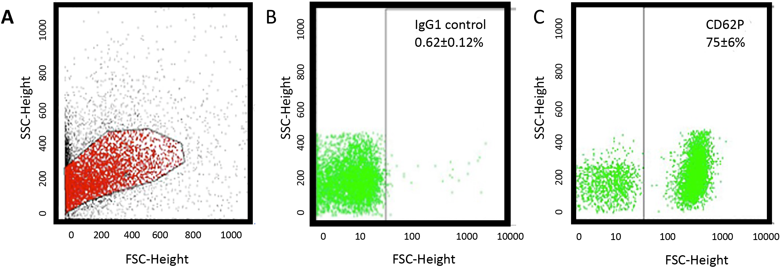
Fig. 1.

Flow cytometry (FACS) analysis. (A) A representative sample of human platelets isolated from a healthy control shows a common population of cells in forward and sideward scatter and (C) markedly expressed CD62P (B) compared to an IgG control.

Western blot data shows a full-length tau standard with a size of approx. 60 kDa (Fig. 2) using the tau-5 antibody. No staining for platelet tau was found with the tau-5 antibody (Fig. 2). Using the HT-7 antibody, 2 clear bands were visible at approx. 45 and 30 kDa size, while a signal for phospho-tau-396 (approx. 50 kDa) was present in all samples (Fig. 2). A loading control GAPDH displayed a positive signal at 37 kDa (Fig. 2).
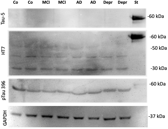
Fig. 2.

Western blot of platelet extracts. Human platelets were isolated, characterized by FACS and 15 samples per group pooled, extracted and 80 μg protein was loaded on a gel and analyzed by Western Blots. As a control, a standard (St) of full-length tau (500 ng) was loaded which gives a strong band at approx. 50–60 kDa when stained with the tau-5 antibody. No staining for platelet tau was found with the tau-5 antibody. Using the HT-7 antibody, 2 clear bands were visible at approx. 45 and 30 kDa size, while a signal for phospho-tau-396 (approx. 50 kDa) was present in all samples. Note that the lanes were not always in line. This is because the gel was maximally loaded (80 μg protein) in order to increase sensitivity. GAPDH served as a loading control and shows a band at approx. 37 kDa.

3.3 qRT-PCR

Platelets are anuclear cells, however, there are reports that mRNAs can be detected in platelets by qRT-PCR. RNA was isolated from human platelets using the Quiagen minikit and we achieved 1.1 ± 0.4 μg/mL RNA (n = 6), which is far lower as isolated from control mouse cortex (209 ± 73 μg/mL, n = 3). Using this RNA, we performed qRT-PCR for tau compared to gapd. Compared to control mouse cortex (3.71 ± 0.06 ΔCT, n = 3), very low amounts of mRNA were found in wildtype mice (0.097 ± 0.076 ΔCT, n = 3), while in human platelets 5 out of 6 samples were undetectable and one sample had 0.003 ΔCT values.

3.4 WANGEL tau Detection

Using our novel WANGEL system, we show that full-length tau migrates to the cathode and elutes from the agarose gel as a 50–60 kDa protein (Fig. 3). However, when the platelet extract (a pool of 32 lanes) was eluted from the WANGEL system, several large and small molecular weight species could be detected, but the majority of the tau+ detection was seen as a 20–30 kDa fragment (Fig. 3).
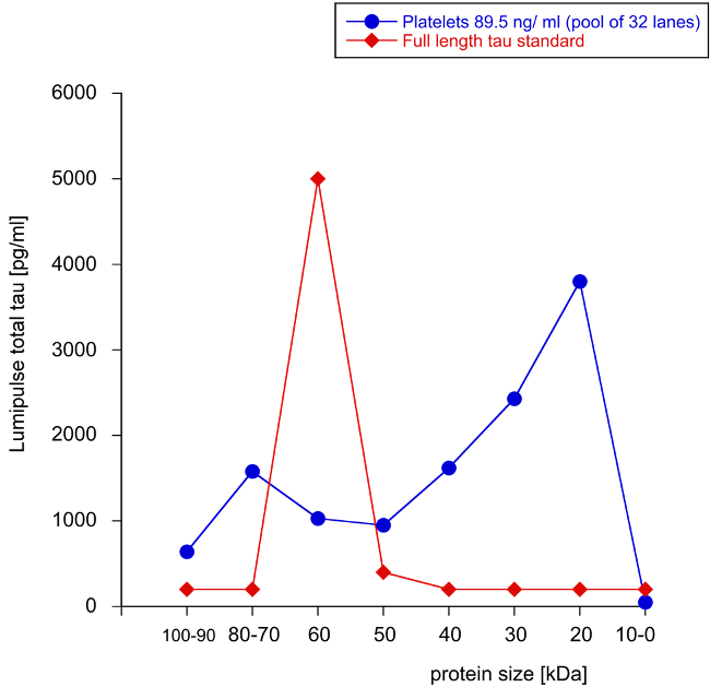
Fig. 3.

Elution of tau from Western Blots linked to agarose gels. Tau protein (100 ng/lane) is separated on a Western Blot gel, then bands with between 0–100 kDa are cut, and tau eluted from agarose gels via the cathode. Note that tau elutes as a 60 kDa protein from Western-agarose gels (red triangles). In order to charcterize tau from platelet, an extract (89.5 ng/mL) was seperated on Western Blots, the lanes cut out and pooled from 32 lanes and analyzed by Lumipulse. Note that many large and small tau fragments were detected, but the majority of tau had a size of 20–30 kDa (blue circles). Values are given as pg/mL total tau measured by Lumipulse assay.

3.5 Detection of Tau in Platelets Using Mass Spectrometry

Applying an immunoprecipitation method with the HT7 antibody on the 2N4R full-length tau standard, 21 peptides could be identified by nanoLC ESI-MS/MS that covered 57% of the amino acid sequence of tau441. In a further experiment with cerebrospinal fluid of an AD patient (1024 pg/mL total tau), four tau peptides were identified with high confidence (data not shown). We used this method to investigate the different human platelet samples. Only a single peptide could be identified in a platelet extract pooled from 15 human patients of the same group (1577 pg/mg tau) with the HT7 antibody. The peptide with the sequence SGYSSPGSPGTPGSR corresponds to amino acid residues 203–217 of tau441, with an m/z = 697.32059 Da, charge state = 2, cross-correlation score (XCorr) = 3.74. The MS/MS spectrum and the assigned y- and b-series ions are shown in Fig. 4. The same peptide was found in the tau standard and in cerebrospinal fluid, respectively (data not shown). Tau could not be detected in all other platelet samples. Applying the tau-5 antibody instead of the HT7 antibody, no tau could be identified, neither in the standard sample nor in CSF or platelets.
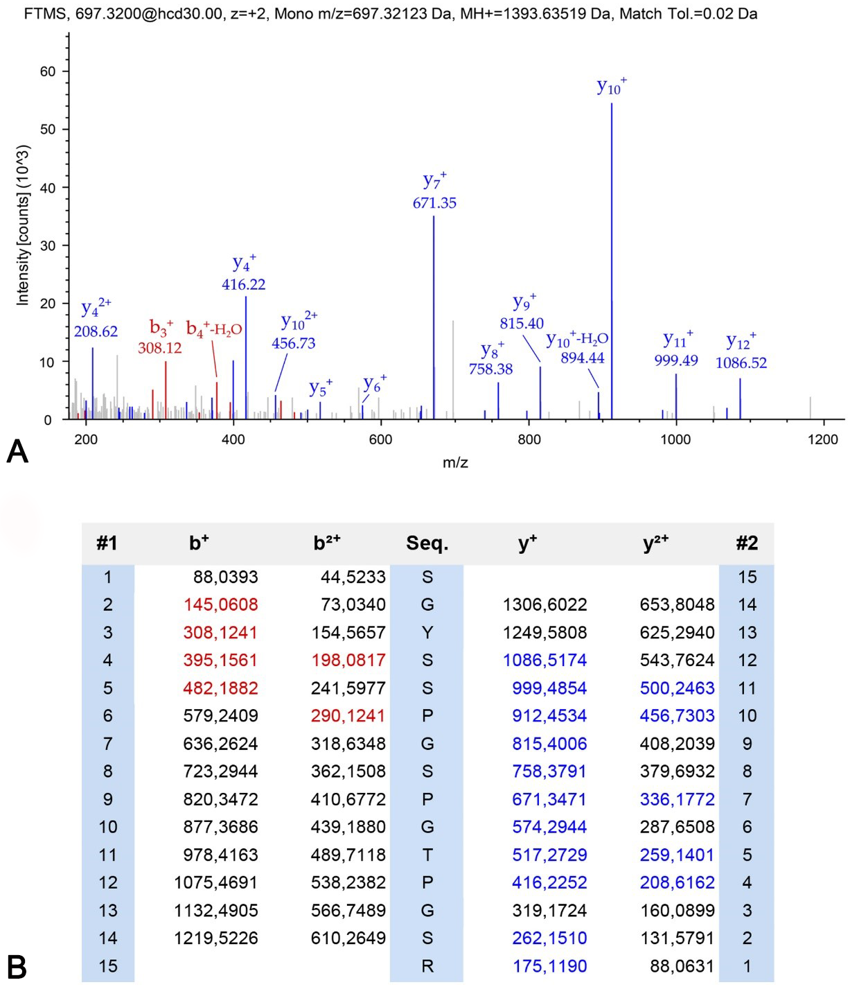
Fig. 4.

Identification of a peptide from tau protein in platelets by mass spectrometry. Immunoprecipitation was performed to enrich tau from pooled platelets. Enriched proteins were analyzed with mass spectrometry and one unique peptide corresponding to amino acid residues 203–217 of tau441 was identified. (A) Shown is the MS/MS spectrum of peptide SGYSSPGSPGTPGSR (B) and the assigned y- and b-series ions.

4. Discussion

In the present study, we show that tau protein is expressed in human platelets. It occurs especially as a fragmented form. We demonstrate that tau protein levels in platelets of AD and depressive patients are significantly changed and reduced tau levels could distinguish AD patients and depressed people from healthy individuals.

Platelets are about 3 μm small cells and are processed from megakaryocytes and do not have a nucleus. The isolation of human platelets from blood has been well described in our hands for several years [14, 38, 39]. For platelets characterization we used FACS analysis for CD62P, which is well established in our laboratory [34]. CD62P is located on the inner membrane of α-granules and released on the outside upon activation where it acts as a receptor and is thus a platelet activation/degranulation marker. A negative control (IgG1) was incorporated in the protocol and the obtained values subtracted from the CD62P positive events. Platelets are sensitive cells and require careful handling during preparation. After decades of study, there is still a general need to establish concurrent methods for the different processing steps of platelets because the wide variation of reported protocols for platelet preparation leads to different biological responses and incomparable data. Several variables (such as, e.g., anticoagulants in collections tubes, time of processing, etc.) in the platelet preparation process need to be standardized to minimize artifactual stimulation during blood collection and sample handling. Thus, as blood was processed within 3 hours, and we are very experienced, we are confident that we analyzed healthy platelets. Blood was centrifuged to obtain platelet-rich plasma and further to give a high pure platelet pellet. In order to extract proteins from platelets, we extracted platelets by sonication and centrifugation, to give a pure extract of soluble proteins in the supernatant. In all cases, protease inhibitors were added to inhibit any degradation process.

Platelets modulate as mediators multiple cellular processes including angiogenesis and inflammation and are involved in a variety of pathologic conditions including AD. It is well known that both inflammatory and vascular processes accompany AD pathology and hence, it seems likely that platelets actively contribute to the progression of AD [9]. In murine models we have shown that platelets are first signs in AD as they concentrate in brain vessels and can also damage brain vessels and thus contribute to the AD pathology [15, 16]. Moreover, platelets contain proteins such as amyloid-precursor protein (APP) and produce beta-amyloid peptides, mainly the shorter 40 amino acid form [10, 17, 18, 19, 20, 40]. It is assumed that platelets use the beta-amyloid(40) peptide to clot the damaged vessel. Regarding the role of tau in platelets and AD, there is a strong mismatch in the literature. Although there are several reports that platelets contain tau (especially C-terminal tau), their role and structure is completely unknown [21, 22, 23, 24]. In the present study, we indeed verify that tau is found in platelets in a fragmented form and slightly decreased in late AD, however, we cannot link tau to any functional process.

Many studies now indicate that platelets in depressed people have a different level of activation [41, 42, 43, 44, 45]. Platelets contain a high concentration of serotonin, which is stored in vesicles and released by stimulation. Since serotonin plays a major role in depression, it could be that serotonin, which comes from platelets, plays a role in the pathogenesis of depression. In a previous study, we used platelets from depressive patients as an additional control, but also because AD and MCI patients complain from depressive episodes. Thus, a bio-profiling of platelets in severely depressed patients treated with medication was performed. We detected that the serotonin levels in blood platelets and different immunomodulatory compounds were significantly altered in these patients [46]. Interestingly, platelets not only contribute to the progression of depression, but also play a role in stress. In two previous papers we reported on differential changes in platelets reactivity induced by acute physical stress compared to persistent mental stress and also showed that stress enhances pro-inflammatory platelets activity [47, 48]. This present study shows that tau protein levels are decreased in depressed people followed by a slight enhanced phospho-tau-181 activation. Future studies are needed to further investigate this finding especially how and what the relationship is between serotonin and tau protein.

In order to characterize tau better, we performed a qRT-PCR, a Western Blot, a WANGEL detection and mass spectrometry. Platelets are anuclear cells; however, there are reports that mRNAs can be detected in platelets [49]. Although it seemed very unrealistic to detect tau mRNA in platelets, we performed a qRT-PCR. Our data supported our suggestion, that no tau mRNA was detectable in human platelets, but only in 1 sample out of 6 a very low tau expression was found by qRT-PCR, which could also be an artefact; this again shows that tau in platelets is expressed at very low concentrations. Thus, in order to catch tau in platelets we pooled up to 15 samples to concentrate tau. Using Western blots we did not see a full length tau protein (of approx. 60 kDa), but rather some smaller tau fragments of 40 and 30 kDa or smaller using the HT7 antibody. Only with an antibody recognizing the phospho-tau 396 form a large protein was seen, where we cannot really determine the specificity. In order to characterize the tau protein better, we performed a WANGEL analysis followed by the high sensitive Lumipulse assay. Here, a pool of 32 Western Blot lanes was extracted into agarose gels and eluted and analyzed and again we found that only very small tau fragments of 20–30 kDa were detectable. And finally applying immunoprecipitation and nanoLC MS, only a single peptide of tau was identified in one of the pooled platelet samples, while it was undetectable in all other platelet samples investigated. A comparison of different sample preparation protocols for the analysis of human platelets showed huge differences in the obtained protein concentrations [50]. A more efficient and replicable method may increase protein isolation and tau concentration, respectively. The fact that tau could not be detected clearly in platelets by mass spectrometry again suggests that it is of low abundance and/or fragmented, which results in a decreased binding to the HT7 antibody.

Taken together, our data highly suggest that tau is indeed present in human platelets, but at very low levels and only in a highly fragmented small (20–40 kDa) form. This was surprising to see but this result fully agrees with a prior study [51]. They report that multiple N-terminal and mid-domain fragments of tau were detected in pooled cerebrospinal fluid (CSF) with apparent sizes ranging from 20 kDa to approx. 40 kDa. The pattern of tau fragments in AD and control samples were similar. They also did not detect full-length tau and C-terminal fragments in their CSF samples. In order to concentrate the CSF, they used a RP-HPLC followed by a Western Blot and also used the same HT7 antibody (as we did) and found tau in fractions 3–7 representing small (20–40 kDa) tau fragments [51]. This is partly surprising, as tau is a well-established biomarker in CSF for AD with a pathological concentration of >500 pg/mL. The tau analysis is well established using different ELISAs or also our Lumipulse assay. The question now comes up, why is tau so highly fragmented, and why is no full length tau detectable in CSF (but also in platelets). More work is necessary to study the metabolism and stability of the tau protein and the functional role of smaller fragmented tau forms.

Due to a new generation of ultrasensitive immunoassays, phosphorylated tau epitopes are detectable in blood plasma. The currently best validated epitopes are phospho-tau-181 and phospho-tau-127. Plasma phosphorylated tau-181, for example shows 1.5 to 3.5 times higher concentrations in AD patients compared to controls [52]. The recently published plasma-phospho-tau-231 is also a promising new biomarker for incipient AD [53]. Furthermore, plasma phospho-tau-181 identified individuals who were Aβ-PET-positive regardless of the clinical diagnosis and correlated with cortical tau protein deposition measured by 18F-flortaucipir PET [52]. Furthermore, the altered tau in blood platelets very probably only represents a small part of the AD pathology. Similar to AD, this also applies to depression. However, for depression, further studies should show whether there is a connection between tau pathology and the serotonin system. Plasma phospho-tau-181 also allows individualised prediction of AD in patients with mild cognitive impairments [54]. Future longitudinal studies will show how tau protein changes in those MCI patients who convert to AD.

The question now arises, if platelet tau may become a good biomarker for dementia or depression. The answer is definitely no. As we have shown tau and phospho-tau-181 are expressed in platelets at very very low concentrations. This limits our tools to quantify tau. In order to overcome such a methodological problem, more sensitive assays must be used. We thus highly suggest to use SIMOA (single molecule array technology) to ultimately enhance the sensitivity for low abundance biomarkers up to the single molecule level. It will be highly interesting to use this technique, which is, however, very expensive and not useful for routine diagnosis. On the other hand, our data show that the differences between AD/depression and healthy controls are very low (1.3× reduction for tau) with a significance of p 0.05. Our experience in several clinical biomarker studies show, that at least a signifance of p 0.001 and a difference of at least 3–5 variances is necessary to establish a biomarker in the clinical routine. The same also holds for phospho-tau-181 and the increase in depression compared to controls (1.3× enhanced). Thus the present study will not discover a new biomarker but will definitely contribute to the idea that tau plays a role in platelet function in dementia and depression.

Definitely, this study has several limits. (1) The changes of tau in the brain of people suffering from severe neurocognitive disorder are well studied. However, in platelets, a functional role of tau is still largely unexplored. More work in humans, or in mouse models including tau transgenic will be necessary. (2) In platelets, the concentration of tau protein is very low. Therefore, future studies are necessary to examine whether it is possible to detect tau levels reliably at a specified level of confidence. The reliable reproducibility of the results are the prerequisite that the change in tau levels could establish as a future biomarker. More work is necessary to link tau to other vascular-related diseases. (3) As mentioned, tau exists as a small fragmented protein. This is really surprising, but it confirms work in human CSF and also questions why tau is so highly fragmented [51]. This fragmentation, however, does not limit the use as a biomarker in CSF for AD, as this method is well established internationally. It however opens the question, why tau is fragmented, and if is this only due to degradation and metabolism or has fragmented tau also a physiological role in the brain, e.g., to enter the nucleus. (4) In this regard, we suggest that full-length tau can be very instable and is metabolized to smaller fragmented peptides. This high fragmentation also limits our approach to detect tau by mass spectrometry.

5. Conclusions

Taken together we show that tau is expressed in human platelets, but exists mainly as a very highly small fragmented form. We show that tau in platelets is reduced in AD and depression and may differentiate them from healthy controls. We show that tau is very low in platelets and display a high heterogeneity. Thus, we conclude that tau in platelets may have a prominent function, but will not serve as a novel biomarker.

Author Contributions

CH designed the study, wrote the manuscript, and acquired funding. DSK performed the Western Blots. JM collected blood and diagnosed patients. BS performed the MS analysis. All authors contributed to editorial changes in the manuscript. All authors read and approved the final manuscript.

Ethics Approval and Consent to Participate

The study was performed according to local ethical approval protocol no. AN2015-0159 351/4.7 405/5.5 (4484a). Informed consent was obtained from all subjects enrolled in the study. The study was in accordance with the guidelines of the ethical commission of the Medical University Innsbruck.

Acknowledgment

We thank Karin Albrecht for excellent technical help.

Funding

This research received no external funding.

Conflict of Interest

The authors declare no conflict of interest.

References
[1]
Selkoe DJ. Alzheimer’s Disease is a Synaptic Failure. Science. 2002; 298: 789–791.
[2]
Morris GP, Clark IA, Vissel B. Inconsistencies and controversies surrounding the Amyloid Hypothesis of Alzheimer’s disease. Acta Neuropathologica Communications. 2014; 2: 135.
[3]
Tanzi RE, Moir RD, Wagner SL. Clearance of Alzheimer’s Abeta peptide: The many roads to perdition. Neuron. 2004; 43: 505–608.
[4]
Iadecola C. Neurovascular regulation in the normal brain and in Alzheimer’s disease. Nature Reviews Neuroscience. 2004; 5: 347–360.
[5]
Kalaria RN. Small vessel disease and Alzheimer’s dementia: pathological considerations. Cerebrovascular Diseases. 2002; 13: 48–52.
[6]
de la Torre JC. Alzheimer’s disease as a vascular disorder. Stroke. 2002; 33: 1152–1162.
[7]
Humpel C. Chronic mild cerebrovascular dysfunction as a cause for Alzheimer’s disease? Experimental Gerontology. 2011; 46: 225–232.
[8]
Attems J, Jellinger K, Thal DR, Van Nostrand W. Review: Sporadic cerebral amyloid angiopathy. Neuropathology and Applied Neurobiology. 2011; 37: 75–93.
[9]
Greenberg SM, Bacskai BJ, Hernandez-Guillamon M, Pruzin J, Sperling R, van Veluw SJ. Cerebral amyloid angiopathy and Alzheimer disease — one peptide, two pathways. Nature Reviews Neurology. 2020; 16: 30–42.
[10]
Humpel C. Platelets: their Potential Contribution to the Generation of Beta-amyloid Plaques in Alzheimer’s Disease. Current Neurovascular Research. 2017; 14: 290–298.
[11]
Plagg B, Humpel C. Platelets in Alzheimer’s Disease. In Kerrigan S, Niamh Moran N (eds.) The Non-Thrombotic Role of Platelets in Health and Disease. 1st edn. InTech: London, United Kingdom. 2015.
[12]
Lindemann S, Gawaz M. The Active Platelet: Translation and Protein Synthesis in an Anucleate Cell. Seminars in Thrombosis and Hemostasis. 2007; 33: 144–150.
[13]
Zucker MB, Nachmias VT. Platelet activation. Arteriosclerosis. 1985; 5: 2–18.
[14]
Kniewallner K, Ehrlich D, Kiefer A, Marksteiner J, Humpel C. Platelets in the Alzheimer’s Disease Brain: do they Play a Role in Cerebral Amyloid Angiopathy? Current Neurovascular Research. 2015; 12: 4–14.
[15]
Kniewallner KM, Wenzel D, Humpel C. Thiazine Red+ platelet inclusions in Cerebral Blood Vessels are first signs in an Alzheimer’s Disease mouse model. Scientific Reports. 2016; 6: 28447.
[16]
Kniewallner KM, Foidl BM, Humpel C. Platelets isolated from an Alzheimer mouse damage healthy cortical vessels and cause inflammation in an organotypic ex vivo brain slice model. Scientific Reports. 2018; 8: 15483.
[17]
Evin G. Platelets and Alzheimer’s disease: Potential of APP as a biomarker. World Journal of Psychiatry. 2012; 2: 102.
[18]
Di Luca M. Differential Level of Platelet Amyloid beta Precursor Protein Isoforms: an Early Marker for Alzheimer Disease. Archives of Neurology. 1998; 55: 1195–1200.
[19]
Ehrlich D, Hochstrasser T, Humpel C. Effects of oxidative stress on amyloid precursor protein processing in rat and human platelets. Platelets. 2013; 24: 26–36.
[20]
Marksteiner J, Humpel C. Platelet-derived secreted amyloid-precursor protein-beta as a marker for diagnosing Alzheimers disease. Current Neurovascular Research. 2013; 10: 297–303.
[21]
Mukaetova-Ladinska EB, Abdell-All Z, Andrade J, da Silva JA, Boksha I, Burbaeva G, et al. Platelet Tau Protein as a Potential Peripheral Biomarker in Alzheimer’s Disease: an Explorative Study. Current Alzheimer Research. 2018; 15: 800–808.
[22]
Farías G, Pérez P, Slachevsky A, Maccioni RB. Platelet Tau Pattern Correlates with Cognitive Status in Alzheimer’s Disease. Journal of Alzheimer’s Disease. 2012; 31: 65–69.
[23]
Neumann K, Farías G, Slachevsky A, Perez P, Maccioni RB. Human Platelets Tau: a Potential Peripheral Marker for Alzheimer’s Disease. Journal of Alzheimer’s Disease. 2011; 25: 103–109.
[24]
Slachevsky A, Guzmán-Martínez L, Delgado C, Reyes P, Farías GA, Muñoz-Neira C, et al. Tau Platelets Correlate with Regional Brain Atrophy in Patients with Alzheimer’s Disease. Journal of Alzheimer’s Disease. 2017; 55: 1595–1603.
[25]
Nisbet RM, Polanco J, Ittner LM, Götz J. Tau aggregation and its interplay with amyloid-β. Acta Neuropathologica. 2015; 129: 207–220.
[26]
Spillantini MG, Goedert M. Tau pathology and neurodegeneration. The Lancet Neurology. 2013; 12: 609–622.
[27]
Liu C, Götz J. How it all Started: Tau and Protein Phosphatase 2a. Journal of Alzheimer’s Disease. 2013; 37: 483–494.
[28]
Wang Y, Mandelkow E. Tau in physiology and pathology. Nature Reviews Neuroscience. 2016; 17: 22–35.
[29]
Hanger DP, Anderton BH, Noble W. Tau phosphorylation: the therapeutic challenge for neurodegenerative disease. Trends in Molecular Medicine. 2009; 15: 112–119.
[30]
Blasko I, Lederer W, Oberbauer H, Walch T, Kemmler G, Hinterhuber H, et al. Measurement of Thirteen Biological Markers in CSF of Patients with Alzheimer’s Disease and other Dementias. Dementia and Geriatric Cognitive Disorders. 2006; 21: 9–15.
[31]
Humpel C, Benke T. Cerebrospinal Fluid Levels of 14-3-3 Gamma: what does it Tell us about Sporadic Creutzfeldt-Jakob Disease? Pharmacology. 2017; 100: 243–245.
[32]
Lecordier S, Pons V, Rivest S, ElAli A. Multifocal cerebral microinfarcts modulate early Alzheimer’s disease pathology in a sex-dependent manner. Frontiers in Immunology. 2022; 12: 813536.
[33]
Petersen RC, Doody R, Kurz A, Mohs RC, Morris JC, Rabins PV, et al. Current Concepts in Mild Cognitive Impairment. Archives of Neurology. 2001; 58: 1985.
[34]
Plagg B, Marksteiner J, Kniewallner KM, Humpel C. Platelet dysfunction in hypercholesterolemia mice, two Alzheimer’s disease mouse models and in human patients with Alzheimer’s disease. Biogerontology. 2015; 16: 543–558.
[35]
Ucar B, Stefanova N, Humpel C. Spreading of aggregated α-synuclein in sagittal organotypic mouse brain slices. Biomolecules. 2022; 12: 163.
[36]
Halfinger B, Hammerer-Lercher A, Amplatz B, Sarg B, Kremser L, Lindner HH. Unraveling the Molecular Complexity of O-Glycosylated Endogenous (N-Terminal) pro–B-Type Natriuretic Peptide Forms in Blood Plasma of Patients with Severe Heart Failure. Clinical Chemistry. 2017; 63: 359–368.
[37]
Amplatz B, Sarg B, Faserl K, Hammerer-Lercher A, Mair J, Lindner HH. Exposing the High Heterogeneity of Circulating Pro B-Type Natriuretic Peptide Fragments in Healthy Individuals and Heart Failure Patients. Clinical Chemistry. 2020; 66: 1200–1209.
[38]
Foidl B., Oberacher H., Marksteiner J, Humpel C. Platelet and plasma phosphatidylcholines as biomarkers to diagnose cerebral amyloid angiopathy. Frontiers in Neurology. 2020; 11: 359.
[39]
Oberacher H, Arnhard K, Linhart C, Diwo A, Marksteiner J, Humpel C. Targeted Metabolomic Analysis of Soluble Lysates from Platelets of Patients with Mild Cognitive Impairment and Alzheimer’s Disease Compared to Healthy Controls: is PC aeC40:4 a Promising Diagnostic Tool? Journal of Alzheimer’s Disease. 2017; 57: 493–504.
[40]
Ehrlich D. Platelets in psychiatric disorders. World Journal of Psychiatry. 2012; 2: 91.
[41]
Marazziti D, Landi P, Baroni S, Vanelli F, Bartolommei N, Picchetti M, et al. The Role of PlateletLymphocyte Serotonin Transporter in Depression and beyond. Current Drug Targets. 2013; 14: 522–530.
[42]
von Kanel R. Platelet hyperactivity in clinical depression and the beneficial effect of antidepressant drug treatment: how strong is the evidence? Acta Psychiatrica Scandinavica. 2004; 110: 163–177.
[43]
Parakh K, Sakhuja A, Bhat U, Ziegelstein RC. Platelet Function in Patients with Depression. Southern Medical Journal. 2008; 101: 612–617.
[44]
Ziegelstein RC, Parakh K, Sakhuja A, Bhat U. Platelet function in patients with major depression. Internal Medicine Journal. 2009; 39: 38–43.
[45]
Bruce EC, Musselman DL. Depression, Alterations in Platelet Function, and Ischemic Heart Disease. Psychosomatic Medicine. 2005; 67: S34–S36.
[46]
Hüfner K, Kandler C, Koudouovoh-Tripp P, Egeter J, Hochstrasser T, Stemer B, et al. Bioprofiling of platelets in medicated patients with depression. Journal of Affective Disorders. 2014; 172: 81–88.
[47]
Hüfner K, Koudouovoh-Tripp P, Kandler C, Hochstrasser T, Malik P, Giesinger J, et al. Differential changes in platelet reactivity induced by acute physical compared to persistent mental stress. Physiology & Behavior. 2015; 151: 284–291.
[48]
Koudouovoh-Tripp P, Hüfner K, Egeter J, Kandler C, Giesinger JM, Sopper S, et al. Stress Enhances Proinflammatory Platelet Activity: the Impact of Acute and Chronic Mental Stress. Journal of Neuroimmune Pharmacology. 2020; 16: 500–512.
[49]
Sacchi E, Pinotti M, Marchetti G, Merati G, Tagliabue L, Mannucci PM, et al. Protein S mRNA in Patients with Protein S Deficiency. Thrombosis and Haemostasis. 1995; 73: 746–749.
[50]
Semančíková E, Tkáčiková S, Talian I, Pálová E, Sabo J. Comparison of Sample Preparation Protocols for the Analysis of the Human Platelet Proteome from whole Blood. Analytical Letters. 2017; 50: 1521–1530.
[51]
Meredith JE Jr, Sankaranarayanan S, Guss V, Lanzetti AJ, Berisha F, Neely RJ, et al. Characterization of novel CSF Tau and ptau biomarkers for Alzheimer’s disease. PLoS ONE. 2013; 8: e76523.
[52]
Thijssen EH, La Joie R, Wolf A, Strom A, Wang P, Iaccarino L, et al. Diagnostic value of plasma phosphorylated tau181 in Alzheimer’s disease and frontotemporal lobar degeneration. Nature Medicine. 2020; 26: 387–397.
[53]
Ashton NJ, Pascoal TA, Karikari TK, Benedet AL, Lantero-Rodriguez J, Brinkmalm G, et al. Plasma p-tau231: a new biomarker for incipient Alzheimer’s disease pathology. Acta Neuropathologica. 2021; 141: 709–724.
[54]
Cullen NC, Leuzy A, Janelidze S, Palmqvist S, Svenningsson AL, Stomrud E, et al. Plasma biomarkers of Alzheimer’s disease improve prediction of cognitive decline in cognitively unimpaired elderly populations. Nature Communications. 2021; 12: 3555.
Share
Back to top