


 , Árpád Kormányos 1, Gergely Rácz 1, Nóra Ambrus 1, Imelda Marton 2,3, Zita Borbényi 2
1 Department of Medicine, Albert Szent-Györgyi Medical School, University of Szeged, 6725 Szeged, Hungary
2 Division of Haematology, Department of Medicine, Albert Szent-Györgyi Medical School, University of Szeged, 6725 Szeged, Hungary
3 Department of Transfusiology, Albert Szent-Györgyi Medical School, University of Szeged, 6725 Szeged, Hungary
Abstract
Background: Hypereosinophilic syndrome (HES) is a peripheral
eosinophilia characterized by elevated absolute eosinophil cell count
(
Keywords
- annulus
- echocardiography
- hypereosinophilic syndrome
- mitral
- speckle-tracking
- three-dimensional
- tricuspid
Hypereosinophilic syndrome (HES) is a peripheral eosinophilia featured by
elevated absolute eosinophil cell count (
There were 17 subjects in the HES group and 24 patients in the group of healthy
controls. HES patients were recruited between May 2012 and December 2021
prospectively in the Division of Haematology at the University of Szeged. Two
patients were excluded from study in the HES group due to suboptimal image
quality with 3D echocardiography. From the 15 patients participating in the
study, 14 patients had idiopathic HES and 1 patient had HES associated with acute
T-lymphoma (mean age: 61.7
2D gray-scale images were performed using a broadband PST-30BT phased-array transducer (1–5 MHz) with a Toshiba Artida® (Toshiba Medical Systems, Tokyo, Japan) echocardiographic tool. The following echocardiographic parameters were measured in all patients: diameter of the left atrium, diameter and volume of the LV respecting the cardiac cycle, thickness of the interventricular septum and thickness of the LV posterior wall, LV ejection fraction measured using the Simpson’s method [11]. With pulsed Doppler transmitral flow velocities were measured in diastole and their ratio (E/A) were calculated. With tissue Doppler imaging E/E’ was measured as a ratio of early diastolic transmitral flow velocity (E) and mitral annular velocity (E’). With Doppler echocardiography tricuspid regurgitation pressure gradient was estimated, as well.
3D data acquisitions were completed by a Toshiba Artida
The end-diastole was considered when peak R wave was on electrocardiogram. The end-systole was considered as the first frame when the aortic valve was closed (at the end of T wave on electrocardiogram). MA/TA assessments were made using optimized image planes on the endpoints of the MA/TA on apical two- and four-chamber views and on C7 short-axis view. Several MA/TA measures and features of their function were calculated at end-diastole and at end-systole (Fig. 1) [15, 16]:
MA/TA dimensions:
MA/TA functional properties:
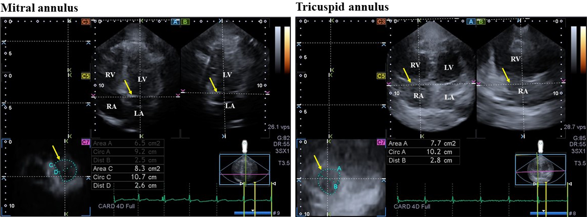 Fig. 1.
Three-dimensional (3D) echocardiographic assessment of mitral and tricuspid annuli in a patient with hypereosinophilic syndrome. (A) apical four-chamber view, (B) apical two-chamber view and cross-sectional view (C7) of the mitral and tricuspid annuli optimalised on mitral and tricuspid annular images. Yellow arrows indicate plane of the mitral and tricuspid annuli. Abbreviations: Area, mitral/tricuspid annular area; Circ, mitral/tricuspid annular perimeter; Dist, mitral/tricuspid annular diameter; LA, left atrium; LV, left ventricle; RA, right atrium; RV, right ventricle.
While categorical data were expressed in counts and percentages (%), continuous
variables were demonstrated in mean
The incidence of hypertension and hyperlipidaemia was higher in the HES group, other demographic parameters were similar between the groups examined (Table 1).
| Controls (n = 24) | HES patients (n = 15) | p value | |
|---|---|---|---|
| Clinical data | |||
| Age (years) | 55.2 |
61.7 |
0.08 |
| Male gender (%) | 12 (50) | 10 (67) | 0.34 |
| Hypertension (%) | 0 (0) | 8 (53) | 0.0001 |
| Diabetes mellitus (%) | 0 (0) | 1 (7) | 0.38 |
| Hyperlipidaemia (%) | 0 (0) | 4 (27) | 0.02 |
| 2D echocardiography | |||
| LA diameter (mm) | 38.4 |
42.1 |
0.15 |
| LA volume index (mL/m |
27.1 |
31.3 |
0.17 |
| LV end-diastolic diameter (mm) | 48.8 |
51.7 |
0.27 |
| LV end-diastolic volume (mL) | 110.7 |
116.0 |
0.65 |
| LV end-systolic diameter (mm) | 31.8 |
34.5 |
0.31 |
| LV end-systolic volume (mL) | 38.6 |
42.9 |
0.41 |
| Interventricular septum (mm) | 9.5 |
10.8 |
0.008 |
| LV posterior wall (mm) | 9.8 |
9.7 |
0.93 |
| LV ejection fraction (%) | 65.0 |
63.4 |
0.48 |
| E (cm/s) | 69.3 |
76.8 |
0.39 |
| A (cm/s) | 71.7 |
72.7 |
0.92 |
| E/A | 1.0 |
1.1 |
0.50 |
| E/E’ | 9.4 |
10.2 |
0.46 |
| Tricuspid regurgitation pressure gradient (mm Hg) | 15.8 |
15.4 |
0.93 |
Abbreviations: 2D, two-dimensional; HES, hypereosinophilic syndrome; E and A, early and late diastolic transmitral flow velocities; E’, early diastolic mitral annular velocity; LA, left atrium; LV, left ventricular.
Absolute eosinophil count (7.4
Table 1 was used for demonstration of 2D echocardiographic parameters of HES patients and controls. Only interventricular septum was significantly thickened in patients with HES, no other parameter differed significantly between the groups examined. Only one HES patient showed grade 2 mitral regurgitation, other HES patients and controls did not show larger than grade 1 valvular insufficiency or had significant valvular stenoses.
Increased end-diastolic and end-systolic MA diameters, areas and perimeters together with reduced MAFAC and MAFS could be detected in HES patients as compared to those of controls. From TA morphological parameters, only end-diastolic TA area and end-systolic TA diameter were significantly increased in HES patients who had preserved TA functional parameters (Tables 2,3). Comparative analysis of MA and TA parameters was done between HES cases with vs. without hypertension without significant differences between these parameters (Table 4).
| Controls (n = 24) | HES patients (n = 15) | p value | ||
|---|---|---|---|---|
| Morphological parameters | ||||
| end-diastolic MA diameter (cm) | 2.4 |
2.6 |
0.03 | |
| end-diastolic MA area (cm |
7.5 |
9.5 |
0.01 | |
| end-diastolic MA perimeter (cm) | 10.4 |
11.6 |
0.03 | |
| end-systolic MA diameter (cm) | 1.7 |
2.2 |
0.001 | |
| end-systolic MA area (cm |
3.8 |
6.7 |
0.001 | |
| end-systolic MA perimeter (cm) | 7.4 |
9.9 |
0.001 | |
| Functional parameters | ||||
| MAFAC (%) | 47.7 |
29.6 |
0.001 | |
| MAFS (%) | 28.9 |
16.6 |
0.004 | |
Abbreviations: HES, hypereosinophilic syndrome; MA, mitral annulus; MAFAC, mitral annular fractional area change; MAFS, mitral annular fractional shortening.
| Controls (n = 24) | HES patients (n = 15) | p value | ||
|---|---|---|---|---|
| Morphological parameters | ||||
| end-diastolic TA diameter (cm) | 2.3 |
2.6 |
0.08 | |
| end-diastolic TA area (cm |
7.5 |
9.1 |
0.04 | |
| end-diastolic TA perimeter (cm) | 10.7 |
11.1 |
0.5 | |
| end-systolic TA diameter (cm) | 1.9 |
2.3 |
0.01 | |
| end-systolic TA area (cm |
5.9 |
6.9 |
0.1 | |
| end-systolic TA perimeter (cm) | 9.3 |
9.9 |
0.2 | |
| Functional parameters | ||||
| TAFAC (%) | 22.6 |
23.4 |
0.9 | |
| TAFS (%) | 17.9 |
13.2 |
0.08 | |
Abbreviations: HES, hypereosinophilic syndrome; TA, tricuspid annulus; TAFAC, tricuspid annular fractional area change; TAFS, tricuspid annular fractional shortening.
| Controls (n = 24) | HES patients with hypertension (n = 8) | HES patients without hypertension (n = 7) | ||
|---|---|---|---|---|
| Morphological MA parameters | ||||
| end-diastolic MA diameter (cm) | 2.4 |
2.6 |
2.7 | |
| end-diastolic MA area (cm |
7.5 |
9.4 |
9.6 | |
| end-diastolic MA perimeter (cm) | 10.4 |
11.7 |
11.4 | |
| end-systolic MA diameter (cm) | 1.7 |
2.2 |
2.1 | |
| end-systolic MA area (cm |
3.8 |
6.5 |
6.8 | |
| end-systolic MA perimeter (cm) | 7.4 |
10.1 |
9.6 | |
| Functional MA parameters | ||||
| MAFAC (%) | 47.7 |
29.9 |
29.2 | |
| MAFS (%) | 28.9 |
14.8 |
18.5 | |
| Morphological TA parameters | ||||
| end-diastolic TA diameter (cm) | 2.3 |
2.5 |
2.8 | |
| end-diastolic TA area (cm |
7.5 |
8.3 |
10.0 | |
| end-diastolic TA perimeter (cm) | 10.7 |
10.3 |
12.1 | |
| end-systolic TA diameter (cm) | 1.9 |
2.2 |
2.4 | |
| end-systolic TA area (cm |
5.9 |
6.4 |
7.5 | |
| end-systolic TA perimeter (cm) | 9.3 |
9.5 |
10.2 | |
| Functional TA parameters | ||||
| TAFAC (%) | 22.6 |
22.0 |
25.0 | |
| TAFS (%) | 17.9 |
12.6 |
13.8 | |
Abbreviations: HES, hypereosinophilic syndrome; MA, mitral annulus; MAFAC, mitral annular fractional area change; MAFS, mitral annular fractional shortening; TA, tricuspid annulus; TAFAC, tricuspid annular fractional area change; TAFS, tricuspid annular fractional shortening.
*p = 0.04 vs. Controls; **p = 0.03 vs. Controls; ***p = 0.001 vs. Controls; ****p = 0.01 vs. Controls; *****p = 0.02 vs. Controls.
No correlations were found either between 2D and 3D echocardiography-derived
parameters, or with any of the laboratory findings in HES patients. The logistic
regression model identified presence of HES as an independent predictor of
reduced MAFAC (hazard ratio (HR) 1.80, 95% CI of HR: 1.21 to 3.45, p
3D echocardiography-derived end-diastolic and end-systolic MA/TA dimensions were
measured twice by the same observer (intraobserver agreement) and by two
independent observers (interobserver agreement), the values were expressed as
mean
| Intraobserver agreement | Interobserver agreement | |||
|---|---|---|---|---|
| Mean |
ICC between measurements of the same examiner | Mean |
ICC between independent measurements of 2 examiners | |
| Mitral annular dimensions | ||||
| End-diastolic MA diameter | 0.02 |
0.94 (p |
0.03 |
0.95 (p |
| End-diastolic MA area | –0.03 |
0.95 (p |
0.02 |
0.97 (p |
| End-diastolic MA perimeter | –0.05 |
0.96 (p |
–0.08 |
0.96 (p |
| End-systolic MA diameter | –0.02 |
0.96 (p |
0.03 |
0.96 (p |
| End-systolic MA area | –0.03 |
0.97 (p |
–0.05 |
0.97 (p |
| End-systolic MA perimeter | 0.05 |
0.96 (p |
0.04 |
0.96 (p |
| Tricuspid annular dimensions | ||||
| End-diastolic TA diameter | 0.03 |
0.95 (p |
0.03 |
0.97 (p |
| End-diastolic TA area | –0.03 |
0.96 (p |
0.03 |
0.97 (p |
| End-diastolic TA perimeter | –0.06 |
0.95 (p |
–0.11 |
0.97 (p |
| End-systolic TA diameter | –0.03 |
0.96 (p |
0.03 |
0.98 (p |
| End-systolic TA area | –0.03 |
0.97 (p |
–0.05 |
0.95 (p |
| End-systolic TA perimeter | 0.06 |
0.96 (p |
0.05 |
0.96 (p |
Abbreviations: ICC, interclass correlation coefficient; MA, mitral annular; TA, tricuspid annular; SD, standard deviation.
Two out of 17 HES patients (12%) were excluded from the study as image quality was poor (inadequate for visual qualitative analysis with or without artifacts). The overall feasibility of MA/TA measurements proved to be 88%.
To our knowledge, the present study is the first in which MA and TA abnormalities in HES patients are presented by 3D echocardiography. Although both MA and TA showed signs of dilation in HES, MA abnormalities proved to be more pronounced, which accompanied by its functional impairment. Similar alterations for TA could not be detected. These results could highlight our attention on differences between left and right heart abnormalities in HES. Moreover, the novelty of the research was to demonstrate 3D echocardiography in the assessment of atrioventricular annuli on an easy-to-learn non-invasive way.
Aortic stiffness is increased in HES, but little is known about HES-related remodeling of heart chambers before the development of Loeffler endocarditis [7, 8, 9, 10, 19]. The novel echocardiographic technique, 3D echocardiography is a non-invasive method, and it seems to be optimal to quantify changes of atria and ventricles respecting the cardiac cycle [12, 13, 14]. A 3D speckle-tracking echocardiography-derived virtual 3D LV cast was used to detect the potential impairment in LV rotational mechanics demonstrating deteriorated apical rotation and twist and lack of LV twist (LV rigid body rotation) in 17% of HES cases mostly in the early necrotic phase [9]. LV longitudinal strain, one of the quantitative features of LV contractility was reduced as well suggesting subclinical functional impairment of LV function in HES [10]. Association was found between deteriorated LV function and elevated LA volumes respecting the cardiac cycle and increased total and active LA stroke volumes without impairment of LA emptying fractions. Moreover, LA circumferential strain was reduced as well [7]. The present findings provided more information about HES-related left heart abnormalities demonstrating dilated MA accompanied by its reduced function. The exact pathophysiology of these findings is not known, but subclinical involvement and infiltration of the walls of left heart chambers may be responsible. Moreover, the effects of different risk factors and aging could also play a role with haemodynamic effects of the aorta and its stiffened walls [19]. Moreover, the above mentioned functional abnormalities of the LV and LA could also have effects on each other resulting in MA dilation and functional impairment, as well [7, 9, 10].
Due to left heart abnormalities, changes may be present in the right heart as well despite absence of any cardiovascular symptoms. In a recent study, elevated RA volumes and mild RA functional abnormalities not affecting RA strains could be demonstrated in HES [8]. Although LA abnormalities found in HES were more significant compared with the RA, TA was found to be somewhat dilated, but obvious functional impairment could not be detected. Other studies should confirm our results and should evaluate HES-related RV abnormalities.
The following important limitations were present:
– Assessing any strains, rotational or dyssynchrony parameters of any chambers by 3D (speckle-tracking) echocardiography was not the aim of this study [12, 13, 14].
– HES is a rare disease, therefore we were able to collect clinical and 3D echocardiography-derived data of only relatively few HES patients [1, 2, 3, 4, 5, 6].
– There was a higher ratio of hypertension and hyperlipidemia in HES patients, which could affect result. However, comparative analysis between HES patients with vs. without hypertension did not find any differences in MA and TA data.
– In addition to the currently available technical development, lower temporal and spatial resolution features 3D echocardiography as compared to 2D echocardiography. This methodologic limitation could affect measurements [12, 13, 14].
The extent of the dilation of the MA is more pronounced than that of the TA in HES. MA functional impairment is present in HES.
All data are available.
AN—Conceptualization, writing – original draft, writing – review & editing; ÁK and GR—Methodology, investigation, data curation, drafted and revised the work critically for important intellectual content; NA—Writing – review & editing, conception and design of the work, acquisition, analysis, and interpretation of data for the work; IM and ZB—Resources, conception and design of the work, acquisition, analysis, and interpretation of data for the work, drafted and revised the work critically for important intellectual content. All authors read and approved the final manuscript. All authors have participated sufficiently in the work and agreed to be accountable for all aspects of the work.
All procedures performed in studies involving human participants were in accordance with the ethical standards of the institutional and/or national research committee and with the 1964 Helsinki declaration and its later amendments or comparable ethical standards (registration number: 71/2011). Informed consent was obtained from all individual participants included in the study.
We would like to express our gratitude to all those who helped us during the writing of this manuscript.
This research received no external funding.
The authors declare no conflict of interest. Attila Nemes is serving as one of the Editorial Board members of this journal. We declare that Attila Nemes had no involvement in the peer review of this article and has no access to information regarding its peer review. Full responsibility for the editorial process for this article was delegated to Zhonghua Sun and Yung-Liang Wan.
References
Publisher’s Note: IMR Press stays neutral with regard to jurisdictional claims in published maps and institutional affiliations.