IMR Press / JIN / Volume 23 / Issue 4 / DOI: 10.31083/j.jin2304086
Open Access Review
Pharmacotherapeutic Potential of Bitter Gourd (Momordica charantia) in Age-related Neurological Diseases
Show Less
1 School of Medicine, Shahid Beheshti University of Medical Sciences, 19839-63113 Tehran, Iran
2 Department of Food Science and Technology, University of Tehran, 14155-6619 Tehran, Iran
3 Student Research Committee, University of Social Welfare and Rehabilitation Sciences, 1985713871 Tehran, Iran
4 Student Research Committee, Vita-Salute San Raffaele University, 20132 Milan, Italy
5 Student Research Committee, Shahroud University of Medical Sciences, 3614773955 Shahroud, Iran
6 Student Research Committee, Mashhad University of Medical Sciences, 91778 99191 Mashhad, Iran
7 School of Medicine, Zanjan University of Medical Sciences, 4513956111 Zanjan, Iran
8 Student Research Committee, Arak University of Medical Sciences, 3818146851 Arak, Iran
9 Student Research Committee, Azerbaijan Medical University, AZ1022 Baku, Azerbaijan
10 Student Research Committee, Faculty of Medicine, Mashhad University of Medical Sciences, 91778 99191 Mashhad, Iran
*Correspondence: niloofarderavi@sbmu.ac.ir (Niloofar Deravi)
These authors contributed equally.
J. Integr. Neurosci. 2024, 23(4), 86; https://doi.org/10.31083/j.jin2304086
Submitted: 1 July 2023 | Revised: 29 November 2023 | Accepted: 7 December 2023 | Published: 23 April 2024
Copyright: © 2024 The Author(s). Published by IMR Press.
This is an open access article under the CC BY 4.0 license.
Abstract

Due to the growth of the elderly population, age-related neurological disorders are an increasing problem. Aging begins very gradually and later leads to several neurological issues such as lower neurotransmitter levels, oxidative stress, neuronal inflammation, and continual neuronal loss. These changes might contribute to brain disorders such as Alzheimer’s disease (AD), dementia or mild cognitive impairment, and epilepsy and glioma, and can also aggravate these disorders if they were previously present. Momordica charantia (bitter gourd), a member of the Cucurbitaceae family, is a good source of carbohydrates, proteins, vitamins, and minerals. It is used for diabetes and known for its hypoglycemic and antioxidant effects. In this review, we discuss the pharmaceutical effects of M. charantia on age-related neurological disorders. We searched several databases, including PubMed and Google Scholar, using MeSH terms. We searched articles published up until 2022 regardless of publication language. M. charantia is rich in luteolin, which increases acetylcholine in neurons by binding to enzymes in acetylcholine metabolism pathways, including butyrylcholinesterase and acetylcholinesterase. This binding inhibits the hyperphosphorylation of tau protein by restraining its kinase enzyme. Furthermore, this substance can lower serum cholesterol and has multi-target activity in AD and memory loss. M. charantia can also improve memory by decreasing tau protein and it also has potent antioxidant activity and anti-inflammatory effects. This review highlights that M. charantia has effects on many age-related neurological disorders, and can be a cost-effective supplement with minimal side effects.

Keywords
bitter gourd
M. charantia
age-related neurological diseases
Alzheimer's disease
Parkinson's disease
1. Introduction

Aging has a considerable impact on the central nervous system (CNS). Due to the recognition of diseases related to the CNS, recent estimates show that the neurological diseases included in the Global Burden of Disease (GBD) Study, such as Alzheimer’s disease (AD), multiple sclerosis (MS), epilepsy, Parkinson’s disease (PD), as well as migraine, medication-overuse headache (MOH), and tension-type headache (TTH), account for 3% of the universal burden of illness. Nevertheless, this accounts for a considerable amount overall. Dementia, especially AD, migraine, epilepsy, and stroke are among the 50 most important agents of disability-adjusted life years (DALYs) [1]. Despite characteristic etiologies, interestingly, aging is the principal risk factor for all the afore mentioned diseases [2, 3, 4]. Based on the significant impacts of aging on neurological diseases, to make real progress in terms of medicines, treatment, and control of these diseases, the molecular causes must be accurately characterized. The molecular mechanisms comprise genome instability through mutations, telomere friction, and epigenetic changes. The connections and collaborations between these mechanisms induce the functional decrease in aging organisms [5]. Appropriate therapies and medicines for the diseases mentioned above have not yet been developed [6]. Considering the previous data and the importance of treating and controlling these diseases that affect health and quality of life, alternative and preventive treatment methods should be sought. Many medicinal plants have been investigated for the prevention and treatment of these diseases [7, 8, 9, 10, 11, 12, 13, 14, 15, 16, 17]. One of these plants is Momordica charantia, otherwise known as bitter gourd or bitter melon. Fig. 1 outlines current knowledge regarding the potential pharmacotherapeutic effects and mechanisms of M. charantia in age-related neurological diseases (ANDs).
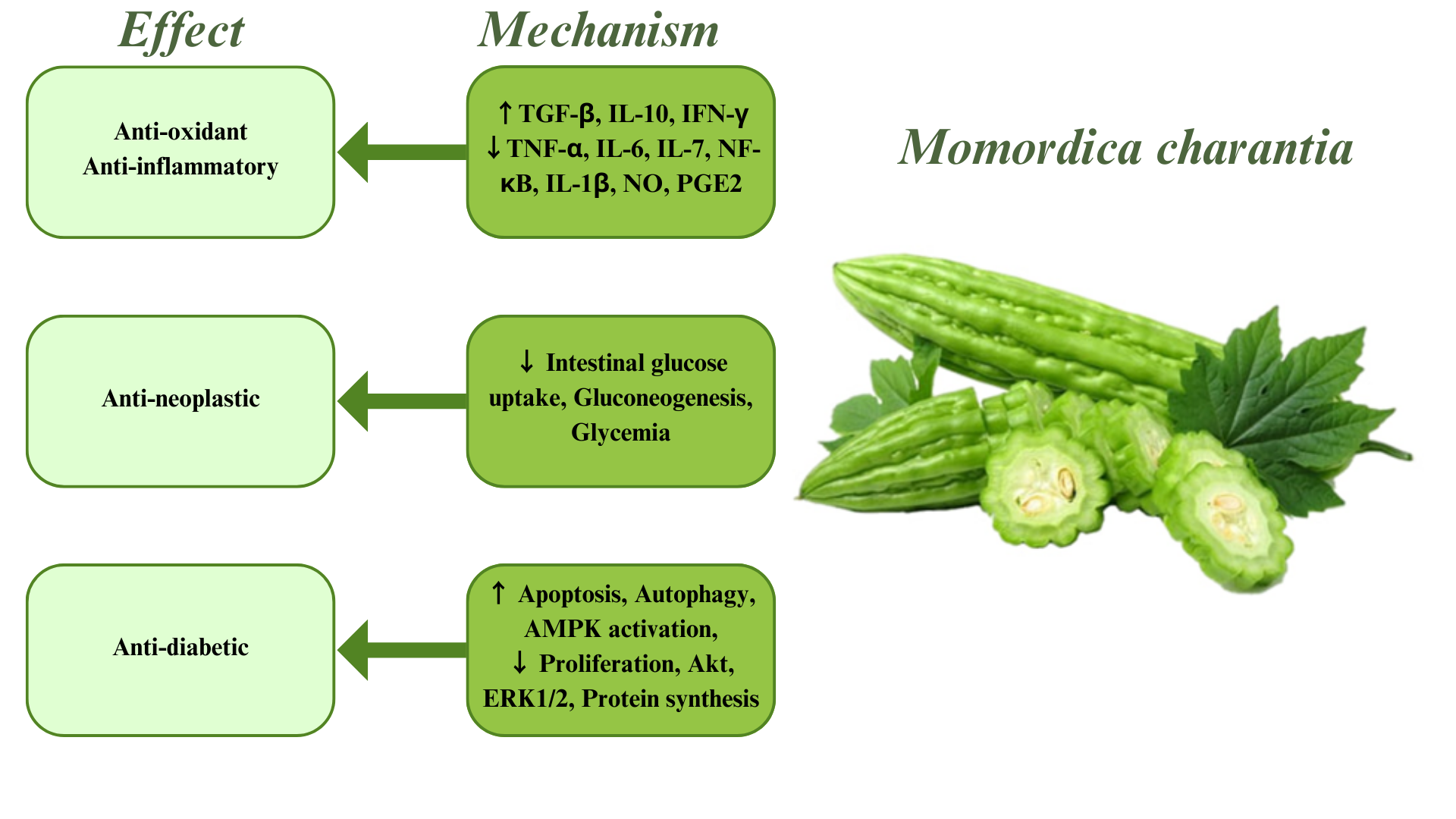
Fig. 1.

Effects and related mechanisms of Momordica charantia. TGF-β, transforming growth factor beta; IL-10, interlukine-10; IFN-γ, interferon gamma; NK-κB, nuclear factor kappa-light-chain-enhancer of activated B cells; NO, nitric oxide; PGE2, prostaglandin E2; TNF-α, tumor necrosis factor-alpha; IL-1β, interleukin-1 beta; AMPK, adenosine monophosphate-activated protein kinase; Akt, protein kinase B; ERK1/2, extracellular signal-regulated kinase 1/2.

2. Retrieval Strategy

We used the online databases Scopus, Google Scholar, and PubMed to retrieve all articles related to the connection between M. charantia and neurological diseases, such as AD, dementia, PD, brain tumors (glioma and glioblastoma), neuroinflammation and neurotoxicity, oxidative stress, epilepsy, and seizure among the elderly, without regard for article date. The search was carried out on March 7th, 2022. For each of the databases we used momordica charantia (MeSH) terms with a specific strategy, e.g.,

([bitter gourd] or [momordica charantia]) and ([Alzheimer disease] or [Alzheimer dementia])

([bitter gourd] or [momordica charantia]) and ([glioma] or [glial cell tumor])

([bitter gourd] or [momordica charantia]) and ([Parkinson disease] or [idiopathic Parkinson’s disease])

([bitter gourd] or [momordica charantia]) and ([neurotoxicity syndrome] or [neurotoxin disorder])

#1 Bitter Gourd

#2 Momordica charantia

#3 Momordica charantias

#3 #1 or #2

#4 Alzheimer’s disease

#5 Alzheimer’s dementia

#6 Memory

#7 dementia

#8 glioma

#9 glial cell tumor

#10 glioblastoma

#11 Parkinson’s disease

#12 Idiopathic Parkinson’s disease

#13 neurotoxicity syndrome

#14 neurotoxin disorder

#15 neuroinflammation

#16 Neuroinflammatory diseases

#17 neuroprotective

#18 neuroprotection

#19 seizure

#20 epilepsy

#21 #4 or #5 or #6 or #7 or #8 or #9 or #10 or #11 or #12 or #13 or #14 or #15 or #16 or #17 or #18 or #19 or #20

#22 #21 and #3

(((bitter gourd [Title/Abstract]) OR (Momordica charantia [Mesh]) OR (Momordica charantia [Title/Abstract]) OR (Momordica charantias [Title/Abstract]) OR (charantias, Momordica [Title/Abstract]) OR (Bitter Melon [Title/Abstract]) OR (Bitter Melons [Title/Abstract]) OR (Melon, Bitter [Title/Abstract]) OR (Melons, Bitter [Title/Abstract]) OR (Gourd, Bitter [Title/Abstract]) OR (Bitter Gourd [Title/Abstract]) OR (Bitter Gourds [Title/Abstract]) OR (Gourds, Bitter [Title/Abstract]) OR (Karela [Title/Abstract]))) AND (((Alzheimer Disease [Mesh]) OR (Alzheimer Disease [Title/Abstract]) OR (Alzheimer [Title/Abstract]) OR (Alzheimer dementia [Title/Abstract]) OR (memory [Title/Abstract]) OR (dementia [Title/Abstract]) OR (Glioma [Mesh]) OR (Glioma [Title/Abstract]) OR (glial cell tumor [Title/Abstract]) OR (Glioblastoma [Title/Abstract) OR (Parkinson Disease [Mesh]) OR (Parkinson [Title/Abstract]) OR (idiopathic Parkinson’s disease [Title/Abstract]) OR (Neurotoxicity Syndromes [Mesh]) OR (Neurotoxicity [Title/Abstract]) OR (neuroinflammation [Title/Abstract]) OR (Neuroinflammatory Diseases [Mesh]) OR (Neuroprotective [Title/Abstract]) OR (“ALS” [Title/Abstract]) OR (“MS” [Title/Abstract]) OR (“Multiple Sclerosis” [Mesh]) OR (“Amyotrophic Lateral Sclerosis” [Mesh]) OR (Neuroprotection [Title/Abstract]) OR (seizure [Title/Abstract]) OR (epilepsy [Title/Abstract])))

In total, 119 articles were retrieved and 106 articles remained after removing duplicates. We then reviewed the titles and abstracts of these articles, and 64 articles were eligible for assessment of the full text. After full text assessment, 30 articles did not meet the criteria as being an original article or review presenting data on the effectiveness of M. charantia on the listed conditions and were excluded. Thirty-four studies remained and were included in this review (Fig. 2).
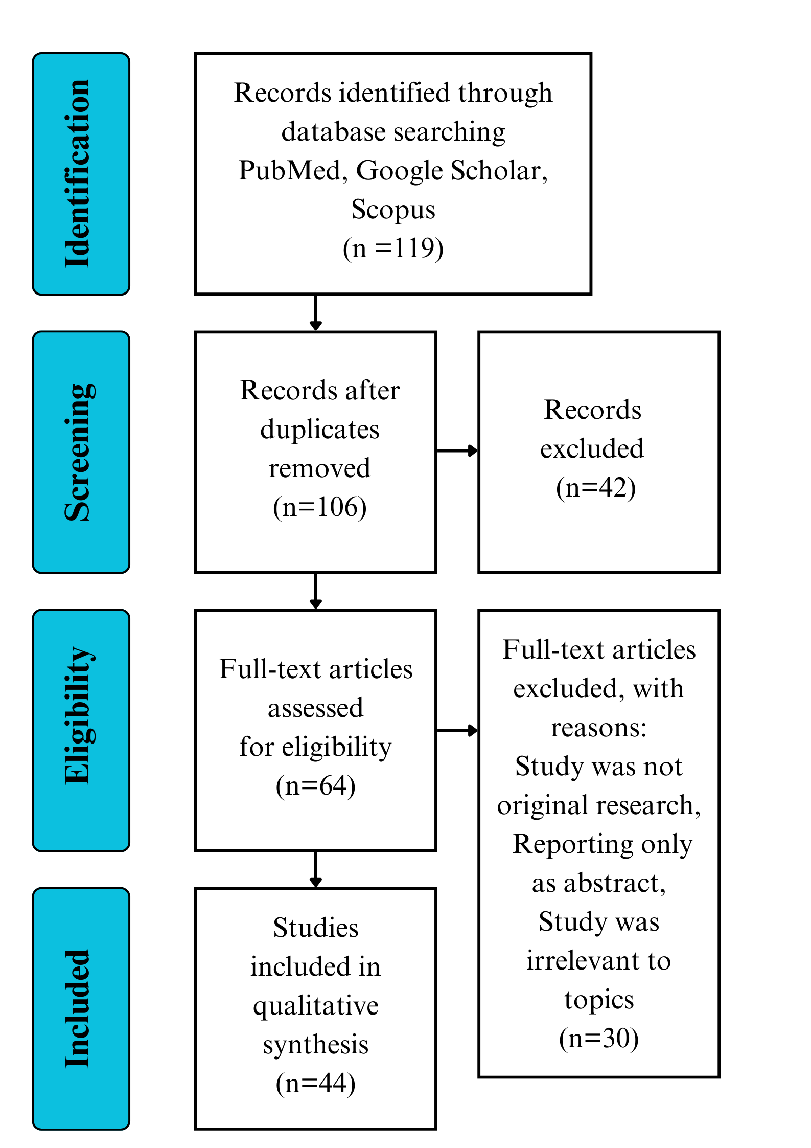
Fig. 2.

Outline of the review process.

3. Findings
3.1 Active Components of M. charantia

M. charantia belongs to the genus Momordica and the family Cucurbitaceae. M. charantia can grow at high temperatures and is used as an edible vegetable in Asia. This plant grows in a tropical and warm climate; its tree is found in parts of Asia, the Americas, Africa, and the Caribbean. This plant may have therapeutic effects on diabetes, cancer, obesity, and bacterial and viral infections [18].

For centuries, people have used medicinal plants to make products that have been used to prevent and treat diseases. M. charantia is widely distributed in tropical and subtropical regions of the world. It is used in folk medicine to treat diabetes, and its fruit has been used as a vegetable for thousands of years [19].

M. charantia contains many substances, including various biological molecules such as proteins and carbohydrates. It also contains minerals such as different vitamins, essential oils, and alkaloids. Other substances found in M. charantia include phenolic acids, quinines, and triterpenoids. Fig. 3 shows the structure of some of the small molecules in this plant [20].
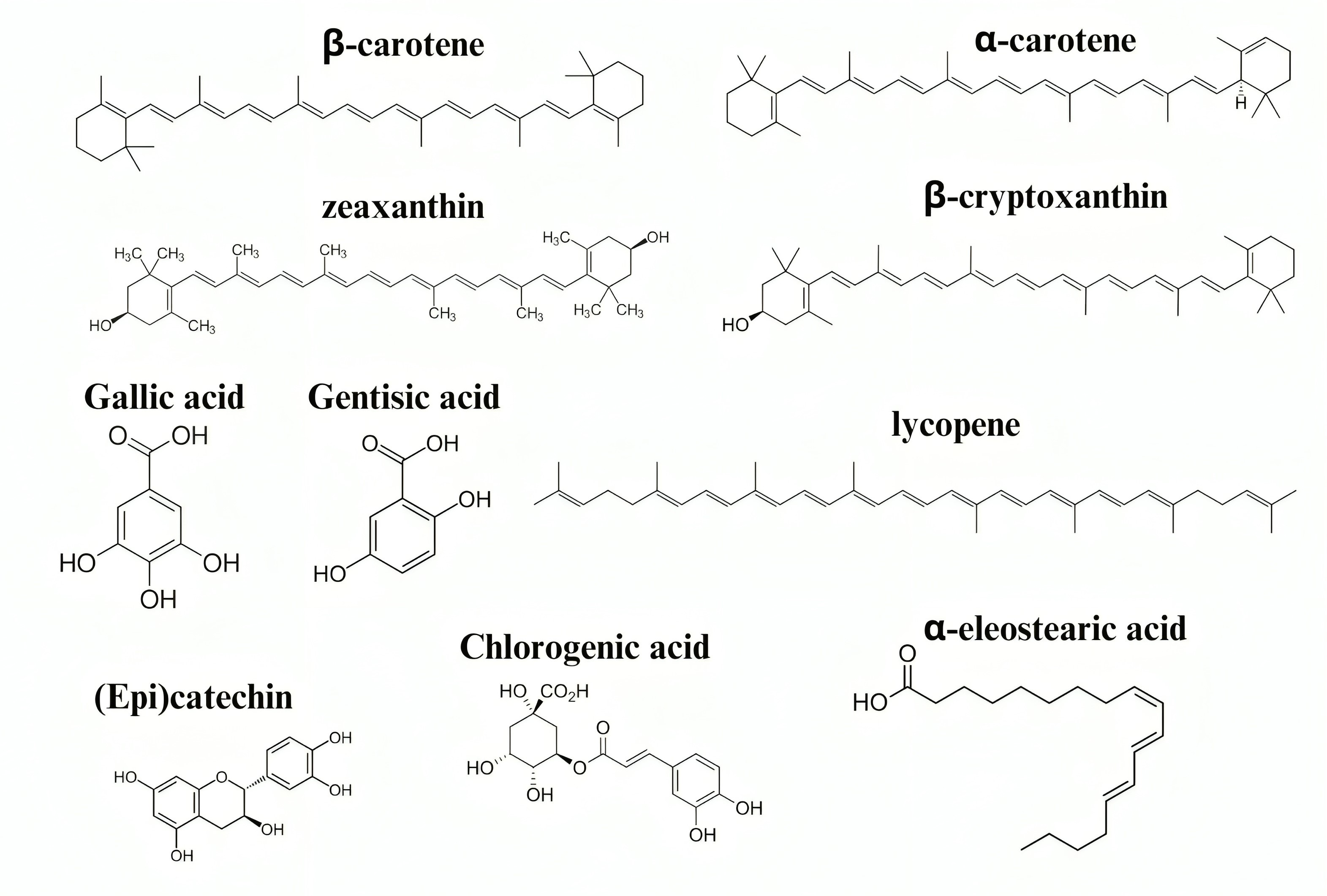
Fig. 3.

Structure of some of the active compounds found in M. charantia that act against age-related neurological diseases.

M. charantia can inhibit cell death via phosphoinositide 3-kinase/protein kinase B (PI3K/Akt) pathway blockade in glioma and glioblastoma. In addition, in AD, it suppresses amyloid-beta (Aβ) accumulation, reduces tau protein, and positively affects acetylcholine (ACh) and serotonin. In neuroinflammation, it has a role in inhibiting nuclear factor kappa-light-chain-enhancer of activated B cells (NF-κB), which can produce reactive oxygen species (ROS), inducible nitric oxide synthase (iNOS), cyclooxygenase (COX), and inflammatory cytokines. Moreover, M. charantia can suppress acetylcholinesterase and Aβ accumulation, which contribute to ROS production in neurotoxicity.

M. charantia has an anti-cancer effect through the modulation of α- and β-momorcharin, which activates caspase-3 and caspase-9, activating cytochrome C in mitochondria and increasing intracellular Ca2+, enhancing apoptosis of tumor cells.

M. charantia has neuroprotective roles through the enhancement of soluble guanylate cyclase. In PD, it inhibits 1-methyl-4-phenyl-1,2,3,6-tetrahydropyridine (MTPT), which produces inflammatory cytokines, and also affects toll-like receptor 4 (TLR4), inhibiting the Myeloid differentiation primary response 88 (MyD88)/NF-κB pathway. It can also positively affect dopamine in PD. M. charantia can improve memory by reducing tau protein and increasing microtubule-associated protein 2 (MAP2), postsynaptic density protein 95 (PSD95), and beta-site amyloid precursor protein cleaving enzyme 1 (BACE-1).

M. charantia has therefore been used for many different neurological disorders, as shown in Table 1 [21, 22, 23, 24, 25, 26, 27, 28, 29].

Table 1.Summary of studies investigating neurological disorders and M. charantia included in our review.
Author Year Type of study Neurological disorder Outcome
Qiaoli Li et al. [21] 2018 In vitro Epilepsy Momordica charantia polysaccharide (MCP) treatment significantly reduced malondialdehyde levels, increased superoxide dismutase and catalase activities, and mitigated Kainic acid (KA)-induced neuronal loss in the Cornu Ammonis 1 (CA1) and CA3 regions of hypocampus.
Pratibha V Nerurkar et al. [22] 2011 In vivo mouse Neuroinflammation Blue mussel treatment improved blood–brain barrier permeability, normalized neuroinflammatory markers, reduced oxidative stress, and normalized plasma antioxidant enzymes and pro-inflammatory cytokines in mice fed a high-fat diet, reducing glial cell activation, forkhead box O (FoxO), sirtuin 1 (Sirt1) protein expression, and up-regulation of Sirt3 mRNA expression.
Seung Mi Sin et al. [23] 2021 In vivo mouse model Alzheimer’s disease (AD) M. charantia’s butanol (BuOH) fraction improved learning and memory by reducing time in the Morris water maze test. It also reduced lipid peroxidation and nitric oxide levels, thus reducing cognitive impairment caused by A25-35.
Hei-Jen Huang et al. [24] 2018 In vivo mouse AD The neuroprotective effect of momordica charantia (MC5), MC3, MC2, and MC5523 is mediated by hyperglycemia or tau hyperphosphorylation. A combination of MC5523 and lithium chloride (LiCl) was administered to ovariectomized mice, boosting survival, increasing neuroprotection, and preventing memory deficits. This suggests that MC5523 and LiCl could be a potential treatment for AD.
Dengjun Guo et al. [25] 2021 In vitro mouse Parkinson’s disease 1-methyl-4-phenyl-1,2,3,6 tetrahydropyridine (MPTP) treatment can cause brain damage, alter neurotransmitter metabolism, cause inflammation, and induce apoptosis. However, M. charantia polysaccharide treatment can reverse these changes, regulating Toll-like receptor 4 (TLR4)/Myeloid differentiation primary response 88 (MyD88)/Nuclear factor kappa-light-chain-enhancer of activated B cells (NF-κB) pathway activation, proving an anti-inflammatory mechanism.
Gunasekar Manoharan [26] 2019 In vivo Glioma M. charantia’s anti-carcinogenic properties were tested on five cancer cell lines. The extract, combined with paclitaxel, significantly decreased cell viability, a significant decrease compared with paclitaxel alone, suggesting its potential for xenobiotic metabolism and oxidative stress.
Zafar Ahmad Malik et al. [27] 2011 In vivo mouse Cerebral ischemia M. charantia juice significantly reduced oxidative stress, damage, and neurological impairments in brains, and exhibited dose-dependent antihyperglycemic action in diabetic mice.
Ankur Joshi et al. [28] 2017 In vivo rat Memory enhancing activity M. charantia significantly corrected scopolamine-induced amnesia and decreased cholinesterase (ChE) activity in rat brain after 7 and 14 days of administration.
Tamilanban Thamaraikani et al. [29] 2020 In silico AD The study found that MC5523 and LiCl, when combined with streptozotocin, increased neuroprotection and anti-gliosis in ovariectomized (OVX) 3Tg-AD mice. This treatment also prevented memory deficits and enhanced synaptic-related protein expression. Luteolin, a compound responsible for AD, showed high affinity for its target protein, indicating its potential as a potential treatment. The study suggests that MC5523 and LiCl could be a potential treatment for AD.
3.2 Protective Mechanisms

Neuroprotection is defined as the ability of a therapy to prevent neuronal cell death by intervening in and inhibiting the pathogenetic cascade that results in cell dysfunction and eventual death [30].

Ghanta et al. [31] analyzed the docking of the phytoconstituents of M. charantia with the catalytic domain of guanylate cyclase. A lyase enzyme known as soluble guanylate cyclase is vital in treating cardiovascular and neurodegenerative disorders. Reduced glutamate excitotoxicity caused by modulating soluble guanylate cyclase activity benefits PD [32]. The soluble guanylate cyclase is required for cyclic guanosine monophosphate (GMP) synthesis from guanosine triphosphate (GTP) [33, 34]. A heme-containing heterodimer with alpha and beta subunits is also essential [35]. Several subunit isoforms exist: alpha 1, alpha 2, beta 1, and beta 2 [36]. Alpha 1 and beta 1 are the two most common subunits in the human brain [37]. In humans, the heme part of the beta 1 subunit acts as the active site [32].

Numerous studies have been performed to identify the allosteric binding sites of the enzyme that regulates soluble guanylate cyclase activity. The search for binding sites for allosteric enzymes is limited to their catalytic domain. Defects or inhibitions in this region can lead to enzyme dysfunction. 1H-[1,2,4]oxadiazolo-[4,3-a]quinoxalin-1-one (ODQ), a specific inhibitor targeting the catalytic domain of soluble guanylate cyclase, was proven to show antiparkinsonian activity [38].

A water extraction alcohol precipitation process used to process unripe fruit extract from M. charantia showed complete inhibition of soluble guanylate cyclase [39]. Several subsequent studies have confirmed M. charantia’s ability to inhibit soluble guanylate cyclase [40, 41]. M. charantia extracts showed neuroprotective effects mainly because of their D-galacturonic acid content [42].

Antioxidants scavenge free radicals from the body’s cells and prevent or reduce the damage caused by oxidation. The protective effect of antioxidants continues to be studied around the world. Oxidative stress is caused by the mass production of ROS, primarily because of an imbalance of oxidative and neutralizing processes [43]. Excessive ROS increases age-related disease, triggering inflammation and proatherogenic activities. One of the mechanisms by which antioxidant agents show their effects is reducing the ROS levels in cells. Antioxidants prevent the aging process and age-related and degenerative diseases by helping to balance the production cycle and neutralize free radicals [44, 45, 46, 47].

The polysaccharides in M. charantia are found to be antioxidants. According to an in vivo study, M. charantia polysaccharide could play a role in detoxification, improving immunity, and decreasing blood sugars [48]. Furthermore, the polysaccharide may increase chloramphenicol acetyltransferase (CAT) and superoxide dismutase (SOD) in mouse serum, liver, and spleen. It can also reduce malondialdehyde (MDA) in these tissues [49]. MDA is considered one of the most vital products in the process of membrane lipid peroxidation. The polysaccharides found in M. charantia might therefore possess antioxidant properties [21]. Another study showed that using the polysaccharides found in M. charantia for treating kainic acid-induced epileptic mice could decrease MDA levels and increase the function of SOD and CAT in the rat hippocampus. As a result, the antioxidant and free radical scavenging activities of M. charantia polysaccharides can significantly reduce neuronal damage in the brain and central nervous system [50].

In addition, Chen et al. [50] found that phosphorylated polysaccharides and sulfated polysaccharides of M. charantia have different sugar levels. Introducing phosphate and sulfate can therefore enhance the anti-lipid peroxidation capability and scavenging capacity of superoxide anions in the M. charantia polysaccharides [51].

M. charantia is also rich in phenolic compounds. Gallic acid is the main phenolic acid found in M. charantia. Several studies support that the phenolic compounds of M. charantia have antimutagen and antioxidant effects [52, 53]. The antioxidant effect signified by the Half maximal inhibitory concentration (IC50) index, a 50% decrease in radical absorbance, was higher in solvent and Soxhlet than in subcritical water extraction, meaning the second extraction had better antioxidant activity. Therefore, the authors suggest M. charantia as a good source of antioxidants [52]. Lin et al. [53] examined the antioxidant effects of M. charantia in various aspects over time and at different temperatures. The results showed that the antioxidant activity was significantly reduced after 3 days of storage, and the reduction rate was more significant at higher temperatures. Abbas et al. [46] divided M. charantia into separate groups to achieve higher amounts of antioxidants. The investigators concluded that treating at the seedling stage can affect the amount of antioxidants in M. charantia.

The antioxidant effect of aqueous extract and phenolic extract of M. charantia in protecting against damage from hydrogen peroxide (H2O2) and hypoxanthine-xanthine oxidase (HX-XO) was evaluated in a cell-based assay. The aqueous extract had no toxic effect at the same dose as the phenolic extract. According to this study, M. charantia extract is rich in phenolics; it has vigorous antioxidant activity and effective radical removal [54, 55].

Neurotoxicity occurs when exposure to natural or manufactured toxic substances (neurotoxicants) alters the normal activity of the nervous system. This can eventually disrupt or even kill neurons, key cells that transmit and process signals in the brain and other parts of the nervous system [56]. Neurotoxicity can result from exposure to substances used in chemotherapy, radiation treatment, drug therapies, and organ transplants, as well as exposure to heavy metals such as lead and mercury, certain foods and food additives, pesticides, industrial or cleaning solvents, cosmetics, and some naturally occurring substances. Symptoms may appear immediately after exposure or after a delay [57]. They may include limb weakness or numbness, memory loss, vision. Individuals with certain disorders may be especially vulnerable to neurotoxicants [58].

In 2021, Kumar et al. [58] experimented with the anticholinesterase and antioxidant activity of M. charantia in Danio rerio (zebrafish). They used trimethyltin chloride (TMT) to induce neuronal death. Six groups were used in this in vivo study as follows: control, TMT (day1), TMT (day2), TMT + food and drug administration (FDA)-approved drug for AD (donepezil), TMT + M. charantia, and TMT + (M. charantia + mesoporous silica nanoparticle) [59, 60, 61]. Their results showed that M. charantia causes no adverse changes in behavior and mortality during treatment. Furthermore, no pathological changes were seen in the brain histopathological analysis of M. charantia-treated fish. The results of the acetylcholinesterase activity assay indicated that M. charantia at the concentration of 50 g/mL caused significant inhibitory activity of acetylcholinesterase compared with the positive control group (donepezil-treated group). In addition, the aqueous M. charantia showed significant antioxidant activity compared with ascorbic acid, the standard antioxidant in the positive control, according to 2, 2-diphenyl-1-picrylhydrazyl (DPPH) radical scavenging activity, a method for evaluating antioxidant potential. The results of the total antioxidant assay indicated a significant total antioxidant activity of M. charantia compared with the positive control (ascorbic acid). In addition, the lipid peroxidation assay showed that M. charantia had significant lipid peroxidation activity compared with MDA (as a standard antioxidant, positive control) [62].

3.3 Potential Pharmacological Applications in Neurological Disorders
3.3.1 Role of M. Charantia in Neuroinflammation

Neuroinflammation is a crucial part of the general pathology of several chronic and severe problems in the CNS [63]. The occurrence of neurologic diseases in people over 65 years is 1.8 and 2.6 for people aged 85 to 90 years, per 100 population [64]. The aggregation of damaging products caused by oxidative stress, such as glycated products, oxidized proteins, and lipid peroxidation, results in the destruction of neurons, which is more noticeable in brain disorders. Cerebrovascular diseases are characterized by vascular lesions that cause cognitive decline and dementia in old age [65].

Signaling molecules generated throughout the process of neuroinflammation meditate a number of pro-apoptotic pathways. In operating microglial cells, a transcription factor named NF-κB induces and controls the expression of many vital components in inflammatory and immune feedback systems in the CNS and neurodegenerative disorders [65].

Kim et al. [66] found that protocatechuic acid (PA), an active phenolic compound of M. charantia, reduced the increased levels of NF-κB, cyclooxygenase-2 (COX-2), and iNOS mRNA due to H2O2-induced oxidative stress. In addition, PA attenuated the production of interleukin (IL)-6 and reduced nitric oxide (NO) induced by endotoxin lipopolysaccharide (LPS) and interferon-gamma (IFN-γ) together. Furthermore, it reduced mRNA expression of iNOS and COX-2 induced by LPS and IFN-γ [22].

M. charantia supplementation significantly reduced high-fat diet-induced brain oxidative stress in C57BL/6 mice, reducing blood–brain barrier (BBB) leakage, neuroinflammatory cytokines, NF-κB1 expression, glial cell activation, and oxidative stress, while improving systemic inflammation [67].

Obesity increases the development of T-helper (Th) 17 cell genealogy, leading to inflammation [68]. IFN-γ, IL-17, and IL-22 are major cytokines liberated from Th1 and Th17 CD4+ cells. M. charantia decreases inflammation by reducing the liberation of plasma pro-inflammatory cytokines, IFN-γ, and IL-17 with tumor necrosis factor-alpha (TNF-α), exotoxin-2, lymphotactin, and IL-1β in mice fed with blue mussels (BM). Furthermore, M. charantia adjusted the Th2 cytokines IL-5, IL-10, and IL-13 to preserve obesity-associated inflammation [69]. However, in another study, the results were different, and Th2 cytokines such as IL-4 were shown to aggravate weight gain and high fat diet (HFD)-related hypothalamic inflammation [70]. In some studies, M. charantia adjusted neuroinflammatory and peripheral reciprocation by regulating the BBB [71, 72, 73, 74, 75]. Increased expression of IL-22 in the brains of HFD-fed mice may indicate glial cell activation, and feeding HFD to Sprague-Dawley rats reduced Sirtuin 1 (SIRT1) levels in the cerebral cortex and hippocampus, promoting oxidative stress. The observed 25% decrease of SIRT1 protein in the brains of HFD rats is lower than the 81% decrease reported by Wu et al. [75] in the cerebral cortex and hippocampus of HFD-fed mice. The levels of SIRT1 were different in the studies by Wu et al. [75] and others.

Horax et al. [76] reported that M. charantia contains various flavonoids, bitter triterpene aglycones, non-bitter cucurbitane, and glycosides that transmit antioxidants. Horax et al. [76] also showed that catechin is a major polyphenol in the seeds of BM and pericarp.

Deng et al. [77] used the chronic social defeat stress (CSDS) mouse model to assess the efficacy of Momordica charantia polysaccharide (MCP). MCP prohibits depressive-like behaviors brought on by CSDS. Moreover, MCP diminishes the amount of pro-inflammatory cytokines and restrains The c-Jun-NH(2)-terminal kinase (JNK3) and c-Jun production in the hippocampus. Furthermore, MCP protects against depressive-like behaviors in mice. Consequently, the protective efficacy of MCP in CSDS mice against depressive-like behaviors could be because of reduced neuroinflammation and down-regulation of the JNK3/PI3K/Akt pathway in the hippocampus [78].

In 2020, Kung et al. [78] studied the effects of wild bitter melon (WBM) on mouse models with spinal cord injuries (SCI), which induced secondary neuroinflammation. The investigators also used astrocyte-like cells stimulated by LPS for in vitro studies of astrocyte inflammation. They found that WBM reduces spinal cord injury. Furthermore, WBM, which contains alpha-eleostearic acid (α-ESA), increases the viability and survival ability of astrocyte-like cells.

Mitochondrial malfunction and inflammation can lead to irreparable neuronal deficits. Chitosan/gelatin/sodium hyaluronate (CGSH) iron-sulfur domain 2 is a protein located on the outer membrane of mitochondria and is involved in maintaining the integrity of mitochondria and inhibiting apoptosis [79, 80]. Furthermore, CGSH iron-sulfur domain 2 reduces inflammatory responses caused by CNS injuries. Aging downregulates the expression of CGSH iron-sulfur domain 2, which aggravates mitochondrial malfunction and increases inflammatory responses [81, 82]. Spinal cord injury-induced downregulation of IL-4 is reduced by WBM because losing IL-4 increases microglial polarization from M2 to M1 [83]. WBM can increase the volume of M2 microglia, which are anti-inflammatory. The anti-inflammatory effects of WBM are therefore facilitated through the increase of CGSH iron-sulfur domain 2 in mice with spinal cord injuries and astrocyte-like cells stimulated with LPS. In addition, its protective effects in mice are shown by the deactivation of astrocytes and the downregulation of NF-κB [83].

3.3.2 M. Charantia and Headaches, Amyotrophic Lateral Sclerosis, and MS

There have been reports of headaches following the consumption of M. Charantia seeds; however, there is no information regarding amyotrophic lateral sclerosis and headache severity or duration, or effectiveness as a treatment [84].

3.3.3 M. Charantia and MS

Macrophages are part of the first line of defense against injury, mediating the innate non-specific immune response via inflammation, and playing an important role not only in host defense but also in tissue homeostasis, repair, and pathology development, including MS. RAW 264.7 mouse derived macrophages have been widely used as an in vitro model to study the modulatory effects of various compounds using LPS for activation. It has been observed that treatment with Bitter Gourd (BG)-4 peptide extracted from M. Charantia has anti-inflammatory effects. In the investigation conducted by Nieto-Veloza et al. [84], BG-4 doses up to 375 g/mL had no effect on macrophage viability. It is known that LPS can activate NF-κB, which acts as a master regulator of the inflammatory response by promoting the release of signaling and effector molecules such as pro-inflammatory cytokines (IL-6, TNF-α, IL-1β) and NO that act as mediators of inflammation during the host immune response. MS and other inflammatory problems can be caused by the destruction of normal and healthy tissue caused by a continuous inflammatory condition; therefore, reducing the quantity of these pro-inflammatory produced molecules can lead to the management and treatment of these diseases. BG-4 dose-dependently inhibits NO and IL-6 production in LPS-activated RAW 264.7 macrophages. Moreover, BG-4 reduces the expression of iNOS and COX-2 [85].

3.3.4 M. Charantia and AD

AD is among the most prevalent of neurological diseases. Oxidative stress and the abnormal increases in Aβ are the most common causes of AD. Dementia is seen in patients with AD and occurs when Aβ accumulates in brain tissue, including in the cerebral cortex. Common treatments are cholinesterase inhibitors and receptor antagonists, but they cannot efficiently treat patients [23, 86, 87].

According to an in vivo study by Sin et al. [23], 100 and 200 mg/kg/day of butanol (BuOH) fraction derived from M. charantia can improve memory and learning in the Aβ (25-26)-induced AD mouse model. Furthermore, oral prescription decreased HNO3 and lipid peroxidation in the brain compared with the control group [24]. Combination therapy including M. charantia and lithium chloride in ovariectomized AD female mice reduced gliosis, neuronal loss, and tau hyperphosphorylation, suggesting the potential for AD treatment [88].

In an in-silico study [89], luteolin, extracted from M. charantia, increased ACh in neuronal cells by attaching to acetylcholinesterase and butyrylcholinesterase. The authors suggested luteolin as a multi-target molecule against AD. Luteolin can therefore effectively act as a multi-target molecule against AD. The summarized effects of M. charantia on age-related neurological disorders is shown in Figs. 4,5.
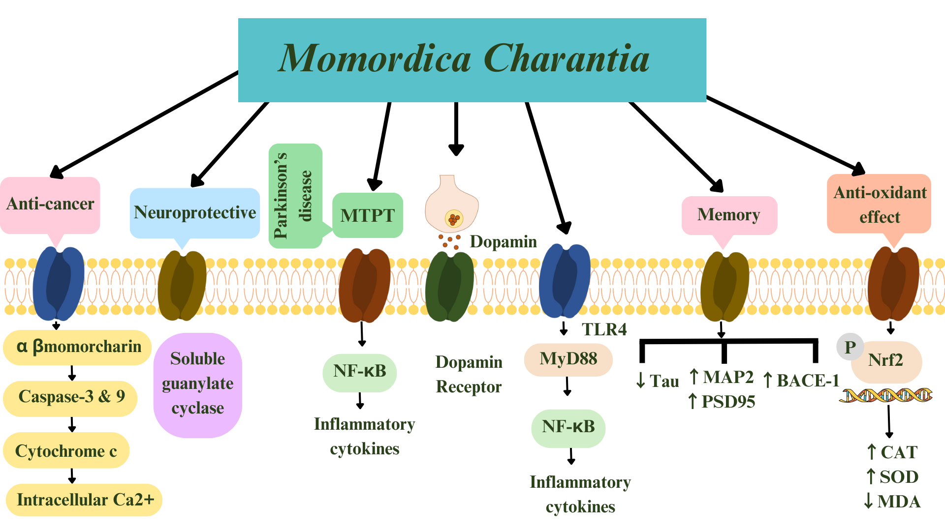
Fig. 4.

Summary of Momordica charantia’s effects including anti-oxidant, anti-cancer, anti-glioma, anti-Alzheimer’s disease, and anti-Parkinson’s disease. Arrows indicate mechanism of actions of M. charantia. Keap1 is the factor accommodating Nrf2. MTPT, 1-methyl-4-phenyl-1,2,3,6-tetrahydropyridine; NF
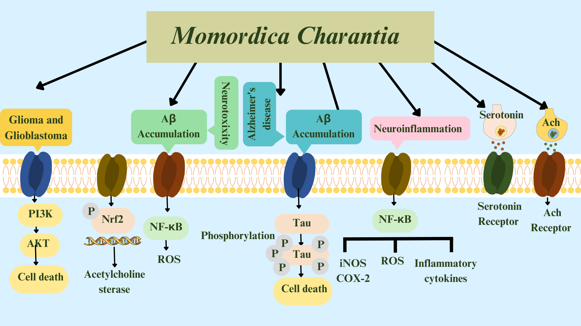
-κB, nuclear factor kappa-light-chain-enhancer of activated B cells; TLR4, toll-like receptor 4; MAP2, microtubule-associated protein 2; BACE-1, beta-site amyloid precursor protein cleaving enzyme 1; PSD95, postsynaptic density protein 95; Nrf2, nuclear factor erythroid 2-related factor 2; CAT, chloramphenicol acetyltransferase; SOD, superoxide dismutase; MDA, malondialdehyde; MyD88, myeloid differentiation primary response 88; P, phosphorylation; CAT, chloramphenicol acetyltransferase; Tau, tubulin associated unit.

Fig. 5.

Summary of pathways: Momordica charantia acts in Gliomas, Glioblastomas, neurotoxicity, and Alzheimer’s disease, and on neuroinflammation, serotonin, and acetylcholine. Glutamate-activated AMPA and NMDA receptors mediate Aβ accumulation. ROS, reactive oxygen species; ACh, acetylcholine; NF-κB, nuclear factor kappa-light-chain-enhancer of activated B cells; iNOS, inducible nitric oxide synthase; COX-2, cyclooxygenase-2; AMPA, α-amino-3-hydroxy-5-methyl-4-isoxazolepropionic acid; NMDA, N-methyl-D-aspartate; PI3K, phosphoinositide 3-kinases; Akt, protein kinase B; P, phosphorylation; Aβ, amyloid-beta.

A study [90] reviewed the therapeutic role of α-ESA, an isomer of conjugated linolenic acids produced from wild bitter melon (Momordica charantia L. var. abbreviata Seringe), and curcumin in CNS illnesses. In addition, the authors highlighted the probable role of glia-mediated neuroinflammation, mitochondrial dysfunction, CDGSH iron-sulfur domain 2 (CISD2) loss, and NF-κB activation in CNS traumas and disorders. Their review enhances our understanding of the CNS pathology–CISD2–NF-κB axis and clarifies the possible therapeutic role of these natural chemicals [90].

A study [90] investigated the neuroprotective benefits of sulforaphane (SFN) on cognitive illnesses such as AD, PD, Huntington’s disease, amyotrophic lateral sclerosis, MS, autistic spectrum disorder, and schizophrenia in a separate review. they described the anti-AD-like action of SFN and how it reduced levels of AD biomarkers, including Aβ, tau, inflammation, oxidative stress, and neurodegeneration in AD-like animal and cell models. In addition, the authors concentrated on the probable mechanisms behind the neuroprotective benefits of SFN. This review reveals that SFN has multiple neuroprotective effects on the pathophysiology of AD, highlighting the necessity to continue SFN research [90].

Yoshinori Okada and Mizue Okada [90] evaluated the protective effects of plant seeds against Aβ-induced neurotoxicity in hippocampal neurons. They examined the aqueous extracts of 15 plant grains for their ability to inhibit Aβ (25-35)-induced cell death using hippocampal neurons. The aqueous juices showed antioxidant properties. Furthermore, intracellular cumulative ROS resulting from Aβ declined when cells were cured with some of these extracts. Kale, bitter melon, red shiso, kaiware radish, and corn inhibited TNF-α in Aβ-stimulated neurons and, in all instances, Japanese honeywort displayed better cell survival. This study showed that some plant seed juices offer protection against Aβ-mediated death in cells.

Katsouri et al. [91] showed promising results regarding a lithium chloride (LiCl) compound, including increased cognition and short-term memory, as well as decreased amounts of oligomer, tau protein phosphorylation, and BACE-1 expression, and improved expression of synaptic plasticity-related proteins. Other neuroprotective results have been reported by synthesizing LiCl with L-dopa or other histone deacetylase inhibitors [67, 92, 93, 94, 95, 96, 97, 98, 99, 100, 101]. Many studies showed that synthesizing therapies with multifold drugs has better therapeutic potential for AD [102, 103, 104, 105].

3.3.5 M. Charantia and Epilepsy

Epilepsy is a common condition that affects the brain and causes frequent seizures. Seizures are bursts of electrical activity in the brain that temporarily affect how it works, causing many symptoms. Epilepsy can start at any age but usually it starts in childhood or in people over 60 years [106]. The drugs that are currently available to treat epilepsy might adversely affect human health. The herbal medicines that have been used in the past and traditional medicines have fewer side effects [107].

Soliman et al. [107] investigated the anticonvulsant potential of M. charantia in rats. Thirty minutes after treatment, ear electrodes were used to give rats a 150 mA shock. According to the reported results, M. charantia might have tremendous anticonvulsant effects against maximal electroshock-induced seizures and reduce the duration and delay the onset of seizures [108].

3.3.6 M. Charantia and PD

PD is the second most common neurodegenerative disease after AD [109, 110]. The cause of PD is not yet well understood, but genetic and environmental factors, including poisons, are proven to contribute to its development [111]. In PD, dopaminergic neurons in the substantia nigra compact area are lost, and Lewy bodies accumulate in the brain [112]. The active metabolite of 1-methyl-4-phenyl-1,2,3,6 tetrahydropyridine (MPTP) is N-methyl 4-phenylpyridinium (MPP+). MPTP is a neurotoxin that specifically targets dopaminergic neurons and causes PD [113]. Evidence indicates that inflammatory damage and oxidative stress can accelerate the disease [25].

Natural ingredients such as polysaccharides in plants delay aging and protect the brain. Momordica charantia polysaccharides (MCPs) have been studied for their antioxidant, anti-inflammatory, anti-tumor, hypoglycemic, and anti-diabetic effects [19]. Little is known about their role in the regulation of neurogenesis [114]. However, MCPs reduce nerve damage after stroke by scavenging free radicals [42].

MCPs showed neuroprotective effects in MPTP- and MPP+-induced PD in mice. A study on mice reported that MCPs alleviated the exercise instability and loss of coordination caused by MPTP, inhibited the release of inflammatory factors and oxidative stress products in the brain, and, as a result, increased dopamine levels. Regarding cell function, MCPs inhibit apoptosis and oxidative stress induced by MPP+. The authors also observed that MCPs can protect against oxidative stress by inhibiting the transcription of Toll-like receptor 4 (TLR4)/Myeloid differentiation primary response 88 (MyD88)/NF-κB [114]. The TLR4/MyD88/NF-κB pathway is thought to play a vital role in the inflammatory process [115, 116]. Observing the changes in this signaling pathway showed that MCPs can affect the status of the TLR4/MyD88/NF-κB pathway. There are various TLR4 inhibitors, among which Resatorvid (TAK-242) is a bioavailable TLR4 inhibitor that inhibits extensive inflammation [117]. As a result of the combined use of MCPs and TAK-242 in the PD condition, the investigators found that TAK-242 could reverse the protective effects of MCPs, therefore proving the effectiveness of MCPs on the TLR4/MyD88/NF-κB signaling pathway [114].

3.3.7 Anti-Cancer Effects of M. Charantia

Increasing the average life expectancy is one of the most significant achievements of the past century [118, 119]. However, a healthy lifestyle and life expectancy free of disease have not increased as much [118, 120]. In addition to disability and reduced physical strength, aging is a risk factor for chronic diseases and cancer, and it has become a pervasive challenge [118, 121, 122]. Research to identify the causes of these destructive changes in the body that occur with the aging process and find a solution to reduce these changes will therefore help to increase quality of life and personal productivity. Cancer and tumors are a group of age-related diseases that we are facing more often due to increases in the average age of society. Prolonged exposure to endogenous and exogenous factors contributing to oxidative stress can eventually lead to gene mutations and inflammatory processes [123]. Antioxidant therapy is therefore accepted as one of the main methods to limit cell damage caused by oxidative stress [124, 125]. Many studies have been conducted to find effective natural and artificial antioxidants for fighting excess free radicals. Currently, people older than 65 years comprise around 60% of all patients with malignant tumors, and they make up 69% of whole cancer deaths [126]. Based on etiology, some highlighted common causes of aging and cancers include oxidative damage and deoxyribonucleic acid (DNA) damage [127, 128, 129, 130], cellular senescence [131], and insulin/insulin-like growth factor-1 (IGF-1) signaling [132].

In a study by Manoharan et al. [132], the impacts of α- and β-momorcharin (200-800 µM) on WERI-Rb-1, SK-MEL, 1321N1, COR-L23, and U87-MG cancer cells lines were compared with the normal and balanced L6 myocytes line. α- and β-momorcharin decreased viability, increased cytochrome c release, and enhanced calcium concentration in treated cells. The authors reported 800 µM as the most effective concentration. Furthermore, other studies have suggested that α- and β-momorcharin can trigger a receptor on the membrane surface of cancer cells or may infiltrate the cell by its osmotic effect [133, 134, 135]. This substance is also assumed to damage the cancer cells’ mitochondria, leading to apoptosis by improving the caspase-3 and caspase-9 processes, releasing cytochrome c and intracellular Ca2+ [136, 137]. In conclusion, M. charantia, which contains α- and β-momorcharin, can effectively lead to cancer cell apoptosis and may be a suitable anti-cancer option.

M. charantia is a plant with medical effects, such as anti-inflammatory [138, 139, 140, 141, 142, 143, 144, 145] and anti-cancer effects, as well as anti-diabetic and antiviral activities [146, 147]. Its evaluation is therefore valuable for discovering its influential factors. Previous studies identified some of its beneficial components, including the ribosome-inactivating proteins α- and β-momorcharin, cucurbitacin B, and momordin, and the chemical analog MAP30 protein [148, 149, 150, 151]. A few study found two new triterpenoids, D [146] and E [147] charantagenin. A novel sterol, 7-oxostigmasta-5,25-diene-3-O-β-D-glucopyranoside, was isolated [152] in addition to eight known compositions [153, 154, 155, 156, 157, 158, 159]. The results confirmed that this novel composition was more abundant than the others and that another (guaglycoside D) contained an O-Methyl (OMe) substituent in the side chain, which was effective against cancer cells and had impressive cytotoxic effects. In addition, they had lower IC50 values compared with other components. It is worth noting that IC50 is applied to measure drug efficacy, as it corresponds to the potency of the drug. These findings suggest the level of OMe may be associated with the cytotoxic activity of cucurbitane-type triterpenoids. The cucurbitane-type triterpenes, especially from M. charantia, therefore probably have potential anti-cancer effects similar to chemotherapy.

Glioblastoma multiforme (GBM), also referred to as a grade four astrocytoma, is a fast-growing and aggressive brain tumor. It invades the nearby brain tissue but does not spread to distant organs. Gliomas are tumors that have a peak incidence in middle-aged humans [160]. Gliomas account for over half of all intracranial tumors [161]. The average annual age-adjusted incidence rate of glioma is estimated at 6 per 100,000 population [162]. These tumors are the most common primary tumors in the brain, originating from different glial cells, including oligodendrocytes, astrocytes, and ependymal cells [163]. Standard of care includes surgery, radiation therapy, and chemotherapy. However, due to the exceedingly invasive capability of glioblastoma cells, tumors develop over time and integrate into surrounding brain tissue [164].

Wang et al. [161] found that M. charantia impedes viability and reduces the multiplication of U251 glioma cells, repressing their influx, which has an anti-glioma effect. However, it had no considerable efficacy in the apoptosis of these cells. Moreover, M. charantia-derived extracellular vesicle-like nanovesicles exert anti-glioma effects by adjusting the PI3K/Akt signaling route [165]. In an in vitro study the effects of the anti-tumor activity of M. charantia (MAP30) on proliferation, migration, and invasion of the U87 and U251 cell lines were assessed [166, 167]. MAP30 inhibited U87 and U251 cell viability in a dose‑ and time‑dependent manner and decreased colony formation of these cell lines. It also induced apoptosis and S-phase cell cycle arrest by breaking the bonds of the adenine‑ribose glycoside. The invading proportions of the cells treated with MAP30 were significantly lower than their control counterparts. Western blot analysis indicated decreased leucine rich repeat containing G protein-coupled receptor 5 (LGR5) expression and increased Smac (activator of intrinsic apoptosis) expression in cells treated with MAP30. The Wnt/β‑catenin and LGR5 signaling pathways play a vital role in the tumorigenesis of gliomas [26, 168, 169, 170, 171]. Manoharan [26] investigated the performance of α- and β- momorcharin obtained from M. charantia by combining cyclophosphamide with the cellular mechanisms. Compared with cyclophosphamide’s effect, the results showed significant decreases in cell viability for each cell line in the presence of active substances [172].

Furthermore, 800 µg of the crude water-soluble M. charantia extraction in combination with 250 µg of paclitaxel showed a significant decline in cell viability of five cell lines mentioned earlier [173]. Another study on the U87G Glioblastoma cell line showed that M. charantia extraction displays a cytotoxic and anti-proliferative role and might be helpful as a therapeutic agent against GBM [174]. According to these studies, M. charantia can be considered a plant with pharmacological and nutritional properties. Its compounds make this plant a potential anti-carcinogenic agent and therapeutic aid for the treatment of glioma.

3.3.8 M. Charantia and Ischemia

Stroke is the second leading cause of disability and death worldwide, and has the most concerning and excessive burden in countries with revenue deficiency. The universal number of incidents was 13.7 million in 2016, and it is estimated that 87% of those were ischemic stroke [27]. Furthermore, stroke is one of the world’s most prevalent vascular ailments and remains the fifth leading cause of death in the United States [175]. Brain ischemia could be focal or multifocal and is caused by an abrupt cessation or diameter reduction of the artery supply of a region in the brain. The decreased brain blood supply leads to hemodynamic dysfunction, which contributes to damage to the brain tissue [28]. There are some uncommon causative agents of ischemia. Rarely, dissection of cervical blood vessels causes brain ischemia and may cause stroke in younger patients. Another rare cause of brain ischemia is vasospasm. An infection can also lead to stroke.

It should be noted that an increased stroke incidence has occurred with the COVID-19 pandemic [174]. Aging and related conditions, including diabetes, could be important factors in ischemia manifestation. Special attention must therefore be paid to ischemia as an age-related disorder.

The potential neuroprotective effect of the freeze-dried juice of fresh M. charantia in cerebral injury caused by ischemia-reperfusion was studied by Malik et al. [27] using cerebral infarct size, measuring thiobarbituric acid reactive substances (TBARS) and immediate memory and motor activity. Cerebral oxidative stress and damage with a shortfall in neurological functions were observed related to the dosage. The authors reported that the manifestations were extenuated by lyophilized M. charantia juice pre-treatment. M. charantia might therefore be an efficient option with neuroprotective activity in treating patients with stroke [27].

4. Conclusions

The aging process begins with molecular changes, such as epigenetic changes, telomere weakening, and the buildup of mutations, which causes genomic instability. These defects increase rapidly over time, with a “snowball effect”, and finally lead to a functional and morphological worsening of the brain, which includes excessive inflammation, reduced amounts of neurotransmitters, progressive neuronal damage, and damaged integrity of vessels, leading to microbleeds and infarction. Furthermore, the reduced effectiveness of the DNA repair systems makes us more vulnerable to spontaneous mutagenesis and ROS, leading to age-related neoplasia. In addition, the malabsorption and malnutrition usually seen in older people may lead to a deficiency in folic acid and vitamin B12, leading to vascular damage. These factors cause brain injuries in the elderly and increase the risk of CNS diseases such as epilepsy, dementia, PD, stroke, and AD [175].

M. charantia is used for diabetes, AD, glioma, neuro-inflammation, seizure, and PD. Its antioxidant compounds, including luteolin, increase acetylcholine in neurons, reduce cholesterol, and treat memory loss [28, 29]. It seems that M. charantia can also improve memory by decreasing tau protein and increasing MAP2, postsynaptic density protein 95 (PSD95), and beta-site amyloid precursor protein cleaving enzyme 1 (BACE-1) [166]. The neurotoxin MPTP targets dopaminergic neurons and causes PD [61]. MCPs reduce exercise instability and coordination loss caused by MPTP, inhibit inflammatory factors and oxidative stress products in the brain, and increase dopamine levels. MCPs also inhibit apoptosis and oxidative stress caused by MPP+, an active metabolite of MPTP. Additionally, MCPs protect against oxidative stress by inhibiting the transcription of TLR4/MyD88/NF-κB [152]. M. charantia contains different types of phenolic acid, including gentisic acid, gallic acid, epicatechin, and catechin, which are more abundant in its flesh than in its seeds. Phenolic acids inhibit lipid oxidation and have potent antioxidant activity [52, 53]. Moreover, M. charantia extracts were found to possess neuroprotective properties partly because of their high level of D-galacturonic acid [42].

M. charantia liposomes are specific for brain tumor cell lines such as U87-MG, GOS-3, and astrocytoma cell line1321N1 and not human astroglial cells (SVGP12), the standard glial cell line. When compared with paclitaxel (an anti-cancer medicine), the side effects of M. charantia were far less, and whereas paclitaxel could inhibit 44–66% of glioma cells without affecting normal glial cells, the M. charantia liposomes inhibited 60–80% of them [176]. Extracts of the seeds of M. charantia contain ethanolic, which can be used for treating maximal electroshock and pentylenetetrazole seizures. These anticonvulsant effects are due to their phytochemical constituents [108].

This review has highlighted that M. charantia has effects on many ANDs, and it can be a cost-effective drug with minimal side effects. We recommend further in vitro and in vivo studies to fully understand its mechanisms, in addition to clinical trials to investigate the effects of this plant on patients.

Author Contributions

Study concept and design: ND. Acquisition of data: SMHHA, OJKA, AA, RT, SB, MF, MR, MSS, SH, SP, DA, MP, HA, SAM, RK. Drafting of the Manuscript: SMHHA, OJKA, AA, RT, SB, MF, MR, MSS, SH, SP, DA, MP, HA. Critical revision of the manuscript for important intellectual content: SAM, MF, RK. Study supervision: ND. All authors contributed to editorial changes in the manuscript. All authors read and approved the final manuscript. All authors have participated sufficiently in the work and agreed to be accountable for all aspects of the work.

Ethics Approval and Consent to Participate

Not applicable.

Acknowledgment

Not applicable.

Funding

This research received no external funding.

Conflict of Interest

The authors declare no conflict of interest.

References
[1]
Thakur KT, Albanese E, Giannakopoulos P, Jette N, Linde M, Prince MJ, et al. Neurological Disorders. Disease control priorities. 2016, 4: 87–107.
[2]
Martínez A, Portero-Otin M, Pamplona R, Ferrer I. Protein targets of oxidative damage in human neurodegenerative diseases with abnormal protein aggregates. Brain Pathology. 2010; 20: 281–297.
[3]
Ferrer I. Defining Alzheimer as a common age-related neurodegenerative process not inevitably leading to dementia. Progress in Neurobiology. 2012; 97: 38–51.
[4]
Ferrer I. Altered mitochondria, energy metabolism, voltage-dependent anion channel, and lipid rafts converge to exhaust neurons in Alzheimer’s disease. Journal of Bioenergetics and Biomembranes. 2009; 41: 425–431.
[5]
Kowalska M, Owecki M, Prendecki M, Wize K, Nowakowska J, Kozubski W, et al. Aging and neurological diseases. Senescence-physiology or pathology. Intechopen: Poland. 2017.
[6]
Bhullar KS, Rupasinghe HPV. Polyphenols: multipotent therapeutic agents in neurodegenerative diseases. Oxidative Medicine and Cellular Longevity. 2013; 2013: 891748.
[7]
Valarmathi N, Sree RS, Rajan TJ. Neuroprotective effects of Momordica charantia: A review from preclinical studies. International Journal of Research in Pharmaceutical Sciences. 2020; 11: 1902–1907.
[8]
Yadav MK, Singh SK, Singh M, Mishra SS, Singh AK, Tripathi JS, et al. Neuroprotective Activity of Evolvulus alsinoides & Centella asiatica Ethanolic Extracts in Scopolamine-Induced Amnesia in Swiss Albino Mice. Open Access Macedonian Journal of Medical Sciences. 2019; 7: 1059–1066.
[9]
Yadav M, Singh S, Singh M, Mishra S, Singh A, Tripathi J, et al. Comparative acute and sub-acute toxicity study of hydro-alcoholic extracts of centella asiatica and evolvulus alsinoides in swiss albino mice. International Journal of Pharmaceutical Sciences and Research. 2019; 10: 4694–4699.
[10]
Abdullah NT, Koneri R. Screening of Simarouba gluaca for antidiabetic and antioxidant activities. International Journal of Pharmaceutical Sciences and Research. 2019; 10: 294–302.
[11]
Rai SN, Tiwari N, Singh P, Mishra D, Singh AK, Hooshmandi E, et al. Therapeutic Potential of Vital Transcription Factors in Alzheimer’s and Parkinson’s Disease With Particular Emphasis on Transcription Factor EB Mediated Autophagy. Frontiers in Neuroscience. 2021; 15: 777347.
[12]
Singh AK, Rai SN, Maurya A, Mishra G, Awasthi R, Shakya A, et al. Therapeutic potential of phytoconstituents in management of Alzheimer’s disease. Evidence-Based Complementary and Alternative Medicine. 2021; 2021: 1–19.
[13]
Singh AK, Singh SS, Rathore AS, Singh SP, Mishra G, Awasthi R, et al. Lipid-Coated MCM-41 Mesoporous Silica Nanoparticles Loaded with Berberine Improved Inhibition of Acetylcholine Esterase and Amyloid Formation. ACS Biomaterials Science & Engineering. 2021; 7: 3737–3753.
[14]
Singh AK, Mishra SK, Mishra G, Maurya A, Awasthi R, Yadav MK, et al. Inorganic clay nanocomposite system for improved cholinesterase inhibition and brain pharmacokinetics of donepezil. Drug Development and Industrial Pharmacy. 2020; 46: 8–19.
[15]
Singh AK, Mishra G, Maurya A, Awasthi R, Kumari K, Thakur A, et al. Role of TREM2 in Alzheimer’s Disease and its Consequences on β- Amyloid, Tau and Neurofibrillary Tangles. Current Alzheimer Research. 2019; 16: 1216–1229.
[16]
Singh AK, Singh SK, Nandi MK, Mishra G, Maurya A, Rai A, et al. Berberine: A Plant-derived Alkaloid with Therapeutic Potential to Combat Alzheimer’s disease. Central Nervous System Agents in Medicinal Chemistry. 2019; 19: 154–170.
[17]
Yadav MK, Singh SK, Singh M, Mishra SS, Singh A, Tripathi J, et al. Neurocognitive Values of Evolvulus alsinoides and Centella asiatica on Scopolamine Induced Amnesia in Mice. American Journal of Ethnomedicine. 2018; 6: 15.
[18]
Kung WM, Lin MS. Beneficial Impacts of Alpha-Eleostearic Acid from Wild Bitter Melon and Curcumin on Promotion of CDGSH Iron-Sulfur Domain 2: Therapeutic Roles in CNS Injuries and Diseases. International Journal of Molecular Sciences. 2021; 22: 3289.
[19]
Jia S, Shen M, Zhang F, Xie J. Recent Advances in Momordica charantia: Functional Components and Biological Activities. International Journal of Molecular Sciences. 2017; 18: 2555.
[20]
Wang S, Li Z, Yang G, Ho CT, Li S. Momordica charantia: a popular health-promoting vegetable with multifunctionality. Food & Function. 2017; 8: 1749–1762.
[21]
Li Q, Chen N, Cai H, Tang Y, Zhou X, Huang Y, et al. Analysis of momordica charantia polysaccharide components and their effects on ka-induced oxidative stress and neuronal loss in the hippocampus of epileptic rats. World Journal of Neuroscience. 2018; 8: 113.
[22]
Nerurkar PV, Johns LM, Buesa LM, Kipyakwai G, Volper E, Sato R, et al. Momordica charantia (bitter melon) attenuates high-fat diet-associated oxidative stress and neuroinflammation. Journal of Neuroinflammation. 2011; 8: 64.
[23]
Sin SM, Kim JH, Cho EJ, Kim HY. Cognitive improvement effects of Momordica charantia in amyloid beta-induced Alzheimer’s disease mouse model. Journal of Applied Biological Chemistry. 2021; 64: 299–307.
[24]
Huang HJ, Chen SL, Chang YT, Chyuan JH, Hsieh-Li HM. Administration of Momordica charantia Enhances the Neuroprotection and Reduces the Side Effects of LiCl in the Treatment of Alzheimer’s Disease. Nutrients. 2018; 10: 1888.
[25]
Guo D, Zhou J, Zhang M, Taximaimaiti R, Wang X, Wang H. Momordica Charantia Polysaccharides Attenuates MPP+-Induced Injury in Parkinson’s Disease Mice and Cell Models by Regulating TLR4/MyD88/NF-κB Pathway. International Journal of Polymer Science. 2021; 2021: 1–15.
[26]
Manoharan G. Effects of alpha, beta momorcharin fruit extract with the combination of paclitaxel in the treatment of glioma cancer in-vivo. International Journal of Current Research in Physiology and Pharmacology (IJCRPP). 2019; 25: 2–6.
[27]
Malik ZA, Singh M, Sharma PL. Neuroprotective effect of Momordica charantia in global cerebral ischemia and reperfusion induced neuronal damage in diabetic mice. Journal of Ethnopharmacology. 2011; 133: 729–734.
[28]
Joshi A, Soni P, Malviya S, Kharia A. Memory enhancing activity of Momordica charantia by scopolamine induced amnesia in rats. Indian Journal of Clinical Anatomy and Physiology. 2017; 2: 11–18.
[29]
Tamilanban T, Kumar VN, Narayanan J, Prathusa S, Dhivya N, Manasa K. In silico Molecular docking of Luteolin from Momordica charantia for dementia in Alzheimer’s disease. Research Journal of Pharmacy and Technology. 2020; 13: 2381–2386.
[30]
Sirén AL, Ehrenreich H. Erythropoietin–a novel concept for neuroprotection. European Archives of Psychiatry and Clinical Neuroscience. 2001; 251: 179–184.
[31]
Ghanta M, Panchanathan E, Lakkakula BVKS, Narayanaswamy A, Abhinand PA, Antony S. Molecular docking analysis of phytoconstituent from Momordica charantia with Guanylate Cyclase catalytic domain. Bioinformation. 2018; 14: 378–383.
[32]
Denninger JW, Marletta MA. Guanylate cyclase and the .NO/cGMP signaling pathway. Biochimica et Biophysica Acta. 1999; 1411: 334–350.
[33]
Lucas KA, Pitari GM, Kazerounian S, Ruiz-Stewart I, Park J, Schulz S, et al. Guanylyl cyclases and signaling by cyclic GMP. Pharmacological Reviews. 2000; 52: 375–414.
[34]
Yuen PS, Potter LR, Garbers DL. A new form of guanylyl cyclase is preferentially expressed in rat kidney. Biochemistry. 1990; 29: 10872–10878.
[35]
Kamisaki Y, Saheki S, Nakane M, Palmieri JA, Kuno T, Chang BY, et al. Soluble guanylate cyclase from rat lung exists as a heterodimer. The Journal of Biological Chemistry. 1986; 261: 7236–7241.
[36]
Burette A, Zabel U, Weinberg RJ, Schmidt HHHW, Valtschanoff JG. Synaptic localization of nitric oxide synthase and soluble guanylyl cyclase in the hippocampus. The Journal of Neuroscience: the Official Journal of the Society for Neuroscience. 2002; 22: 8961–8970.
[37]
Tseng KY, Caballero A, Dec A, Cass DK, Simak N, Sunu E, et al. Inhibition of striatal soluble guanylyl cyclase-cGMP signaling reverses basal ganglia dysfunction and akinesia in experimental parkinsonism. PLoS ONE. 2011; 6: e27187.
[38]
Vesely DL, Graves WR, Lo TM, Fletcher MA, Levey GS. Isolation of a guanylate cyclase inhibitor from the balsam pear (Momordica charantia abreviata). Biochemical and Biophysical Research Communications. 1977; 77: 1294–1299.
[39]
Takemoto DJ, Dunford C, McMurray MM. The cytotoxic and cytostatic effects of the bitter melon (Momordica charantia) on human lymphocytes. Toxicon: Official Journal of the International Society on Toxinology. 1982; 20: 593–599.
[40]
Takemoto DJ, Kresie R, Vaughn D. Partial purification and characterization of a guanylate cyclase inhibitor with cytotoxic properties from the bitter melon (Momordica charantia). Biochemical and Biophysical Research Communications. 1980; 94: 332–339.
[41]
Gong J, Sun F, Li Y, Zhou X, Duan Z, Duan F, et al. Momordica charantia polysaccharides could protect against cerebral ischemia/reperfusion injury through inhibiting oxidative stress mediated c-Jun N-terminal kinase 3 signaling pathway. Neuropharmacology. 2015; 91: 123–134.
[42]
Zuo L, Zhou T, Pannell BK, Ziegler AC, Best TM. Biological and physiological role of reactive oxygen species–the good, the bad and the ugly. Acta Physiologica. 2015; 214: 329–348.
[43]
Cheng HY, Lin TC, Yu KH, Yang CM, Lin CC. Antioxidant and free radical scavenging activities of Terminalia chebula. Biological & Pharmaceutical Bulletin. 2003; 26: 1331–1335.
[44]
Slater TF. Free Radical Mechanisms in Tissue Injury. Cell function and disease. Biochem J: London. 1988.
[45]
Comporti M. Lipid peroxidation and cellular damage in toxic liver injury. Laboratory Investigation; a Journal of Technical Methods and Pathology. 1985; 53: 599–623.
[46]
Abbas M, Sharif S, Baig IS, Anjum R, Riaz M, Rafique MK, et al. Biochemical Stress Markers, Antioxidants, and Infectious Wound-Healing Potential of UV Irradiation and Salt Stress Effects on the Pre-Treated Seed of Bitter Melon (Momordica charantia L.). Dose-Response . 2021; 19: 15593258211044062.
[47]
Yang X, Chen F, Huang G. Extraction and analysis of polysaccharide from Momordica charantia. Industrial Crops and Products. 2020; 153: 112588.
[48]
Chen F, Huang G, Huang H. Preparation, analysis, antioxidant activities in vivo of phosphorylated polysaccharide from Momordica charantia. Carbohydrate Polymers. 2021; 252: 117179.
[49]
Chen H, Hang T, Tian X. Study on antioxidant activity of Momordica polysaccharide. Food Science and Technology. 2009; 34: 166–169.
[50]
Chen F, Huang G, Yang Z, Hou Y. Antioxidant activity of Momordica charantia polysaccharide and its derivatives. International Journal of Biological Macromolecules. 2019; 138: 673–680.
[51]
Budrat P, Shotipruk A. Extraction of phenolic compounds from fruits of bitter melon (Momordica charantia) with subcritical water extraction and antioxidant activities of these extracts. Chiang Mai Journal of Science. 2008; 35: 123–30.
[52]
Horax R, Hettiarachchy N, Islam S. Total phenolic contents and phenolic acid constituents in 4 varieties of bitter melons (Momordica charantia) and antioxidant activities of their extracts. Journal of Food Science. 2005; 70: C275–C280.
[53]
Lin YS, Huang WY, Ho PY, Hu SY, Lin YY, Chen CY, et al. Effects of Storage Time and Temperature on Antioxidants in Juice from Momordica charantia L. and Momordica charantia L. var. abbreviata Ser. Molecules. 2020; 25: 3614.
[54]
Kumar R, Balaji S, Sripriya R, Nithya N, Uma TS, Sehgal PK. In vitro evaluation of antioxidants of fruit extract of Momordica charantia L. on fibroblasts and keratinocytes. Journal of Agricultural and Food Chemistry. 2010; 58: 1518–1522.
[55]
Sayre LM, Perry G, Smith MA. Oxidative stress and neurotoxicity. Chemical Research in Toxicology. 2008; 21: 172–188.
[56]
Grisold W, Carozzi VA. Toxicity in Peripheral Nerves: An Overview. Toxics. 2021; 9: 218.
[57]
Kuroi K, Shimozuma K. Neurotoxicity of taxanes: symptoms and quality of life assessment. Breast Cancer. 2004; 11: 92–99.
[58]
Kumar B, Tharumasivam SV, Boominathan V, Perumal E, Dhandapani P, Kaliyaperumal K, et al. A Pilot Study on Nanotherapy of Momordica charantia against Trimethyltin Chloride-Induced Neurotoxicity in Danio rerio (Zebrafish). Journal of Nanomaterials. 2021; 2021: 2180638.
[59]
Saary MJ, House RA. Preventable exposure to trimethyl tin chloride: a case report. Occupational Medicine. 2002; 52: 227–230.
[60]
Tang X, Yang X, Lai G, Guo J, Xia L, Wu B, et al. Mechanism underlying hypokalemia induced by trimethyltin chloride: Inhibition of H+/K+-ATPase in renal intercalated cells. Toxicology. 2010; 271: 45–50.
[61]
Mukhara D, Oh U, Neigh GN. Neuroinflammation. Handbook of Clinical Neurology. 2020; 175: 235–259.
[62]
Liong M, Lu J, Kovochich M, Xia T, Ruehm SG, Nel AE, et al. Multifunctional inorganic nanoparticles for imaging, targeting, and drug delivery. ACS Nano. 2008; 2: 889–896.
[63]
de Rijk MC, Launer LJ, Berger K, Breteler MM, Dartigues JF, Baldereschi M, et al. Prevalence of Parkinson’s disease in Europe: A collaborative study of population-based cohorts. Neurologic Diseases in the Elderly Research Group. Neurology. 2000; 54: S21–S23.
[64]
Baierle M, Nascimento SN, Moro AM, Brucker N, Freitas F, Gauer B, et al. Relationship between inflammation and oxidative stress and cognitive decline in the institutionalized elderly. Oxidative Medicine and Cellular Longevity. 2015; 2015: 804198.
[65]
Shabab T, Khanabdali R, Moghadamtousi SZ, Kadir HA, Mohan G. Neuroinflammation pathways: a general review. The International Journal of Neuroscience. 2017; 127: 624–633.
[66]
Kim JH, Choi JR, Cho EJ, Kim HY. Protective effect of protocatechuic acid, phenolic compound of Momordica charantia, against oxidative stress and neuroinflammation in C6 glial cell. Journal of Korean Medicine for Obesity Research. 2020; 20: 10–19.
[67]
Winer S, Paltser G, Chan Y, Tsui H, Engleman E, Winer D, et al. Obesity predisposes to Th17 bias. European Journal of Immunology. 2009; 39: 2629–2635.
[68]
Miller AM, Asquith DL, Hueber AJ, Anderson LA, Holmes WM, McKenzie AN, et al. Interleukin-33 induces protective effects in adipose tissue inflammation during obesity in mice. Circulation Research. 2010; 107: 650–658.
[69]
Oh-I S, Thaler JP, Ogimoto K, Wisse BE, Morton GJ, Schwartz MW. Central administration of interleukin-4 exacerbates hypothalamic inflammation and weight gain during high-fat feeding. American Journal of Physiology. Endocrinology and Metabolism. 2010; 299: E47–E53.
[70]
Shin JH, Shin DW, Noh M. Interleukin-17A inhibits adipocyte differentiation in human mesenchymal stem cells and regulates pro-inflammatory responses in adipocytes. Biochemical Pharmacology. 2009; 77: 1835–1844.
[71]
Fabry Z, Schreiber HA, Harris MG, Sandor M. Sensing the microenvironment of the central nervous system: immune cells in the central nervous system and their pharmacological manipulation. Current Opinion in Pharmacology. 2008; 8: 496–507.
[72]
Liu L, Yang P, He H, Lin X, Jiang L, Chi W, et al. Leptin increases in Vogt-Koyanagi-Harada (VKH) disease and promotes cell proliferation and inflammatory cytokine secretion. The British Journal of Ophthalmology. 2008; 92: 557–561.
[73]
Murphy AC, Lalor SJ, Lynch MA, Mills KHG. Infiltration of Th1 and Th17 cells and activation of microglia in the CNS during the course of experimental autoimmune encephalomyelitis. Brain, Behavior, and Immunity. 2010; 24: 641–651.
[74]
Peron JPS, Yang K, Chen ML, Brandao WN, Basso AS, Commodaro AG, et al. Oral tolerance reduces Th17 cells as well as the overall inflammation in the central nervous system of EAE mice. Journal of Neuroimmunology. 2010; 227: 10–17.
[75]
Wu A, Ying Z, Gomez-Pinilla F. Oxidative stress modulates Sir2alpha in rat hippocampus and cerebral cortex. The European Journal of Neuroscience. 2006; 23: 2573–2580.
[76]
Horax R, Hettiarachchy N, Chen P. Extraction, quantification, and antioxidant activities of phenolics from pericarp and seeds of bitter melons (Momordica charantia) harvested at three maturity stages (immature, mature, and ripe). Journal of Agricultural and Food Chemistry. 2010; 58: 4428–4433.
[77]
Deng Z, Yuan C, Yang J, Peng Y, Wang W, Wang Y, et al. Behavioral defects induced by chronic social defeat stress are protected by Momordica charantia polysaccharides via attenuation of JNK3/PI3K/AKT neuroinflammatory pathway. Annals of Translational Medicine. 2019; 7: 6.
[78]
Kung WM, Lin CC, Kuo CY, Juin YC, Wu PC, Lin MS. Wild Bitter Melon Exerts Anti-Inflammatory Effects by Upregulating Injury-Attenuated CISD2 Expression following Spinal Cord Injury. Behavioural Neurology. 2020; 2020: 1080521.
[79]
Chang NC, Nguyen M, Bourdon J, Risse PA, Martin J, Danialou G, et al. Bcl-2-associated autophagy regulator Naf-1 required for maintenance of skeletal muscle. Human Molecular Genetics. 2012; 21: 2277–2287.
[80]
Lin CC, Chiang TH, Sun YY, Lin MS. Protective Effects of CISD2 and Influence of Curcumin on CISD2 Expression in Aged Animals and Inflammatory Cell Model. Nutrients. 2019; 11: 700.
[81]
Lin CC, Chiang TH, Chen WJ, Sun YY, Lee YH, Lin MS. CISD2 serves a novel role as a suppressor of nitric oxide signalling and curcumin increases CISD2 expression in spinal cord injuries. Injury. 2015; 46: 2341–2350.
[82]
Liu X, Liu J, Zhao S, Zhang H, Cai W, Cai M, et al. Interleukin-4 Is Essential for Microglia/Macrophage M2 Polarization and Long-Term Recovery After Cerebral Ischemia. Stroke. 2016; 47: 498–504.
[83]
Basch E, Gabardi S, Ulbricht C. Bitter melon (Momordica charantia): a review of efficacy and safety. American Journal of Health-system Pharmacy: AJHP: Official Journal of the American Society of Health-System Pharmacists. 2003; 60: 356–359.
[84]
Nieto-Veloza A, Wang Z, Zhong Q, Krishnan HB, Dia VP. BG-4 from Bitter Gourd (Momordica charantia) Differentially Affects Inflammation In Vitro and In Vivo. Antioxidants. 2019; 8: 175.
[85]
Masters CL, Bateman R, Blennow K, Rowe CC, Sperling RA, Cummings JL. Alzheimer’s disease. Nature Reviews. Disease Primers. 2015; 1: 15056.
[86]
Jang MH, Piao XL, Kim JM, Kwon SW, Park JH. Inhibition of cholinesterase and amyloid-beta aggregation by resveratrol oligomers from Vitis amurensis. Phytotherapy Research: An International Journal Devoted to Pharmacological and Toxicological Evaluation of Natural Product Derivatives. 2008; 22: 544–549.
[87]
Ray B, Chauhan NB, Lahiri DK. The “aged garlic extract:” (AGE) and one of its active ingredients S-allyl-L-cysteine (SAC) as potential preventive and therapeutic agents for Alzheimer’s disease (AD). Current Medicinal Chemistry. 2011; 18: 3306–3313.
[88]
Tamilanban T, Kumar VN, Narayanan J, Prathusa S, Dhivya N, Manasa K. In silico Molecular docking of Luteolin from Momordica charantia for dementia in Alzheimer’s disease. Research Journal of Pharmacy and Technology. 2020; 13: 2381–2386.
[89]
Brunetti L. Pharmacological Studies on Neuromodulatory Effects of Plant Extracts. International Journal of Molecular Sciences. 2023; 24: 10653.
[90]
Okada Y, Okada M. Protective effects of plant seed extracts against amyloid β-induced neurotoxicity in cultured hippocampal neurons. Journal of Pharmacy & Bioallied Sciences. 2013; 5: 141–147.
[91]
Katsouri L, Lim YM, Blondrath K, Eleftheriadou I, Lombardero L, Birch AM, et al. PPARγ-coactivator-1α gene transfer reduces neuronal loss and amyloid-β generation by reducing β-secretase in an Alzheimer’s disease model. Proceedings of the National Academy of Sciences of the United States of America. 2016; 113: 12292–12297.
[92]
Feng HL, Leng Y, Ma CH, Zhang J, Ren M, Chuang DM. Combined lithium and valproate treatment delays disease onset, reduces neurological deficits and prolongs survival in an amyotrophic lateral sclerosis mouse model. Neuroscience. 2008; 155: 567–572.
[93]
Xia Y, Rao J, Yao A, Zhang F, Li G, Wang X, et al. Lithium exacerbates hepatic ischemia/reperfusion injury by inhibiting GSK-3β/NF-κB-mediated protective signaling in mice. European Journal of Pharmacology. 2012; 697: 117–125.
[94]
Lamari FN, Papasotiropoulos V, Tsiris D, Bariamis SE, Sotirakis K, Pitsi E, et al. Phytochemical and genetic characterization of styles of wild Crocus species from the island of Crete, Greece and comparison to those of cultivated C. sativus. Fitoterapia. 2018; 130: 225–233.
[95]
Kurze E, Lo Scalzo R, Campanelli G, Schwab W. Effect of tomato variety, cultivation, climate and processing on Sola l 4, an allergen from Solanum lycopersicum. PLoS ONE. 2018; 13: e0197971.
[96]
Sagor AT, Chowdhury MRH, Tabassum N, Hossain H, Rahman MM, Alam MA. Supplementation of fresh ucche (Momordica charantia L. var. muricata Willd) prevented oxidative stress, fibrosis and hepatic damage in CCl4 treated rats. BMC Complementary and Alternative Medicine. 2015; 15: 115.
[97]
Durairajan SSK, Liu LF, Lu JH, Chen LL, Yuan Q, Chung SK, et al. Berberine ameliorates β-amyloid pathology, gliosis, and cognitive impairment in an Alzheimer’s disease transgenic mouse model. Neurobiology of Aging. 2012; 33: 2903–2919.
[98]
Ly PTT, Wu Y, Zou H, Wang R, Zhou W, Kinoshita A, et al. Inhibition of GSK3β-mediated BACE1 expression reduces Alzheimer-associated phenotypes. The Journal of Clinical Investigation. 2013; 123: 224–235.
[99]
Xu J, de Winter F, Farrokhi C, Rockenstein E, Mante M, Adame A, et al. Neuregulin 1 improves cognitive deficits and neuropathology in an Alzheimer’s disease model. Scientific Reports. 2016; 6: 31692.
[100]
Angelo M, Plattner F, Giese KP. Cyclin-dependent kinase 5 in synaptic plasticity, learning and memory. Journal of Neurochemistry. 2006; 99: 353–370.
[101]
Das S, Basu S. Multi-targeting Strategies for Alzheimer’s Disease Therapeutics: Pros and Cons. Current Topics in Medicinal Chemistry. 2017; 17: 3017–3061.
[102]
Jalili-Baleh L, Babaei E, Abdpour S, Nasir Abbas Bukhari S, Foroumadi A, Ramazani A, et al. A review on flavonoid-based scaffolds as multi-target-directed ligands (MTDLs) for Alzheimer’s disease. European Journal of Medicinal Chemistry. 2018; 152: 570–589.
[103]
Wenzel TJ, Klegeris A. Novel multi-target directed ligand-based strategies for reducing neuroinflammation in Alzheimer’s disease. Life Sciences. 2018; 207: 314–322.
[104]
Anastasio TJ. Computational identification of potential multi-drug combinations for reduction of microglial inflammation in Alzheimer disease. Frontiers in Pharmacology. 2015; 6: 116.
[105]
Vu LC, Piccenna L, Kwan P, O’Brien TJ. New-onset epilepsy in the elderly. British Journal of Clinical Pharmacology. 2018; 84: 2208–2217.
[106]
Liu W, Ge T, Pan Z, Leng Y, Lv J, Li B. The effects of herbal medicine on epilepsy. Oncotarget. 2017; 8: 48385–48397.
[107]
Soliman GA, Yusufoglu H, Tatli-Çankaya I, Abdel-Rahman RF, Anul SA, Akaydn G. The potential anticonvulsant activity of the ethanolic extracts of Achillea nobilis and Momordica charantia in rats. Journal of Pharmacy & Pharmacognosy Research. 2016; 4: 107–114.
[108]
Poewe W, Seppi K, Tanner CM, Halliday GM, Brundin P, Volkmann J, et al. Parkinson disease. Nature Reviews. Disease Primers. 2017; 3: 17013.
[109]
Rai SN, Singh P. Advancement in the modelling and therapeutics of Parkinson’s disease. Journal of Chemical Neuroanatomy. 2020; 104: 101752.
[110]
Dunn AR, O’Connell KMS, Kaczorowski CC. Gene-by-environment interactions in Alzheimer’s disease and Parkinson’s disease. Neuroscience and Biobehavioral Reviews. 2019; 103: 73–80.
[111]
Balestrino R, Schapira AHV. Parkinson disease. European Journal of Neurology. 2020; 27: 27–42.
[112]
Jiang X, Jin T, Zhang H, Miao J, Zhao X, Su Y, et al. Current Progress of Mitochondrial Quality Control Pathways Underlying the Pathogenesis of Parkinson’s Disease. Oxidative Medicine and Cellular Longevity. 2019; 2019: 4578462.
[113]
Singh SS, Rai SN, Birla H, Zahra W, Rathore AS, Singh SP. NF-κB-Mediated Neuroinflammation in Parkinson’s Disease and Potential Therapeutic Effect of Polyphenols. Neurotoxicity Research. 2020; 37: 491–507.
[114]
Dexter DT, Jenner P. Parkinson disease: from pathology to molecular disease mechanisms. Free Radical Biology & Medicine. 2013; 62: 132–144.
[115]
Zhao G, Zhang T, Ma X, Jiang K, Wu H, Qiu C, et al. Oridonin attenuates the release of pro-inflammatory cytokines in lipopolysaccharide-induced RAW264.7 cells and acute lung injury. Oncotarget. 2017; 8: 68153–68164.
[116]
Kashani B, Zandi Z, Bashash D, Zaghal A, Momeny M, Poursani EM, et al. Small molecule inhibitor of TLR4 inhibits ovarian cancer cell proliferation: new insight into the anticancer effect of TAK-242 (Resatorvid). Cancer Chemotherapy and Pharmacology. 2020; 85: 47–59.
[117]
Partridge L, Deelen J, Slagboom PE. Facing up to the global challenges of ageing. Nature. 2018; 561: 45–56.
[118]
Sander M, Oxlund B, Jespersen A, Krasnik A, Mortensen EL, Westendorp RGJ, et al. The challenges of human population ageing. Age and Ageing. 2015; 44: 185–187.
[119]
Crimmins EM. Lifespan and Healthspan: Past, Present, and Promise. The Gerontologist. 2015; 55: 901–911.
[120]
Ogura S, Jakovljevic MM. Editorial: Global Population Aging - Health Care, Social and Economic Consequences. Frontiers in Public Health. 2018; 6: 335.
[121]
Christensen K, Doblhammer G, Rau R, Vaupel JW. Ageing populations: the challenges ahead. Lancet. 2009; 374: 1196–1208.
[122]
Berben L, Floris G, Wildiers H, Hatse S. Cancer and Aging: Two Tightly Interconnected Biological Processes. Cancers. 2021; 13: 1400.
[123]
Wang SY, Wu JH, Shyur LF, Kuo YH, Chang SH. Antioxidant Activity of Abietane-Type Diterpenes from Heartwood of Taiwania cryptomerioides Hayata. Holzforschung, 2002; 56, 487-492.
[124]
Avery SV. Molecular targets of oxidative stress. The Biochemical Journal. 2011; 434: 201–210.
[125]
Yancik R. Epidemiology of cancer in the elderly. Current status and projections for the future. Rays. 1997; 22: 3–9.
[126]
Hamilton ML, Van Remmen H, Drake JA, Yang H, Guo ZM, Kewitt K, et al. Does oxidative damage to DNA increase with age? Proceedings of the National Academy of Sciences of the United States of America. 2001; 98: 10469–10474.
[127]
von Zglinicki T, Bürkle A, Kirkwood TB. Stress, DNA damage and ageing – an integrative approach. Experimental Gerontology. 2001; 36: 1049–1062.
[128]
Kawanishi S, Hiraku Y, Oikawa S. Mechanism of guanine-specific DNA damage by oxidative stress and its role in carcinogenesis and aging. Mutation Research. 2001; 488: 65–76.
[129]
Singer B, Grunberger D. Molecular Biology of Mutagens and Carcinogens. Springer Science & Business Media: New York, USA. 2012.
[130]
Shay JW, Roninson IB. Hallmarks of senescence in carcinogenesis and cancer therapy. Oncogene. 2004; 23: 2919–2933.
[131]
Anisimov VN. Biology of aging and cancer. Cancer Control: Journal of the Moffitt Cancer Center. 2007; 14: 23–31.
[132]
Manoharan G, Jaiswal SR, Singh J. Effect of α, β momorcharin on viability, caspase activity, cytochrome c release and on cytosolic calcium levels in different cancer cell lines. Molecular and Cellular Biochemistry. 2014; 388: 233–240.
[133]
Feng PC, Haynes LJ, Magnus KE, Plimmer JR, Sherratt HSA. Pharmacological screening of some West Indian medicinal plants. The Journal of Pharmacy and Pharmacology. 1962; 14: 556–561.
[134]
Heinrich M, Bremner P. Ethnobotany and ethnopharmacy–their role for anti-cancer drug development. Current Drug Targets. 2006; 7: 239–245.
[135]
Hajnóczky G, Csordás G, Madesh M, Pacher P. The machinery of local Ca2+ signalling between sarco-endoplasmic reticulum and mitochondria. The Journal of Physiology. 2000; 529 Pt 1: 69–81.
[136]
Sun Y, Huang PL, Li JJ, Huang YQ, Zhang L, Huang PL, et al. Anti-HIV agent MAP30 modulates the expression profile of viral and cellular genes for proliferation and apoptosis in AIDS-related lymphoma cells infected with Kaposi’s sarcoma-associated virus. Biochemical and Biophysical Research Communications. 2001; 287: 983–994.
[137]
Licastro F, Franceschi C, Barbieri L, Stirpe F. Toxicity of Momordica charantia lectin and inhibitor for human normal and leukaemic lymphocytes. Virchows Archiv. B, Cell Pathology Including Molecular Pathology. 1980; 33: 257–265.
[138]
Ng TB, Liu WK, Sze SF, Yeung HW. Action of alpha-momorcharin, a ribosome inactivating protein, on cultured tumor cell lines. General Pharmacology. 1994; 25: 75–77.
[139]
Battelli MG, Polito L, Bolognesi A, Lafleur L, Fradet Y, Stirpe F. Toxicity of ribosome-inactivating proteins-containing immunotoxins to a human bladder carcinoma cell line. International Journal of Cancer. 1996; 65: 485–490.
[140]
Ganguly C, De S, Das S. Prevention of carcinogen-induced mouse skin papilloma by whole fruit aqueous extract of Momordica charantia. European Journal of Cancer Prevention: the Official Journal of the European Cancer Prevention Organisation. 2000; 9: 283–288.
[141]
Takemoto DJ, Dunford C, Vaughn D, Kramer KJ, Smith A, Powell RG. Guanylate cyclase activity in human leukemic and normal lymphocytes. Enzyme inhibition and cytotoxicity of plant extracts. Enzyme. 1982; 27: 179–188.
[142]
Yasui H, Kato A, Yazawa M. Antifeedants to Armyworms, Spodoptera litura and Pseudaletia separata, from Bitter Gourd Leaves, Momordica charantia. Journal of Chemical Ecology. 2004; 24: 803–813.
[143]
Singh A, Singh SP, Bamezai R. Momordica charantia (Bitter Gourd) peel, pulp, seed and whole fruit extract inhibits mouse skin papillomagenesis. Toxicology Letters. 1998; 94: 37–46.
[144]
Huang PL, Sun Y, Chen HC, Kung HF, Lee-Huang S. Proteolytic fragments of anti-HIV and anti-tumor proteins MAP30 and GAP31 are biologically active. Biochemical and Biophysical Research Communications. 1999; 262: 615–623.
[145]
Tsao SW, Ng TB, Yeung HW. Toxicities of trichosanthin and alpha-momorcharin, abortifacient proteins from Chinese medicinal plants, on cultured tumor cell lines. Toxicon: Official Journal of the International Society on Toxinology. 1990; 28: 1183–1192.
[146]
Lee-Huang S, Huang PL, Chen HC, Huang PL, Bourinbaiar A, Huang HI, et al. Anti-HIV and anti-tumor activities of recombinant MAP30 from bitter melon. Gene. 1995; 161: 151–156.
[147]
Lee-Huang S, Huang PL, Huang PL, Bourinbaiar AS, Chen HC, Kung HF. Inhibition of the integrase of human immunodeficiency virus (HIV) type 1 by anti-HIV plant proteins MAP30 and GAP31. Proceedings of the National Academy of Sciences of the United States of America. 1995; 92: 8818–8822.
[148]
Chang CI, Chen CR, Liao YW, Cheng HL, Chen YC, Chou CH. Cucurbitane-type triterpenoids from Momordica charantia. Journal of Natural Products. 2006; 69: 1168–1171.
[149]
Akihisa T, Higo N, Tokuda H, Ukiya M, Akazawa H, Tochigi Y, et al. Cucurbitane-type triterpenoids from the fruits of Momordica charantia and their cancer chemopreventive effects. Journal of Natural Products. 2007; 70: 1233–1239.
[150]
Chen JC, Lu L, Zhang XM, Zhou L, Li ZR, Qiu MH. Eight New Cucurbitane Glycosides, Kuguaglycosides A–H, from the Root of Momordica charantia L. Helvetica Chimica Acta. 2008; 91: 920–929.
[151]
Okabe H, Miyahara Y, Yamauchi T. Studies on the constituents of Momordica charantia L. IV. Characterization of the new cucurbitacin glycosides of the immature fruits.(2) Structures of the bitter glycosides, momordicosides K and L. Chemical and Pharmaceutical Bulletin. 1982; 30: 4334–4340.
[152]
Murakami T, Emoto A, Matsuda H, Yoshikawa M. Medicinal foodstuffs. XXI. Structures of new cucurbitane-type triterpene glycosides, goyaglycosides-a, -b, -c, -d, -e, -f, -g, and -h, and new oleanane-type triterpene saponins, goyasaponins I, II, and III, from the fresh fruit of Japanese Momordica charantia L. Chemical & Pharmaceutical Bulletin. 2001; 49: 54–63.
[153]
Sucrow W. Constituents of Momordica charantia II, two new ΔT-sterols from Momordica charantia. Chemische Berichte. 1966; 99: 3559–3567.
[154]
Zhang Y, Cui J, Piao H, Zhao Y. Novel compounds in Momordica charantia. Chinese Traditional and Herbal Drugs. 2009; 40: 509–512.
[155]
Okabe H, Miyahara Y, Yamauchi T. Studies on the constituents of Momordica charantia L. III. Characterization of new cucurbitacin glycosides of the immature fruits. Structures of momordicosides G, F1, F2 and I. Chemical and Pharmaceutical Bulletin. 1982; 30: 3977–3986.
[156]
Schneider T, Mawrin C, Scherlach C, Skalej M, Firsching R. Gliomas in adults. Deutsches Arzteblatt International. 2010; 107: 799–807.
[157]
Deighton RF, McGregor R, Kemp J, McCulloch J, Whittle IR. Glioma pathophysiology: insights emerging from proteomics. Brain Pathology. 2010; 20: 691–703.
[158]
Ostrom QT, Cote DJ, Ascha M, Kruchko C, Barnholtz-Sloan JS. Adult Glioma Incidence and Survival by Race or Ethnicity in the United States From 2000 to 2014. JAMA Oncology. 2018; 4: 1254–1262.
[159]
Xiao G, Zhang X, Zhang X, Chen Y, Xia Z, Cao H, et al. Aging-related genes are potential prognostic biomarkers for patients with gliomas. Aging. 2021; 13: 13239–13263.
[160]
Seker-Polat F, Pinarbasi Degirmenci N, Solaroglu I, Bagci-Onder T. Tumor Cell Infiltration into the Brain in Glioblastoma: From Mechanisms to Clinical Perspectives. Cancers. 2022; 14: 443.
[161]
Wang B, Guo XJ, Cai H, Zhu YH, Huang LY, Wang W, et al. Momordica charantia-derived extracellular vesicles-like nanovesicles inhibited glioma proliferation, migration, and invasion by regulating the PI3K/AKT signaling pathway. Journal of Functional Foods. 2022; 90: 104968.
[162]
Allen M, Bjerke M, Edlund H, Nelander S, Westermark B. Origin of the U87MG glioma cell line: Good news and bad news. Science Translational Medicine. 2016; 8: 354re3.
[163]
Heckler M, Osterberg N, Guenzle J, Thiede-Stan NK, Reichardt W, Weidensteiner C, et al. The nitric oxide donor JS-K sensitizes U87 glioma cells to repetitive irradiation. Tumour Biology: the Journal of the International Society for Oncodevelopmental Biology and Medicine. 2017; 39: 1010428317703922.
[164]
Fang EF, Zhang CZY, Wong JH, Shen JY, Li CH, Ng TB. The MAP30 protein from bitter gourd (Momordica charantia) seeds promotes apoptosis in liver cancer cells in vitro and in vivo. Cancer Letters. 2012; 324: 66–74.
[165]
Fulda S. Promises and Challenges of Smac Mimetics as Cancer Therapeutics. Clinical Cancer Research: an Official Journal of the American Association for Cancer Research. 2015; 21: 5030–5036.
[166]
Marschall V, Fulda S. Smac mimetic-induced upregulation of interferon-β sensitizes glioblastoma to temozolomide-induced cell death. Cell Death & Disease. 2015; 6: e1888.
[167]
Morris SAL, Huang S. Crosstalk of the Wnt/β-catenin pathway with other pathways in cancer cells. Genes & Diseases. 2016; 3: 41–47.
[168]
Jiang Y, Miao J, Wang D, Zhou J, Liu B, Jiao F, et al. MAP30 promotes apoptosis of U251 and U87 cells by suppressing the LGR5 and Wnt/β-catenin signaling pathway, and enhancing Smac expression. Oncology Letters. 2018; 15: 5833–5840.
[169]
Manoharan G. Effects of Momordica charantia fruit extract with the combination of paclitaxel in the treatment of glioma cancer in-vivo. East African Scholars Journal of Biotechnology and Genetics. 2019; 3:7–12.
[170]
Erdogan K, Eroglu O. Investigation of the Effects of Momordica charantia Extract on Cell Survival and Migration in U87G Glioblastoma Cell Line. Multidisciplinary Digital Publishing Institute Proceedings. 2019; 40: 18.
[171]
Saini V, Guada L, Yavagal DR. Global Epidemiology of Stroke and Access to Acute Ischemic Stroke Interventions. Neurology. 2021; 97: S6–S16.
[172]
Benjamin EJ, Muntner P, Alonso A, Bittencourt MS, Callaway CW, Carson AP, et al. Heart Disease and Stroke Statistics-2019 Update: A Report From the American Heart Association. Circulation. 2019; 139: e56–e528.
[173]
Formisano L, Guida N, Mascolo L, Serani A, Laudati G, Pizzorusso V, et al. Transcriptional and epigenetic regulation of ncx1 and ncx3 in the brain. Cell Calcium. 2020; 87: 102194.
[174]
Zhou Y, Li W, Wang D, Mao L, Jin H, Li Y, et al. Clinical time course of COVID-19, its neurological manifestation and some thoughts on its management. Stroke and Vascular Neurology. 2020; 5: 177–179.
[175]
Kowalska M, Owecki M, Prendecki M, Wize K, Nowakowska J, Kozubski W, et al. Aging and neurological diseases. Senescence-Physiology or Pathology. Intechopen: Poland. 2017.
[176]
Jaiswal SR. Liposomes generated from proliposomes for treatment of glioma using Momordica charantia extracts [doctoral thesis]. University of Central Lancashire. 2013.

Publisher’s Note: IMR Press stays neutral with regard to jurisdictional claims in published maps and institutional affiliations.

Share
Back to top