IMR Press / JIN / Volume 22 / Issue 3 / DOI: 10.31083/j.jin2203069
Open Access Review
Presence of Small-Conductance Calcium-Activated Potassium (SK) Channels in the Central and Peripheral Nervous Systems and Their Role in Health and Disease
Show Less
1 Laboratório de Propriedades Mecânicas e Biologia Celular (PropBio), Departamento de Prótese e Materiais Dentários, Faculdade de Odontologia, Universidade Federal do Rio de Janeiro (UFRJ), RJ 21941-617 Rio de Janeiro, Brazil
2 Laboratório de Morfogênese Celular (LMC), Instituto de Ciências Biomédicas (ICB), Universidade Federal do Rio de Janeiro (UFRJ), RJ 21941-590 Rio de Janeiro, Brazil
3 Programa de Pós-Graduação em Neurociência Translacional, Instituto Nacional de Neurociência Translacional (INNT-UFRJ), RJ 21941-590 Rio de Janeiro, Brazil
4 Programa de Pós-Graduação em Odontologia (PPGO), Universidade Federal do Rio de Janeiro (UFRJ), RJ 21941-617 Rio de Janeiro, Brazil
5 Department of Life Sciences & Health, Faculty of Health Sciences, Oslo Metropolitan University, 0130 Oslo, Norway
6 Department of Health Science & Technology, Faculty of Medicine, Aalborg University, 9260 Gistrup, Denmark
*Correspondence: parisaga@oslomet.no (Parisa Gazerani)
J. Integr. Neurosci. 2023, 22(3), 69; https://doi.org/10.31083/j.jin2203069
Submitted: 11 January 2023 | Revised: 25 February 2023 | Accepted: 14 March 2023 | Published: 9 May 2023
Copyright: © 2023 The Author(s). Published by IMR Press.
This is an open access article under the CC BY 4.0 license.
Abstract

Potassium (K+) channels establish and maintain the resting potential of most living cells. Their activity is predominantly regulated by the membrane voltage or the K+ gradient across the cell membrane. However, many cells also express small-conductance calcium-activated potassium (SK) channels, which have the unique ability to translate changes in the level of the intracellular second messenger, Ca2+ to changes in the membrane K+ conductance and, therefore, the resting membrane potential. This article reviews the structure, presence, distribution, and function of SK channels, their pharmacological modulation, and their role in health and disease, emphasizing nociception and pain.

Keywords
calcium-activated potassium channels
small-conductance calcium-activated potassium channels
SK channels
pain
nociception
central nervous system
peripheral nervous system
1. Introduction

Small-Conductance Calcium-Activated Potassium (SK) Channels are a subtype of Potassium (K+) channels gated by calcium [1, 2]. These channels were first described in 1958 by Gardos [3], in a simple experiment with erythrocytes. Gardos reported that when glycolysis is inhibited, a process of ion equilibration occurs in the blood. He tested four plasma conditions and only in those where calcium was added, a potassium output could be detected [3]. Twelve years later, this topic was revisited by Meech [4]. In his experiments, small quantities of calcium chloride or calcium acetate were injected into giant neurons from the abdominal ganglion of the Aplysia californica, causing a dramatic reduction of the membrane resistance due to an increase in potassium conductance [4]. Later studies were performed in squid axons, and the results showed that potassium channels are activated by an increase in Ca2+ concentration on the inner surface of the cell membrane. It was also discovered that the increase in the concentration of the intracellular Ca2+ generated by the influx of Ca2+ during a brief depolarizing pulse is sufficient to activate some of those channels. Based on these observations, Meech postulated the existence of Calcium-Activated Potassium Channels [5]. Following technical advancements in subsequent studies, differences were observed in Ca2+ sensitivity, voltage dependence, pharmacological properties, and channel conductance [6]. These properties have been used to distinguish the different channels in these subtypes. The unit conductance of large or big conductance (BK) channels ranges from 100 to 220 pS, while intermediate conductance (IK) channels have a unit conductance from 20 to 85 pS, and SK channels, have a unit conductance between 2 to 20 pS. BK channels are gated by the actions of membrane potential and internal calcium ions [7]. IK and SK are more sensitive to calcium and are only gated by internal calcium ions [8]. These channels are ubiquitously distributed in various cells and tissues. They play a role in the secretion of glands, repetitive firing, and in the phenomenon of after-hyperpolarization that occurs in some neurons and myotubes [7]. In this review, we focus on SK channels and their role in the context of health and disease, with a particular focus on their involvement in mechanisms underlying nociception and pain.

2. Structure of SK Channels

Molecular cloning and mutagenesis experiments have demonstrated that all K
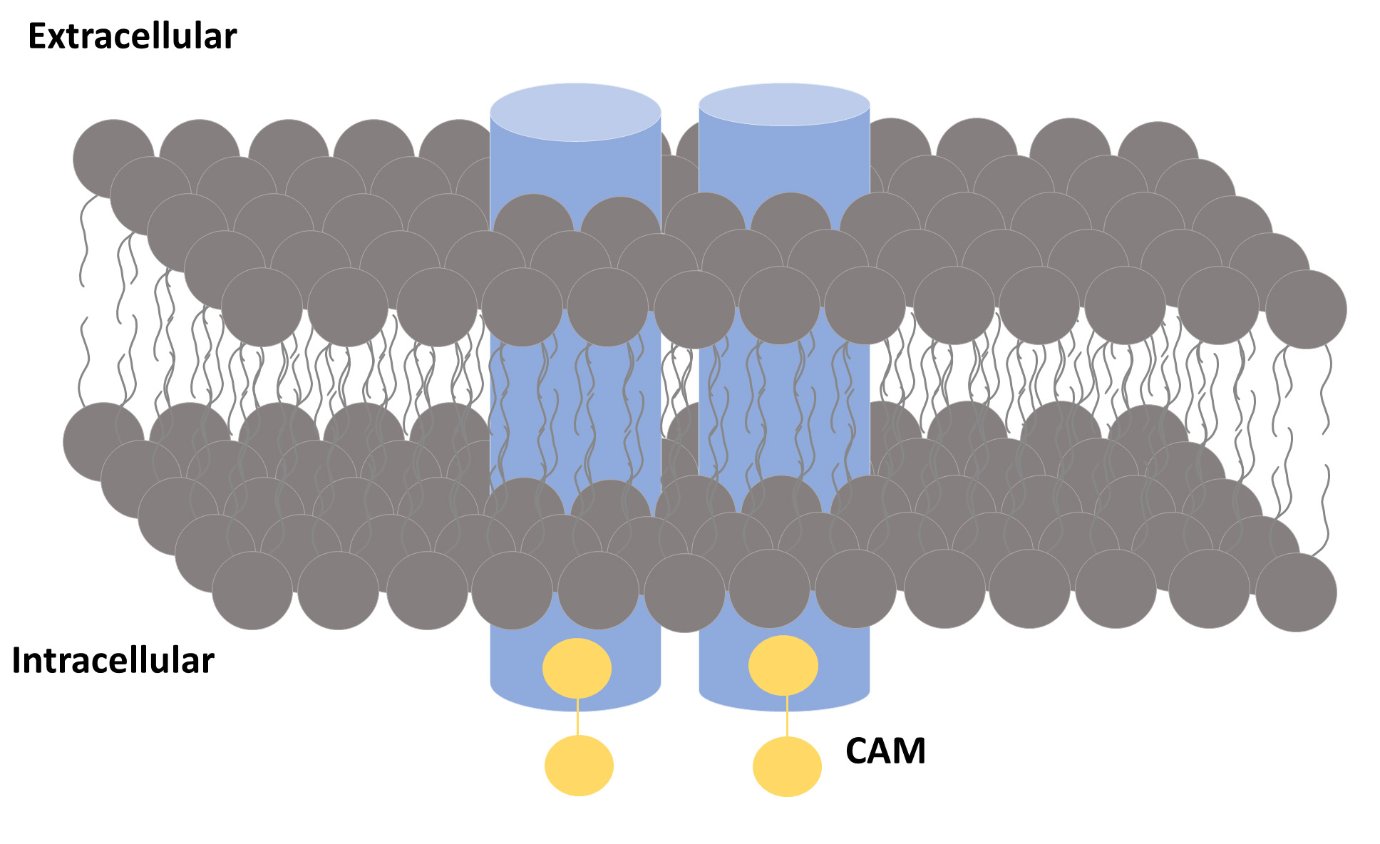
+ channels contain a critical amino acid sequence of eight amino acids, referred to as the “K+ channel signature sequence” [9, 10]. SK channels have a similar topology to the members of the K+ channel family (Fig. 1). A tetramer is formed by four associated subunits (Fig. 2). This structure consists of six transmembrane segments (S1 to S6) with a pore located between segments 5 and 6. This pore-forming P-loop located between the domains S5 and S6 is associated with potassium ion selectivity [11]. The SK channel structure has an N-terminus (before the S1 subunit) and a C-terminus (after the S6 subunit). Nevertheless, the structural characterization alone fails to explain how Ca2+ acts on these channels.

Fig. 1.

A general view of an SK channel. An SK channel is formed by four subunits (represented by blue cylinders). Each subunit binds to a calmodulin (CaM) molecule.
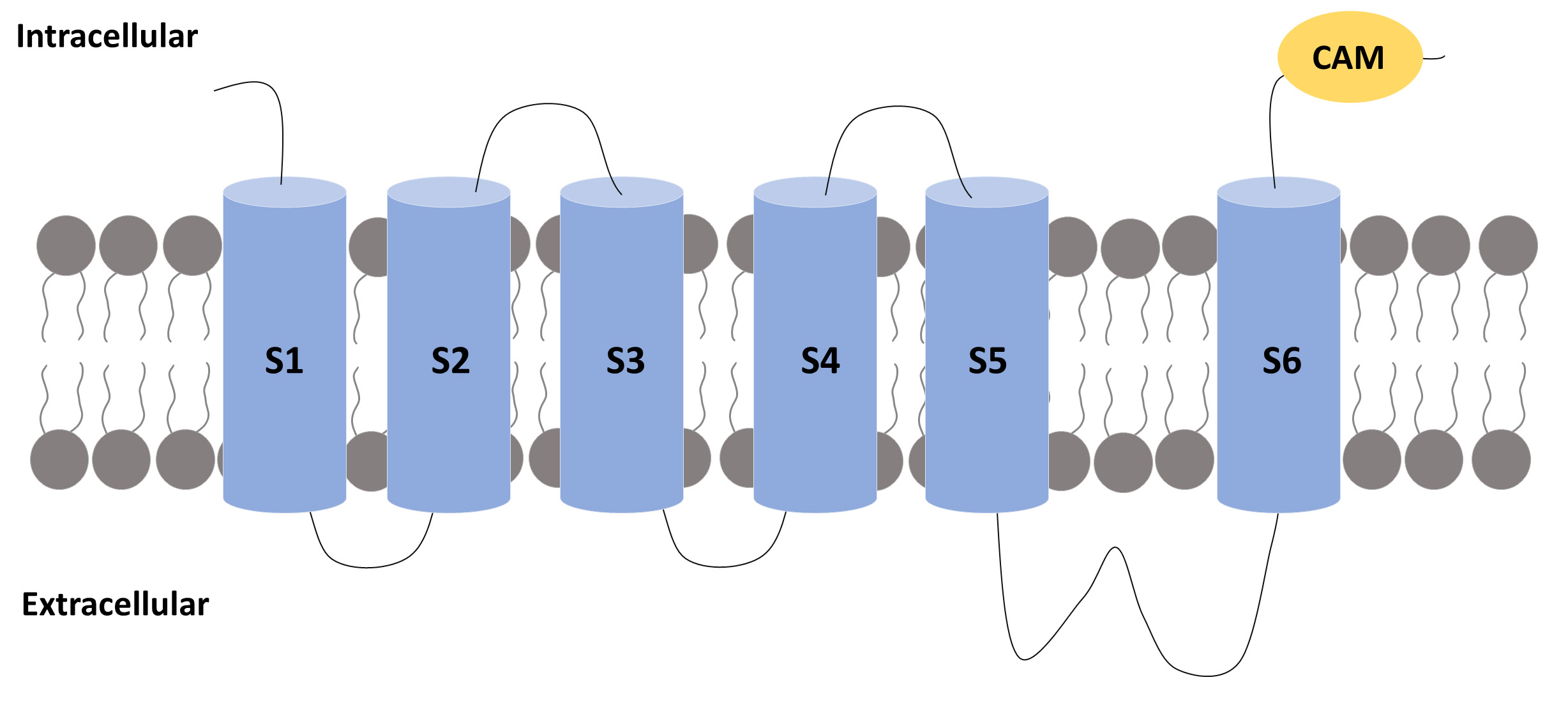
Fig. 2.

A subunit of an SK channel. A subunit of an SK channel comprises six transmembrane segments (S1 to S6) with the pore located between segments 5 and 6. The S4 segment is the one that confers voltage sensitivity to the K+ channel. The C-terminus bind to a calcium sensor called calmodulin (CaM).

Studies were conducted to evaluate clusters of conserved negatively charged residues found between S2–S3 and in the C-terminus. No change was found in the S2–S3 cluster neutralization. However, neutralizations in the C-terminus blocked the channel activity. It was also found that this domain could be modelled as a series of three or four α-helices of highly positively charged amino acids [12]. This “two-hybrid approach” showed that the C-terminus interacts with the ubiquitous calcium sensor calmodulin (CaM) to form a very stable complex [13].

Each CaM molecule binds two calcium ions to activate an SK channel. Biochemical experiments confirmed that the C-terminus interacts with CaM independently of the presence of calcium, while a subdomain only interacts with the C-terminus domain in the presence of calcium [14]. This site on the C-terminus is called the CaM-binding domain (CaMBD) [15].

The CaMBD binds two CaM. Each CaM wraps around three
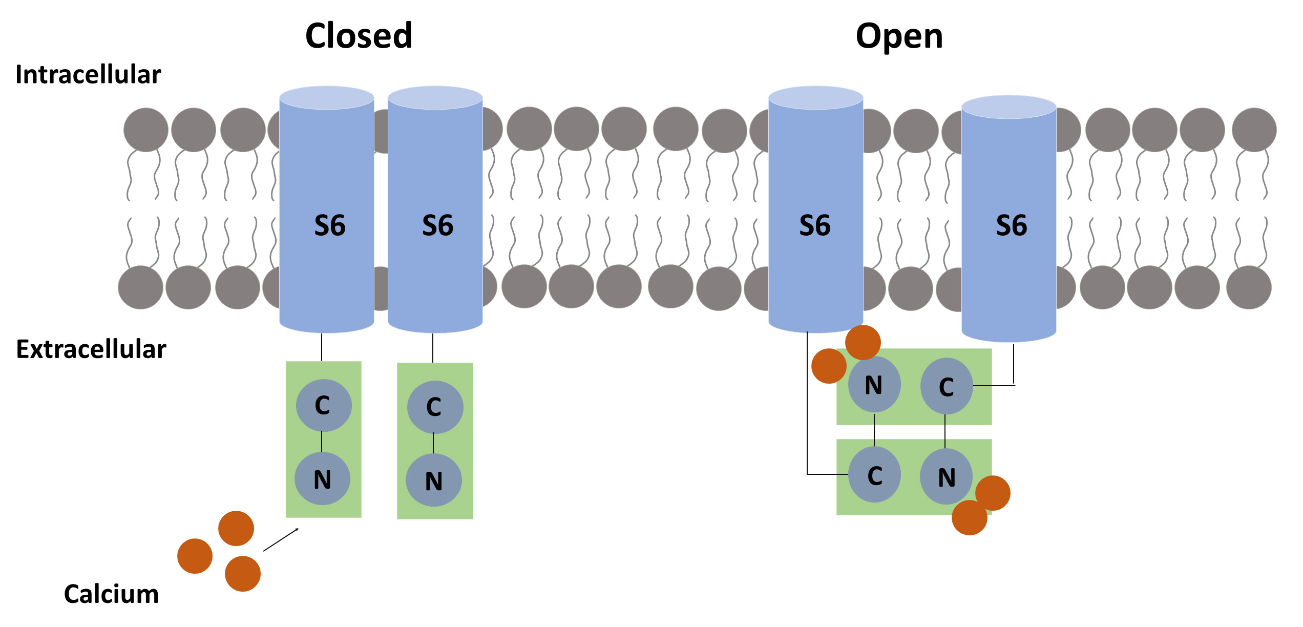
 α-helices: two from one CaMBD and one from the other CaM. CaM is a flexible molecule that can undergo an open-closed conformation and contains two main domains: the N-terminus and the C-terminus [16]. Biochemical experiments suggest a model for Ca2+ gating to CaM. In this model, calcium binds to each CaM N-terminus, exposing its hydrophobic region and allowing it to interact with an adjacent CaMBD monomer. When this occurs, each N-lobe attaches to the other CaM C-lobe, resulting in a rotatory force to the attached S6 pore helices in the gate region, resulting in the opening of the pore [17, 18]. The SK channel SK2 has other subunits. SK2 binds either to the protein kinase 2 (casein kinase 2) (CK2), in the C-terminus or to the protein phosphatase 2A (PP2A), in the N-terminus. Both modulate the SK affinity to calcium. When CK2 is phosphorylated, calcium sensitivity is reduced. This effect is counterbalanced by the protein phosphatase 2 (PP2A) [19]. Fig. 3 illustrates the possible calcium-related mechanism for the pore opening.

Fig. 3.

The suggested calcium-related mechanism for pore opening. Each calmodulin (CaM) molecule binds two calcium ions to activate the SK channel. When calcium binds to the N-terminal of CaM, a hydrophobic region is exposed, allowing the CaM-binding domain (CaMBD) to link to another CaMBD, causing a conformational change that results in pore opening.

3. Classification of SK Channels

Experiments performed in skeletal muscle cells have demonstrated that the process of after-hyperpolarization is mediated by SK channels that lack voltage sensitivity. It was also demonstrated that they differ pharmacologically. For instance, some Ca2+ currents registered in cultured skeletal muscle cells are blocked by apamin, whereas others (e.g., in hippocampal pyramidal neurons) are not [20].

Overall, SK channels are classified into three isoforms: SK1, SK2, and SK3, based on their gene origin and their sensitivity to the bee venom, apamin [21, 22]. An overall view of these subtypes is presented in Table 1 (Ref. [23]). Detailed information on each SK channel is provided in Tables 2,3,4 (Ref. [23]). These three small-conductance channels are blocked by apamin, distinguishing them from all other Ca2+ channels [23].

Table 1.Small-Conductance Calcium-Activated Potassium Channels, according to the International Union of Pharmacology (IUPHAR) and the HUGO Gene Nomenclature Committee (HGNC) (adapted from [23]).
IUPHAR HGNC Other
KCa2.1 KCNN1 SKCa1; SK1
KCa2.2 KCNN2 SKCa2; SK2

The third column refers to other commonly used nomenclatures.

Table 2.General overview of the SK1 channel (adapted from [23]).
Channel name KCa2.1
Molecular information Human: 543aa, NM_002248, chr. 19p13.1, KCNN1
Mouse: 580aa, NM_032397, chr. 8
Rat: 536aa, NM_019313, chr. 16p14
Conductance 9.2 pS (symmetric K+), 2–3 pS (normal Ringer)
Activators Ca2+, 1-EBIO (630 μM), NS309 (30 nM), riluzole (2 μM)
Blockers UCL1684 (1 nM), apamin (8 nM), tamapin (42 nM), leiurotoxin/scyllatoxin (325 nM), dequalinium (400 nM), leiurotoxin-Dab7 (6 μM), fluoxetine (7 μM), ubocurarine (23 μm), biciculline (1.1 μM)
Channel distribution Brain (amygdala > hippocampus, caudate nucleus, fetal brain > cerebellum > thalamus, substantia nigra, spinal cord, pituitary gland), oligodendroglioma, glioblastoma, gastric tumor, aorta)
Mutations and pathophysiology Not established
Pharmacological significance Modulators of SK channel subtypes may have potential use in the treatment of myotonic muscular dystrophy, gastrointestinal dysmotility, memory disorders, epilepsy narcolepsy, and alcohol intoxication
Table 3.General overview of the SK2 channel (adapted from [23]).
Channel name KCa2.2
Molecular information Human: 579aa, NM_021614 (transcript variant 1), chr. 5q22.3, KCNN2
Mouse: 574aa, NM_080465, chr. 18
Rat: 580aa, NM_019314, chr. 18q11
Conductance 9.9 pS (symmetric K+), 2–3 pS (normal Ringer)
Activators 1-EBIO, chlorzoxazone, zoxazolamine, NS309 (30 nM), riluzole (2 μM)
Blockers Tamapin (24 pM), apamin (60–200 pM), leiurotoxin/scyllatoxin (200 pM), leiuritoxin-Dab7 (3.8 nM), PO5 (22 nM), Tskappa (80 nM), Pi1-OH (>1 μM), Pi1-NH2 (100 nM), and maurotoxin (1 μM), 11 UCL1684 (250pM), tubocurarine (5 μM); with micromolar affinity: amitriptyline, carbamazepine, chlorpromazine, cyproheptadine, fluoxetine, imipramine, tacrine, trifluperazine, biciculline (1.1 μM)
Channel distribution Brain (spinal cord > hippocampus, cerebellum > amygdala > fetal brain > corpus callosum, thalamus, caudate nucleus, substantia nigra), pituitary gland, melanocyte, melanoma, germ cell tumor, prostate, oligodendroglioma, lung, Jurkat T cells, liver, heart, skeletal muscle, myometrium
Mutations and pathophysiology Dominant-negative suppression of KCa2.2 channels in deep cerebellar nuclei in a transgenic mouse causes cerebellar ataxia
Pharmacological significance Modulators of SK channel subtypes may have potential use in the treatment of myotonic muscular dystrophy, gastrointestinal dysmotility, memory disorders, epilepsy, narcolepsy, and alcohol intoxication; KCa2.2 openers have been proposed for the treatment of cerebellar ataxia
Table 4.General overview of the SK3 channel (adapted from [23]).
Channel name KCa2.3
Molecular information Human: 736aa, NM_002249 (transcript variant 1), chr. 1q21.3, KCNN3
Mouse: 731aa, NM_080466, chr. 3
Rat: 732aa, NM_019315, chr. 2q34
Conductance Not determined
Activators 1-EBIO, riluzole (3 μM), NS309 (30 nM)
Blockers Leiurotoxin/scyllatoxin (1.1 nM), apamin (10 nM), PO5 (25 nM), Tskappa (197 nM), Pi1-OH (330 nM), and Pi1-NH2 (250 nM), UCL1684 (9.5 nM); with micromolar affinity: bicuculline, amitriptyline, fluoxetine, desipramine, imipramine, nortriptyline, fluphenazine, promethazine, chlorpromazine
Channel distribution Brain (substantia nigra > amygdala, caudate nucleus, thalamus, hippocampus, ventral tegmental area, cerebellum, spinal cord > corpus callosum, fetal brain), lymphocytes (germinal center B cells, tonsillar B cells, Burkitt’s lymphoma, microglia), skeletal muscle (increased denervated muscle, myotonic dystrophy), myometrium, prostate, kidney, heart, pituitary gland, liver, pancreas, colon, germinal cells, head, neck, ovary, vascular endothelium
Mutations and pathophysiology Longer polyglutamine repeats are over-represented in schizophrenic (especially negative-symptom form) individuals and patients with anorexia nervosa and spinocerebellar ataxia; a four-base deletion has been found in a patient with schizophrenia that truncates the protein just before the S1 segment and causes dominant-negative suppression of endogenous SK channels; protein and mRNA levels are increased in skeletal muscle following denervation and patients with myotonic muscular dystrophy; involved in the endothelium-mediated vasodilation (EDHF response); conditional knockout of KCa2.3 leads to hypertension and bladder instability
Pharmacological significance Modulators of SK channel subtypes may have potential use in the treatment of myotonic muscular dystrophy, gastrointestinal dysmotility, memory disorders, epilepsy, narcolepsy, hypertension, and urinary incontinence

The SK1 channel is moderately sensitive to apamin, and its gene is located on chromosome 19, whereas the SK2 channel has the strongest affinity for apamin and is located on chromosome 5. SK3 is moderately sensitive to apamin, and its gene is located on chromosome 1 [21].

Modulators of SK channel subtypes can be used to treat many diseases due to their different effects. For instance, NS309 promotes endothelial protection [24]; chlorzoxazone improves motor coordination [25]; DCEBIO reduces the number of evoked spikes, the firing frequency and increases the afterhyperpolarizing potential of infralimbic neurons, suggesting the potential use of SK channels modulators to treat patients with anxiety disorders [26]; 1-EBIO decreases epileptiform activity and positively affects cerebral ischemia [27, 28], while riluzole improves symptoms related to cerebellar ataxia [29] and significantly reduces acute arthritis [30]. On the other hand, apamin acts by inhibiting SK channels. It suppresses hepatic fibrosis [31, 32], attenuates cytokine production [33], and may reduce attentional deficits related to Alzheimer’s disease [34].

SK channels are encoded by the KCNN gene family [35], which is abundantly conserved in different species, suggesting an orthologous relationship. The KCNN1 and KCNN3 genes exhibit alternative splicing [36, 37], a mechanism that increases the complexity of gene expression [38]. A pre-mRNA containing introns and exons can generate different mRNA. In theory, 32 transcripts can be formed by the KCNN1 gene and 20 have already been found in the mouse brain [36]. However, the function of alternative splicing in SK channels has not yet been clarified.

4. Expression of SK Channels in the Central (CNS) and Peripheral Nervous System (PNS)

SK channels are widely distributed in humans and animals, in central nervous system (CNS) and in peripheral tissue, among them, epithelium, smooth muscle and endothelium [39]. The aim of this study in characterize the SK channels throughout the brain. SK1 and SK2 channels are co-expressed by the neurons of the neocortex, cerebellum, hippocampus, and brain stem, and SK3 channels are located in the midbrain and hypothalamus [39]. Stocker and Pedarzani [40] performed a study using in situ hybridization to characterize the distribution pattern of SK channels in the rat brain [40]. They found that SK1 channels are mainly expressed in the neocortex. The CA1–3 layers of the hippocampus and the thalamic reticular nucleus have high expressions of both SK1 and SK2 channels. In contrast, the supraoptic nucleus has predominantly SK3 channels, while the inferior olivary nucleus has equivalent expressions of SK2 and SK3.

Rimini et al. [41] evaluated the distribution of SK channels in the CNS and peripheral tissues through real-time reverse transcription polymerase chain reaction. In their evaluation, all SK channels were expressed in the human brain and, only SK1 was not detected in the peripheral nervous system. SK3 exhibited the highest expression among the SK channels, followed by SK2 and SK1, respectively. SK1 was mainly found in the amygdala, the hippocampus, and the caudate nucleus. SK2 was primarily expressed in the pituitary gland and the liver. The highest expression of SK3 was detected in the substantia nigra of the midbrain. An immunocytochemistry analysis found that SK3 is specifically localized in the presynaptic terminals of the neuromuscular junctions [42]. Although the SK channel distribution has been well-mapped in previous studies, there are still controversies as to the cellular distribution of these channels.

5. SK Expression in the Spinal Cord and Sensory Ganglia

All SK channels are expressed in the spinal cord and the dorsal root ganglion (DRG). SK1 and SK2 are preferentially located in the cytoplasm and cellular membrane of small cell bodies of DRG neurons. In contrast, SK3 is found in the cellular membrane and granular cytoplasmic clusters of small and large DRG neurons [43]. In addition, it has been demonstrated that SK1 and SK2 are expressed in both peptidergic and non-peptidergic nociceptors [43].

SK3 expression is higher in the laminae I–III of the spinal cord [44]. However, SK3 is also expressed in ependymal cells. This finding suggests that this channel might be expressed in other epithelial cell types [45]. SK3 is also expressed in motoneurons and may determine the apamin-sensitive after-hyperpolarization in this cell type [46, 47].

SK3 is found in both peripherin-positive and peripherin-negative neurons, indicating that this specific channel could play a role in the process of the transduction of nociceptive signals. The participation of SK channels in pain and nociception is further discussed. This immunoreactivity of SK3 in peripheral neurons is also compatible with the distribution of SK3 throughout the Rexed laminae (I–III) of the spinal cord [44].

It has been reported that SK3 channels are also found in non-neuronal glial cells, such as the satellite glial cells (SGCs). A study that evaluated the role of trigeminal ganglion SGCs in the mechanisms of trigeminal neuropathic pain using a model of chronic constriction of the infraorbital nerve reported a co-labeling of SK3 and glial cell markers, e.g., the inwardly rectifying K+ channel, subunit 4.1, and the glutamate-aspartate transporter (GLAST) [48]. A feature of peripheral nerve injury is the increase of neuronal activity that, in turn, requires a greater demand on SGCs to uptake K+. A deficiency of SGC-mediated K+ buffering results in an increased extracellular concentration of K+ and neuronal hyperexcitability, a phenomenon that is proposed to underlie the development of neuropathic pain.

The expression of SK3 in peripheral neurons and SGCs has been a matter of debate. According to some studies, SK3 is a specific marker of SGCs in vivo [49, 50]. SK3 immunolabeling has also been found in early-cultured cells [49]. Our group has evaluated changes that occur in SK3 expression in cultured trigeminal ganglion cells, and found important variations [51]. It was possible to detect a high expression of SK3, with some cells double-staining with ßIII-tubulin at 4h in culture. This was followed by a decrease in SK3 expression at 24 h and an increase at 48 h, indicating great plasticity of SGCs, especially in vitro. We hypothesized that a differentiation process occurred in the time frame that was analyzed. Therefore, the specific expression of SK3 in peripheral neural cells in vitro and in vivo must be explored in-depth with further studies.

6. Functional role of SK Channels in CNS and PNS

SK channels are predominantly expressed in the nervous system. These channels are found in neurons and other cell types and contribute to a large variety of physiological functions. Functional studies show that SK channels are diverse, and their activity depends on neuronal types, developmental stages, and animal species. The activation of SK channels in the midbrain and cerebellum neurons is involved in muscle coordination and movement. In the hippocampus and amygdala, SK channels of pyramidal neurons regulate the amplitude of excitatory postsynaptic potentials (EPSPs). SK channel activation may also influence N-methyl-D-aspartate (NMDA) receptor activation, consequently playing a role in synaptic plasticity. Interestingly, blocking the activation of SK channels in hippocampus pyramidal neurons and the amygdala increases long-term potentiation (LTP), which can point to the involvement of SK channels in learning and memory. SK channels expressed in the mitochondrial membranes are proposed to be neuroprotective, for example, in protecting against glutamate toxicity or inhibition of reactive oxygen species (ROS) production [52].

It has been well-accepted that SK channels are essential for neuronal excitability. An action potential is characterized by a quick change in the voltage of the cellular membrane due to an influx of ions determined by the ionic concentration and the cellular membrane permeability [53, 54]. The action potential is followed in many cells by an after-hyperpolarization (AHP) period that prevents repetitive neuronal activation. SK channels are related to the phenomenon of after-hyperpolarization (especially in non-innervated skeletal muscle and in some nerve cells) since apamin (an SK channel blocker) abolishes the process of this phenomenon [55]. Considering that SK channels are blocked by apamin while BK channels are not, it has been proposed that small conductance calcium channels are involved in a larger variety of cells [55].

The regulation of neuronal excitability is crucial for healthy brain function. The equilibrium between depolarizing and hyperpolarizing ion channels determines the neuron’s firing activity. Potassium channels constitute the most prevalent ion channels. Since they mediate the outward potassium currents that repolarize and hyperpolarize the membrane potential, they are one of the most important elements of neuronal excitability [56].

Several categories of potassium channels mediate the after-hyperpolarization process. This process repolarizes the neuronal membrane, limits both the amplitude and width of the spikes, also controling neuronal activation. Therefore, neurotoxicity related to excessive neuronal firing is avoided [57]. Because of their distinct activation-inactivation kinetics and Ca2+ sensitivity, each K+ channel mediates after-hyperpolarization at a distinct time scale. This separates the course of the after-hyperpolarization into three components: 1-fast (activated in 1–5 s); 2-medium (mAHP, activated between 10–300 ms), and 3-slow (sAHP, activated between 0.5-multiple seconds) after-hyperpolarization [57, 58]. Several studies have shown that the SK channels, as well as the hyperpolarization-activated cyclic nucleotide channels, and voltage-gated K+ channel 7, are important elements of the mAHP that takes place in the neurons [59, 60]. A detailed description of the mAHP-mediating ion channels is provided in this review. These ion channels control anomalies that are associated with cells and cellular excitability, both of which are related to several neurological disorders. Therefore, the pharmacological manipulation of these channels is of great interest and has immense therapeutic potential [61].

The SK channels of the CNS neurons are activated by high concentrations of intracellular Ca2+. The generation of an mAHP, activated within milliseconds and decays within hundreds of milliseconds, is amplified by the outflow of K+ [62, 63]. Apamin-sensitive SK channels have been recorded in several brain regions, indicating a large distribution of apamin-binding sites and SK channels [40, 64].

Studies that evaluated the distribution of SK channel subunits and their pharmacological properties through specific toxins have led to the molecular identification of the channels that underlie the process of after-hyperpolarization in multiple brain regions.

High levels of SK2 subunits are associated with the presence of an apamin-sensitive after-hyperpolarization current in the pyramidal neurons of CA1 and CA3 in the hipocampus [65, 66]. The after-hyperpolarization currents in the CA1 pyramidal neurons of rats are blocked by apamin at a concentration approximately eight-fold higher than those required to block recombinant SK2 channels [67]. Given that SK1 and SK2 subunits are co-expressed in pyramidal CA1 neurons [68], and that SK2 channels are apamin-insensitive [69], the low apamin sensitivity of currents after the after-hyperpolarization is probably related to the development of heteromeric SK1/SK2 channels. In fact, there is evidence that rat heteromeric SK1 and SK2 channels are less apamin sensitive in heterologous expression systems [70]. Homomeric SK2 channels also underlie an apamin-sensitive after-hyperpolarization current that occurs in the cerebellum, which is critical for the intrinsic firing pattern of the Purkinje neurons [67, 71].

In the brainstem, the after-hyperpolarization of the motor neurons of the dorsal vagus nucleus influences spontaneous firing and is involved in the regulation of esophageal, gastric, and pancreatic functions [72] and is also blocked by apamin. The after-hyperpolarization current of the dopaminergic neurons of the midbrain is controlled by spontaneous firing activity, and SK3 channels are involved in this process [73].

The hypothalamus is another region of the brain with an increased expression of SK3 [40]. In the rostral hypothalamic basal region, SK3 expression is hormonally regulated [74]. The secretion of oxytocin and vasopressin from nerve terminals of magnocellular neurosecretory cells is partially regulated by an SK3-mediated after-hyperpolarization current that regulates phasic firing [40, 75]. SK3 channels also mediate the mAHP in the suprachiasmatic nucleus. Therefore, it regulates the firing rate, which undergoes diurnal modulation by the circadian rhythm [40, 76]. SK3 channels underlie the after-hyperpolarization in superior cervical ganglion neurons, which also regulate circadian rhythm via its connections with the pineal gland [46].

The overexpression of SK3 channels produces abnormal breathing patterns during a hypoxic challenge and compromises parturition [77]. After-hyperpolarization was selectively abolished in CA1 pyramidal neurons from knockout mice that lacked SK2 channels. However, this current was intact in mice that lacked SK1 and SK3 channels [78], which confirmed the predominant role of SK2 subunits in hippocampal pyramidal neurons [65, 66] and raised questions regarding the function of SK1. The splice variant SK3-1B, is known to suppress SK2 and SK3 channels [79, 80] and was recently used to generate a transgenic mouse model [81]. The expression of SK3-1B under the control of a neuronal promoter suppressed the expression of SK channels in several regions of the brain [81]. These transgenic mice showed severe cerebellar ataxia related to the downregulation of SK1 and SK2 expressions in the deep cerebellar nuclei neurons, resulting in the loss of the after-hyperpolarization current and, consequently, in increased spontaneous firing and hyperexcitability [81]. A strong and selective reduction in the apamin-sensitive after-hyperpolarization was also observed in layer V of neocortical neurons, where overexpression of SK1 or SK2 led to a selective enhancement of this current component [80]. These studies support the role of SK channels as regulators of intrinsic neuronal excitability and firing. Further studies are needed to evaluate and scrutinize their impact on cellular and network functions and to correlate them with specific changes in animal behavior [55].

It has been demonstrated that SK channels are important in controlling the action potential firing frequency and regulating dendritic excitability, synaptic transmission, and synaptic plasticity. In accordance with their role in modulating synaptic plasticity, SK channels exert control over several learning and memory tasks and may also participate in the mechanisms of several neurological disorders [82].

Modulating dendritic excitability and activation of SK channels by calcium influx during action potentials regulates the frequency of action potential discharge in most neurons. The location of the channel underlying this effect is unknown, but it is generally believed to be somatic near the action potential initiation site. Nevertheless, it has been shown that apamin-sensitive channels are also present in the dendritic tree and are activated by calcium surges from sources other than voltage-gated calcium channels. For example, calcium released from intracellular stores in dopaminergic [73] and cortical pyramidal neurons [66, 83] activates apamin-sensitive conductances, leading to hyperpolarizing potentials.

In Lamprey spinal motor neurons, SK channels can also be activated following dendritic activation of NMDA receptors, terminating the resulting dendritic plateau potential [84]. SK dendritic channels in these neurons also contribute to after-hyperpolarization and reroute excitatory inputs when triggered after-hyperpolarization. This shunting requires potential activation of SK channels, since blockade of SK channels alone does not affect individual excitatory postsynaptic potentials (EPSPs) or episodes of EPSPs [85]. Finally, activation of NMDA receptors in mitral cells of the olfactory bulb also activates dendritic SK channels, modulating dendritic excitability [86]. The regulation of synaptic transmission and the role of SK channels in synaptic transmission was first described in neurons in the substantia nigra and ventral tegmental area, where SK channels contribute to inhibitory post-synaptic potentials. Activation of SK channels follows the release of calcium from intracellular stores triggered by glutamate acting on metabotropic glutamate receptors [87].

An inhibitory post-synaptic conductance in auditory outer hair cells following activation by calcium influx through calcium-permeable nicotinic acetylcholine receptor is also mediated by SK channels [88]. Recently, SK channels have been shown to bypass fast excitatory synaptic transmission in the lateral amygdala and hippocampal pyramidal neurons [85, 88]. Calcium influx in these neurons via NMDA receptors during basal synaptic transmission activates SK channels co-localized in the spines of hippocampal and amygdala pyramidal neurons [85, 88]. The resulting hyperpolarization bypasses EPSP and increases the magnesium blockade of NMDA receptors. Apamin use reverses this effect and increases NMDA receptor-mediated calcium transients in the cephalic spine [88]. In the pyramidal neurons of the lateral amygdala, shunting of excitatory synaptic transmission by SK channels reduces the amount of depolarization during repetitive stimulation of cortical afferents, thus reducing the extent of the long-term potentiation (LTP) at these synapses [83]. Similarly, the blockade of hippocampal SK channels enhances the LTP after low-frequency tetanic stimulation of Schaffer collaterals [89] and lowers the threshold for LTP in the hippocampal CA1 pyramidal cells [90]. These effects have been attributed to a depression of the mAHP and the consequent increase of the action potential discharge. However, it has become clear that these effects are most likely due to the SK channel-mediated shunt on excitatory synaptic transmission rather than to the relatively minor regulation of the firing frequency [91]. Further defining the role of SK channels in limiting the LTP, it has been found that overexpression of SK2 channels in the hippocampus reduces the LTP in the CA1 neurons [92].

SK channels are involved in the processes of learning and memory through the modulation of basal excitatory synaptic transmission and plasticity [85]. The blockade of SK channels by a systemic administration of apamin in rats enhances learning in an object-recognition task [93]. In addition, the administration of apamin reversed the spatial navigation deficit induced by lesions in the medial septum and the hippocampus of mice in the Morris water maze spatial memory task [94, 95]. Corroborating the findings of these studies, apamin induced the expression of the immediate early genes c-fos and c-jun in the hippocampus, which are considered the initial markers for memory formation [96, 97].

Conversely, the overexpression of SK2 channels led to an impairment in the performance of rats in the Morris water maze, contextual fear conditioning, and amygdala-dependent cued fear conditioning [92, 98]. In addition, apamin enhanced learning in an appetite-motivated bar-pressing response in mice [93] and an olfactory discrimination learning task [93]. Finally, increases in the SK3 expression underlie an age-related deficit in hippocampal-mediated learning tasks [92, 99]. Together, these findings indicate that SK channels play a key role in negatively regulating learning and memory formation.

7. Modulation of SK Channels

Several ion channels have phosphorylation sites for protein kinases. The phosphorylation by sites for protein kinases present in several ion channels that modulates the functioning or trafficking across those channels. However, only some studies examined the process of SK channel modulation. Despite the presence of many possible sites for the phosphorylation of the protein kinases A (PKA) and C, the biophysical evidence for the modulation of SK channels via these kinases still needs to be determined. PKA’s phosphorylation of the C-terminus of the SK2 channel has been demonstrated, and this process reduces the cell surface of this channel subtype [100]. According to previous studies, this indicates that SK channels may be regulated by membrane trafficking, similar to what occurs with voltage-dependent potassium channels [101].

8. Role of SK Channels Modulators as Potential Targets for the Treatment of Diseases

As previously discussed, SK channels play a role in memory and learning. Therefore, modulators of SK channels that improve performance in learning tasks may be useful therapeutic agents in treating cognitive dysfunction and memory disorders. Nevertheless, agents that block SK channels, such as apamin, currently have a narrow therapeutic window. Therefore, there is a need for new agents that offer fewer side effects regarding therapeutic purposes [101]. High doses of apamin can induce epileptic-like activity, and agents that enhance the activity of SK channels (called SK channel activators), such as 1-ethyl-2-benzimidazolinone (1-EBIO) or NS309, may be useful in treating epilepsy. NS309 also modulates the activity of both pro-inflammatory and anti-inflammatory cytokines by regulating the TSG-6/NF-κB pathway. For example, the administration of NS309 in an animal model of traumatic brain injury led to a decrease in the levels of IL-1β, IL-6, tumor Necrosis Factor-alpha (TNF-α), Monocyte Chemoattractant Protein 1 (MCP-1), Macrophage Inflammatory Protein 2 (MIP-2), and RANTES (CCL-5) and an increase in the levels of IL-4, IL-10, and transforming growth factor beta (TGF-β1), which affects microglia but not astrocytes [102]. SK activators also affect other conditions, such as visceral hypersensitivity, a complex condition associated with the activity of different CNS regions and SK2 channels. It has been demonstrated that Intratechal administration of the SK activators CyPPA or 1-EBIO can avoid the development of visceral hypersensitivity induced by colorectal distension with a decrease of the related after-hyperpolarization current [103].

SK channel potentiators may be useful in treating depression, phobias, and other psychiatric disorders. For example, enhancing the activity of SK channels may raise fear conditioning thresholds [92] and apamin enhances performance in the forced swim test and increases measures of antidepressant activity [76]. It has been found that the binding of apamin to SK channels is high in the prefrontal cortex and low in the hippocampus of rats exposed to chronic unpredictable stress, indicating dysfunctional activity. In addition, this can be reversed by selective inhibition of SK3 channels with selective SK3 channel negative allosteric modulators or in knockout mice [104].

SK channels are also present in dopaminergic neurons in the midbrain and control firing patterns. Burst firing of these neurons triggers the release of dopamine, which is depleted in Parkinson’s disease. Blockade of SK channels causes the sudden firing of these neurons [105], suggesting that treating midbrain dopaminergic neurons with SK channel blockers may alleviate some symptoms of Parkinson’s disease. In fact, a preclinical study showed that the systemic administration of apamin increases the levels of striatal extracellular dopamine, which could not be detected after nigrostriatal lesions induced by the neurotoxin 6-hydroxydopamine (6-OHDA) in rats [105].

Furthermore, SK channels play an important role in endothelium-dependent hyperpolarization and thus have an important influence on the regulation of vasodilation. Therefore, SK channels are highly involved in regulating the vascular tone related to health and disease. For instance, it has been demonstrated that administering the SK channel activator NS309 before and during cardioplegic hypoxia protects the coronary microvasculature against cardioplegic-hypoxia and reoxygenation injury. However, this effect is reduced in diabetic coronary microvasculature [24]. Calcium-Activated Potassium Channels also influence the occurrence of preeclampsia, an important pregnancy-related complication. Li et al. 2020 [106], found that the dysfunction of SK and IK channels might be associated with deficient blood vessel formation and inadequate remodeling of spiral arteries, which are related to the development of preeclampsia.

9. Role of SK in Mechanisms Underlying Nociception and Pain

SK channels’ role in nociception mechanisms has also been explored. In literature, some studies use drosophila models to understand nociceptive behavior. Activation of SK channels reduces nociceptive behavior in response to these stimuli [107], suggesting that these channels play essential roles in regulating the sensitivity of nociceptive neurons. Walcott et al. 2018 [107] evaluated the influence of SK channels on the thermal regulation of nociceptors, mechanical nociception behavior, and stability regulation. The nociceptor-specific requirement for SK was assessed using the SK-M transcript under the control of the ppk-GAL4 driver in the SK mutant background. This released the hypersensitive nociception phenotype of the SK mutant animals, confirming the site of action for SK in nociceptor neurons. It was found that, compared to the short isoforms, the long SK isoforms may be more important in controlling the thermal sensitivity of nociceptors. In Regulation Mechanical Nociception Behavior, the effects of SK were more specific for the nociception pathway, regulating nociceptor activity in mechanical stimuli and response to noxious thermal [107].

SK channels have been found to be capable of regulating the tonic firing of nociceptive neurons. The behavior of substantia gelatinosa (SG) neurons, crucial to the nociceptive transmission, was studied through whole-cell recordings. The results showed that 1-EBIO attenuates spike discharges and increases the AHP amplitudes of the neurons located in the SG, an effect mimicked by a high Ca2+ external solution. Apamin increased neuronal excitability and decreased the AHP amplitudes, an effect that was mimicked by a Ca2+ free external solution. Thermal-induced nociception was attenuated by the systemic administration of 1-EBIO. Apamin also increased excitatory synaptic transmission and this was dependent on NMDA receptors, extracellular Mg2+, and intracellular Ca2+ [108].

It has been widely accepted that the expression of postsynaptic NMDA receptors is associated with LTP and the development of chronic pain. However, the role of expression of presynaptic expression of NMDA on chronic pain has not been fully evaluated. One study demonstrated that presynaptic expression of NMDA receptors in spinal cord neurons modulates the synaptic transmission in a nociceptive tone-dependent form [109]. The results of that study indicate that this process is determined by the influx of Ca2+ through presynaptic NMDA receptors which depended on the activity of SK channels. Therefore, SK channels are probably related to synaptic depression in the presynaptic neurons of the nociceptive pathway. In addition, tissue inflammation has been shown to lead to downstream SK2 downregulation, via the Brain-Derived Neurotrophic Factor [109]. A colocalization of NMDA receptors and SK channels, especially SK2, has been reported in the DRG [110] which might help to explain these findings. Another study has shown a co-expression of SK3 and NMDA receptors in the dorsal horn of the spinal cord [111]. The same study also demonstrated that intrathecal administration of 6,7-Dichloro-1H-indole-2,3-dione 3-oxime (NS309), an SK channel activator, attenuates the mechanical hypersensitivity driven by Freund’s adjuvant and that the co-administration of NS309 with an antagonist of the NMDA receptor decreases the dose of the NMDA receptor antagonist, DL-2-Amino-5-phosphonopentanoic acid needed to produce antinociceptive effects. This corroborates the concept of a functional interaction between SK channels and NMDA receptors in the mechanisms of nociceptive regulation.

A recent study has also reported the expression of SK channels (in this case SK3) in the trigeminal ganglion [112]. Furthermore, a decreased expression of SK3 has been found following infraorbital nerve ligation, a classical model of trigeminal neuropathic pain in rodents. The administration of the SK channels agonist (CyPPA) significantly increased the pain thresholds (resulting in mechanical allodynia), while the administration of the SK channel antagonist apamin significantly increased the pain thresholds [112]. These findings indicate that SK channels, especially SK3, could be potential pharmacological targets in the treatment of trigeminal neuralgia and other types of trigeminal neuropathic pain. There is a considerable number of nonselective SK channel blockers such as apamin, scyllatoxin, UCL 1684, and UCL 1848, and a few SK2 selective blockers such as the peptide inhibitor Lei-Dab (7) [113], the scorpion toxin tapamin. The discovery and development of novel classes of selective inhibitors of SK1, SK2, and SK3 are crucial to determine the specific contribution of each SK channel in nociception mechanisms and pain [114].

According to one preclinical study, SK3 is confined to the SGCs and not expressed by the neurons of the trigeminal ganglion [50]. However, this concept must be taken cautiously when interpreting the results of in vitro studies, since phenotypic changes involving SK channels can occur in cell cultures of the trigeminal ganglion [51]. However, these results shed light on the expression of SK channels in the trigeminal ganglion cells and suggest that targeting SK channels could be of great value in the treatment of orofacial pain, especially those of neuropathic origin. In fact, the expression of SK and IK channels can be modulated by neuropathic factors as demonstrated by the significant increase in the percentage of SK1-positive cells after the treatment of DRG cultures with nerve growth factor and the stimulation of IK1 expression upon the administration of the neurotrophin 3, NT-3 [96]. Thus, the modulation of potassium channels with different compounds might be a promising therapy in the treatment of different types of pain.

Chronic pain results in a negative impact on the quality of life and compromises healthcare systems worldwide. The burden of chronic pain and the limited therapeutic options for these patients, drives the search for novel pharmacological therapies that could offer more efficient and safer treatments. Riluzole, a drug used in the management of amyotrophic lateral sclerosis (ALS), has shown positive antinociceptive effects in preclinical models of both inflammatory [115] and neuropathic pain [116]. Changes in regions related to somatosensory processing (e.g., thalamic nuclei and postcentral gyrus) have been found in patients with ALS. Future studies will evaluate how these findings relate to the effects of riluzole on the brain network altered in ALS and chronic pain [117]. One study that used brain slice electrophysiology and behavioral evaluation in an experimental model of neuropathic pain through spinal nerve ligation, found that riluzole has a positive effect in reducing neuropathic pain. This occurs through SK channel-dependent and independent mechanisms in the amygdala. These results also indicate that this action could be related to central amygdala output nucleus (CeA) neurons, which in turn, have high levels of SK channels [118]. The participation of the CeA in the mechanisms of riluzole-induced pain modulation through the activation of SK channels were also demonstrated by another study that used an experimental model of knee arthritis induced by kaolin/carrageenan. This study found that riluzole inhibits supraspinally pain behaviors by activating SK channels, but not BK channels in the CeA amygdala [119]. These findings suggest that riluzole is a promising compound to treat chronic pain and that this mechanism of action is probably related to the activation of SK channels located in the CeA. However, clinical studies combined with neuroimaging analysis will be necessary to verify these hypotheses. In addition to efficacy, the long-term safety of this compound, and newer compounds for chronic pain needs to be determined.

10. Conclusions

SK channels are known to translate changes in the level of the intracellular second messenger, Ca2+. They play a pivotal role in the phenomenon of after-hyperpolarization, and their interaction with NMDA receptors contributes to the LTP and, consequently, to the mechanisms of learning and memory. The functional coupling of SK channels and NMDA receptors is important to physiological activities and participates in the mechanisms of diseases, including chronic pain. The modulation of SK channels through antagonists and agonists appears to interfere with the process of nociception and pain, which opens opportunities for developing novel compounds to treat chronic pain based on the modulation of SK channels, and exploring their interactions with NMDA receptors. Riluzole, an already existing agent with other indications, emerges as a promising drug to modulate chronic pain mainly due to its ability to affect the activity of SK channels of the CeA. The multifaceted effects of riluzole, particularly on neuromodulation and excitotoxicity, and its controlled safety, make this drug attractive to treat different conditions such as traumatic and non-traumatic spinal cord injury [120]. Nonetheless, further studies are essential to substantiate the current findings for the efficacy and safety of SK activators that inhibit neuronal hyperexcitability to reduce chronic pain.

Author Contributions

MFD, and PG designed the study. LGAF, CPV and CKS performed the research, developed the search strategy and literature search. MFD, LGAF, CPV, CKS, and PG wrote the manuscript. All authors contributed to editorial changes in the manuscript. All authors read and approved the final manuscript.

Ethics Approval and Consent to Participate

Not applicable.

Acknowledgment

Not applicable.

Funding

This research received no external funding.

Conflict of Interest

Parisa Gazerani is serving as one of the Editorial Board members and Guest editors of this journal. We declare that Parisa Gazerani had no involvement in the peer review of this article and has no access to information regarding its peer review. Full responsibility for the editorial process for this article was delegated to Gernot Riedel. The other authors declare no conflict of interest.

References
[1]
Bond CT, Maylie J, Adelman JP. Small-conductance calcium-activated potassium channels. Annals of the New York Academy of Sciences. 1999; 868: 370–378.
[2]
Coetzee WA, Amarillo Y, Chiu J, Chow A, Lau D, McCormack T, et al. Molecular diversity of K+ channels. Annals of the New York Academy of Sciences. 1999; 868: 233–285.
[3]
Gardos G. The function of calcium in the potassium permeability of human erythrocytes. Biochimica et Biophysica Acta. 1958; 30: 653–654.
[4]
Meech RW. Intracellular calcium injection causes increased potassium conductance in Aplysia nerve cells. Comparative Biochemistry and Physiology. A, Comparative Physiology. 1972; 42: 493–499.
[5]
Meech RW. Calcium-dependent potassium activation in nervous tissues. Annual Review of Biophysics and Bioengineering. 1978; 7: 1–18.
[6]
Blatz AL, Magleby KL. Calcium-activated potassium channels. Trends in Neurosciences. 1987; 10: 463–467.
[7]
Latorre R, Oberhauser A, Labarca P, Alvarez O. Varieties of calcium-activated potassium channels. Annual Review of Physiology. 1989; 51: 385–399.
[8]
Ishii TM, Silvia C, Hirschberg B, Bond CT, Adelman JP, Maylie J. A human intermediate conductance calcium-activated potassium channel. Proceedings of the National Academy of Sciences of the United States of America. 1997; 94: 11651–11656.
[9]
Doyle DA, Morais Cabral J, Pfuetzner RA, Kuo A, Gulbis JM, Cohen SL, et al. The structure of the potassium channel: molecular basis of K+ conduction and selectivity. Science. 1998; 280: 69–77.
[10]
Heginbotham L, Lu Z, Abramson T, MacKinnon R. Mutations in the K+ channel signature sequence. Biophysical Journal. 1994; 66: 1061–1067.
[11]
Nam YW, Orfali R, Liu T, Yu K, Cui M, Wulff H, et al. Structural insights into the potency of SK channel positive modulators. Scientific Reports. 2017; 7: 17178.
[12]
Xia XM, Fakler B, Rivard A, Wayman G, Johnson-Pais T, Keen JE, et al. Mechanism of calcium gating in small-conductance calcium-activated potassium channels. Nature. 1998; 395: 503–507.
[13]
Luján R, Maylie J, Adelman JP. New sites of action for GIRK and SK channels. Nature Reviews. Neuroscience. 2009; 10: 475–480.
[14]
Maylie J, Bond CT, Herson PS, Lee WS, Adelman JP. Small conductance Ca2+-activated K+ channels and calmodulin. The Journal of Physiology. 2004; 554: 255–261.
[15]
Gu M, Zhu Y, Yin X, Zhang DM. Small-conductance Ca2+-activated K+ channels: insights into their roles in cardiovascular disease. Experimental & Molecular Medicine. 2018; 50: 1–7.
[16]
Chou JJ, Li S, Klee CB, Bax A. Solution structure of Ca(2+)-calmodulin reveals flexible hand-like properties of its domains. Nature Structural Biology. 2001; 8: 990–997.
[17]
Gifford JL, Walsh MP, Vogel HJ. Structures and metal-ion-binding properties of the Ca2+-binding helix-loop-helix EF-hand motifs. The Biochemical Journal. 2007; 405: 199–221.
[18]
Schumacher MA, Rivard AF, Bächinger HP, Adelman JP. Structure of the gating domain of a Ca2+-activated K+ channel complexed with Ca2+/calmodulin. Nature. 2001; 410: 1120–1124.
[19]
Allen D, Fakler B, Maylie J, Adelman JP. Organization and regulation of small conductance Ca2+-activated K+ channel multiprotein complexes. The Journal of Neuroscience. 2007; 27: 2369–2376.
[20]
Lancaster B, Adams PR. Calcium-dependent current generating the afterhyperpolarization of hippocampal neurons. Journal of Neurophysiology. 1986; 55: 1268–1282.
[21]
Katherine Z, Shawn K, Frank S, Jun F. SK Channels and Heart Disease. In Zuzana Sevcikova T (ed.) Ion Transporters. IntechOpen: Rijeka. 2022.
[22]
Vergara C, Latorre R, Marrion NV, Adelman JP. Calcium-activated potassium channels. Current Opinion in Neurobiology. 1998; 8: 321–329.
[23]
Wei AD, Gutman GA, Aldrich R, Chandy KG, Grissmer S, Wulff H. International Union of Pharmacology. LII. Nomenclature and molecular relationships of calcium-activated potassium channels. Pharmacological Reviews. 2005; 57: 463–472.
[24]
Zhang Z, Shi G, Liu Y, Xing H, Kabakov AY, Zhao AS, et al. Coronary endothelial dysfunction prevented by small-conductance calcium-activated potassium channel activator in mice and patients with diabetes. The Journal of Thoracic and Cardiovascular Surgery. 2020; 160: e263–e280.
[25]
Egorova PA, Gavrilova AV, Bezprozvanny IB. In vivo analysis of the spontaneous firing of cerebellar Purkinje cells in awake transgenic mice that model spinocerebellar ataxia type 2. Cell Calcium. 2021; 93: 102319.
[26]
Criado-Marrero M, Santini E, Porter JT. Modulating fear extinction memory by manipulating SK potassium channels in the infralimbic cortex. Frontiers in Behavioral Neuroscience. 2014; 8: 96.
[27]
Khandai P, Forcelli PA, N’Gouemo P. Activation of small conductance calcium-activated potassium channels suppresses seizure susceptibility in the genetically epilepsy-prone rats. Neuropharmacology. 2020; 163: 107865.
[28]
Soboleva EB, Amakhin DV, Sinyak DS, Zaitsev AV. Modulation of seizure-like events by the small conductance and ATP-sensitive potassium ion channels. Biochemical and Biophysical Research Communications. 2022; 623: 74–80.
[29]
Romano S, Coarelli G, Marcotulli C, Leonardi L, Piccolo F, Spadaro M, et al. Riluzole in patients with hereditary cerebellar ataxia: a randomised, double-blind, placebo-controlled trial. The Lancet. Neurology. 2015; 14: 985–991.
[30]
Thompson J, Ji G, Neugebauer V. (355) Riluzole actions on SK channels in the amygdala in acute arthritic and chronic neuropathic pain. The Journal of Pain. 2016; 17: S64.
[31]
Lee WR, Kim KH, An HJ, Kim JY, Lee SJ, Han SM, et al. Apamin inhibits hepatic fibrosis through suppression of transforming growth factor β1-induced hepatocyte epithelial-mesenchymal transition. Biochemical and Biophysical Research Communications. 2014; 450: 195–201.
[32]
Kim JY, An HJ, Kim WH, Park YY, Park KD, Park KK. Apamin suppresses biliary fibrosis and activation of hepatic stellate cells. International Journal of Molecular Medicine. 2017; 39: 1188–1194.
[33]
Bae GS, Heo KH, Park KC, Choi SB, Jo IJ, Seo SH, et al. Apamin attenuated cerulein-induced acute pancreatitis by inhibition of JNK pathway in mice. Digestive Diseases and Sciences. 2013; 58: 2908–2917.
[34]
Proulx É, Power SK, Oliver DK, Sargin D, McLaurin J, Lambe EK. Apamin Improves Prefrontal Nicotinic Impairment in Mouse Model of Alzheimer’s Disease. Cerebral Cortex. 2020; 30: 563–574.
[35]
Köhler M, Hirschberg B, Bond CT, Kinzie JM, Marrion NV, Maylie J, et al. Small-conductance, calcium-activated potassium channels from mammalian brain. Science. 1996; 273: 1709–1714.
[36]
Shmukler BE, Bond CT, Wilhelm S, Bruening-Wright A, Maylie J, Adelman JP, et al. Structure and complex transcription pattern of the mouse SK1 K(Ca) channel gene, KCNN1. Biochimica et Biophysica Acta. 2001; 1518: 36–46.
[37]
Wittekindt OH, Visan V, Tomita H, Imtiaz F, Gargus JJ, Lehmann-Horn F, et al. An apamin- and scyllatoxin-insensitive isoform of the human SK3 channel. Molecular Pharmacology. 2004; 65: 788–801.
[38]
Wang Y, Liu J, Huang BO, Xu YM, Li J, Huang LF, et al. Mechanism of alternative splicing and its regulation. Biomedical Reports. 2015; 3: 152–158.
[39]
Qi MM, Qian LL, Wang RX. Modulation of SK Channels: Insight Into Therapeutics of Atrial Fibrillation. Heart, Lung & Circulation. 2021; 30: 1130–1139.
[40]
Stocker M, Pedarzani P. Differential distribution of three Ca(2+)-activated K(+) channel subunits, SK1, SK2, and SK3, in the adult rat central nervous system. Molecular and Cellular Neurosciences. 2000; 15: 476–493.
[41]
Rimini R, Rimland JM, Terstappen GC. Quantitative expression analysis of the small conductance calcium-activated potassium channels, SK1, SK2 and SK3, in human brain. Brain Research. Molecular Brain Research. 2000; 85: 218–220.
[42]
Obermair GJ, Kaufmann WA, Knaus HG, Flucher BE. The small conductance Ca2+-activated K+ channel SK3 is localized in nerve terminals of excitatory synapses of cultured mouse hippocampal neurons. The European Journal of Neuroscience. 2003; 17: 721–731.
[43]
Mongan LC, Hill MJ, Chen MX, Tate SN, Collins SD, Buckby L, et al. The distribution of small and intermediate conductance calcium-activated potassium channels in the rat sensory nervous system. Neuroscience. 2005; 131: 161–175.
[44]
Bahia PK, Suzuki R, Benton DCH, Jowett AJ, Chen MX, Trezise DJ, et al. A functional role for small-conductance calcium-activated potassium channels in sensory pathways including nociceptive processes. The Journal of Neuroscience. 2005; 25: 3489–3498.
[45]
Rhodes JD, Collison DJ, Duncan G. Calcium activates SK channels in the intact human lens. Investigative Ophthalmology & Visual Science. 2003; 44: 3927–3932.
[46]
Hosseini R, Benton DC, Dunn PM, Jenkinson DH, Moss GW. SK3 is an important component of K(+) channels mediating the afterhyperpolarization in cultured rat SCG neurones. The Journal of Physiology. 2001; 535: 323–334.
[47]
Deardorff AS, Romer SH, Deng Z, Bullinger KL, Nardelli P, Cope TC, et al. Expression of postsynaptic Ca2+-activated K+ (SK) channels at C-bouton synapses in mammalian lumbar -motoneurons. The Journal of Physiology. 2013; 591: 875–897.
[48]
Vit JP, Ohara PT, Bhargava A, Kelley K, Jasmin L. Silencing the Kir4.1 potassium channel subunit in satellite glial cells of the rat trigeminal ganglion results in pain-like behavior in the absence of nerve injury. The Journal of Neuroscience. 2008; 28: 4161–4171.
[49]
Jager S. Satellite Glial Cell involvement in Neuropathic Pain. Aarhus University: Denmark. 2018.
[50]
Vit JP, Jasmin L, Bhargava A, Ohara PT. Satellite glial cells in the trigeminal ganglion as a determinant of orofacial neuropathic pain. Neuron Glia Biology. 2006; 2: 247–257.
[51]
Veríssimo CP, Acosta Filha LG, Moreira da Silva FJ, Westgarth H, Coelho Aguiar JDM, Pontes B, et al. Short-Term Functional and Morphological Changes in the Primary Cultures of Trigeminal Ganglion Cells. Current Issues in Molecular Biology. 2022; 44: 1257–1272.
[52]
Kshatri AS, Gonzalez-Hernandez A, Giraldez T. Physiological Roles and Therapeutic Potential of Ca2+ Activated Potassium Channels in the Nervous System. Frontiers in Molecular Neuroscience. 2018; 11: 258.
[53]
Blatz AL, Magleby KL. Single apamin-blocked Ca-activated K+ channels of small conductance in cultured rat skeletal muscle. Nature. 1986; 323: 718–720.
[54]
Grider MH, Jessu R, Kabir R. Physiology, Action Potential. StatPearls: Treasure Island (FL). 2022.
[55]
Stocker M. Ca(2+)-activated K+ channels: molecular determinants and function of the SK family. Nature Reviews. Neuroscience. 2004; 5: 758–770.
[56]
Ranjan R, Logette E, Marani M, Herzog M, Tâche V, Scantamburlo E, et al. A Kinetic Map of the Homomeric Voltage-Gated Potassium Channel (Kv) Family. Frontiers in Cellular Neuroscience. 2019; 13: 358.
[57]
Zhang L, McBain CJ. Potassium conductances underlying repolarization and after-hyperpolarization in rat CA1 hippocampal interneurones. The Journal of Physiology. 1995; 488: 661–672.
[58]
Storm JF. Potassium currents in hippocampal pyramidal cells. Progress in Brain Research. 1990; 83: 161–187.
[59]
Gu N, Vervaeke K, Hu H, Storm JF. Kv7/KCNQ/M and HCN/h, but not KCa2/SK channels, contribute to the somatic medium after-hyperpolarization and excitability control in CA1 hippocampal pyramidal cells. The Journal of Physiology. 2005; 566: 689–715.
[60]
Mateos-Aparicio P, Murphy R, Storm JF. Complementary functions of SK and Kv7/M potassium channels in excitability control and synaptic integration in rat hippocampal dentate granule cells. The Journal of Physiology. 2014; 592: 669–693.
[61]
Dwivedi D, Bhalla US. Physiology and Therapeutic Potential of SK, H, and M Medium AfterHyperPolarization Ion Channels. Frontiers in Molecular Neuroscience. 2021; 14: 658435.
[62]
Schwindt PC, Spain WJ, Foehring RC, Stafstrom CE, Chubb MC, Crill WE. Multiple potassium conductances and their functions in neurons from cat sensorimotor cortex in vitro. Journal of Neurophysiology. 1988; 59: 424–449.
[63]
Storm JF. An after-hyperpolarization of medium duration in rat hippocampal pyramidal cells. The Journal of Physiology. 1989; 409: 171–190.
[64]
Gehlert DR, Gackenheimer SL. Comparison of the distribution of binding sites for the potassium channel ligands [125I]apamin, [125I]charybdotoxin and [125I]iodoglyburide in the rat brain. Neuroscience. 1993; 52: 191–205.
[65]
Sailer CA, Hu H, Kaufmann WA, Trieb M, Schwarzer C, Storm JF, et al. Regional differences in distribution and functional expression of small-conductance Ca2+-activated K+ channels in rat brain. The Journal of Neuroscience. 2002; 22: 9698–9707.
[66]
Stocker M, Krause M, Pedarzani P. An apamin-sensitive Ca2+-activated K+ current in hippocampal pyramidal neurons. Proceedings of the National Academy of Sciences of the United States of America. 1999; 96: 4662–4667.
[67]
Womack MD, Khodakhah K. Somatic and dendritic small-conductance calcium-activated potassium channels regulate the output of cerebellar Purkinje neurons. The Journal of Neuroscience. 2003; 23: 2600–2607.
[68]
Stocker M, Hirzel K, D’hoedt D, Pedarzani P. Matching molecules to function: neuronal Ca2+-activated K+ channels and afterhyperpolarizations. Toxicon. 2004; 43: 933–949.
[69]
D’hoedt D, Hirzel K, Pedarzani P, Stocker M. Domain analysis of the calcium-activated potassium channel SK1 from rat brain. Functional expression and toxin sensitivity. The Journal of Biological Chemistry. 2004; 279: 12088–12092.
[70]
Benton DCH, Monaghan AS, Hosseini R, Bahia PK, Haylett DG, Moss GWJ. Small conductance Ca2+-activated K+ channels formed by the expression of rat SK1 and SK2 genes in HEK 293 cells. The Journal of Physiology. 2003; 553: 13–19.
[71]
Cingolani LA, Gymnopoulos M, Boccaccio A, Stocker M, Pedarzani P. Developmental regulation of small-conductance Ca2+-activated K+ channel expression and function in rat Purkinje neurons. The Journal of Neuroscience. 2002; 22: 4456–4467.
[72]
Pedarzani P, Kulik A, Muller M, Ballanyi K, Stocker M. Molecular determinants of Ca2+-dependent K+ channel function in rat dorsal vagal neurones. The Journal of Physiology. 2000; 527 Pt 2: 283–290.
[73]
Wolfart J, Neuhoff H, Franz O, Roeper J. Differential expression of the small-conductance, calcium-activated potassium channel SK3 is critical for pacemaker control in dopaminergic midbrain neurons. The Journal of Neuroscience. 2001; 21: 3443–3456.
[74]
Bosch MA, Kelly MJ, Rønnekleiv OK. Distribution, neuronal colocalization, and 17beta-E2 modulation of small conductance calcium-activated K(+) channel (SK3) mRNA in the guinea pig brain. Endocrinology. 2002; 143: 1097–1107.
[75]
Kirkpatrick K, Bourque CW. Activity dependence and functional role of the apamin-sensitive K+ current in rat supraoptic neurones in vitro. The Journal of Physiology. 1996; 494: 389–398.
[76]
Cloues RK, Sather WA. Afterhyperpolarization regulates firing rate in neurons of the suprachiasmatic nucleus. The Journal of Neuroscience. 2003; 23: 1593–1604.
[77]
Bond CT, Sprengel R, Bissonnette JM, Kaufmann WA, Pribnow D, Neelands T, et al. Respiration and parturition affected by conditional overexpression of the Ca2+-activated K+ channel subunit, SK3. Science. 2000; 289: 1942–1946.
[78]
Bond CT, Herson PS, Strassmaier T, Hammond R, Stackman R, Maylie J, et al. Small conductance Ca2+-activated K+ channel knock-out mice reveal the identity of calcium-dependent afterhyperpolarization currents. The Journal of Neuroscience. 2004; 24: 5301–5306.
[79]
Tomita H, Shakkottai VG, Gutman GA, Sun G, Bunney WE, Cahalan MD, et al. Novel truncated isoform of SK3 potassium channel is a potent dominant-negative regulator of SK currents: implications in schizophrenia. Molecular Psychiatry. 2003; 8: 524–35, 460.
[80]
Villalobos C, Shakkottai VG, Chandy KG, Michelhaugh SK, Andrade R. SKCa channels mediate the medium but not the slow calcium-activated afterhyperpolarization in cortical neurons. The Journal of Neuroscience. 2004; 24: 3537–3542.
[81]
Shakkottai VG, Chou CH, Oddo S, Sailer CA, Knaus HG, Gutman GA, et al. Enhanced neuronal excitability in the absence of neurodegeneration induces cerebellar ataxia. The Journal of Clinical Investigation. 2004; 113: 582–590.
[82]
Sun J, Liu Y, Baudry M, Bi X. SK2 channel regulation of neuronal excitability, synaptic transmission, and brain rhythmic activity in health and diseases. Biochimica et Biophysica Acta. Molecular Cell Research. 2020; 1867: 118834.
[83]
Pedarzani P, Mosbacher J, Rivard A, Cingolani LA, Oliver D, Stocker M, et al. Control of electrical activity in central neurons by modulating the gating of small conductance Ca2+-activated K+ channels. The Journal of Biological Chemistry. 2001; 276: 9762–9769.
[84]
Alpert MH, Alford S. Synaptic NMDA receptor-dependent Ca2+ entry drives membrane potential and Ca²⁺ oscillations in spinal ventral horn neurons. PLoS ONE. 2013; 8: e63154.
[85]
Faber ESL, Delaney AJ, Sah P. SK channels regulate excitatory synaptic transmission and plasticity in the lateral amygdala. Nature Neuroscience. 2005; 8: 635–641.
[86]
Maher BJ, Westbrook GL. SK channel regulation of dendritic excitability and dendrodendritic inhibition in the olfactory bulb. Journal of Neurophysiology. 2005; 94: 3743–3750.
[87]
Riegel AC, Williams JT. CRF facilitates calcium release from intracellular stores in midbrain dopamine neurons. Neuron. 2008; 57: 559–570.
[88]
Oliver D, Klöcker N, Schuck J, Baukrowitz T, Ruppersberg JP, Fakler B. Gating of Ca2+-activated K+ channels controls fast inhibitory synaptic transmission at auditory outer hair cells. Neuron. 2000; 26: 595–601.
[89]
Lin MT, Luján R, Watanabe M, Adelman JP, Maylie J. SK2 channel plasticity contributes to LTP at Schaffer collateral-CA1 synapses. Nature Neuroscience. 2008; 11: 170–177.
[90]
Kumar A. Long-Term Potentiation at CA3-CA1 Hippocampal Synapses with Special Emphasis on Aging, Disease, and Stress. Frontiers in Aging Neuroscience. 2011; 3: 7.
[91]
Voglis G, Tavernarakis N. The role of synaptic ion channels in synaptic plasticity. EMBO Reports. 2006; 7: 1104–1110.
[92]
Hammond RS, Bond CT, Strassmaier T, Ngo-Anh TJ, Adelman JP, Maylie J, et al. Small-conductance Ca2+-activated K+ channel type 2 (SK2) modulates hippocampal learning, memory, and synaptic plasticity. The Journal of Neuroscience. 2006; 26: 1844–1853.
[93]
Fournier C, Kourrich S, Soumireu-Mourat B, Mourre C. Apamin improves reference memory but not procedural memory in rats by blocking small conductance Ca(2+)-activated K(+) channels in an olfactory discrimination task. Behavioural Brain Research. 2001; 121: 81–93.
[94]
Ikonen S, Schmidt B, Riekkinen P, Jr. Apamin improves spatial navigation in medial septal-lesioned mice. European Journal of Pharmacology. 1998; 347: 13–21.
[95]
Inostroza M, Cid E, Brotons-Mas J, Gal B, Aivar P, Uzcategui YG, et al. Hippocampal-dependent spatial memory in the water maze is preserved in an experimental model of temporal lobe epilepsy in rats. PLoS ONE. 2011; 6: e22372.
[96]
Boettger MK, Till S, Chen MX, Anand U, Otto WR, Plumpton C, et al. Calcium-activated potassium channel SK1- and IK1-like immunoreactivity in injured human sensory neurones and its regulation by neurotrophic factors. Brain. 2002; 125: 252–263.
[97]
Alberini CM. Transcription factors in long-term memory and synaptic plasticity. Physiological Reviews. 2009; 89: 121–145.
[98]
Schumacher MA, Crum M, Miller MC. Crystal structures of apocalmodulin and an apocalmodulin/SK potassium channel gating domain complex. Structure. 2004; 12: 849–860.
[99]
Blank T, Nijholt I, Kye MJ, Radulovic J, Spiess J. Small-conductance, Ca2+-activated K+ channel SK3 generates age-related memory and LTP deficits. Nature Neuroscience. 2003; 6: 911–912.
[100]
Nam YW, Kong D, Wang D, Orfali R, Sherpa RT, Totonchy J, et al. Differential modulation of SK channel subtypes by phosphorylation. Cell Calcium. 2021; 94: 102346.
[101]
Faber ESL, Sah P. Functions of SK channels in central neurons. Clinical and Experimental Pharmacology & Physiology. 2007; 34: 1077–1083.
[102]
Chen T, Zhu J, Hang CH, Wang YH. The Potassium SK Channel Activator NS309 Protects Against Experimental Traumatic Brain Injury Through Anti-Inflammatory and Immunomodulatory Mechanisms. Frontiers in Pharmacology. 2019; 10: 1432.
[103]
Song Y, Zhu JS, Hua R, Du L, Huang ST, Stackman RW, Jr, et al. Small-Conductance Ca2+-Activated K+ Channel 2 in the Dorsal Horn of Spinal Cord Participates in Visceral Hypersensitivity in Rats. Frontiers in Pharmacology. 2018; 9: 840.
[104]
Nashed MG, Waye S, Hasan SMN, Nguyen D, Wiseman M, Zhang J, et al. Antidepressant activity of pharmacological and genetic deactivation of the small-conductance calcium-activated potassium channel subtype-3. Psychopharmacology. 2022; 239: 253–266.
[105]
Chen L, Deltheil T, Turle-Lorenzo N, Liberge M, Rosier C, Watabe I, et al. SK channel blockade reverses cognitive and motor deficits induced by nigrostriatal dopamine lesions in rats. The International Journal of Neuropsychopharmacology. 2014; 17: 1295–1306.
[106]
Li F, Xie Y, He M, Fan Y, Yang M, Wang S, et al. Ik_Ca and SK_Ca might participate in preeclampsia through regulating placental angiogenesis. Pregnancy Hypertension. 2020; 21: 90–95.
[107]
Walcott KCE, Mauthner SE, Tsubouchi A, Robertson J, Tracey WD. The Drosophila Small Conductance Calcium-Activated Potassium Channel Negatively Regulates Nociception. Cell Reports. 2018; 24: 3125–3132.e3.
[108]
Yang K. Regulation of excitability in tonic firing substantia gelatinosa neurons of the spinal cord by small-conductance Ca(2+)-activated K(+) channels. Neuropharmacology. 2016; 105: 15–24.
[109]
Xie RG, Chu WG, Liu DL, Wang X, Ma SB, Wang F, et al. Presynaptic NMDARs on spinal nociceptor terminals state-dependently modulate synaptic transmission and pain. Nature Communications. 2022; 13: 728.
[110]
Pagadala P, Park CK, Bang S, Xu ZZ, Xie RG, Liu T, et al. Loss of NR1 subunit of NMDARs in primary sensory neurons leads to hyperexcitability and pain hypersensitivity: involvement of Ca(2+)-activated small conductance potassium channels. The Journal of Neuroscience. 2013; 33: 13425–13430.
[111]
Hipólito L, Fakira AK, Cabañero D, Blandón R, Carlton SM, Morón JA, et al. In vivo activation of the SK channel in the spinal cord reduces the NMDA receptor antagonist dose needed to produce antinociception in an inflammatory pain model. Pain. 2015; 156: 849–858.
[112]
Zhao J, Zhang Y, Liu X, Rao Y, Fu J, Hua L, et al. Activation of SK3 channel plays a pivotal role in modulation of trigeminal neuralgia. Neurological Research. 2021; 43: 1005–1012.
[113]
Shakkottai VG, Regaya I, Wulff H, Fajloun Z, Tomita H, Fathallah M, et al. Design and characterization of a highly selective peptide inhibitor of the small conductance calcium-activated K+ channel, SkCa2. The Journal of Biological Chemistry. 2001; 276: 43145–43151.
[114]
Blank T, Nijholt I, Kye MJ, Spiess J. Small conductance Ca2+-activated K+ channels as targets of CNS drug development. Current Drug Targets. CNS and Neurological Disorders. 2004; 3: 161–167.
[115]
Blackburn-Munro G, Ibsen N, Erichsen HK. A comparison of the anti-nociceptive effects of voltage-activated Na+ channel blockers in the formalin test. European Journal of Pharmacology. 2002; 445: 231–238.
[116]
Sung B, Lim G, Mao J. Altered expression and uptake activity of spinal glutamate transporters after nerve injury contribute to the pathogenesis of neuropathic pain in rats. The Journal of Neuroscience. 2003; 23: 2899–2910.
[117]
Chipika RH, Mulkerrin G, Murad A, Lope J, Hardiman O, Bede P. Alterations in somatosensory, visual and auditory pathways in amyotrophic lateral sclerosis: an under-recognised facet of ALS. Journal of Integrative Neuroscience. 2022; 21: 88.
[118]
Thompson JM, Yakhnitsa V, Ji G, Neugebauer V. Small conductance calcium activated potassium (SK) channel dependent and independent effects of riluzole on neuropathic pain-related amygdala activity and behaviors in rats. Neuropharmacology. 2018; 138: 219–231.
[119]
Thompson JM, Ji G, Neugebauer V. Small-conductance calcium-activated potassium (SK) channels in the amygdala mediate pain-inhibiting effects of clinically available riluzole in a rat model of arthritis pain. Molecular Pain. 2015; 11: 51.
[120]
Fehlings MG, Badhiwala JH, Ahn H, Farhadi HF, Shaffrey CI, Nassr A, et al. Safety and efficacy of riluzole in patients undergoing decompressive surgery for degenerative cervical myelopathy (CSM-Protect): a multicentre, double-blind, placebo-controlled, randomised, phase 3 trial. The Lancet. Neurology. 2021; 20: 98–106.

Publisher’s Note: IMR Press stays neutral with regard to jurisdictional claims in published maps and institutional affiliations.

Share
Back to top