


1 Department of Human Anatomy, Wannan Medical College, 241002 Wuhu, Anhui, China
2 Department of Medical Technology, Qingdao Binhai University, 266555 Qingdao, Shandong, China
†These authors contributed equally.
Abstract
Objective: To investigate the potential mechanisms underlying the migration of endogenous neural stem cells (eNSCs) to the frontal cortex to differentiate into neurons, and to monitor the effect of electroacupuncture (EA) regulation of focal cerebral ischemia (FCI) in rats on the expression of growth arrest-specific protein 7 (Gas7) and nerve growth factor (NGF) in the prefrontal cortex (PFC). Methods: Randomly, forty-eight male Sprague-Dawley rats were divided into four groups: Normal, Sham operation, Model, and EA. The right middle cerebral artery was embolized utilizing the thread-embolism technique. In the EA group, “Baihui” and “Zusanli” points were treated with electroacupuncture for 30 minutes, once a day, for 21 days. Nissl staining revealed the neuronal morphology of the PFC. Using immunohistochemistry and Western blot, the expression of Gas7 and NGF in the right PFC was observed. Results: Nissl staining showed clear PFC neurons with centered nuclei and distinct nucleoli in the Normal and Sham groups. In the Model group, the PFC nuclei were distinctively smaller. The neuronal morphology in the EA group resembled that of the Normal group. Results from Western blot and immunohistochemistry were comparable. The expression of Gas7 and NGF in the Sham surgery group did not differ significantly from the Normal group. However, the expression of Gas7 and NGF in the Model group was significantly lower than in the Normal group. The expression of Gas7 and NGF was significantly higher in the EA group than in the Model group. Conclusions: EA can increase the expressions of Gas7 and NGF in the ischemic prefrontal cortex, which may be one of the mechanisms by which EA promotes the differentiation of eNSCs into neurons in the injured area.
Keywords
- electroacupuncture
- focal cerebral ischemia
- growth arrest-specific protein 7
- Gas7
- nerve growth factor
- NGF
- prefrontal cortex
Previous studies have shown that electroacupuncture (EA) can increase the proliferation of endogenous neural stem cells (eNSCs) in the prefrontal cortex (PFC) around the ischemia focus in rats with focal cerebral ischemia (FCI), and promote their differentiation into neurons and migration to the injured area, thereby improving neural function and exhibiting significant therapeutic effects on ischemic brain injury [1, 2]. However, the mechanism by which EA promotes the migration of eNSCs to the injured area and differentiation into neurons remains unclear. Growth arrest-specific protein 7 (Gas7) has been shown to enhance nerve growth factor (NGF)-mediated differentiation of PC12 cells, thereby promoting the re-establishment of neuronal connections in areas of a brain injury, such as those caused by a stroke, and enhancing the neuronal differentiation capacity. Hence, Gas7 has become a potential therapeutic target [3]. NGF plays an important role in regulating the development, differentiation, plasticity, cell death, and survival, of neurons [4]. EA stimulation can induce NGF to play a therapeutic role in cerebral ischemia. Therefore, EA can be used as a new strategy to deliver treatment to the central nervous system [5]. Uncertain is whether EA can regulate the Gas7-induced production of NGF, encourage the differentiation and migration of eNSCs to the damaged area, and restore neural connections. This work seeks to investigate this issue and investigate the potential mechanism by which EA enhances the differentiation of eNSCs into neurons in the damaged region following cerebral ischemia.
Male Sprague-Dawley rats, n = 48, weighing 180~220 g (Suzhou Industrial Park Airmate Technology Company, Suzhou, China), were kept in clean cages and housed in a room at a temperature of 21–25 °C and humidity of 60–65% in a natural light/dark cycle. All rats underwent a 12-h fast prior to surgery, with free access to water. Randomly and equitably, the rats were separated into four groups: Normal, Sham operation, Model, and EA. In accordance with the recommendations and protocols for the use of laboratory animals in China, all procedures were conducted in accordance with animal welfare standards. All surgical procedures were conducted under anesthesia with sodium pentobarbital, and every attempt was made to reduce animal suffering.
As reported previously, the right middle cerebral artery embolism model was generated utilizing the thread-embolization method [6, 7]. In brief, rats were anesthetized via intraperitoneal injection of sodium pentobarbital (30 mg/kg, AM00469, Beijing Chemical Reagent Company, Beijing, China) and were placed supine on the operating table with their necks shaved and disinfected. A median incision was made to expose and isolate the right common carotid artery (CCA), the external carotid artery (ECA), and the extracranial segment of the internal carotid artery (ICA). ECA and the proximal end of CCA were ligated. A tiny incision was made close to the bifurcation of the right CCA in order to insert a 0.26 mm nylon thread to a depth of 18–19 mm. The wound was sutured layer by layer with surgical sutures. At the same time, the Sham operation group, the procedure was the same except that CCA, ECA and ICA on the right side were only separated bluntly without filament insertion and artery occlusion, there was no intervention in the normal group.
One week after the successful modeling, the EA group treatment was based on the experimental animal acupoint map formulated by the Experimental Acupuncture Research Association of the Chinese Acupuncture and Moxibustion Society. A 30-ga 1-inch needle was used to puncture the “Baihui” point obliquely, 1 mm into the scalp, and 2 mm perpendicularly into the left “Zusanli” point. The needles were connected to a Huatuo electronic acupuncture instrument (model SDZ-II, Suzhou Medical Supplies Factory Co., Ltd., Suzhou, China), and low-frequency (2 Hz) current stimulation was performed using 2 V intensity, 1 ms wave width, and 30 min duration. Stimulation was performed once a day for 21 consecutive days. No intervention procedures were conducted on the other 3 groups.
At the end of EA treatment (on the 28th day of the experiment), intraperitoneal injections of 1% sodium pentobarbital (30 mg/kg) were used to anesthetize the rats. Immunohistochemical tests were performed using six rats per group. After thoracotomy, saline was perfused into the left ventricle-ascending aorta, followed by 4% paraformaldehyde. Following perfusion, the brain was prepared by fixing it with 4% formaldehyde and embedding it in paraffin. Another 6 rats were used for Western blot experiments. The frontal cortex around the right ischemic focus was quickly separated in a low-temperature environment, placed in a cryotube, and kept for later use in a –80 °C freezer.
To examine the morphology of the neurons in the rat frontal cortex, the
Nissl-staining method was used. Tissue was obtained from the area surrounding the
frontal cortex’s ischemia region according to a stereotaxic map of the rat brain
[7] and sliced at a thickness of 5
The right frontal cortex’s Gas7- and NGF-positive neurons were identified using
immunohistochemical techniques. Slices were prepared from the frontal cortex,
sectioned at 5
Using the Western blot technique, the expression of Gas7 and NGF proteins in the right frontal cortex was determined. The frontal cortex proteins of each group were extracted and quantified, followed by: (a) Gel electrophoresis; (b) Film transfer; (c) Shaking in 5% skimmed milk at room temperature for 1–2 h; (d) Incubation with TBST-diluted Gas7 primary antibody (1:1000, sc-34364, Santa Cruz Biotechnology, Inc.) and NGF (1:500, BM4100, Wuhan Boster Biotechndogy Co., Ltd.) or glyceraldehyde-3-phosphate dehydrogenase (GAPDH) secondary antibody (1:1000, AF7021, Affinity Biosciences, Cincinnati, OH, USA); and (e) Shaking at room temperature for 2 h. At the end of each of the above steps, samples were thoroughly washed 3 times with 0.01 mol/L PBS for 10 min each. Enhanced chemiluminescence (ECL) was used for imaging. After scanning the film, the obtained images were measured in the gel imaging analysis system to measure the average absorbance values of Gas7, NGF, and GAPDH protein bands in the right frontal cortex of each group of rats to obtain the relative expression.
All data are expressed using the mean and standard deviation format. The
statistical analysis was conducted using SPSS 16.0. (IBM Corp., Chicago, IL, USA)
ANOVA with one-way analysis of variance was utilized for comparison, and the least significance difference (LSD)
test was employed for further comparison between two groups. p
In the Normal and Sham groups, Nissl-positive neurons were densely distributed in the PFC; the nuclei were centered, the nucleoli were visible, and the cell structures were entire and well-defined. The distribution of Nissl-positive neurons in the Model group was sparse, showing large intracellular vacuoles, shifted nuclei, and unclear nucleoli. In the Model group, there were fewer Nissl-positive neurons than in the Normal group. The neuron cell body morphology of the EA group was comparable to that of the Normal group. Significantly more Nissl-positive neurons were seen in the EA group than in the Model group (see Fig. 1).
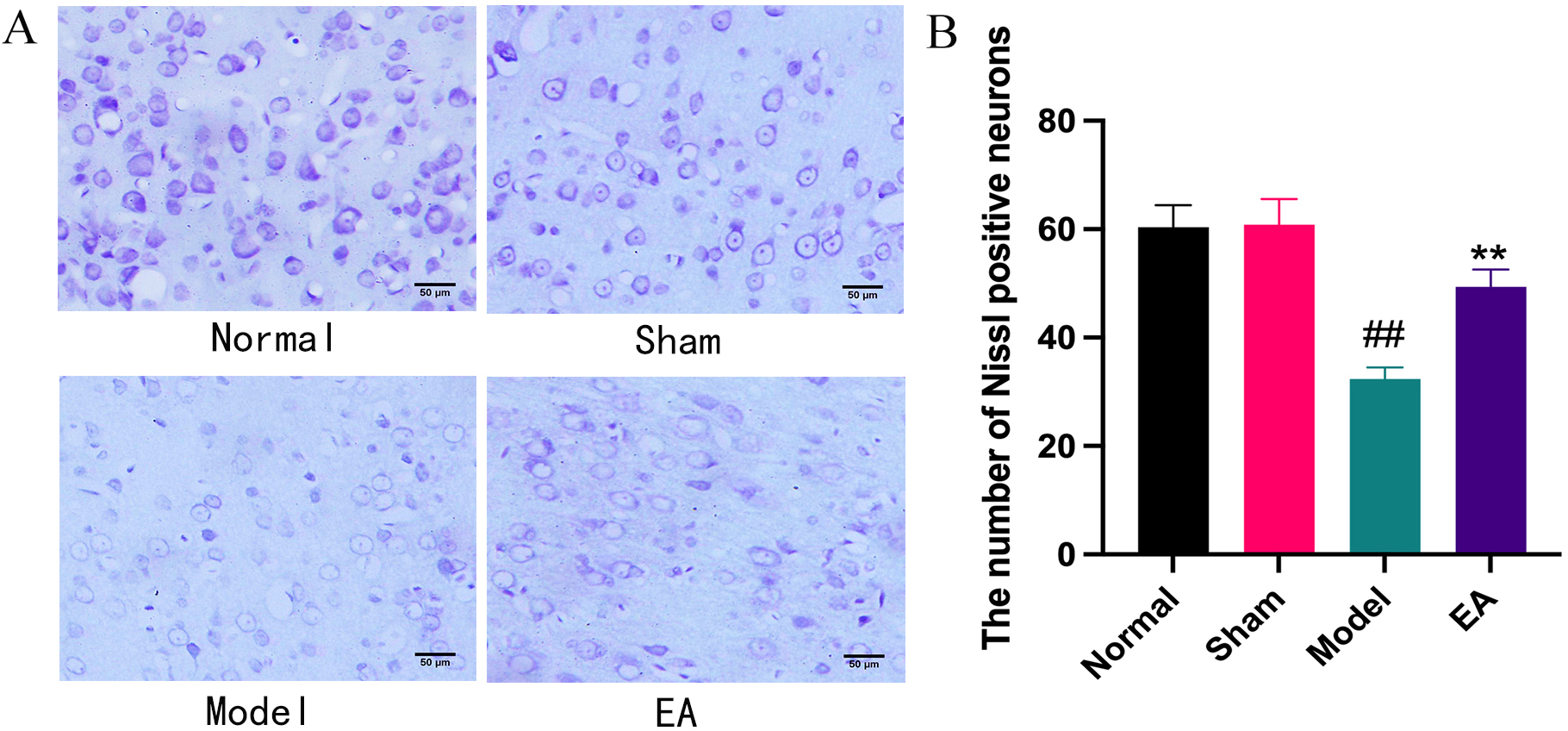 Fig. 1.
Effects of EA on histopathological changes in right PFC of MCAO
rats. (A) Representative pictures of Nissl staining results;
Gas7 and NGF immunoreactive proteins were detected in the PFC of each group, and the positive cells were brownish yellow, mainly colored in the cytoplasm and cell membrane. In the Normal group, the quantity of Gas7 and NGF-positive cells was low, and their distribution was sparse. The number of Gas7 and NGF-positive cells did not differ substantially between the Sham and Normal groups, whereas the number of Gas 7Gas7 and NGF-positive cells in the Model group was significantly bigger than either. Significantly more Gas7 and NGF-positive cells were seen in the EA group than in the Model group (see Fig. 2).
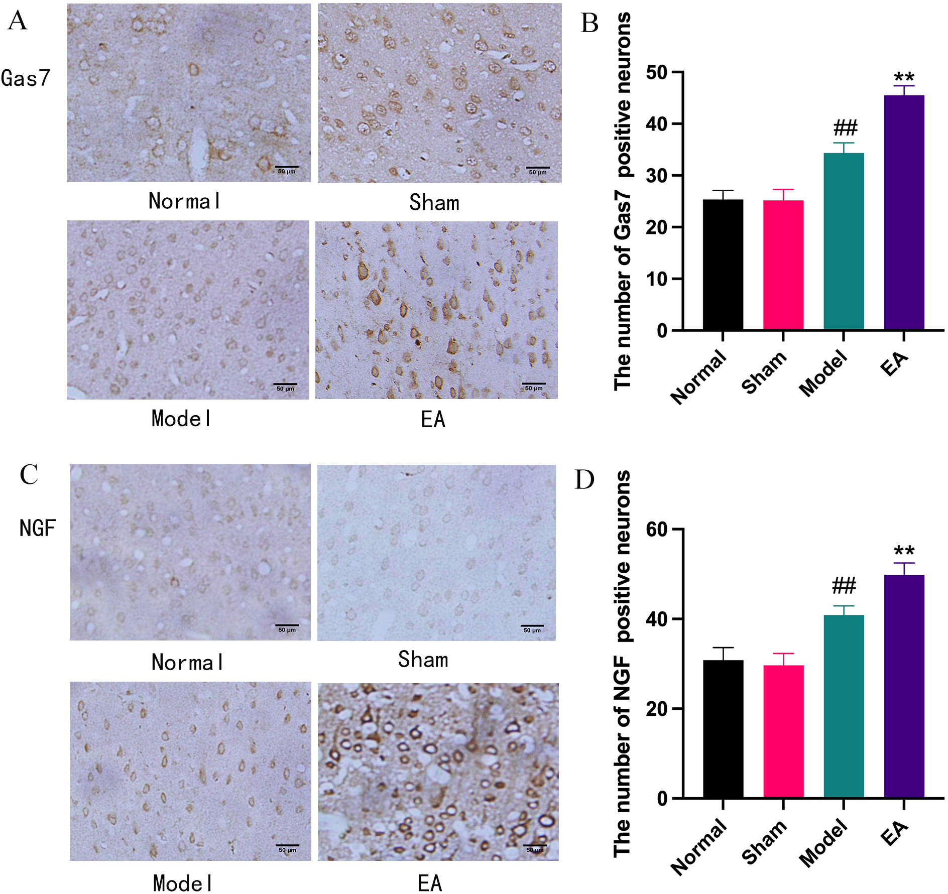 Fig. 2.
Effect of EA on Gas7 and NGF protein expression was examined
through immunohistochemical staining in the right PFC. (A) Representative
pictures of Gas7 immunohistochemical staining results;
Western blot results showed that Gas7 and NGF proteins were expressed at different levels in the PFC of rats in each group. The expressions of Gas7 and NGF proteins in the PFC of rats in each group were normalized by GAPDH. In the Model group, the expression of Gas7 and NGF proteins in the right (ischemic side) PFC was considerably higher than in the Normal group. In comparison to the Model group, the EA group’s PFC on the ischemia side had considerably higher expression levels of the proteins Gas7 and NGF. The right PFC of the Sham surgery group did not statistically express more Gas7 and NGF proteins than did the Normal group (see Fig. 3).
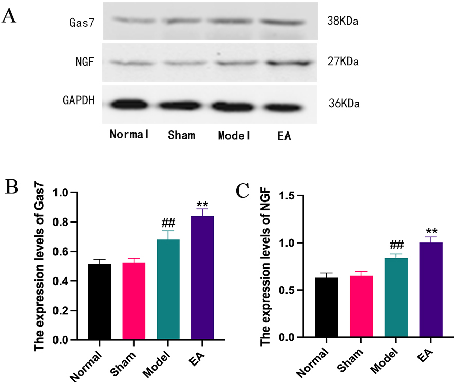 Fig. 3.
Effect of EA on Gas7 and NGF protein expression was examined
through Western blot. (A) Western blot experiment representative band.
(B) Histogram of Gas7 protein expression level comparison;
Gas7 is a member of the family of growth-arrest genes. It is expressed predominantly in the brain. It is an important regulatory factor related to cell differentiation. Studies have shown that it is involved in the growth of nerve axons in vitro. Increased expression of Gas7 can promote the migration of neurons; inhibition of its expression can suppress the migration process of neurons [8, 9, 10]. It has been found that a high level of Gas7 expression protects neurons from damage caused by hypoxia. In the chronic compound-stress model, Gas7 can induce the differentiation of neural stem cells in the hippocampal dentate gyrus into nerve cells, and contribute to the damage repair of the nervous system at various levels. The nervous system, especially the cerebral cortex, is extremely sensitive to ischemia and hypoxia. The cortex tends to be the first area affected by ischemic and hypoxic environments, causing cell damage and even apoptosis [11]. Studies have shown that Gas7 protein can enhance NGF-mediated differentiation of PC12 cells to promote the re-growth of neurons in areas of brain injury such as that caused by stroke [3]. NGF is a protein that regulates the development and maturation of the nervous system and maintains neuronal function. Cerebral ischemia itself can up-regulate the expression level of neuronal NGF in the brain, thereby participating in endogenous protection from cerebral ischemia [12, 13]. NGF is a neuronal growth regulator with dual biological functions of neuron nourishment and neurite outgrowth; it can boost the survival and growth of neurons following ischemia, protect them from harm, and alleviate their diseased state. It promotes neuronal regeneration and differentiation and plays a crucial role in injury repair [14]. The expression levels of Gas7 and NGF proteins in the PFC neurons of the ischemic side of the Model group were considerably higher than those of the Normal and Sham groups, as determined by immunohistochemical detection and Western blot analysis in the present study, suggesting that after cerebral ischemia, the Gas7-mediated endogenous protective effect of NGF on neurons in the injured area may be initiated.
Previous studies have shown that acupuncture at the “Baihui” and “Zusanli” points has the effects of stimulating the “mind”, regulating the spleen and stomach, and “clearing the meridians and collaterals” [15, 16]. “Baihui” belongs to the governor channel and is closely related to the central nervous system. “Zusanli” is tightly associated with blood vessels and nerves. The traditional acupoint combination of “Du Channel” and “Yangming Meridian” has a good therapeutic effect on cerebral ischemia [17, 18]. In rats with cerebral ischemia-reperfusion, electroacupuncture at the “Baihui” and “Zusanli” sites drastically decreased the volume of cerebral infarction and enhanced neurological function. After 72 hours of perfusion, the endoplasmic reticulum chaperone (GRP78) mRNA expression level dramatically increased, while the expression levels of pro-apoptotic proteins significantly decreased [19]. Clinical studies have shown that acupuncture has an effect on improving the disability rate of patients with cerebral infarction. In the frontal cortex around the ischemia area of MCAO rats, our laboratory has demonstrated that electroacupuncture can greatly increase the expression of nestin and stem cell factors [1]. The proliferation of neural stem cells has a protective effect on the frontal cortex neurons around the ischemic focus [1]. The results of Nissl staining in this experiment show that stimulation of “Baihui” and “Zusanli” points with electric acupuncture can significantly lessen the atrophy of ischemic PFC neurons in cerebral ischemia rats. Additionally, electroacupuncture was found to raise the levels of Gas7 and NGF protein expression in neurons in the ischemic PFC, according to immunohistochemistry and Western blot data.
Combining previous findings with those of the current experiment suggests that EA may increase the expression of the Gas7 and NGF in the area of cerebral ischemia injury. and can help eNSCs migrate to the injured area to differentiate into neurons, thereby enhancing the endogenous protective effect.
EA can increase the expressions of Gas7 and NGF in the ischemic prefrontal cortex, which may be one of the mechanisms by which EA promotes the differentiation of eNSCs into neurons in the injured area.
The datasets used and/or analyzed during the current study are available from the corresponding author on reasonable request.
TM, JD, FW—study design; TM, WD, YZ—acquisition and analysis of the Nissl staining data; TM, WD, YZ, JD, FW—acquisition and analysis of the Immunohistochemistry and Western blot data. All authors have made contributions to data interpretation, manuscript drafting and revising, and have approved the final version of the manuscript. All authors have participated sufficiently in the work and agreed to be accountable for all aspects of the work.
The protocol was approved by the Experimental Animal Welfare and Ethics Committee of Wannan Medical College (approval number: LLSC-2021-025).
Not applicable.
This work was supported by the domestic visit and training program for outstanding young backbone talents of colleges and universities in Anhui Province (No. gxgnfx2018016).
The authors declare no conflict of interest.
References
Publisher’s Note: IMR Press stays neutral with regard to jurisdictional claims in published maps and institutional affiliations.