IMR Press / JIN / Volume 22 / Issue 3 / DOI: 10.31083/j.jin2203058
Open Access Review
New Insights into Molecular Mechanisms Underlying Neurodegenerative Disorders
Show Less
1 Department of Neurosurgery, Neuroscience Center, Integrated Hospital of Traditional Chinese Medicine, Southern Medical University, 510310 Guangzhou, Guangdong, China
2 Department of Neurology, Affiliated Zhongda Hospital, School of Medicine, Southeast University, 210009 Nanjing, Jiangsu, China
3 Department of Pathology, Integrated Hospital of Traditional Chinese Medicine, Southern Medical University, 510310 Guangzhou, Guangdong, China
4 Department of Neurosurgery, The Second People's Hospital of Pingnan, 537300 Pingnan, Guangxi, China
5 Department of Orthopedics and Traumatology, The University of Hong Kong, 999077 Hongkong, China
*Correspondence: neuro_clk@hotmail.com (Lukui Chen)
These authors contributed equally.
J. Integr. Neurosci. 2023, 22(3), 58; https://doi.org/10.31083/j.jin2203058
Submitted: 11 January 2023 | Revised: 17 February 2023 | Accepted: 21 February 2023 | Published: 6 May 2023
Copyright: © 2023 The Author(s). Published by IMR Press.
This is an open access article under the CC BY 4.0 license.
Abstract

As a large and heterogeneous group of disorders, neurodegenerative diseases are characterized by the progressive loss of structure or function in neurons, finally leading to neuronal death. Neurodegenerative diseases cause serious threat to a patient’s quality of life and the most common are Alzheimer’s disease and Parkinson’s disease. Currently, little is known of the detailed etiology of these disorders; as such, there are no effective treatments available. Furthermore, the lack of targeted, effective, and resolvable therapy for neurodegenerative diseases, represents an expanding research field for the discovery of new therapeutic strategies. Investigations of the potential pathogenesis of neurodegenerative diseases will become the basis of preventing the occurrence and development of neurodegenerative diseases and finding effective therapies. Existing theories and mechanisms, such as genetic and environmental factors, abnormal protein accumulation, and oxidative stress, are intricately associated with each other. However, there is no molecular theory that can entirely explain the pathological processes underlying neurodegenerative diseases. Due to the development of experimental technology and the support of multidisciplinary integration, it has been possible to perform more in-depth research on potential targets for neurodegenerative diseases and there have been many exciting discoveries in terms of original theories and underlying mechanisms. With this review, we intend to review the existing literature and provide new insights into the molecular mechanisms underlying neurodegenerative diseases.

Keywords
neurodegenerative diseases
molecular mechanism
Alzheimer's disease
Parkinson's disease
Huntington's disease
amyotrophic lateral sclerosis
1. Introduction

Neurodegenerative diseases (NDDs) are typically adult-onset processive disorders that affect the function and plasticity of neuron that arise through a host of one or more genetic and environmental factors [1]. Typical NDDs are Alzheimer’s disease (AD), Parkinson’s disease (PD), amyotrophic lateral sclerosis (ALS), and Huntington’s disease (HD).The number of patients under NDDs is increasing annually. Besides, the lack of effective therapies for NDDs causes considerable burden and economic impact for society [2].

Thus far, many NDD biomarkers have been discovered around the world; these are being used to investigate the mechanisms responsible for the occurrence and development of NDDs [3, 4]. This research has generated a number of advanced theories for the factors responsible for NDDS, including genetic factors [5, 6], oxidative stress [7], accumulation of abnormal protein [8], mitochondrial dysfunction [9] etc. Besides, the association between NDDs and immunology, microbiology and other disciplines has become a new research focus [10, 11]. These factors are involved in the physiological and pathological process of NDDs and promote their occurrence and development. Furthermore, the mechanisms responsible for the transmission of NDDs are under intense investigation [12]. In this review, we provide new insight in our understanding of the pathological mechanism underlying NDDs.

2. Molecular Mechanisms
2.1 Genetic Factor

Genetic Factors are important risk factors to the common NDDs. Genes, formed by nucleotides (DNA, RNA), are the fundamental unit of all biological process. DNA shows differences in terms of base pairs among individuals, thus leading to genetic variation and individual differences in specific trait [13]. The cell types of the nervous system are highly complex, with astrocytes, microglia, and oligodendrocytes present in various neuronal subtypes. The gene expression profiles of these different types of cells play an important role in neurodegenerative diseases and form complex gene expression networks. The survival and function of neurons are coordinated by these intricate gene expression networks.

AD is the most familiar disease of all neurodegenerative diseases and is characterized by the impairment of executive function and memory followed by progressive, global cognitive decline. In developed countries, AD is responsible for the sixth highest death rate [14]. Early-onset AD occurs before the age of 65 years and less than 10% of AD cases represent early-onset (EOAD), a condition that is caused by autosomal dominant mutations in APP, PS1, or PS2, each of which increases Aβ production and thus the extent of amyloid plaques. Of these, less than 13% of EOAD cause demonstrate a fully penetrant autosomal dominant inheritance. The remaining 90% of AD cases were late-onset (LOAD) or sporadic cases. Extensive research efforts have led to the identification of many genes that increase the risk of AD [15]. Human apolipoprotein E (APOE) apolipoproteins have three isoforms (APOEε2, APOEε3, and APOEε4) that differ by only two residues; these stimulated APP transcription and amyloid beta (Aβ) secretion to different degrees. The strongest genetic risk factor of sporadic late onset AD is the ε4 allele of apolipoprotein E [16]. The APOEε4 allele is present in 60–80% of AD cases and increases the risk of AD in a dose-dependent manner. APOE is the most abundant apolipoprotein in the brain where it plays a fundamental role in transport and metabolism of cholesterol and lipid [17]. Over recent years, researchers have performed meta-analyses and genome-wide association studies (GWAS) and identified 29 risk loci related to AD. Apolipoprotein E is one of these 29 risk loci [18].

PD is characterized by the presence of Lewy bodies and Lewy neurons featuring the accumulation of α-synuclein; familial PD is a single mutation and α-synuclein variants are more common in idiopathic cases of PD [19]. Parkinson’s disease only has an effect on 2% of the people over the age of sixty. Over 30 variants approximate genes, like TMEM175, CTSB, LRRK2 (leucine-rich repeat kinase 2), have been reported in meta-analyses from GWAS studies including patients with PD [20]. In addition, family-based genetic studies have identified 23 genes that are associated with the development of PD; these genes exert differing functionalities, including deficiency of synaptic transmission, lysosomal dysfunction vesicular recycling, and mitophagy. Some of the genes involved include PARKIN, autosomal recessive juvenile parkinsonism, α-synuclein, inherited in an autosomal dominant, and PTEN-induced kinase (PINK1), and recessive early onset [21].

HD is an autosomal dominant disorder and is caused by a cytosine-adenine-guanine (CAG) trinucleotide repeat amplification in mutant Huntington genes [22]. Some gene variants, which are related to HD, are similar to the UBR5, FAN1, RRM2B, MTRM10 and MLH1 genes, and are associated with structure-specific DNA handling, DNA mismatch repair, oxidative stress and mitochondrial energetics [23]. Studies have shown that Brain-derived neurotrophic factor (BDNF) plays a role in the development of HD degeneration. A mutant gene product known as huntingtin expresses an extended polyglutamine chain and results in the suppression of BDNF synthesis via dysregulation of translation. Transgenic mice expressing mutant huntingtin have very similar expression impairments to those observed in BDNF knockout mice [24].

ALS can attack the motor neurons in the brain and spinal cord cells; this can lead to paralysis in patients. More than 90% of ALS cases are sporadic. The loss of C9orf72 function may affect the communication between motor neurons and muscles in patients. Mutations in the C9orf72 gene represent the main genetic cause of ALS. A hexanucleotide repeat expansion in the C9orf72 gene is the most common genetic alteration associated with ALS [25]. Mutations in the C9orf72 gene usually include six DNA base (GGGGCC) expansions, typically ranging from a few copies to more than 1000 copies; this compares to less than 20 copies in healthy individuals. Mutations in this gene cause a gain of toxicity including RNA aggregates and toxic dipeptides and are responsible for part of inherited ALS cases without a family history [26]. In the past few years, new breakthroughs in the genetics of ALS have focused on mutations in several malfunctional proteins, such as TAR DNA-binding protein 43 (TDP-43), fusion sarcoma, and SOD1. Family-superoxide dismutase 1 (SOD1) is the main enzyme that scavenges reactive oxygen species (ROS). In fact, SOD1 mutations were identified as the first genetic factor associated with ALS and are observed in 15–30% of families and 1.2–1.5% of sporadic ALS cases. Under normal circumstances, SOD1 can protect cells; however, when a gene mutation is thought to make SOD1 toxic, this form of toxic protein may be directly related to the genetic form of ALS pathogenesis. Abnormal mutations in SOD1 only in neuronal cell death was found in the spinal cord area, which means that the abnormal protein may be related to cell death [27]. TAR DNA binding protein 43 (TDP-43) and fusion sarcoma are related to ALS and frontotemporal dementia, indicating a new pathological mechanism associated with RNA metabolism [28, 29, 30]. Gene-TE pairs have been found to be associated with several cell biological processes that might contribute directly to ALS. Of these, mutations in the TDP-43 can cause transposable elements (TEs) and DNA sequences capable of transposing within the genome to become dysregulated and transcribed. Genes in the extracellular matrix and RNA processing are closely related to TEs. Thus, all the regulatory pathways can be affected in ALS [31].

In summary, GWAS analysis of evidence-based medicine has clearly shown that the expression of neurodegenerative diseases is regulated by similar functional genes, including oxidative stress, immune regulation, mitochondrial abnormalities, and metal ion damage (Fig. 1).
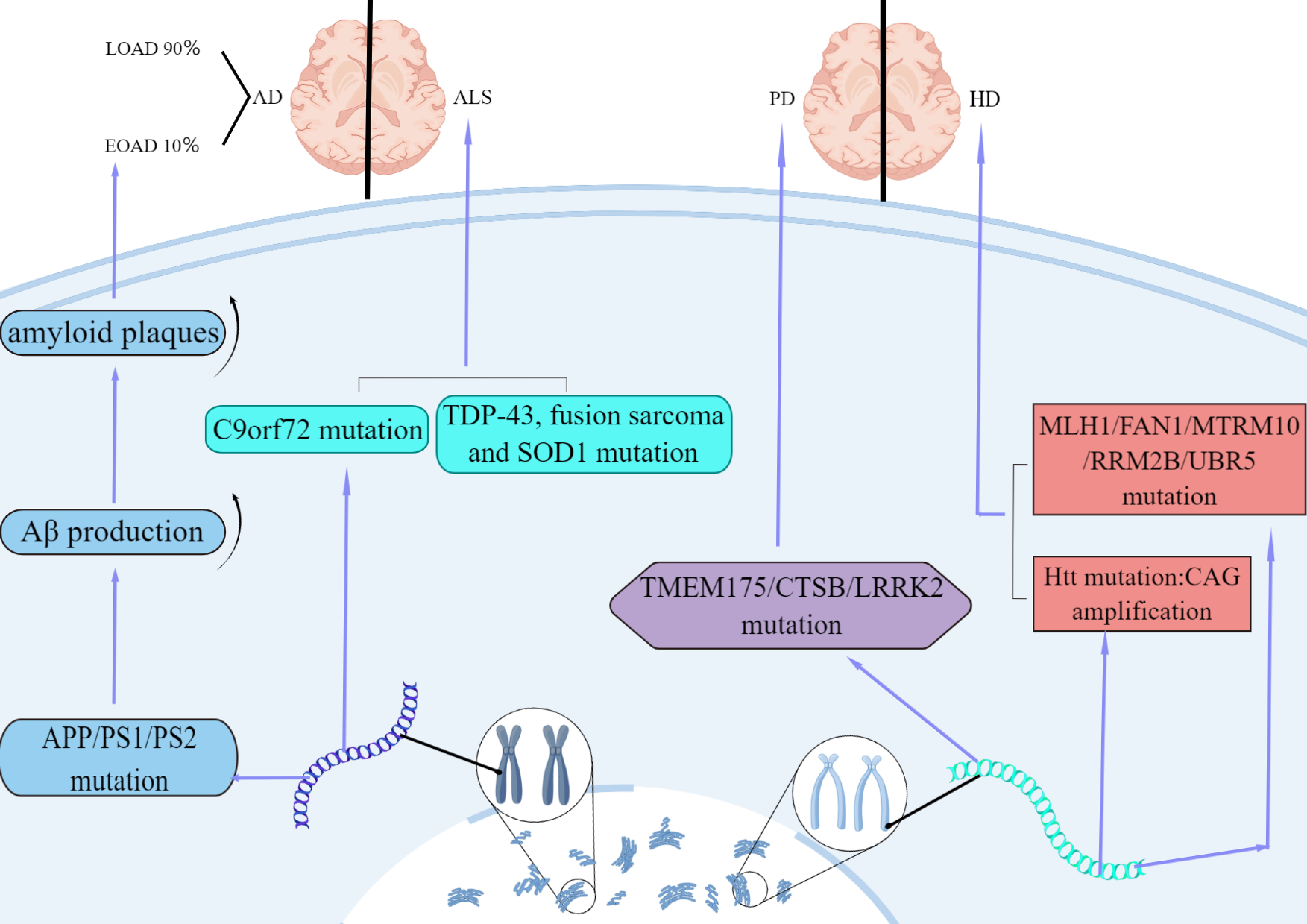
Fig. 1.

Features of AD, ALS, HD and PD in genetic factor. AD is divided into early-onset (EOAD) and late-onset (LOAD). 10% of AD cases are early-onset caused by autosomal dominant mutations in APP, PS1, or PS2. The mutations increase Aβ production and thus amyloid plaques. Besides, UBR5, FAN1, RRM2B, MTRM10 and MLH1 genes and CAG trinucleotide repeat amplification in mutant Huntington (Htt) genes can be detected in HD. PD is characterized by the presence of TMEM175, CTSB, LRRK2 mutation. The main genetic cause of ALS is mutations in the C9orf72 gene. And a new breakthrough focuses on mutations in several malfunctional proteins, such as TAR DNA-binding protein 43 (TDP-43), fusion sarcoma and SOD1.

2.2 Oxidative Stress

Oxidative stress is mostly a reactive process in neurodegenerative diseases. Oxidative stress is caused by the accumulation of free radicals because of environmental changes, including chronic inflammation and mitochondrial dysfunction [32]. A crucial pathogenesis of neurodegenerative diseases is the dysfunction of specific regions of nerve cells due to oxidative stress, the main clinical manifestation of which is the dysfunction of memory and learning [33]. It has been demonstrated that oxidative damage to neuronal DNA is closely related to cognitive deficits and occurs early in the pathological changes of various diseases [34]. Reactive oxygen species (ROS) are fundamental free radicals that can aggravate tissue dysfunction and exacerbate oxidative stress. Under normal circumstances, the generation and scavenging of ROS presents a dynamic balance in the body. Appropriate ROS levels are considered to activate certain signaling pathways (such as AMPK/Ras pathway, epidermal growth factor receptor (EGFR) pathway and protein kinase C (PKC) pathway), adjust cell metabolism, and can also stimulate cell proliferation. However, when ROS scavenging is inferior to production capacity, ROS can accumulate and affect cells, thus resulting in DNA [nuclear and mitochondrial DNA (mtDNA)] damage, chromosomal instability, and protein misfolding [32].

Oxidative stress is an important factor in the pathogenesis of AD. The pathophysiology of AD is primarily related to the formation of extracellular amyloid beta (Aβ) plaques and intracellular tau neurofibrillary tangles (NFT). ROS-induced over-production is believed to play a critical role in the aggregation and secretion of Aβ in AD [35]. ROS and oxidative stress levels are elevated in AD patients while the levels of antioxidants and antioxidant enzymes are reduced. These changes may lead to abnormal functionality due to impaired protein folding in the endoplasmic reticulum, reduced autophagy-mediated clearance and protease activity in damaged proteins, and the increased accumulation of tau proteins and Aβ (Fig. 2) [36]. In a mouse model of AD, peroxidase was shown to significantly improve cognitive performance and spatial learning memory, while also reducing the deposition of Aβ plaques in the cerebral cortex and hippocampus [37].

Fig. 2.

Factors of Oxidative stress involve in AD. Levels of ROS and oxidative stress are improved, and levels of antioxygen and anti-oxidase are decreased in AD patients that lead to function of impaired protein folding in the endoplasmic reticulum, reduced autophagy-mediated clearance and protease of damaged proteins, and increased accumulation of tau proteins and Aβ.

Several lines of evidence suggest that oxidative stress and ROS may be one of the major factors responsible for the progression of neurodegeneration. The neurotoxin metabolite 1-methyl-4-phenylpyridine ion is transported via dopamine transporters to neuronal mitochondria where they accumulate and inhibit complex I activity in the mitochondrial respiratory chain. This leads to a reduction in ATP production and an increase in ROS release, thus resulting in the degeneration of nigrostriatal and striatal dopaminergic neurons (Fig. 3).

Fig. 3.

Mechanism of metabolite 1-methyl-4-phenylpyridine ion (MPP+) in PD. MPP+ is transported via dopamine transporters to neuronal mitochondria. Then, they accumulate and inhibit the complex I activity in the chain of mitochondrial respiratory. This leads to a decrease in ATP production and an increase in ROS release.

This leads to degenerative necrosis of dopaminergic neurons in the striatum and substantia nigra, and ultimately to the development of PD [38]. In addition, metal-mediated toxicity in PD patients also plays a key role in the neuronal damage, the enhancement of oxidative stress in the cellular environment, and elevation of ROS [39]. Like other neurodegenerative diseases, oxidative stress and ROS play a substantial part in the progression of HD. ROS generated during DNA damage, lipid peroxidation and especially protein carbonylation are particularly active in HD. Free radicals cause the peroxidation of intracellular DNA, proteins and membrane lipids. Oxidative DNA damage induces DNA repair pathways, and the behavior leads to restoration of normal DNA function and structure and removal of oxidized bases. Repairment of damaged DNA may cause instability and expansion of CAG trinucleotide repeats in Huntington mutants (Fig. 4) [40]. Furthermore, the levels of iron increase; this can cause an increase in the production of very harmful hydroxyl radicals, thus facilitating interactions with the ferrous iron with H2O2 [35].

Fig. 4.

Factors of Oxidative stress involve in HD. Free radicals lead to peroxidation of DNA, proteins and membrane lipids. Oxidative DNA damage induces DNA repair pathways, and the behavior leads to restoration of normal DNA function and structure. And oxidized bases are removed. Repairment of damaged DNA may cause instability and expansion of CAG trinucleotide repeats in Huntington mutant.

The key biomarker for ALS is oxidative stress; it is widely considered that oxidative stress may contribute to neuronal death and the progression of ALS. SOD1 is a member of the superoxide dismutase family and is the main enzyme for scavenging reactive oxygen species. The second most commonly known genetic cause of ALS is mutations in the SOD1 gene; these account for approximately 12% of familial cases and 1% of sporadic cases [41]. However, these mutations result in increased misfolding or toxic function of the SOD1 protein. In a previous study, Garg et al. [42] found that the direct interaction between mutant misfolded SOD1 and TRAF6 could lead to increased SOD1 functional toxicity.

2.3 Accumulation of Abnormal Protein

Proteins are responsible to the most of organismal and cellular functions. The synthesis of protein involves multiple procedures; information held by a gene is transcribed into mRNA which is then translated by the ribosomes into a protein. Most proteins need to fold appropriately into a specific three-dimensional structure in order to function. Errors may occur at any time during this process. When errors occur, we can use chaperones and degradation machinery to help prevent their aggregation and folding [43]. Numerous studies have shown that protein misfolding and aggregation occur in many neurodegenerative diseases [44]. The misfolding of proteins can exert detrimental effects on their normal biological function and can generate harmful oligomers. These processes are associated with the binding of proteases and molecular chaperones during translation in cells [45]. The appearance of aggregated nuclei is the beginning of aggregation of protein [46]. Once nucleated, intracellular protein monomers with a tendency to aggregate undergo random folding to produce misfolded, non-functional protein oligomers, which further aggregate to form fibrils (also known as aggregates) [47]. Oligomers and multimers can be degraded by protein molecular chaperones, either by the ubiquitin-proteasome or by autophagy; damaged oligomers and multimers produce cytotoxicity, thus causing cell death and inflammation [47, 48].

AD, PD, and HD are closely related to the deposition and aggregation of proteins that are misfolded, for example Aβ and neurofibrillary tangle in the brain of AD patients; α-synuclein aggregation forming Lewy bodies in the brain of PD; and Huntington protein in HD. Furthermore, GSK3-b is considered as a key regulator in AD because its dysregulation might cause the production of amyloid-b, dysfunctional neurogenesis, and Tau phosphorylation. Studies have shown that the overexpression of lncRNA-ZBTB20-AS1 can inhibit ZBTB20 expression and promote GSK-3b expression and Tau phosphorylation, thus contributing to the development of AD. These findings identified potential molecular mechanisms and provided reference guidelines for the treatment of AD [49]. However, drugs that are developed by major international pharmaceuticals companies on the basis of removing protein polymers or amyloid plaques cannot significantly improve cognitive decline and the progression of disease. Therefore, it is important to investigate how the aggregation and abnormal folding of Huntington proteins leads to neurological death or the onset of neurodegeneration results in protein aggregation. We also need to investigate whether the earliest pathological changes originate in the nucleus accumbens of the brainstem or in the peripheral input pathway intestine [34].

2.4 Immunology

The link between neurodegeneration and immunity was established some time ago. However, little is known about neurodegeneration [50]. The immune cells in central nervous system are mainly microglia, oligodendrocyte and astrocytes [51]. These three types of cells undergo different physiological changes during neurodegeneration. Acute Neurodegeneration is associated with a relatively supernal rate of disease and a relatively low rate of immune-related development. However, chronic neurodegeneration is more persistent than immune-related development.

AD is characterized by progressive memory loss and cognitive impairment. Astrocyte also secretes and ingests Aβ and α-synuclein. Extracellular Aβ plaques in the brain, intracellular neurofibrillary tangle, and neuronal loss in the central nervous system (CNS) gray matter with hyperphosphorylated tau protein are all known to be key features of AD. Microglia, a major player in the immune system of the central nervous system, has a strong phagocytic effect on Aβ peptide. To this end, Hettmann et al. [52] designed an antibody that targets the pyroglutamic acid modified end of the Aβ protein. This antibody only binds to this protein in the center and has no binding effects with non-pathological proteins in other regions. Synaptic plasticity, synaptic pruning, apoptosis, and neurogenesis, are associated with the complement system and other functions of microglia, especially C1q complement, which mediates the phagocytosis of microglia [53]. Specific antibodies can inhibit a significant portion of activation in complement, which in turn inhibits activation of the microglia in the CNS. Hickman et al. [54] showed that the microglial expression of chemokine receptor CX3CR1 which allows more microglia to be recruited to the site of neuroinflammation. During the late stages of mouse development, a lack of CX3CR1 led to poorer mental function and plaque accumulation in this part of the brain [54]. A reduction in Aβ levels in the brain is associated with improvements in cognitive function. Thus, it is possible that this pathway could act as a regulator of gene expression to increase the rate of Aβ clearance in neurons. Today, the most promising AD trials are based on the Aβ cascade hypothesis, and uses a monoclonal antibody to target a β peptide. Most monoclonal antibodies clear Aβ peptide, but do not prevent cognitive decline. Most monoclonal antibodies are already in phase III, and some may reduce the level of the marker protein tau [55].

In the case of PD, there is evidence that Parkinson is associated with Specific HLA variants (HLA-DRA and HLA-DRB1) [56], thus suggesting an association with the autoimmune system. As a special case, idiopathic Parkinson’s syndrome is diagnosed when the body detects the inflammation induced by T cells that target α-synuclein [57]. α-synuclein is present in normal body fluids, although there are indications that it is involved in immunity, especially in spontaneous immunity. In addition, the activation of microglia and the expression of related anti-inflammatory factors (IFN-γ and doludinD1), were also detected in mice expressing human α-synuclein [58]. LRRK2 affects the normal morphology of endogenous α-synuclein physiological tetramer, thus affecting the process of disease [59]. The expression of LRRK2 involves immune signals in macrophages and microglia from human-induced pluripotent stem cells, especially IFN-γ, which are induced in a highly significant manner [60]. In addition, the patients’ B cells and T cells express higher levels of LRRK2 in patients than in a control group. The key difference between LRRK2-deficient and non-LRRK2-deficient mice was that LRRK2-G2019S transgenic mice injected with α-synuclein preformed fibrils in the striatum showed different functional effects when compared with control mice [61]. Some microstructural changes of demyelination were observed in PD patients. Studies in transgenic mice have shown that oligodendrocyte dysfunction exacerbates neuronal degeneration [34, 50].

Although astrocytes secrete and ingest α-synuclein and β-synuclein, they are clearly associated with AD. However, there is also a strong similarity to the ALS (amyotrophic lateral sclerosis) model. The neurotoxic effects of astrocytes are attributed to impaired neuronal responses to the release of Ephb1 [50]. Astrocytes are important players in the pathogenesis of ALS. Astrocytes take effect through multiple gain-of-toxicity and loss-of-support mechanisms. Furthermore, astrocytes attempt to counteract toxicity through upregulation of the WNT/β-catenin pathway in motor neurons [62]. In a previous study, Chen et al. [63] analyzed differentially expressed proteins (DEPs) in the spinal cord and validated 48 proteins from immunity and inflammation-related pathways by parallel reaction monitoring (PRM) analysis. Most of these were involved in the following pathways: antigen processing and presentation, complement and coagulation cascades, NF-kappa B signaling pathway, extracellular matrix-receptor (ECM- receptor) interactions, retinoic acid-inducible gene I (RIG)-I-like receptor signaling pathway, phagosome focal adhesion, and lysosomes. These results expand our understanding of immunity and inflammation with regards to ALS [63].

2.5 DNA Damage Repair

In a previous study, Ross et al. [64] reported that DNA damage was a common pathway responsible for neurodegenerative diseases. Furthermore, recent studies have shown that DNA damage can be divided into nuclear DNA damage and mitochondrial DNA damage [65]. Although relevant pathological data and the underlying reasons are open to question, Heather et al. [66] stated that dying or damaged cells released damage-related molecular proteins that can cause specific inflammatory cascades. Both have similar outcomes for neurodegenerative diseases.

There are now two main options, one is to increase free radical scavenging, such as with superoxide dismutase 1 (SOD1), which protects neurons from Aβ-mediated neurotoxicity in Alzheimer’s disease [67]. The other is to repair DNA.

In a previous study, Cai et al. [68] found that mitochondrial DNA may be a key factor associated with aging in neurodegenerative diseases. Tau tangles and Aβ in AD patients cannot be cleaned-up; furthermore, excess free radicals can lead to mitochondrial damage [69]. Fang et al. [70] reported that the incidence of AD and the level of inflammation in a mouse model of mitophagy were reduced.

In addition to key proteins, related abnormalities have also been detected in exosomes. A recent study reported that extracellular vesicles contain misfolded SOD1 enzymes that are delivered to other normal cells, where they can spread to other normal cells [71]. Furthermore, exosomes have also been found in AD and PD. SOD1 has also been associated with mitochondrial DNA damage. Therefore, exosomes may represent a new mechanism to improve mitochondrial DNA damage.

2.6 Mitochondrial Dysfunction

The regulation of the body’s energy metabolism is essential to ensure the functioning of the organism. Mitochondria, as active controllers, play a central role in whole-body energy homeostasis [72]; neurons are one of the most energy-consuming cell types in the body and have a highly complex morphology. The normal functionality of neurons depends on mitochondrial morphology and functional integrity. Apart from the fact that mitochondria serve as primary energy providers, there are many other important functions in mediating some other activities of cells, such as calcium homeostasis, oxidative stress, amino and lipids acids metabolism and proliferation of cells [73]. In fact, mitochondrial dysfunction and mitochondrial abnormalities have been recognized as key and co-factors in the pathology of neurodegenerative diseases, which can disrupt the normal activity of cells and ultimately lead to neuronal loss [74].

Compared with glucose utilization, by means of positron emission tomography, we detected a reduction in brain metabolism and an increase of oxidative utilization in the temporoparietal cortex of AD. The proportion of normal mitochondria was significantly lower, and the proportion of cristae-fractured mitochondria was significantly higher in AD neurons when compared with controls of the same age. In addition, mitochondrial size, number, and distribution, were significantly altered in AD-vulnerable neurons [75]. Furthermore, 15 out of 51 members of pathways related to glycolysis, oxidative phosphorylation, and the TCA cycle, were significantly downregulated in AD, as determined by microarray analysis and quantitative RT-PCR studies. Gene set enrichment analysis showed that disruption of mitochondrial import pathways and the downregulation of mitochondrial oxidative phosphorylation were prevalent in AD patients. These changes resulted in marked changes in the activities of enzymes that are involved in the TCA cycle in AD: dehydrogenases (including SDH and MDH) were increased, while dehydrogenases/decarboxylases (including PDHC, ICDH, and KGDHC) were decreased; these phenomena are associated with the clinical status of AD patients [76] (Fig. 5).

Fig. 5.

Features of AD in Mitochondrial dysfunction factor. Disruption of mitochondrial import pathways and downregulation of oxidative phosphorylation (OXPHOS) are prevalent in AD. These changes resulted in marked changes in the activities of enzymes that are involved in the TCA cycle of AD.

Furthermore, other studies have suggested that damaged mitochondria played a key role in the pathophysiology of AD. Mitochondria regularly encounter endogenous stress such as DNA damage, oxidative toxicants and environmental stresses, thus generating structural or functional damage to these vital organelles. Neuronal activity is extremely energy-dependent and sensitive to disturbances in mitochondrial function [35].

Several studies have highlighted the crucial role of proteins and miRNAs in regulating genes that are involved in mitochondrial integrity under different pathological conditions of neurodegenerative diseases [77, 78, 79]. The mtDNA levels in body tissues and fluids, and the ratio of mitochondrial genome to nuclear genome, are usually the evaluative criteria for variations in mtDNA levels, these represent biomarkers of mitochondrial aberrations [80]. Zhou et al. [81] previously described the effects of microRNA-330, which targets the proto-oncogene vav (VAV), on oxidative stress, mitochondrial dysfunction, and Aβ production in AD mice via the MAPK signaling pathway. The multifunctional DNA and RNA-binding protein TDP-43 was found to activate the mitochondrial unfolded protein response, inhibit mitochondrial complex I activity, and reduce mitochondrial ATP synthesis in cellular and animal models [82]. Mitophagy mediated by the PINK1/Parkin pathway has a significant impact on the unwanted proteins deposited in mitochondria, the mitochondrial quality control system, and elimination of misfolded protein [83]. In addition to this, recent studies suggest that mitochondrial damage may affect neurodegenerative diseases by regulating many aspects of ferroptosis, including cellular metabolism, iron homeostasis, and lipid peroxidation [84].

2.7 Metal ion Disorders

The maintenance of metal ion homeostasis is very important for the normal biological functions of the brain, such as the synthesis and metabolism of neurotransmitters and oxygen transport. Na+ and K+ are essential for the conduction of nerve impulses. In addition to acting as the second messenger of cells, Ca2+ also triggers the structural transformation of related proteins to form protein conformations with specific functions. Furthermore, specific metal ions, such as Zn2+, Cu2+ and Fe2+, can perform crucial physiological functions in the physiology in the brain, such as the cerebral cortex, forebrain basal, and hippocampus. Once the levels of metal ions in the brain are disturbed, many biological processes can become abnormal; this eventually leads to the occurrence of neurodegenerative diseases.

Imbalanced intracellular metal homeostasis and toxic metal exposure may play a contributing role in pathology and may represent a cause of neurodegenerative diseases. Karolina et al. [85] reported that an excessive level of trace metal ions could contribute to oxidative stress, endoplasmic reticulum stress, the activation of apoptosis, mitochondrial dysfunction, and the dysregulation of autophagy.

With regards to AD, there is evidence that it is associated with iron homeostasis. And there are potential links with oxidative stress and glutathione [86]; these associations may involve high levels of ACSL4 [87]. Researchers previously demonstrated that the upregulation of xCT perturbed glutathione metabolism and lipid peroxidation in ferroptosis in AD patients. Metal ions, such as Zn2+ and Fe2+, can bind to Aβ and impact on amyloid aggregation in AD patients [86]. These metal ions such as Zn2+ and Fe2+ can bind to Aβ and impact on amyloid aggregation in AD patients. The ability of Aβ to reduce Fe (III) to Fe (II) has been repeatedly confirmed in extensive studies. This process leads to the generation of superoxide anions, thus leading to oxidative damages [88]. Song et al. [89] has shown that Iron can be involved in propagation of lipid peroxidation and free radical formation. An abnormal increase in labile iron pools through dysregulation of transferrin and transferrin receptors (iron from the extracellular environment) or autophagy of ferritin (namely, ferritinophagy) mediated by nuclear receptor coactivator 4 releases iron bounds to ferritin when iron homeostasis in the body is out of balance. Then through peroxy radicals and the Fenton reaction produces hydroxyl, then extracts oxygen atoms from PUFA diallyl carbon and induces PUFA-PLs peroxidation, finally induced ferroptosis. Patients with PD are characterized by nigrostriatal damage and dopamine depletion [39]. Typically, the dopamine released is taken-up by presynaptic neurons, or taken up by adjacent astrocytes and metabolized by mitochondria to HoMovanillic acid (HVA). Dopamine can be temporarily oxidized in a spontaneous manner. This process can be facilitated by highly reactive metals to facilitate the depletion of dopamine [90]. Similarly, metal ions can also disrupt mitochondrial function and structure, and can produce pathological features in cascades.

HD is another neurodegenerative disorder that is closely related to oxidative reactions catalyzed by transition metals. A growing number of studies have highlighted oxidative stress and the dysregulation of metals in the pathogenesis of HD. Studies of the effects of free radicals on HD have revealed a considerable increase in carbonylation in the striatum of the brain after HD. Similarly, changes in catalytic activity or the protein levels of antioxidant enzymes have been observed [91].

3. Conclusions and Perspectives

Neurodegenerative diseases are the most common diseases of the elderly. The mechanisms underlying such disease are both intricate and complex. Different theories have arisen that have their own advantages and disadvantages; however, precise mechanisms have yet to be elucidated. Furthermore, genetic factors, oxidative stress, and immunology all play roles in the pathogenesis process to a greater or lesser extent, and may complement each other. The current research focuses in neurodegenerative disease is mainly Parkinson’s disease, Alzheimer’s disease, and amyotrophic lateral sclerosis. Due to recent in-depth studies of the enteric nervous system, we are now beginning to explore gut-related microbes, starting from the enteric neural network, to regulate disease and act as a target to improve regulation of disease as a target to improve disease. A current research hotspot relates to autophagy and mitochondria/lysosomes. These various theories may pave the way for a better understanding of the etiology or mechanisms of neurodegenerative diseases. With the advancement of global research, and increasing collaboration across different research topics, the mechanisms underlying neurodegenerative diseases will become increasingly clear and contribute to the development of clinical treatment plans.

Author Contributions

All authors declared that they materially participated in the article preparation. LC, YH and JLong launched the viewpoint of manuscript. JLiu and WD wrote the manuscript and conceived the structure of Figures. JLiu finished the contents of Figures. QZ, CG and HC summarized the manuscript. RL, WA, YD and YQ revised the manuscript and collected the supplementary contents at the revision stage. YH polished the contents of manuscript at the revision stage. All authors contributed to editorial changes in the manuscript. All authors read and approved the final manuscript. All authors have participated sufficiently in the work and agreed to be accountable for all aspects of the work.

Ethics Approval and Consent to Participate

Not applicable.

Acknowledgment

We thank Fig.Draw (https://www.figdraw.com/) for editing figures.

Funding

This work was sponsored by National Key R&D Program of China (2022YFA1104900 & 2022YFA1104904), Natural Science Foundation of Guangdong Province (2021A1515010013), Department of Education of Guangdong Province (2021ZDZX2011), Traditional Chinese Medicine Bureau of Guangdong Province (20221275), Guangzhou Municipal Science and Technology Project (202201011760) and President Foundation of Integrated Hospital of Traditional Chinese Medicine of Southern Medical University (1202101003 & 1202103007).

Conflict of Interest

Lukui Chen is serving as one of the Guest editors of this journal. We declare that Lukui Chen had no involvement in the peer review of this article and has no access to information regarding its peer review. Full responsibility for the editorial process for this article was delegated to Gernot Riedel. The other authors declare no conflict of interest.

References
[1]
Takalo M, Salminen A, Soininen H, Hiltunen M, Haapasalo A. Protein aggregation and degradation mechanisms in neurodegenerative diseases. American Journal of Neurodegenerative Disease. 2013; 2: 1–14.
[2]
Mao K, Zhang G. The role of PARP1 in neurodegenerative diseases and aging. The FEBS Journal. 2022; 289: 2013–2024.
[3]
Sharma HS, Chopp M, Chen L, Sarnowska A, Xue M, Ao Q, et al. The 2021 yearbook of Neurorestoratology. Journal of Neurorestoratology. 2022; 10: 100008.
[4]
Huang H, Chen L, Chopp M, Young W, Robert Bach J, He X, et al. The 2020 Yearbook of Neurorestoratology. Journal of Neurorestoratology. 2021; 9: 1–12.
[5]
Sawcer S, Hellenthal G, Pirinen M, Spencer CCA, Patsopoulos NA, Moutsianas L, et al. Genetic risk and a primary role for cell-mediated immune mechanisms in multiple sclerosis. Nature. 2011; 476: 214–219.
[6]
Taylor JP, Brown RH, Jr, Cleveland DW. Decoding ALS: from genes to mechanism. Nature. 2016; 539: 197–206.
[7]
Uttara B, Singh AV, Zamboni P, Mahajan RT. Oxidative stress and neurodegenerative diseases: a review of upstream and downstream antioxidant therapeutic options. Current Neuropharmacology. 2009; 7: 65–74.
[8]
Taylor JP, Hardy J, Fischbeck KH. Toxic Proteins in Neurodegenerative Disease. Science. 2002; 296: 1991–1995.
[9]
Chen H, Chan DC. Mitochondrial dynamics-fusion, fission, movement, and mitophagy-in neurodegenerative diseases. Human Molecular Genetics. 2009; 18: R169–R176.
[10]
Marsh SE, Abud EM, Lakatos A, Karimzadeh A, Yeung ST, Davtyan H, et al. The adaptive immune system restrains Alzheimer’s disease pathogenesis by modulating microglial function. Proceedings of the National Academy of Sciences. 2016; 113: E1316–E1325.
[11]
Cekanaviciute E, Yoo BB, Runia TF, Debelius JW, Singh S, Nelson CA, et al. Gut bacteria from multiple sclerosis patients modulate human T cells and exacerbate symptoms in mouse models. Proceedings of the National Academy of Sciences of the United States of America. 2017; 114: 10713–10718.
[12]
Jaunmuktane Z, Mead S, Ellis M, Wadsworth JDF, Nicoll AJ, Kenny J, et al. Evidence for human transmission of amyloid-β pathology and cerebral amyloid angiopathy. Nature. 2015; 525: 247–250.
[13]
Flores-Dorantes MT, Díaz-López YE, Gutiérrez-Aguilar R. Environment and Gene Association With Obesity and Their Impact on Neurodegenerative and Neurodevelopmental Diseases. Frontiers in Neuroscience. 2020; 14: 863.
[14]
Wang S, Mustafa M, Yuede CM, Salazar SV, Kong P, Long H, et al. Anti-human TREM2 induces microglia proliferation and reduces pathology in an Alzheimer’s disease model. Journal of Experimental Medicine. 2020; 217: e20200785.
[15]
Wang H, Devadoss D, Nair M, Chand HS, Lakshmana MK. Novel Alzheimer risk factor IQ motif containing protein K is abundantly expressed in the brain and is markedly increased in patients with Alzheimer’s disease. Frontiers in Cellular Neuroscience. 2022; 16: 954071.
[16]
Ertuzun T, Semerci A, Cakir ME, Ekmekcioglu A, Gok MO, Soltys DT, et al. Investigation of base excision repair gene variants in late-onset Alzheimer’s disease. PLoS ONE. 2019; 14: e0221362.
[17]
de Frutos Lucas J, Sewell KR, García-Colomo A, Markovic S, Erickson KI, Brown BM. How does apolipoprotein E genotype influence the relationship between physical activity and Alzheimer’s disease risk? A novel integrative model. Alzheimer’s Research & Therapy. 2023; 15: 22.
[18]
Jansen IE, Savage JE, Watanabe K, Bryois J, Williams DM, Steinberg S, et al. Genome-wide meta-analysis identifies new loci and functional pathways influencing Alzheimer’s disease risk. Nature Genetics. 2019; 51: 404–413.
[19]
Lill CM. Genetics of Parkinson’s disease. Molecular and Cellular Probes. 2016; 30: 386–396.
[20]
Chang D, Nalls MA, Hallgrímsdóttir IB, Hunkapiller J, van der Brug M, Cai F, et al. A meta-analysis of genome-wide association studies identifies 17 new Parkinson’s disease risk loci. Nature Genetics. 2017; 49: 1511–1516.
[21]
Karimi-Moghadam A, Charsouei S, Bell B, Jabalameli MR. Parkinson Disease from Mendelian Forms to Genetic Susceptibility: New Molecular Insights into the Neurodegeneration Process. Cellular and Molecular Neurobiology. 2018; 38: 1153–1178.
[22]
Casella C, Lipp I, Rosser A, Jones DK, Metzler‐Baddeley C. A Critical Review of White Matter Changes in Huntington’s Disease. Movement Disorders. 2020; 35: 1302–1311.
[23]
Correia K, Harold D, Kim K, Holmans P, Jones L, Orth M, et al. The Genetic Modifiers of Motor OnsetAge (GeM MOA) Website: Genome-wide Association Analysis for Genetic Modifiers of Huntington’s Disease. Journal of Huntington’s Disease. 2015; 4: 279–284.
[24]
Zhou Z, Zhong S, Zhang R, Kang K, Zhang X, Xu Y, et al. Functional analysis of brain derived neurotrophic factor (BDNF) in Huntington’s disease. Aging. 2021; 13: 6103–6114.
[25]
van’t Spijker HM, Almeida S. How villains are made: the translation of dipeptide repeat proteins in C9ORF72-ALS/FTD. Gene. 2023; 858: 147167.
[26]
Butti Z, Pan YE, Giacomotto J, Patten SA. Reduced C9orf72 function leads to defective synaptic vesicle release and neuromuscular dysfunction in zebrafish. Communications Biology. 2021; 4: 792.
[27]
Trist BG, Genoud S, Roudeau S, Rookyard A, Abdeen A, Cottam V, et al. Altered SOD1 maturation and post-translational modification in amyotrophic lateral sclerosis spinal cord. Brain. 2022; 145: 3108–3130.
[28]
Sreedharan J, Blair IP, Tripathi VB, Hu X, Vance C, Rogelj B, et al. TDP-43 Mutations in Familial and Sporadic Amyotrophic Lateral Sclerosis. Science. 2008; 319: 1668–1672.
[29]
Kwiatkowski TJ, Jr, Bosco DA, Leclerc AL, Tamrazian E, Vanderburg CR, Russ C, et al. Mutations in the FUS/TLS gene on chromosome 16 cause familial amyotrophic lateral sclerosis. Science. 2009; 323: 1205–1208.
[30]
Vance C, Rogelj B, Hortobágyi T, De Vos KJ, Nishimura AL, Sreedharan J, et al. Mutations in FUS, an RNA Processing Protein, Cause Familial Amyotrophic Lateral Sclerosis Type 6. Science. 2009; 323: 1208–1211.
[31]
Valdebenito-Maturana B, Rojas-Tapia MI, Carrasco M, Tapia JC. Dysregulated Expression of Transposable Elements in TDP-43M337V Human Motor Neurons That Recapitulate Amyotrophic Lateral Sclerosis In Vitro. International Journal of Molecular Sciences. 2022; 23: 16222.
[32]
Wang X, Zhou Y, Gao Q, Ping D, Wang Y, Wu W, et al. The Role of Exosomal microRNAs and Oxidative Stress in Neurodegenerative Diseases. Oxidative Medicine and Cellular Longevity. 2020; 2020: 1–17.
[33]
Gu C, Zhang Q, Li Y, Li R, Feng J, Chen W, et al. The PI3K/AKT Pathway-The Potential Key Mechanisms of Traditional Chinese Medicine for Stroke. Frontiers in Medicine. 2022; 9: 900809.
[34]
Liu S, Han S, Dai Q, Li S, Li J. BICAO-induced ischaemia caused depressive-like behaviours and caspase-8/-9-dependent brain regional neural cell apoptosis in mice. Stroke and Vascular Neurology. 2017; 3: 1–8.
[35]
Rehman MU, Sehar N, Dar NJ, Khan A, Arafah A, Rashid S, et al. Mitochondrial dysfunctions, oxidative stress and neuroinflammation as therapeutic targets for neurodegenerative diseases: an update on current advances and impediments. Neuroscience and Biobehavioral Reviews. 2023; 144: 104961.
[36]
Mohsenzadegan M, Mirshafiey A. The immunopathogenic role of reactive oxygen species in Alzheimer disease. Iranian Journal of Allergy, Asthma, and Immunology. 2012; 11: 203–216.
[37]
Xu J, Wang K, Yuan Y, Li H, Zhang R, Guan S, et al. A Novel Peroxidase Mimics and Ameliorates Alzheimer’s Disease-Related Pathology and Cognitive Decline in Mice. International Journal of Molecular Sciences. 2018; 19: 3304.
[38]
Hu Q, Wang G. Mitochondrial dysfunction in Parkinson’s disease. Translational Neurodegeneration. 2016; 5: 14.
[39]
Monzani E, Nicolis S, Dell’Acqua S, Capucciati A, Bacchella C, Zucca FA, et al. Dopamine, Oxidative Stress and Protein–Quinone Modifications in Parkinson’s and other Neurodegenerative Diseases. Angewandte Chemie International Edition. 2019; 58: 6512–6527.
[40]
Singh A, Kukreti R, Saso L, Kukreti S. Oxidative Stress: A Key Modulator in Neurodegenerative Diseases. Molecules. 2019; 24: 1583.
[41]
Renton AE, Chiò A, Traynor BJ. State of play in amyotrophic lateral sclerosis genetics. Nature Neuroscience. 2014; 17: 17–23.
[42]
Garg P, Semmler S, Baudouin C, Vande Velde C, Plotkin SS. Misfolding-Associated Exposure of Natively Buried Residues in Mutant SOD1 Facilitates Binding to TRAF6. Journal of Molecular Biology. 2022; 434: 167697.
[43]
Bertolotti A. Importance of the subcellular location of protein deposits in neurodegenerative diseases. Current Opinion in Neurobiology. 2018; 51: 127–133.
[44]
Bae EJ, Ho DH, Park E, Jung JW, Cho K, Hong JH, et al. Lipid peroxidation product 4-hydroxy-2-nonenal promotes seeding-capable oligomer formation and cell-to-cell transfer of α-synuclein. Antioxidants & Redox Signaling. 2013; 18: 770–783.
[45]
Yu J, Lyubchenko YL. Early stages for Parkinson’s development: alpha-synuclein misfolding and aggregation. Journal of Neuroimmune Pharmacology. 2009; 4: 10–16.
[46]
Kumar V, Sami N, Kashav T, Islam A, Ahmad F, Hassan MI. Protein aggregation and neurodegenerative diseases: from theory to therapy. European Journal of Medicinal Chemistry. 2016; 124: 1105–1120.
[47]
Wolozin B. Regulated protein aggregation: stress granules and neurodegeneration. Molecular Neurodegeneration. 2012; 7: 56.
[48]
Bandyopadhyay U, Cuervo AM. Chaperone-mediated autophagy in aging and neurodegeneration: lessons from alpha-synuclein. Experimental Gerontology. 2007; 42: 120–128.
[49]
Wang Y, Cai M, Lou Y, Zhang S, Liu X. ZBTB20-as1 promoted Alzheimer’s disease progression through ZBTB20/GSK-3β/Tau pathway. Biochemical and Biophysical Research Communications. 2023; 640: 88–96.
[50]
Rõlova T, Lehtonen Š, Goldsteins G, Kettunen P, Koistinaho J. Metabolic and immune dysfunction of glia in neurodegenerative disorders: Focus on iPSC models. Stem Cells. 2021; 39: 256–265.
[51]
Lee DSW, Rojas OL, Gommerman JL. B cell depletion therapies in autoimmune disease: advances and mechanistic insights. Nature Reviews Drug Discovery. 2021; 20: 179–199.
[52]
Hettmann T, Gillies SD, Kleinschmidt M, Piechotta A, Makioka K, Lemere CA, et al. Development of the clinical candidate PBD-C06, a humanized pGlu3-Aβ-specific antibody against Alzheimer’s disease with reduced complement activation. Scientific Reports. 2020; 10: 3294.
[53]
Bie B, Wu J, Foss JF, Naguib M. Activation of mGluR1 Mediates C1q-Dependent Microglial Phagocytosis of Glutamatergic Synapses in Alzheimer’s Rodent Models. Molecular Neurobiology. 2019; 56: 5568–5585.
[54]
Hickman SE, Allison EK, Coleman U, Kingery-Gallagher ND, El Khoury J. Heterozygous CX3CR1 Deficiency in Microglia Restores Neuronal β-Amyloid Clearance Pathways and Slows Progression of Alzheimer’s Like-Disease in PS1-APP Mice. Frontiers in Immunology. 2019; 10: 2780.
[55]
Cummings J, Aisen P, Lemere C, Atri A, Sabbagh M, Salloway S. Aducanumab produced a clinically meaningful benefit in association with amyloid lowering. Alzheimer’s Research & Therapy. 2021; 13: 98.
[56]
Hollenbach JA, Norman PJ, Creary LE, Damotte V, Montero-Martin G, Caillier S, et al. A specific amino acid motif of HLA-DRB1 mediates risk and interacts with smoking history in Parkinson’s disease. Proceedings of the National Academy of Sciences of the United States of America. 2019; 116: 7419–7424.
[57]
Lindestam Arlehamn CS, Dhanwani R, Pham J, Kuan R, Frazier A, Rezende Dutra J, et al. α-Synuclein-specific T cell reactivity is associated with preclinical and early Parkinson’s disease. Nature Communications. 2020; 11: 1875.
[58]
Krashia P, Cordella A, Nobili A, La Barbera L, Federici M, Leuti A, et al. Blunting neuroinflammation with resolvin D1 prevents early pathology in a rat model of Parkinson’s disease. Nature Communications. 2019; 10: 3945.
[59]
Fonseca-Ornelas L, Stricker JMS, Soriano-Cruz S, Weykopf B, Dettmer U, Muratore CR, et al. Parkinson-causing mutations in LRRK2 impair the physiological tetramerization of endogenous α-synuclein in human neurons. NPJ Parkinson’s Disease. 2022; 8: 118.
[60]
Lee H, Flynn R, Sharma I, Haberman E, Carling PJ, Nicholls FJ, et al. LRRK2 is Recruited to Phagosomes and Co-recruits RAB8 and RAB10 in Human Pluripotent Stem Cell-Derived Macrophages. Stem Cell Reports. 2020; 14: 940–955.
[61]
Bieri G, Brahic M, Bousset L, Couthouis J, Kramer NJ, Ma R, et al. LRRK2 modifies α-syn pathology and spread in mouse models and human neurons. Acta Neuropathologica. 2019; 137: 961–980.
[62]
Stoklund Dittlau K, Terrie L, Baatsen P, Kerstens A, De Swert L, Janky R, et al. FUS-ALS hiPSC-derived astrocytes impair human motor units through both gain-of-toxicity and loss-of-support mechanisms. Molecular Neurodegeneration. 2023; 18: 5.
[63]
Chen L, Wang N, Zhang Y, Li D, He C, Li Z, et al. Proteomics analysis indicates the involvement of immunity and inflammation in the onset stage of SOD1-G93a mouse model of ALS. Journal of Proteomics. 2023; 272: 104776.
[64]
Ross CA, Truant R. DNA repair: A unifying mechanism in neurodegeneration. Nature. 2017; 541: 34–35.
[65]
Gonzalez‐Hunt CP, Sanders LH. DNA damage and repair in Parkinson’s disease: Recent advances and new opportunities. Journal of Neuroscience Research. 2021; 99: 180–189.
[66]
Wilkins HM, Weidling IW, Ji Y, Swerdlow RH. Mitochondria-Derived Damage-Associated Molecular Patterns in Neurodegeneration. Frontiers in Immunology. 2017; 8: 508.
[67]
Wiseman FK, Al-Janabi T, Hardy J, Karmiloff-Smith A, Nizetic D, Tybulewicz VLJ, et al. A genetic cause of Alzheimer disease: mechanistic insights from down syndrome. Nature Reviews Neuroscience. 2015; 16: 564–574.
[68]
Cai Q, Jeong YY. Mitophagy in Alzheimer’s Disease and Other Age-Related Neurodegenerative Diseases. Cells. 2020; 9: 150.
[69]
Babbar M, Basu S, Yang B, Croteau DL, Bohr VA. Mitophagy and DNA damage signaling in human aging. Mechanisms of Ageing and Development. 2020; 186: 111207.
[70]
Fang EF, Hou Y, Palikaras K, Adriaanse BA, Kerr JS, Yang B, et al. Mitophagy inhibits amyloid-β and tau pathology and reverses cognitive deficits in models of Alzheimer’s disease. Nature Neuroscience. 2019; 22: 401–412.
[71]
Silverman JM, Christy D, Shyu CC, Moon K, Fernando S, Gidden Z, et al. CNS-derived extracellular vesicles from superoxide dismutase 1 (SOD1)G93a ALS mice originate from astrocytes and neurons and carry misfolded SOD1. Journal of Biological Chemistry. 2019; 294: 3744–3759.
[72]
Göbel B, Oltmanns KM, Chung M. Linking neuronal brain activity to the glucose metabolism. Theoretical Biology and Medical Modelling. 2013; 10: 50.
[73]
Annesley SJ, Fisher PR. Mitochondria in Health and Disease. Cells. 2019; 8: 680.
[74]
Bader V, Winklhofer KF. Mitochondria at the interface between neurodegeneration and neuroinflammation. Seminars in Cell & Developmental Biology. 2020; 99: 163–171.
[75]
Su B, Wang X, Zheng L, Perry G, Smith MA, Zhu X. Abnormal mitochondrial dynamics and neurodegenerative diseases. Biochimica Et Biophysica Acta (BBA) - Molecular Basis of Disease. 2010; 1802: 135–142.
[76]
Wang W, Zhao F, Ma X, Perry G, Zhu X. Mitochondria dysfunction in the pathogenesis of Alzheimer’s disease: recent advances. Molecular Neurodegeneration. 2020; 15: 30.
[77]
Abuelezz NZ, Nasr FE, AbdulKader MA, Bassiouny AR, Zaky A. MicroRNAs as Potential Orchestrators of Alzheimer’s Disease-Related Pathologies: Insights on Current Status and Future Possibilities. Frontiers in Aging Neuroscience. 2021; 13: 743573.
[78]
Jauhari A, Singh T, Mishra S, Shankar J, Yadav S. Coordinated Action of miR-146a and Parkin Gene Regulate Rotenone-induced Neurodegeneration. Toxicological Sciences. 2020; 176: 433–445.
[79]
Wang H. MicroRNAs, Parkinson’s Disease, and Diabetes Mellitus. International Journal of Molecular Sciences. 2021; 22: 2953.
[80]
Keogh MJ, Chinnery PF. Mitochondrial DNA mutations in neurodegeneration. Biochimica Et Biophysica Acta (BBA) - Bioenergetics. 2015; 1847: 1401–1411.
[81]
Zhou Y, Wang ZF, Li W, Hong H, Chen J, Tian Y, et al. Protective effects of microRNA-330 on amyloid β-protein production, oxidative stress, and mitochondrial dysfunction in Alzheimer’s disease by targeting VAV1 via the MAPK signaling pathway. Journal of Cellular Biochemistry. 2018; 119: 5437–5448.
[82]
Wang P, Deng J, Dong J, Liu J, Bigio EH, Mesulam M, et al. TDP-43 induces mitochondrial damage and activates the mitochondrial unfolded protein response. PLoS Genetics. 2019; 15: e1007947.
[83]
Cen X, Zhang M, Zhou M, Ye L, Xia H. Mitophagy Regulates Neurodegenerative Diseases. Cells. 2021; 10: 1876.
[84]
Zhou J, Jin Y, Lei Y, Liu T, Wan Z, Meng H, et al. Ferroptosis is Regulated by Mitochondria in Neurodegenerative Diseases. Neurodegenerative Diseases. 2020; 20: 20–34.
[85]
Wojtunik-Kulesza K, Oniszczuk A, Waksmundzka-Hajnos M. An attempt to elucidate the role of iron and zinc ions in development of Alzheimer’s and Parkinson’s diseases. Biomedicine & Pharmacotherapy. 2019; 111: 1277–1289.
[86]
Ashraf A, Jeandriens J, Parkes HG, So P. Iron dyshomeostasis, lipid peroxidation and perturbed expression of cystine/glutamate antiporter in Alzheimer’s disease: Evidence of ferroptosis. Redox Biology. 2020; 32: 101494.
[87]
Killion EA, Reeves AR, El Azzouny MA, Yan Q, Surujon D, Griffin JD, et al. A role for long-chain acyl-CoA synthetase-4 (ACSL4) in diet-induced phospholipid remodeling and obesity-associated adipocyte dysfunction. Molecular Metabolism. 2018; 9: 43–56.
[88]
Ha C, Ryu J, Park CB. Metal ions differentially influence the aggregation and deposition of Alzheimer’s beta-amyloid on a solid template. Biochemistry. 2007; 46: 6118–6125.
[89]
Song X, Long D. Nrf2 and Ferroptosis: A New Research Direction for Neurodegenerative Diseases. Frontiers in Neuroscience. 2020; 14: 267.
[90]
Sun Y, Pham AN, Waite TD. Elucidation of the interplay between Fe(II), Fe(III), and dopamine with relevance to iron solubilization and reactive oxygen species generation by catecholamines. Journal of Neurochemistry. 2016; 137: 955–968.
[91]
Kumar A, Ratan RR. Oxidative Stress and Huntington’s Disease: The Good, The Bad, and The Ugly. Journal of Huntington’s Disease. 2016; 5: 217–237.

Publisher’s Note: IMR Press stays neutral with regard to jurisdictional claims in published maps and institutional affiliations.

Share
Back to top