IMR Press / JIN / Volume 19 / Issue 4 / DOI: 10.31083/j.jin.2020.04.243
Open Access Original Research
Fasudil reduces β -amyloid levels and neuronal apoptosis in APP/PS1 transgenic mice via inhibition of the Nogo-A/NgR/RhoA signaling axis
Show Less
1 Institute of Brain Science, Shanxi Key Laboratory of Inflammatory Neurodegenerative Diseases, Shanxi Datong University, Datong, 037009, P. R. China
2 Research Center of Neurobiology, The Key Research Laboratory of Benefiting Qi for Acting Blood Circulation Method to Treat Multiple Sclerosis of State Administration of Traditional Chinese Medicine, Shanxi University of Traditional Chinese Medicine, Jinzhong, 030619, P. R. China
3 Department of Neurology, First Affiliated Hospital, Shanxi Medical University, Taiyuan, 030001, P. R. China
4 Department of Neurology, Datong Fifth People's Hospital, Datong, 037009, P. R. China
*Correspondence: sxdtyjz2020@163.com (Jie-Zhong Yu); macungen2001@163.com (Cun-Gen Ma)
J. Integr. Neurosci. 2020, 19(4), 651–662; https://doi.org/10.31083/j.jin.2020.04.243
Submitted: 13 August 2020 | Revised: 6 October 2020 | Accepted: 20 October 2020 | Published: 30 December 2020
Copyright: © 2020 Guo et al. Published by IMR Press.
This is an open access article under the CC BY 4.0 license ( https://creativecommons.org/licenses/by/4.0/).
Abstract

Recent studies have shown that Nogo-A and the Nogo-A receptor affect β -amyloid metabolism and the downstream Rho GTP enzyme signaling pathway, which may affect the levels of β -amyloid and tau. Nogo-A may play a key role in the pathogenesis of Alzheimer’s disease. However, the underlying molecular mechanisms of Fasudil treatment in Alzheimer’s disease are not yet clear. Our results have found that Fasudil treatment for two months substantially ameliorated behavioral deficits, diminished β -amyloid plaque and tau protein pathology, and alleviated neuronal apoptosis in APP/PS1 transgenic mice. More importantly, two well-established markers for synaptic function, growth-associated protein 43 and synaptophysin, were upregulated after Fasudil treatment. Finally, the levels of Nogo-A, Nogo-A receptor complex NgR/p75NTR/LINGO-1 and the downstream Rho/Rho kinase signaling pathway were significantly reduced. These findings suggest that Fasudil exerts its neuroprotective function in Alzheimer’s disease by inhibiting the Nogo-A/NgR1/RhoA signaling pathway.

Keywords
Fasudil
Alzheimer's disease
β -amyloid
apoptosis
Nogo-A/NgR/RhoA
hyper-phosphorylated tau (p-tau)
1. Introduction

Alzheimer’s disease (AD) two key pathological features are amyloid deposition or plaques and neurofibrillary tangles. The main component of amyloid plaques is β -amyloid-(A β ), and the main component of neurofibrillary tangles is hyper-phosphorylated tau protein (p-tau), both of which act to induce synaptic dysfunction and neural apoptosis, which ultimately results in memory deficit and cognitive dysfunction (Fan et al., 2020; van der Kant et al., 2020). Cognitive impairment decreases the quality of life for AD patients and carries a substantial socioeconomic burden. Although research investigating the pathogenesis of AD has made substantial progress in recent years and there are now well-recognized etiologies of AD (A β toxicity and tau hyperphosphorylation) (van der Kant et al., 2020), the exact molecular mechanisms underlying neurodegeneration are still unknown, and effective treatments capable of delaying or preventing the occurrence of AD have not yet been developed.

A recent study confirmed a significant loss of hippocampus CA1 and CA3 neurons in AD patients (Padurariu et al., 2012). The reduction of neuronal apoptosis improves cognition and reduces anxiety-like behavior in APP/PS1 transgenic mice (Meng et al., 2020). Previous research has demonstrated that caspase-3 is involved in neuronal apoptosis in AD as it cleaves the adaptor protein GGA3, thus elevating A β generation (Vassar, 2007). Further, an imbalance of Bax and Bcl-2 has also been shown to lead to deleterious neurodegenerative disorders such as AD (Obulesu and Lakshmi, 2014).

Nogo-A is a well-known myelin-associated protein that inhibits axonal regeneration. Nogo-A was the earliest identified neurite outgrowth inhibitor, which plays a significant role in developing the central nervous system (Chen et al., 2000; Pernet and Schwab, 2012; Sekine et al., 2020). Nogo-A binds to the Nogo-66 receptor (NgR), which forms a complex with the p75 neurotrophin receptor (p75NTR) and a BK channel regulator (LINGO1) to transduce intracellular signals. This complex activates downstream RhoA/Rho kinase (ROCK) signaling, resulting in inhibition of axonal regeneration and prevention of neurite/axon outgrowth (Fournier et al., 2001; Kempf and Schwab, 2013). Nogo-A is upregulated in the AD hippocampus and participates in synaptic plasticity (Gil et al., 2006). Activation and overexpression of Nogo-A/NgR both inhibit neurite outgrowth by ROCK activation and promote overproduction and release of A β (Xiao et al., 2012).

Fasudil is a potent ROCK inhibitor with multiple functions in the central nervous system (CNS). It has been shown to participate in promoting axonal regeneration and activation of endogenous neural stem cells, inhibition of the neuroinflammatory response, induction of the release of neurotrophic factors and prevention of neurodegeneration caused by A β (Yan et al., 2019). It has previously been demonstrated in APP/PS1 transgenic mice that Fasudil improves memory deficits, reduces the levels of A β and p-tau protein and increases the expression of synaptic protein postsynaptic density 95 (PSD-95) (Yu et al., 2017). This study was designed to further explore Fasudil’s mechanism(s) in AD treatment by investigating β -amyloid levels and neuronal apoptosis in the hippocampus area CA1 of APP/PS1 transgenic mice.

2. Material and methods
2.1 Animals and drug treatment

Eight-month-old male APP/PS1 transgenic mice (APPswe/PSEN1dE9) were purchased from Shanghai Research Center, and age- and sex-matched wild-type (WT) C57BL/6 mice were purchased from Vital River Laboratory Animal Technology Company Limited (Beijing, P. R. China). All animals were housed in the animal house of the Institute of Brain Science, Shanxi Datong University. The Ethics Committee approved all experiments of Shanxi Datong University, Datong, P. R. China.

APP/PS1 double transgenic mice were randomly divided into two groups: An APP/PS1 transgenic mice group (APP group, n = 8) and a Fasudil-treated APP/PS1 transgenic mice group (APP + Fa group, n = 8). Age- and sex-matched wild-type (WT) male C57BL/6J mice served as controls (WT group, n = 8). Groupings were blind during behavioral tests and analysis.

Fasudil (Tianjin Chase Sun Pharmaceutical Co., Ltd.) was dissolved in a sterile saline solution. The APP + Fa group was treated with Fasudil (25 mg/kg day) for two months by intraperitoneal injection, starting at eight months. In the APP and WT groups, mice were injected with the same volume of 0.9% NaCl.

2.2 Morris water maze tests

Morris water maze (MWM) tests were performed as described previously (Yu et al., 2018). Briefly, the MWM was a 90 cm diameter pool, containing a transparent platform (5 × 5 cm) below the water surface in the center of the target quadrant (internal SW zone) of the pool. The pool was filled with opaque water and maintained at around 19 C. Animals individually underwent five consecutive days of training, four times a day, in the pool before testing. If a mouse failed to reach the hidden platform within 60 s, they were guided to the platform to rest for 10 s and learn its location. Each mouse’s swimming trajectory was recorded by an automated video acquisition and analysis system (SMART V3.0 system, Panlab, Barcelona, Spain). A probe trial was carried out 24 hours after the final training trial. The platform was removed, and mice were placed into the pool and swim freely for 60 s. Swimming trajectories within the 60 s were video recorded and analyzed. Latency to target, mean distance to target, and latency to first entrance to the SW zone were measured for each mouse to assess the degree of memory consolidation.

2.3 Y-maze test

The Y-maze test was based on a previously described method (Xu et al., 2018) to test short-term memory functions. The Y-maze consists of three symmetrical opaque arms (30 cm long, 8 cm wide, 15 cm high), randomly designated as novel, start or other arms. During the training period, the novel arm was blocked, and mice freely explored the maze’s remainder for 10 min. Between tests, the maze was wiped with 75% ethanol to eliminate olfactory cues from previous mice. All trials were recorded, and the spontaneous alternation rate between maze arms was calculated using the following equation: alternations (%) = (sequence of arm choices)/(total arm choices - 2) × 100.

2.4 TUNEL assay

An in situ cell death detection kit (Beyotime Biotechnology) was used to detect apoptotic cells with Deoxyuride-5’-triphosphate biotin nick end labeling (TUNEL) assay, as per the manufacturer’s protocol. Half the mice were briefly anesthetized and perfused with saline and 4% paraformaldehyde in phosphate buffer (PBS, 0.01 M, pH 7.4). Brain tissue was collected, frozen in liquid nitrogen and cut into 10 μ m thick coronal sections for the TUNEL assay. Slides were washed for five minutes three times with PBS at 25 C for then permeabilized with 0.3% Triton X-100 for five minutes. Tissues were then submerged in fluorescein TUNEL reagent for 10 minutes at 37 C. Nuclei were counterstained with 4 , 6-diamidino-2-phenylindole (DAPI; 1 μ g/mL) for five minutes then washed. Slides were visualized (FV1200 software) under a confocal laser scanning microscope (CLSM, Olympus, Tokyo, Japan). A quantitative analysis of the number of positive cells was performed (Image-Pro Plus 6.0 software).

2.5 Histology and immunohistochemistry

For double immunohistochemistry, sections were incubated with 0.3% Triton X-100 in 1% bovine serum albumin (BSA) - phosphate buffer saline (PBS) for one hour to block unspecific binding, then incubated at 4 C overnight with the following primary antibodies: anti-NeuN/A β (Cell Signaling Technology), anti-NeuN/NogoA (Cell Signaling Technology), anti-NeuN/NgR (Cell Signaling Technology), anti-NeuN/P75NTR (Cell Signaling Technology) and anti-synaptophysin (Cell Signaling Technology). Sections were then incubated with the corresponding secondary antibodies at room temperature for two hours. Sections were visualized under a confocal laser scanning microscope. Quantitative analysis of the area (polygon) of positive cells was then carried out (Image-Pro Plus software).

2.6 Real-time PCR

Half of the mice were anesthetized and perfused with saline only. Total brain RNA was extracted (Total RNA extraction kit, Promega, USA) and reverse transcribed (Promega, USA) for complementary DNA synthesis. The GoTaq ® Green Master Mix (Promega, USA) was then used to perform polymerase chain reaction (PCR) amplification reactions. Image Lab software (Bio-rad, Hercules, CA, USA) was used to determine the target genes’ relative expression levels. According to previous publications, the primers used were synthesized (Kan et al., 2017; Liddelow et al., 2017). Sequences included: growth-associated protein 43(Gap43): (FWD- AAACAAGCCGATGTGCCT, REV- CTTTACCCTCATCCTGTCG, 181bp), NgR: (FWD- GTTGTGCTGTGGCTTCGG, REV- CCATTGCCTGGTGGAGTGT, 192bp), p75NTR: (FWD- ATTCCTGTCTATTGCTCCATCT, REV- CCTGAGGCAGTCTGCGTAT, 224bp), NogoA: (FWD- AGTAGTAGCACCTGTGAGGGAAGA, REV- AAAGGGAAAGTGTTTGCTGTGG, 309bp), LINGO-1: (FWD- CTGGACATCAGCGAGAAC AAGA, REV- GGAAAGATTGAGGAAACGGAGATA, 444bp), glyceraldehyde-3-phosphate dehydrogenase (GAPDH): (FWD- AAGAGGGATGCTGCCCTTAC, REV- TACGGCCAAATCCGTTCACA, 119bp).

2.7 Western blot analysis

Equal amounts of protein (50 μ g) were separated on sodium dodecyl sulfate (SDS) - polyacrylamide gel electrophoresis (PAGE) gels and transferred onto a polyvinylidene fluoride (PVDF) membrane (Immun-Blot, BD). Membranes were blocked with 5% non-fat milk for two hours at room temperature and incubated at 4 C overnight with the following primary antibodies: anti-A β (Cell Signaling), anti-p-tau (Cell Signaling), anti-NogoA (Cell Signaling), anti-NgR (Cell Signaling), anti-P75NTR (Cell Signaling), anti-LINGO-1 (Cell Signaling), anti-synaptophysin (Cell Signaling), anti-ROCK2 (Cell Signaling), anti-p-ROCK2 (Cell Signaling), anti-Bax (Abcam), anti-Bcl-2 (Abcam), anti-Cleaved caspase-3 (Abcam) and anti-GAPDH (Cell Signaling) antibody. Immunoblots were incubated at room temperature for two hours with HRP-conjugated secondary antibodies and visualized using a chemiluminescence (ECL) kit under the ECL system (Bio-rad, Hercules, CA, USA). Band intensities were quantified with the Image Lab Software (Bio-rad, Hercules, CA, USA).

2.8 Statistical analysis

All statistical analyses employed GraphPad Prism 5.0 (GraphPad Software, San Diego, CA). Data are presented as mean ± SEM. Two-way ANOVA was used for escape latency analysis. One-way analysis of variance followed by Dunnett’s post hoc test was used to compare data between the groups. Statistical significance was assumed at P < 0.05.

3. Results
3.1 Fasudil rescues cognitive deficits in APP/PS1 Tg mice

Initially, an MWM test was conducted to evaluate Fasudil’s effects on cognitive function in APP/PS1 transgenic mice. Representative swimming paths and the behavioral performance of mice in each group are displayed in Fig. 1a. These data indicate that APP/PS1 mice performed more unnecessary swimming. In the five-day initial training period, APP/PS1 Tg mice exhibited an increased latency and mean distance to the target (finding the hidden platform) compared to WT mice. Fasudil-treated mice showed reduced latency and mean distance to target compared to APP/PS1 Tg mice. However, there was no significant difference between groups in mice’s time to locate the hidden platform (Fig. 1b). The platform was removed on the sixth day, and mice were placed into the pool and allowed to swim freely for 60 seconds. APP/PS1 transgenic mice exhibited an increased latency and mean distance to target and latency to first entrance to the SW zone when compared with WT mice (Fig. 1c). This suggests that APP/PS1 transgenic mice exhibited cognitive deficits. However, this effect was partly reversed by Fasudil treatment, as evidenced by the significantly shorter time taken and distance traveled to reach the platform from the starting point on to the platform exhibited by Fasudil-treated APP/PS1 transgenic mice when compared with APP mice (Fig. 1c).
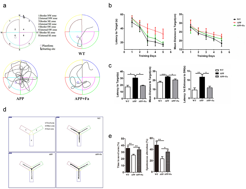
Fig. 1.

Fasudil improves spatial learning of APP/PS1 Tg mice. Eight-month-old APP/PS1 Tg mice were injected with saline (n = 8) or Fasudil (n = 8) for two months. (a) Typical diagram of the Morris water maze test and the corresponding parameters. (b) Latency to target and mean distance to target for five consecutive daily tests. (c) On the sixth day, latency to target, mean distance to target, latency to first entrance to the SW zone were recorded in a retention test session, representing the time spent and distance traveled by animals from the starting point onto the platform or to the SW zone. (d) Typical diagram of the Y maze tests. (e) The percentage of time spent in the novel arm and the spontaneous alternation rate of each group in Y maze tests. Quantitative results for several parameters are mean ± SEM of the eight mice in each group. * P < 0.05, * * P < 0.01, * * * P < 0.001.

Following the MWM trials, mice’s behavioral performance was assessed in the Y maze (Fig. 1d). Results showed that APP mice spent less time in the novel arm and exhibited a lower spontaneous alternation rate than the WT group (Fig. 1e). Following Fasudil treatment, the time spent in the novel arm and the spontaneous alternation rate was increased significantly (Fig. 1e). This suggests that Fasudil could rescue learning and memory deficits.

3.2 Fasudil significantly reduces A β plaques in the CA1 hippocampal area of APP/PS1 mice

The pathogenesis of AD involves an abnormal accumulation of A
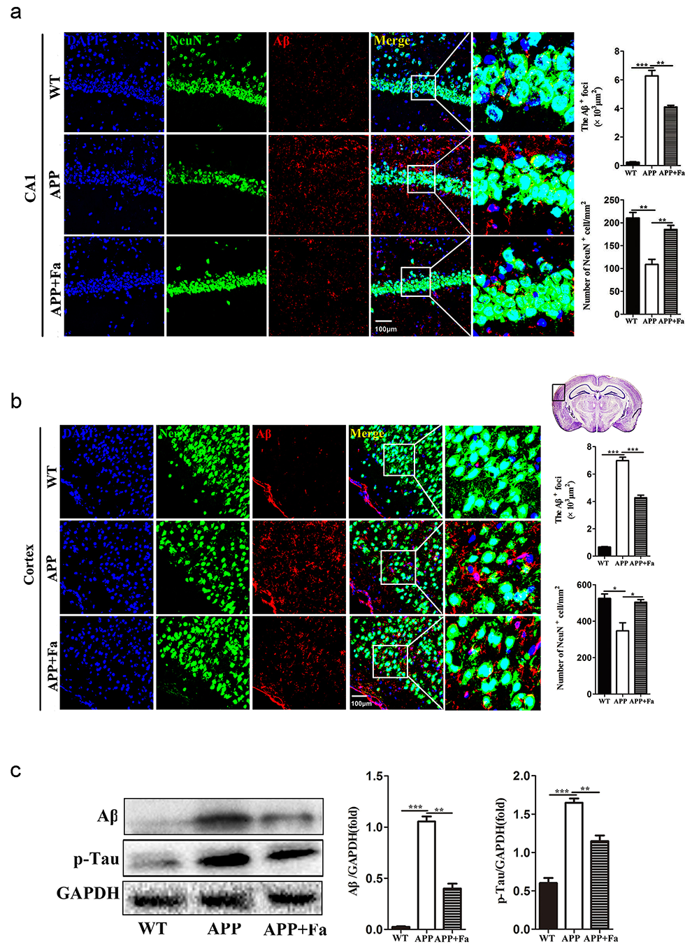
 β and tau in the brain. The formation of A β plaques and tau protein contributes to neuronal apoptosis and learning and memory deficits (van der Kant et al., 2020). To evaluate Fasudil’s effect on the formation of A β and tau, the levels of A β and p-tau in mice that underwent MWM tests were investigated. Using immunofluorescent staining, A β deposits were observed in the cortex and hippocampus area CA1 of APP mice. Fasudil treatment was found to significantly reduce the expression of A β (Fig. 2a and 2b). Additionally, results showed that Fasudil administration increased the number of NeuN immunopositive cells in the cortex and hippocampus area CA1 of APP/PS1 transgenic mice (Fig. 2a and  2b). Finally, Western blots confirmed that Fasudil administration decreased A β and p-tau protein levels in the brain of APP/PS1 transgenic mice (Fig. 2c).

Fig. 2.

Fasudil reduces A β and p-tau deposition and neural apoptosis in hippocampus area CA1 of APP/PS1 mice. (a) Immunofluorescence image of neurons (NeuN, green) and A β (red) in the hippocampus area CA1 of mice. Quantitative analysis of area (polygon) of A β + cells and the number of NeuN immunopositive cells. (b) Immunofluorescence image of neurons (NeuN, green) and A β (red) in mice’s cortical area. Quantitative analysis of area (polygon) of A β + cells and the number of NeuN immunopositive cells. (c) Detection of A β and p-tau in the brain by Western blot. Quantitative results are mean ± SEM from three independent experiments. * P < 0.05, * * P < 0.01, * * * P < 0.001.

3.3 Fasudil inhibits apoptosis in the brain of APP/PS1 mice

Another pathological feature of APP transgenic mice is neuronal apoptosis in the hippocampus (Obulesu and Lakshmi, 2014). TUNEL assay revealed apoptosis in APP mice’s brain tissue was significantly increased compared with the WT mice, while apoptosis was markedly reduced in APP + Fa mice (Fig. 3a). To further explore the anti-apoptotic effects of Fasudil in APP/PS1 transgenic mice, protein levels involved in apoptosis were analyzed via Western blot. Results showed that following Fasudil treatment, expression of the pro-apoptotic proteins cleaved-caspase-3 and Bax decreased significantly, while the expression of anti-apoptotic protein Bcl-2 increased significantly in APP/PS1 Tg mice (Fig. 3b).
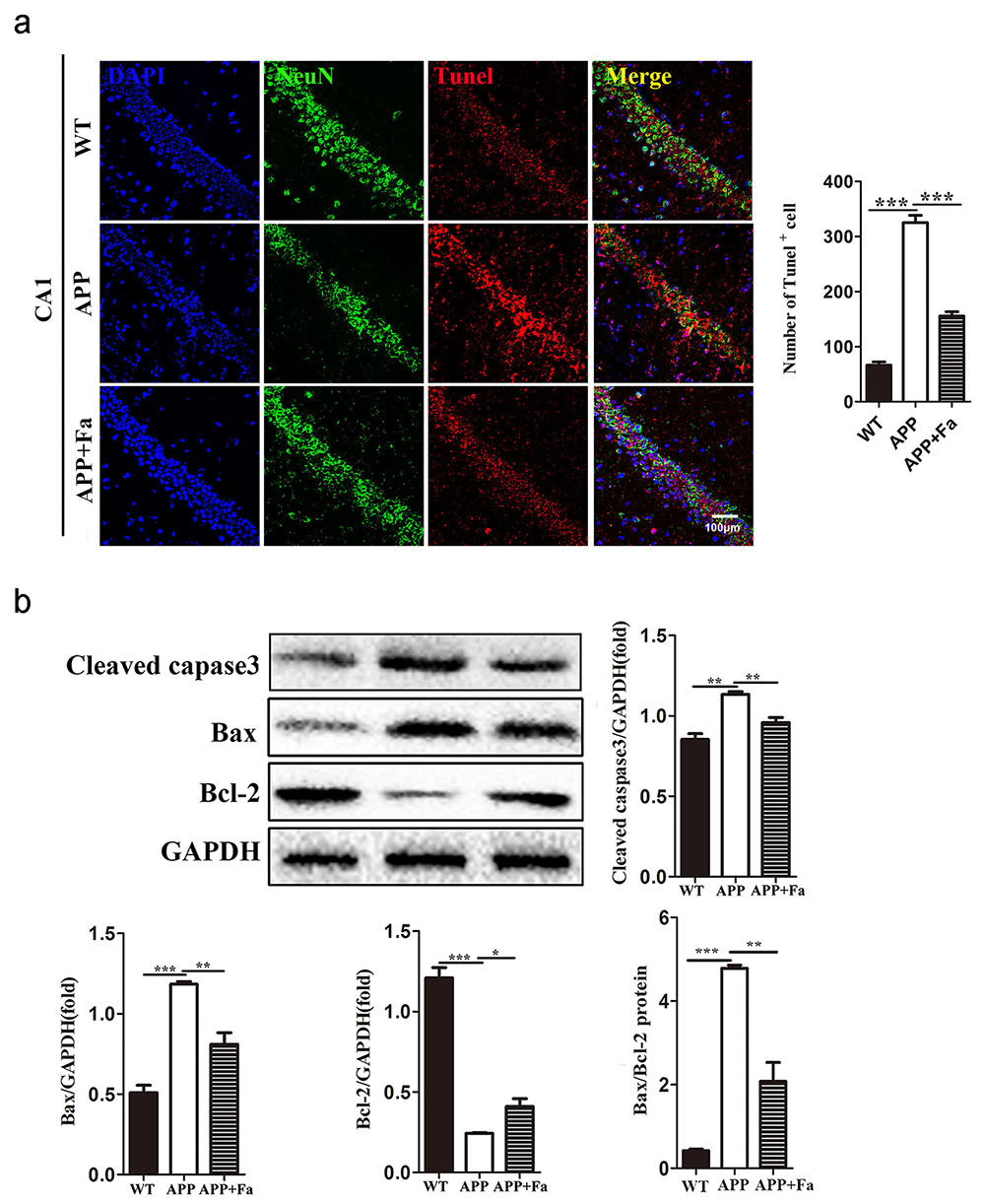
Fig. 3.

Fasudil inhibits apoptosis in the brain of APP/PS1 mice. (a) Representative images of TUNEL staining in hippocampus area CA1 of mice and the number of TUNEL immunopositive cells. (b) Detection of cleaved-caspase-3, Bax and Bcl-2 protein levels in brains by Western blot and quantitative results of Western blot. Quantitative results are mean ± SEM from three independent experiments. * P < 0.05, * * P < 0.01, * * * P < 0.001.

3.4 Effect of Fasudil on synaptophysin and Gap43 expression

Synaptic dysfunction is another pathological characteristic of AD, contributing to cognitive function loss (Bello-Medina et al., 2019). Levels of synaptophysin and Gap43, key markers for functional synapses, are significantly reduced in AD transgenic mouse models (Bereczki et al., 2018; Goetzl et al., 2016). For these reasons, the expression of synaptophysin and Gap43 in the brain were investigated to test whether Fasudil may improve synaptic function. In the APP mouse brain, the expression of synaptophysin was significantly reduced compared to WT mice, while its expression was greatly improved after Fasudil treatment (Fig. 4a). Similarly, Gap43 mRNA and protein expression in APP + Fa mice’s brain was increased compared to untreated APP mice (Fig. 4b). This indicates that Fasudil’s positive effect on learning and memory may be related to an upregulation of synaptophysin and Gap43.
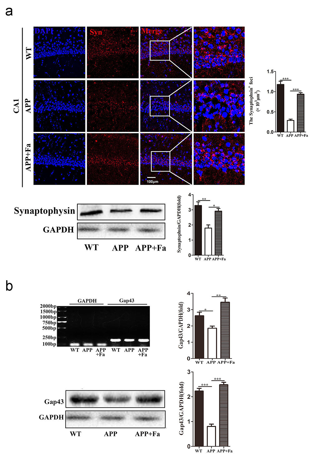
Fig. 4.

Effect of Fasudil on synaptophysin and Gap43 expression. (a) Detection of synaptophysin in hippocampus area CA1 of mice by immunofluorescence staining and Western blot. Quantitative analysis of area (polygon) of synaptophysin + cells and the gray value ratio between synaptophysin and GAPDH. (b) Detection of Gap43 mRNA and protein levels in brains by RT-PCR and Western blot. Quantitative results are mean ± SEM from three independent experiments. * P < 0.05, * * P < 0.01, * * * P < 0.001.

3.5 Fasudil inhibits Nogo-A expression

Previous studies have shown that Nogo-A/NgR is closely related to AD’s pathogenesis by regulating the metabolism of A
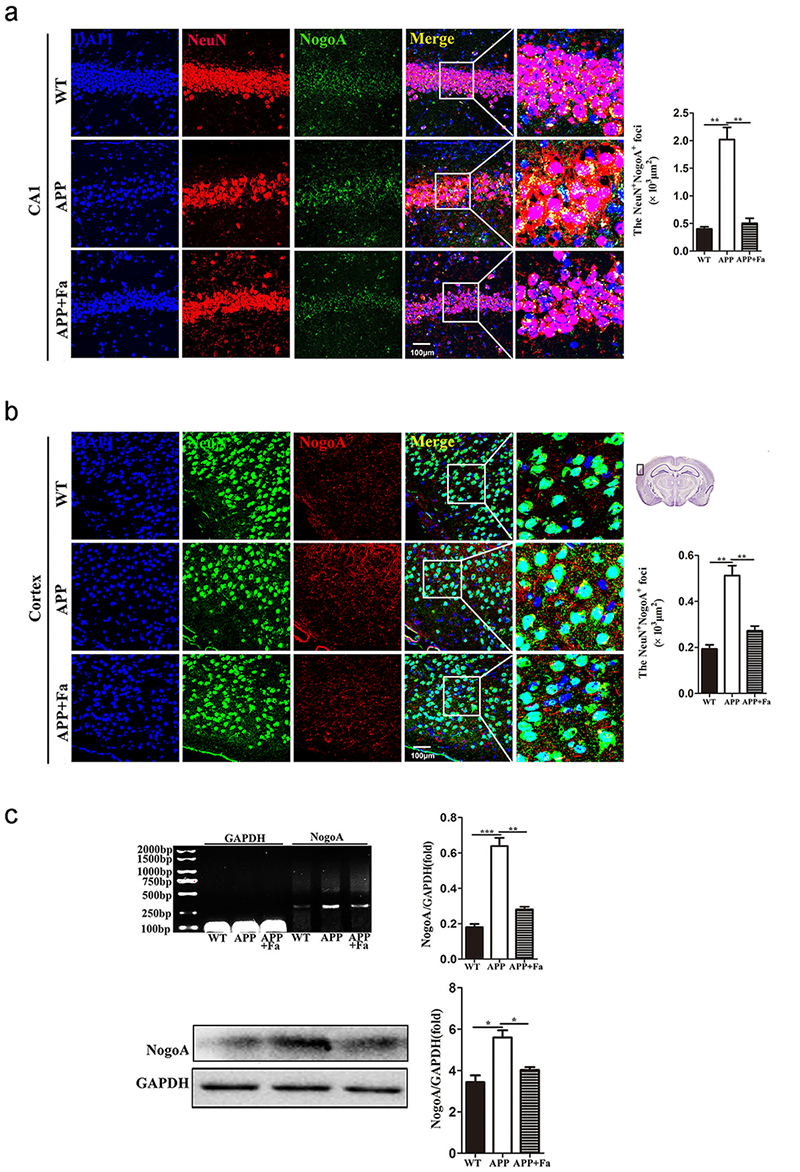
 β and neurodegeneration (Xu et al., 2015). Therefore, Fasudil’s effect on Nogo-A in the brain was explored by immunofluorescence, Western blot and RT-PCR. Immunofluorescence staining revealed that NogoA- expression in the cortex and hippocampus was significantly reduced in the APP + Fa group compared to the untreated APP group (Fig. 5a and 5b). Likewise, the mRNA and protein levels of Nogo-A were also significantly decreased after Fasudil treatment (Fig. 5c).

Fig. 5.

Fasudil inhibits the expression of NogoA. (a) Immunofluorescence image of neurons (NeuN, green) and NogoA (red) in hippocampus area CA1 of mice and quantitative analysis of area (polygon) of NogoA + NeuN + cells. (b) Immunofluorescence image of neurons (NeuN, green) and NogoA (red) in the cortex of mice and quantitative analysis of area (polygon) of NogoA + NeuN + cells. (c) Detection of NogoA mRNA and protein levels in brains by RT-PCR and Western blot. Quantitative results are mean ± SEM from three independent experiments. * P < 0.05, * * P < 0.01, * * * P < 0.001.

3.6 Fasudil inhibits expression of the Ngr/P75ntr/LINGO-1 receptor complex

The expression of the Nogo-A receptor complex, NgR/p75NTR/LINGO-1, was next investigated. Compared with the APP group, the expression of NgR in the cortex and hippocampus was reduced in APP + Fa mice (Fig. 6a and 6b). Similarly, Western blot and quantitative mRNA analysis revealed that the protein and mRNA levels of NgR in the brain were significantly decreased after Fasudil treatment (Fig. 6c). Similar results were found for other components of the Nogo-A receptor complex, including p75NTR and LINGO-1. Furthermore, the protein and mRNA levels of p75NTR and LINGO-1 were significantly increased in APP mice compared with WT mice, but this was rescued by Fasudil treatment (Fig. 6d).
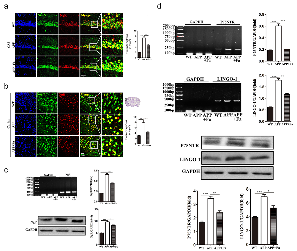
Fig. 6.

Fasudil inhibits the expression of the NgR/p75NTR/LINGO-1 receptor complex. (a) Immunofluorescence image of neurons (NeuN, green) and NgR (red) in hippocampus area CA1 of mice and quantitative analysis of area (polygon) of NgR + NeuN + cells. (b) Immunofluorescence image of neuron cells (NeuN, green) and NgR (red) in the cortical area of mice and quantitative analysis of area (polygon) of NgR + NeuN + cells. (c) Detection of NgR mRNA and protein level in brains by RT-PCR and Western blot. Quantitative results are mean ± SEM from three independent experiments. (d) Detection of p75NTR and LINGO-1 mRNA and protein levels RT-PCR and Western blot. Quantitative results are mean ± SEM from three independent experiments. * P < 0.05, * * P < 0.01, * * * P < 0.001.

3.7 Fasudil suppresses activation of the ROCK signaling pathway

Previous studies have provided substantial evidence that Fasudil inhibits the ROCK pathway and promotes neuroregeneration and remyelination (Li et al., 2017; Wang et al., 2020). Here, ROCK2 and phospho (p)-ROCK2 protein was quantified in the brain of Fasudil-treated and untreated APP mice. Expression of ROCK2 and p-ROCK2 was found to be significantly reduced after Fasudil treatment. Additionally, the activity of ROCK2, measured as the proportion of p-ROCK2 and total ROCK2, was found to be decreased after Fasudil treatment (Fig. 7).
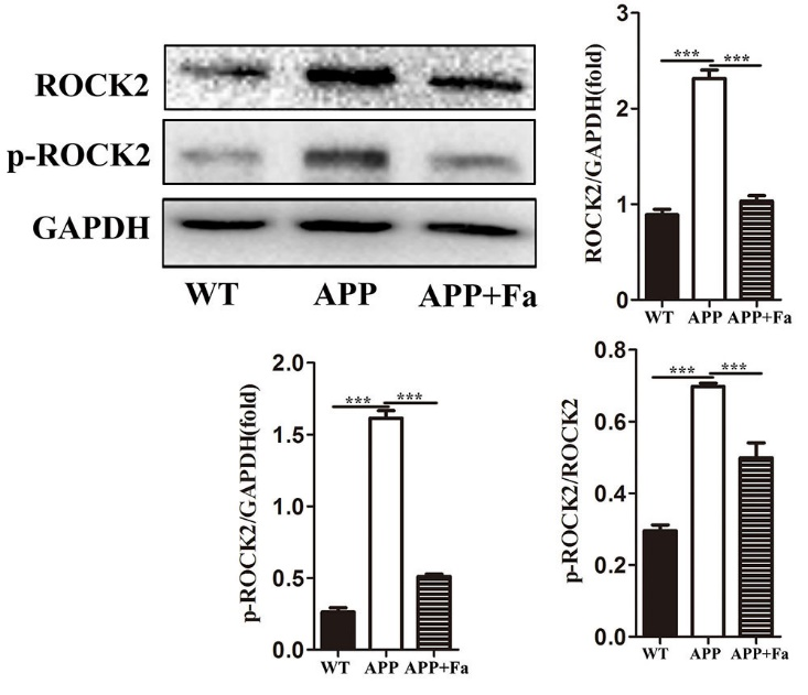
Fig. 7.

Fasudil inhibits the rhoA/ROCK pathway. Detection of ROCK2 and phospho-ROCK2 by Western blot. Quantitative results of Western blot are expressed as fold changes relative to GAPDH bands and ratio of gray values between p-ROCK and ROCK. Quantitative results are mean ± SEM from three independent experiments. * * * P < 0.001.

4. Discussion

Although the pathogenesis of AD is now partially understood, effective treatment strategies for delay or prevention of AD remain to be developed. Therefore, there is an urgent requirement to understand AD’s pathogenesis more clearly to identify effective intervention targets for clinical management. The ROCK signaling pathway is involved in a series of pathological AD processes (Aguilar et al., 2017). Fasudil has been shown to protect against A β -induced neuronal damage by suppressing inflammatory response (Song et al., 2013). A previous study has also shown that Fasudil ameliorates memory deficit and reduces A β deposition and tau protein phosphorylation in APP/PS1 transgenic mice by inhibiting the TLRs-NF- κ B-MyD88 inflammatory cytokine axis (Yu et al., 2017). However, further research is required to determine whether Fasudil affects other targets in APP/PS1 mice. As excepted, we found that Fasudil effectively inhibited the expression of Nogo-A and the Nogo-A receptor complex and the downstream RhoA/ROCK signaling.

APP/PS1 double transgenic mice are known to develop AD-like pathology and are widely used to study AD (Ferguson et al., 2013; Yu et al., 2020). Here, the efficacy of a two-month course of Fasudil treatment (intraperitoneal (i.p.) injection of 25 mg/kg/day) was investigated in the eight-month-old APP/PS1 transgenic mice. It was found that Fasudil treatment improves learning and cognitive abilities and ameliorates both A β and tau pathologies in these mice. This is consistent with previous research (Yu et al., 2017) and collectively supports Fasudil’s strategy as a potential anti-AD agent. The purpose of this study was to further explore the underlying mechanisms of Fasudil treatment and its suitability for AD using the APP/PS1 transgenic mouse model. Fasudil was found to rescue cognitive deficits, reduce A β levels, effectively improve synaptic function and inhibit apoptosis in APP/PS1 transgenic mice via inhibition of the Nogo-A/NgR1/RhoA signaling pathway. These findings provide further evidence for Fasudil as an effective treatment strategy for AD and provide a new therapeutic strategy for inhibiting AD progression by targeting the production of A β and neuronal apoptosis.

Synaptic dysfunction is closely related to AD’s cognitive deficits, and studies have suggested that abnormally elevated A β causes synaptic loss (Selkoe, 2002; Tönnies and Trushina, 2017). Synaptophysin, a marker for synaptic vesicles, was absent in the proximity of neurons with A β clusters, leading to synaptic failure, which is presumed to be the leading cause of cognitive deficit in AD (Ishibashi et al., 2006). Gap43 is highly expressed in neurons, often used as a marker for synaptic reconstruction and nerve regeneration and is significantly reduced in AD (Bogdanovic et al., 2000). However, it is unclear whether Fasudil is capable of rescuing synaptic function. The results reported here show that the expression of synaptophysin and Gap43 in the brain declined in APP/PS1 transgenic mice but upregulated following two months of Fasudil treatment, suggesting that Fasudil may rescue synaptic function by the promotion of synapse-related protein expression.

Another pathological feature of AD is neuronal apoptosis, and apoptosis-related proteins are involved in pathologically neuronal death in AD (Engidawork et al., 2001). In this study, a reduced number of neurons were observed in the hippocampus area CA1 in APP/PS1 transgenic mice; however, Fasudil treatment significantly reduced neuronal apoptosis in this mouse model. Proteins of the caspase family play crucial roles in the process of apoptosis, with caspase-3 acting as a direct effector of apoptosis (Porter and Jänicke, 1999). The results reported here show that cleaved-caspase-3 expression in the brain tissue of APP/PS1 transgenic mice was significantly increased, while Fasudil treatment significantly inhibited caspase-3 expression. The ratio of Bax and Bcl-2 determines the cell (Sun et al., 2012); the data reported here indicate that the ratio of Bax/Bcl-2 expression is altered in APP/PS1 transgenic mice. Fasudil induced resistance to cellular apoptosis by reducing the Bax/Bcl-2 ratio. This demonstrates that Fasudil has an anti-apoptotic effect in APP/PS1 transgenic mice.

The Rho/ROCK pathway is downstream of NogoA/NgR. It is involved in the progression of AD via regulation of APP metabolism, A β generation, reduction of phosphorylated tau levels and inhibition of neuronal regeneration (Xu et al., 2015). Consistent with the previous observation, it was found here that Fasudil treatment reduces both the expression of ROCK2 and the ratio of p-ROCK2/ROCK2 in the brain of APP/PS1 transgenic mice, which is also consistent with the presence of increased synaptophysin and Gap43. These results indicate that inhibition of Fasudil treatment’s ROCK pathway may play a key role in neuro-regeneration during AD.

Nogo-A is a membrane protein abundantly expressed in both neurons and oligodendrocytes that limits synaptic plasticity and neurite outgrowth (Pernet and Schwab, 2012). Nogo-A transduces intracellular signals by binding to a receptor complex consisting of NgR, p75NTR and LINGO-1 and activates the downstream Rho/Rho kinase signaling pathway preventing further axonal growth and inhibiting myelin formation (Fournier et al., 2001; Kempf and Schwab, 2013). Many studies have proposed that Nogo-A promotes the development of AD. For example, one study found that the deletion of the Nogo gene improves learning and memory deficits in APP transgenic mice and restores the levels of some synaptic markers, including synaptophysin and Gap43 (Masliah et al., 2010). Furthermore, it has been suggested that Nogo-A may trigger the occurrence and development of AD by affecting A β metabolism and inhibiting synaptic plasticity (Xu et al., 2015).

Similarly, NgR plays an essential role in axonal and synaptic plasticity and may participate in the pathological process of AD by affecting the metabolism of amyloid precursor protein (Park and Strittmatter, 2008). Further study has demonstrated that NgR knockdown in the perforant path rescues cognitive deficits and synaptic function by decreasing the level of amyloid precursor protein and A β production (Jiang et al., 2020). p75NTR is a co-receptor for NgR, which mediates cellular apoptosis and survival and synapse weakening (Fahnestock and Shekari, 2019). LINGO-1 is involved in the pathophysiology of AD by promoting the production of A β fragments and inhibiting the growth and survival of neurons (Fernandez-Enright and Andrews, 2016). Fasudil, a potent ROCK inhibitor, may inhibit neurodegeneration and promote neuroregeneration by inhibiting the ROCK signaling pathway. But whether Fasudil could also inhibit upstream molecules of the ROCK signaling pathway remained unclear. This study found that Nogo-A and its receptor complex molecules in neurons increase significantly in APP/PS1 transgenic mice, an effect reversed after Fasudil treatment. This indicates that cognitive deficits, neuronal apoptosis and synaptic dysfunction are associated with the Nogo-A/NgR/RhoA axis in APP/PS1 transgenic mice.

From the present study, it can be concluded that Fasudil effectively rescues cognitive deficits, reduces A β plaques and tau protein levels, maintains synaptic function and inhibits neuronal apoptosis in a mouse model of AD. Fasudil’s therapeutic effect on APP/PS1 transgenic mice is likely attributable to its ability to effectively inhibit the expression of Nogo-A and the Nogo-A receptor complex and downstream RhoA/ROCK signaling. These results provide a new therapeutic target for the treatment of AD.

Author contributions

Min-Fang Guo participated in the study’s design, conducted most of the experiments, analyzed the results, and wrote most of the manuscript. Hui-Yu Zhang and Pei-Jun Zhang carried out the mouse behavioral tests, immunoassays and proofread the article. Xiao-Qin Liu, Wen-Yue Wei, Yu-Yin Wang and Bing-Tao Mu performed the RT-PCR and Western blot experiments. Li-Juan Song and Zhi Chai helped with data analysis. Cun-Gen Ma and Jie-Zhong Yu designed the experiments. All datasets generated for this study are included in the article.

Ethics approval and consent to participate

All animals were housed in the animal house of the Institute of Brain Science, Shanxi Datong University. The Ethics Committee approved all experiments of Shanxi Datong University, Datong, P. R. China.

Acknowledgment

This work was supported by grants from the National Natural Science Foundation of P. R. China (No. 81473577 and 81471412), and the Scientific and technological innovation team of integrated Chinese and Western medicine for the prevention and treatment of nervous system diseases, Shanxi University of Chinese Medicine (2018TD-012), Shanxi Applied Basic Research Project (201901D211538), Natural Fund Project of Shanxi Province (201901D111334) and Research Project Supported by Shanxi Scholarship Council of P. R. China (2014-7), Project of Shanxi Province Platform Base (201805D131005 and 201805D111009), Shanxi Province Key R & D Plan (2106ZD0505), Science and Technology Innovation Projects of Universities in Shanxi Province (2020L0484) and Platform Base Plan Project of Datong (2019198).

Conflict of Interest

The authors declare that the research was conducted in the absence of any commercial or financial relationships that could be construed as a potential conflict of interest.

References
[1]
Aguilar, B. J., Zhu, Y. and Lu, Q. (2017) Rho GTPases as therapeutic targets in Alzheimer’s disease. Alzheimer’s Research & Therapy 9, 97.
[2]
Bello-Medina, P. C., González-Franco, D. A., Vargas-Rodríguez, I. and Díaz-Cintra, S. (2019) ‘Oxidative stress, the immune response, synaptic plasticity, and cognition in transgenic models of Alzheimer disease’. Neurologia 9, 30109-30104.
[3]
Bereczki, E., Branca, R. M., Francis, P. T., Pereira, J. B., Baek, J., Hortobágyi, T., Winblad, B., Ballard, C., Lehtiö, J. and Aarsland, D. (2018) Synaptic markers of cognitive decline in neurodegenerative diseases: a proteomic approach. Brain 141, 582-595.
[4]
Bogdanovic, N., Davidsson, P., Volkmann, I., Winblad, B. and Blennow, K. (2000) Growth-associated protein GAP-43 in the frontal cortex and in the hippocampus in Alzheimer’s disease: an immunohistochemical and quantitative study. Journal of Neural Transmission 107, 463-478.
[5]
Chen, M. S., Huber, A. B., van der Haar, M. E., Frank, M., Schnell, L., Spillmann, A. A., Christ, F. and Schwab, M. E. (2000) Nogo-A is a myelin-associated neurite outgrowth inhibitor and an antigen for monoclonal antibody IN-1. Nature 403, 434-439.
[6]
Engidawork, E., Gulesserian, T., Yoo, B. C., Cairns, N. and Lubec, G. (2001) Alteration of caspases and apoptosis-related proteins in brains of patients with Alzheimer’s disease. Biochemical and Biophysical Research Communications 281, 84-93.
[7]
Fahnestock, M. and Shekari, A. (2019) ProNGF and neurodegeneration in Alzheimer’s disease. Frontiers in Neuroscience 13, 129.
[8]
Fan, L., Mao, C., Hu, X., Zhang, S., Yang, Z., Hu, Z., Sun, H., Fan, Y., Dong, Y., Yang, J., Shi, C. and Xu, Y. (2020) New insights into the pathogenesis of Alzheimer’s disease. Frontiers in Neurology 10, 1312.
[9]
Ferguson, S. A., Sarkar, S. and Schmued, L. C. (2013) Longitudinal behavioral changes in the APP/PS1 transgenic Alzheimer’s disease model. Behavioural Brain Research 242, 125-134.
[10]
Fernandez-Enright, F. and Andrews, J. (2016) Lingo-1: A novel target in therapy for Alzheimer’s disease? Neural Regeneration Research 11, 88.
[11]
Fournier, A. E., GrandPre, T. and Strittmatter, S. M. (2001) Identification of a receptor mediating Nogo-66 inhibition of axonal regeneration. Nature 409, 341-346.
[12]
Gil, V., Nicolas, O., Mingorance, A., Ureña, J. M., Tang, B. L., Hirata, T., Sáez-Valero, J., Ferrer, I., Soriano, E. and del Río, J. A. (2006) Nogo-A expression in the human hippocampus in normal aging and in Alzheimer disease. Journal of Neuropathology & Experimental Neurology 65, 433-444.
[13]
Goetzl, E. J., Kapogiannis, D., Schwartz, J. B., Lobach, I. V., Goetzl, L., Abner, E. L., Jicha, G. A., Karydas, A. M., Boxer, A. and Miller, B. L. (2016) Decreased synaptic proteins in neuronal exosomes of frontotemporal dementia and Alzheimer’s disease. The FASEB Journal 30, 4141-4148.
[14]
Ishibashi, K., Tomiyama, T., Nishitsuji, K., Hara, M. and Mori, H. (2006) Absence of synaptophysin near cortical neurons containing oligomer Abeta in Alzheimer’s disease brain. Journal of Neuroscience Research 84, 632-636.
[15]
Jiang, R., Wu, X., Wang, B., Guan, R., Lv, L., Li, A., Lei, L., Ma, Y., Li, N., Li, Q., Ma, Q., Zhao, J. and Li, S. (2020) Reduction of NgR in perforant path decreases amyloid- β peptide production and ameliorates synaptic and cognitive deficits in APP/PS1 mice. Alzheimer’s Research & Therapy 12, 47.
[16]
Kan, Q., Zhang, H., Zhang, Y., Li, X., Xu, Y., Thome, R., Zhang, M., Liu, N., Chu, Y., Zhang, G. and Zhu, L. (2017) Matrine treatment blocks NogoA-induced neural inhibitory signaling pathway in ongoing experimental autoimmune encephalomyelitis. Molecular Neurobiology 54, 8404-8418.
[17]
Kempf, A. and Schwab, M. E. (2013) Nogo-A represses anatomical and synaptic plasticity in the central nervous system. Physiology 28, 151-163.
[18]
Li, Y., Xie, C., Zhang, Y., Li, X., Zhang, H., Wang, Q., Chai, Z., Xiao, B., Thome, R., Zhang, G. and Ma, C. (2017) FSD-C10, a Fasudil derivative, promotes neuroregeneration through indirect and direct mechanisms. Scientific Reports 7, 41227.
[19]
Liddelow, S. A., Guttenplan, K. A., Clarke, L. E., Bennett, F. C., Bohlen, C. J., Schirmer, L., Bennett, M. L., Münch, A. E., Chung, W., Peterson, T. C., Wilton, D. K., Frouin, A., Napier, B. A., Panicker, N., Kumar, M., Buckwalter, M. S., Rowitch, D. H., Dawson, V. L., Dawson, T. M., Stevens, B. and Barres, B. A. (2017) Neurotoxic reactive astrocytes are induced by activated microglia. Nature 541, 481-487.
[20]
Masliah, E., Xie, F., Dayan, S., Rockenstein, E., Mante, M., Adame, A., Patrick, C. M., Chan, A. F. and Zheng, B. (2010) Genetic deletion of Nogo/Rtn4 ameliorates behavioral and neuropathological outcomes in amyloid precursor protein transgenic mice. Neuroscience 169, 488-494.
[21]
Meng, S., Wang, B. and Li, W. (2020) Intermittent hypoxia improves cognition and reduces anxiety-related behavior in APP/PS1 mice. Brain and Behavior 10, e01513.
[22]
Obulesu, M. and Lakshmi, M. J. (2014) Apoptosis in Alzheimer’s disease: An understanding of the physiology, pathology and therapeutic avenues. Neurochemical Research 39, 2301-2312.
[23]
Padurariu, M., Ciobica, A., Mavroudis, I., Fotiou, D. and Baloyannis, S. (2012) Hippocampal neuronal loss in the CA1 and CA3 areas of Alzheimer’s disease patients. Psychiatria Danubina 24, 152-158.
[24]
Park, J. H. and Strittmatter, S. M. (2008) Nogo receptor interacts with brain APP and Abeta to reduce pathologic changes in Alzheimer’s transgenic mice. Current Alzheimer Research 4, 568-570.
[25]
Pernet, V. and Schwab, M. E. (2012) The role of Nogo-A in axonal plasticity, regrowth and repair. Cell and Tissue Research 349, 97-104.
[26]
Porter, A. G. and Jänicke, R. U. (1999) Emerging roles of caspase-3 in apoptosis. Cell Death & Differentiation 6, 99-104.
[27]
Sekine, Y., Lindborg, J. A. and Strittmatter, S. M. (2020) A proteolytic C-terminal fragment of Nogo-A (reticulon-4A) is released in exosomes and potently inhibits axon regeneration. Journal of Biological Chemistry 295, 2175-2183.
[28]
Selkoe, D. J. (2002) Alzheimer’s disease is a synaptic failure. Science 298, 789-791.
[29]
Song, Y., Chen, X., Wang, L., Gao, W. and Zhu, M. (2013) Rho kinase inhibitor fasudil protects against β -amyloid-induced hippocampal neurodegeneration in rats. CNS Neuroscience & Therapeutics 19, 603-610.
[30]
Sun, Z., Ma, X., Yang, H., Zhao, J. and Zhang, J. (2012) Brain-derived neurotrophic factor prevents beta- amyloid-induced apoptosis of pheochromocytoma cells by regulating Bax/Bcl-2 expression. Neural Regeneration Research 7, 347-51.
[31]
Tönnies, E. and Trushina, E. (2017) Oxidative stress, synaptic dysfunction, and Alzheimer’s disease. Journal of Alzheimer’s Disease 57, 1105-1121.
[32]
van der Kant, R., Goldstein, L. S. B. and Ossenkoppele, R. (2020) Amyloid- β -independent regulators of tau pathology in Alzheimer’s disease. Nature Reviews Neuroscience 21, 21-35.
[33]
Vassar, R. (2007) Caspase-3 cleavage of GGA3 stabilizes BACE: Implications for Alzheimer’s disease. Neuron 54, 671-673.
[34]
Wang, J., Sui, R. X., Miao, Q., Wang, Q., Song, L. J., Yu, J. Z., Li, Y. H., Xiao, B. G. and Ma, C. G. (2020) Effect of Fasudil on remyelination following cuprizone-induced demyelination. CNS Neuroscience and Therapeutics 26, 76-89.
[35]
Xiao, F., Lin, L. F., Cheng, X., Gao, Q. and Luo, H. M. (2012) Nogo-66 receptor activation inhibits neurite outgrowth and increases β -amyloid protein secretion of cortical neurons. Molecular Medicine Reports 5, 619-624.
[36]
Xu, T., Zhang, Y., He, J., Luo, D., Luo, Y., Wang, Y., Liu, W., Wu, J., Zhao, W., Fang, J., Guan, L., Huang, S., Wang, H., Lin, L., Zhang, S. and Wang, Q. (2018) Bajijiasu ameliorates β -amyloid-triggered endoplasmic reticulum stress and related pathologies in an Alzheimer’s disease model. Cellular Physiology and Biochemistry 46, 107-117.
[37]
Xu, Y., Sun, Z., Wang, Y., Xiao, F., and Chen, M. (2015) Function of Nogo-A/Nogo-A receptor in Alzheimer’s disease. CNS Neuroscience & Therapeutics 21, 479-485.
[38]
Yan, Y., Yu, J., Gao, Y., Kumar, G., Guo, M., Zhao, Y., Fang, Q., Zhang, H., Yu, J., Jiang, Y., Zhang, H. and Ma, C. (2019) Therapeutic potentials of the Rho kinase inhibitor Fasudil in experimental autoimmune encephalomyelitis and the related mechanisms. Metabolic Brain Disease 34, 377-384.
[39]
Yu, J., Li, Y., Liu, C., Wang, Q., Gu, Q., Wang, H., Zhang, G., Xiao, B. and Ma, C. (2017) Multitarget Therapeutic Effect of Fasudil in APP/PS1transgenic Mice. CNS & Neurological Disorders - Drug Targets 16, 199-209.
[40]
Yu, J., Yan, Y., Gu, Q., Kumar, G., Yu, H., Zhao, Y., Liu, C., Gao, Y., Chai, Z., Chumber, J., Xiao, B., Zhang, G., Zhang, H., Jiang, Y. and Ma, C. (2018) Fasudil in combination with bone marrow stromal cells (BMSCS) attenuates Alzheimer’s disease-related changes through the regulation of the peripheral immune system. Frontiers in Aging Neuroscience 10, 216.
[41]
Yu, N., Huang, Y., Jiang, Y., Zou, L., Liu, X., Liu, S., Chen, F., Luo, J. and Zhu, Y. (2020) Ganoderma lucidum triterpenoids (GLTs) reduce neuronal apoptosis via inhibition of ROCK signal pathway in APP/PS1 transgenic Alzheimer’s disease mice. Oxidative Medicine and Cellular Longevity 2020, 1-11.
Share
Back to top