


 , Tomonori Ishikawa 1, Moeko Onose 3, Rinko Ibi 3, Kenichi Tatsumi 2, Naoyuki Miyasaka 3
1 Department of Perinatal and Maternal Medicine (Ibaraki), Graduate School, Tokyo Medical and Dental University, 113-8510 Tokyo, Japan
2 Umegaoka Women's Clinic, Umegaoka, Setagaya, 154-0022 Tokyo, Japan
3 Department of Comprehensive Reproductive Medicine, Graduate School, Tokyo Medical and Dental University, 113-8510 Tokyo, Japan
Academic Editor: Michael H. Dahan
Abstract
Background: Submucosal myoma is a common gynecological disease that causes menorrhagia and infertility. While hysteroscopic surgery is a minimally invasive and effective method for treating submucosal myomas, its feasibility depends on the size and location of the myomas. Conversely, abdominal procedures enable enucleation of submucosal myomas and preservation of endometrial integrity, but are accompanied by technical difficulties. Herein we report the case of an infertile woman with a submucosal and an intramural myoma who underwent laparoscopic myomectomy using laparoscopic ultrasonography. Case: The patient was a 36-year-old infertile woman. Transvaginal ultrasonography revealed a 15 mm submucosal myoma with a 60% myometrial extension in the anterior uterine wall and a 49 mm intramural myoma in the posterior wall. During myomectomy, the submucosal myoma was not apparent from the external side of the uterus; therefore, laparoscopic ultrasonography was used to detect it. Under ultrasonography, vasopressin was injected between the myoma and the myometrium, thereby separating the layers. Consequently, we were able to resect both myomas without breaching the endometrium. Conclusions: The present case demonstrates the effectiveness of laparoscopic ultrasonography for detecting submucosal myomas and ensuring injection of vasopressin into the proper layer. These advantages allow surgeons to preserve endometrial integrity during laparoscopic myomectomy.
Keywords
- endometrium
- laparoscopic myomectomy
- laparoscopic ultrasonography
- submucosal myoma
Submucosal myoma is a common gynecologic disorder that grows adjacent to the endometrium and causes menorrhagia and implantation failure [1]. Treatment options include hysteroscopic, laparoscopic, and laparotomic surgeries, and the optimal approach depends on the size and position of the myoma [1]. A hysteroscopic approach is less invasive to the patient than a laparoscopic approach and is often applied to infertile women. In fact, removing type 0 and 1 submucosal myomas and some type 2 myomas are safely accomplished by a one-step procedure with hysteroscopy [2]. However, the intramural component of type 2 submucosal myomas is often challenging to treat with hysteroscopic surgery [1, 2, 3]. Additionally, some type 2 myomas are not visible by hysteroscopy despite their coming up to the endometrial cavity. Therefore, the hysteroscopic approach is not always the best treatment for submucosal myomas. Alternatively, abdominal myomectomy enables surgeons to enucleate myomas. However, submucosal myomas are not always apparent from the external side of the uterus, and preserving endometrial integrity during laparoscopic myomectomy is technically difficult.
Laparoscopic ultrasonography (LUS) is used to visualize lesions inside organs during laparoscopy. It requires specialized transducers that fit through conventional laparoscopic trocars and is commonly used in gastrointestinal, hepatobiliary, and urologic surgeries [4]. Because LUS can be used to precisely detect small lesions deep inside organs, it is used for biopsies during laparoscopic surgeries. While LUS is not commonly used in the gynecologic field, this technique may work well with vasopressin injection during myomectomy. Therefore, we report the first case of LUS-guided myomectomy for submucosal myoma. LUS allowed for visualization and successful removal of the myoma without breaching the endometrium.
A 36-year-old nulligravida woman visited our clinic for an evaluation of her infertility. She was healthy, and her menses were regular and normal. Transvaginal ultrasonography revealed a type 2 submucosal myoma with a 60% myometrial extension measuring 15 mm in the anterior uterine wall and a type 4 intramural myoma (classified via guidelines from the International Federation of Gynecology and Obstetrics) measuring 49 mm in the posterior uterine wall. Magnetic resonance imaging suggested that the posterior myoma was a cellular leiomyoma (Fig. 1A). To make a definitive diagnosis regarding the intramural myoma, we planned a laparoscopic myomectomy. Furthermore, we planned to laparoscopically resect the submucosal myoma considering the myoma’s negative effect on implantation. Since the submucosal myoma was not visible from the external side of the uterus, we planned to use LUS to detect it. The location of the submucosal myoma was preoperatively assessed by transvaginal ultrasound sonography and magnetic resonance imaging. We measured the depth from the external side and the distance from the midline and fundus of the uterus. During the surgery, we used an ultrasound machine (Aplio a verifia CUS-AA000, Canon Medical Systems, Otawara, Tochigi, Japan) and a transducer (Linear array transducer PET-805LA, Canon Medical Systems, Otawara, Tochigi, Japan) with the detection frequency set at 7.0 MHz. We inserted the transducer through a 12-mm trocar (Fig. 1B). The submucosal myoma was visualized successfully with LUS immediately after the transducer was placed on the anticipated area of the anterior wall. Vasopressin (2 units diluted in 10 mL of saline) was injected precisely between the myoma and the myometrium under ultrasonography guidance (Fig. 2A,B). LUS also confirmed that the injected vasopressin effectively spread between the myoma and the myometrium (Fig. 2C). Using laparoscopic forceps and monopolar cautery, both the submucosal and the intramural myomas were successfully removed from the uterus without breaching the endometrium (Fig. 3A,B). The myomas were morcellated in a bag so that small fragments would not be scattered into the peritoneal cavity. The myoma beds were sutured to double to triple-layer closure with absorbable surgical thread. The postoperative course was uneventful, and the patient was discharged from the hospital as scheduled. No abnormal signs were detected at hospital visits 1 and 3 months after the surgery, and the initiation of fertility treatment was planned for 6 months after the surgery.
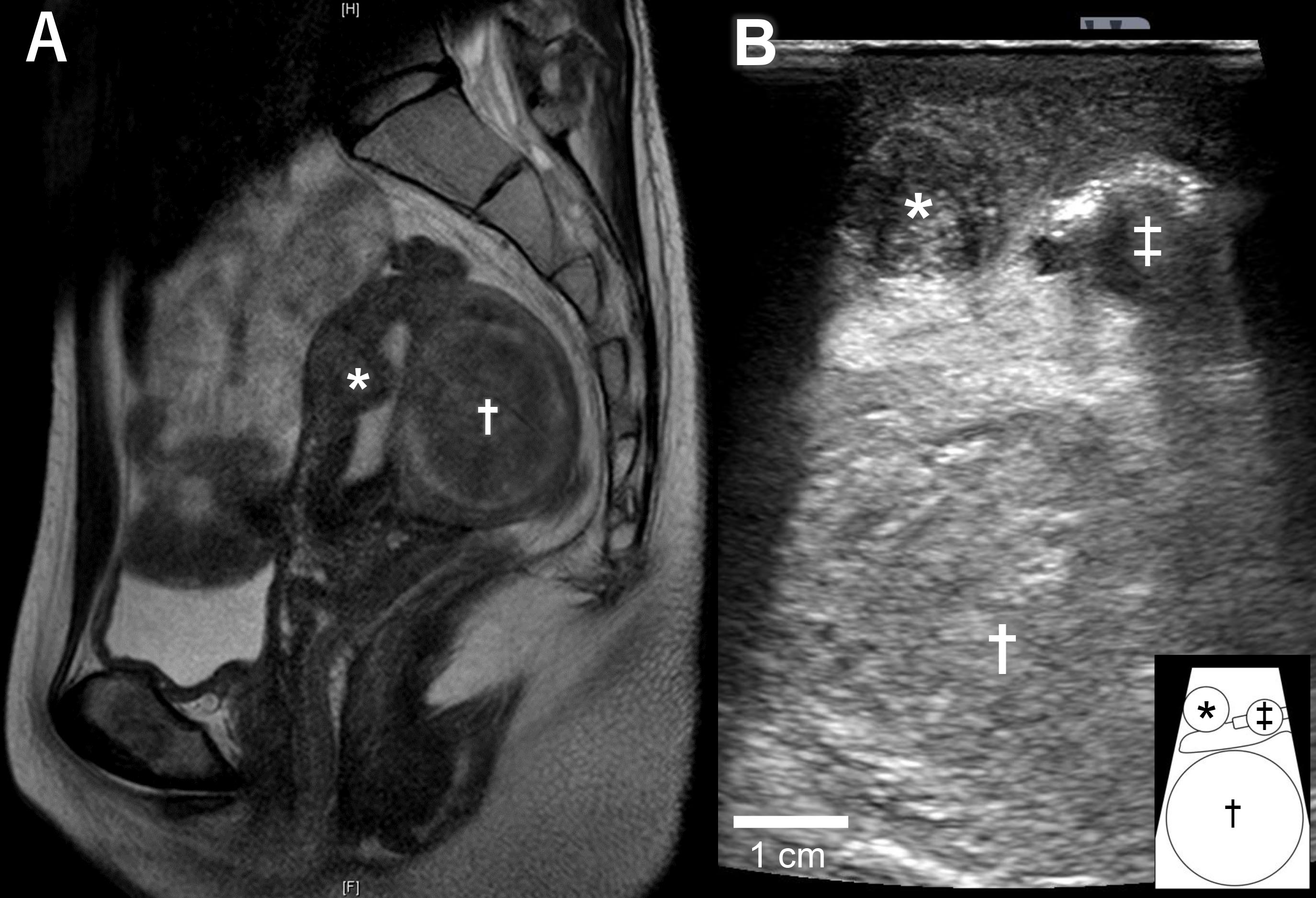 Fig. 1.
Images of submucosal and intramural myomas. (A) Magnetic resonance image of the uterus. The submucosal myoma (*) in the anterior wall of the uterus was 40% intramural. The intramural myoma (†) in the posterior wall of the uterus was suggested as a cellular leiomyoma. (B) Image of laparoscopic ultrasonography during laparoscopic surgery. Transducer put on the anterior wall of the uterus, as shown in Fig. 2A, with a submucosal myoma (*) and an intramural myoma (†) clearly shown. A uterine manipulator is indicated by ‡. The schematic diagram at the bottom right explains the image of ultrasonography.
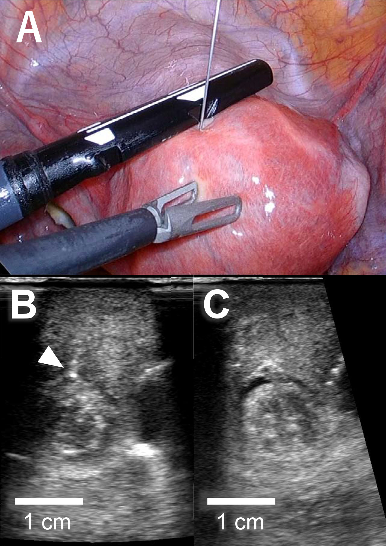 Fig. 2.
Injection of vasopressin during laparoscopic myomectomy. (A) Intraperitoneal image during laparoscopic surgery. Diluted vasopressin was injected under ultrasonography guidance. (B) Injection needle (white arrowhead) was inserted to the boundary between the myoma and the myometrium. (C) Injected vasopressin spread in the layer between the myoma and the myometrium.
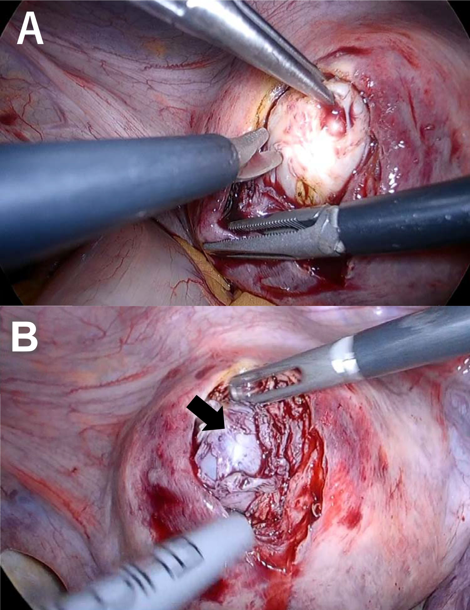 Fig. 3.
Laparoscopic images during myomectomy. (A) Myomas are removed using laparoscopic forceps and monopolar cautery. (B) Endometrial integrity was preserved after removal of the submucosal myomas. Black arrow shows the preserved endometrium.
Our case report demonstrates the advantages of using LUS for laparoscopic myomectomy. First, ultrasonography enables the laparoscopist to detect the location of a submucosal myoma easily and accurately. Second, under ultrasonography guidance, diluted vasopressin can be precisely injected, which then spreads in the layer between the myoma and myometrium. These merits enable the laparoscopist to remove submucosal myomas without breaching the endometrium and thereby impairing subsequent fertility.
Because patients with submucosal myomas suffer from heavy menstrual bleeding,
infertility, recurrent pregnancy loss, or a combination of these, the
gynecologist should be familiar with a given patient’s history, including her
desires with respect to fertility, as well as an appropriately detailed
evaluation of the uterus [1]. Indeed, the therapeutic effect of transcervical
resection seems to depend on the type of submucosal myoma determined by its
extension into the myometrium: type 0 submucosal myoma exists entirely within the
endometrial cavity, type 1 submucosal myoma extends
One big challenge in laparoscopic myomectomy is its technical difficulty regarding the preservation of endometrial integrity during surgery. Because submucosal myomas are adjacent to the endometrium, gentle handling of the myoma and uterus is essential. For the sake of reducing blood loss during surgery and separating the myoma from the surrounding myometrium, diluted vasopressin with saline is often injected between them. While preoperative assessment with transvaginal ultrasonography provides information about the myoma’s size and location, it is still challenging to inject vasopressin between the myoma and myometrium, especially when the myoma is small and located deep in the myometrium. In this respect, LUS facilitates the detection of small myomas during surgery. In addition, the laparoscopist can inject vasopressin under ultrasonography guidance and ensure that the diluted vasopressin spreads in the proper layer between the myoma and the myometrium, thereby separating the layers. Consequently, this technique will help the laparoscopist remove the myoma without breaching the endometrium.
Breaching the endometrium during myomectomy is known to cause intrauterine adhesions, potentially leading to impaired implantation [6]. In addition, the risk of placenta accrete spectrum disorders may increase in cases of entry into the uterine cavity during myomectomy [7]. Therefore, abdominal removal of type 2 submucosal myomas is a good option for patients who wish to become pregnant. Furthermore, evidence suggests that a single intramural fibroid of around 2 cm in diameter may decrease pregnancy rates at in vitro fertilization. As such this technique would be ideal to locate this fibroid and remove it prior to care if desired, while minimizing hospital and recovery time for the patient [8]. Currently, most gynecologists are not familiar with LUS, and manipulating the transducer on the uneven surface of the uterus may be cumbersome. However, a newly developed smaller transducer may help gynecologists perform LUS more easily and subsequently improve diagnosis and surgical procedures. Additionally, this technique may also benefit robotic surgery, in which the sense of touch is limited and visual information is more important to finding myomas when compared to conventional surgeries.
Our case demonstrates that LUS is effective for preserving the integrity of the endometrium during the laparoscopic removal of submucosal myomas for the first time. This technique will help laparoscopists enucleate type 2 submucosal myomas which are not apparent under a hysteroscope.
KS, TI, MO, NM designed and performed the research study. KS, RI, and KT collected data and taken the pictures. KS, TI, and NM wrote the manuscript. All authors contributed to editorial changes in the manuscript. All authors read and approved the final manuscript.
This study is exempt from institutional review board approval. Written informed consent for the case report was obtained from the patient.
We would like to thank Editage (www.editage.com) for English language editing.
This research received no external funding.
The authors declare no conflict of interest.
References
Publisher’s Note: IMR Press stays neutral with regard to jurisdictional claims in published maps and institutional affiliations.