IMR Press / RCM / Volume 24 / Issue 8 / DOI: 10.31083/j.rcm2408226
Open Access Review
A Review of Therapeutic Strategies against Cardiac Fibrosis: From Classical Pharmacology to Novel Molecular, Epigenetic, and Biotechnological Approaches
Show Less
1 Department of Medical Surgical Sciences and Biotechnologies, Sapienza University, 04100 Latina, Italy
2 Institute of Biochemistry and Cell Biology, National Council of Research (IBBC-CNR), 00015 Monterotondo, Italy
3 Mediterranea Cardiocentro, 80122 Napoli, Italy
*Correspondence: isotta.chimenti@uniroma1.it (Isotta Chimenti)
These authors contributed equally.
Rev. Cardiovasc. Med. 2023, 24(8), 226; https://doi.org/10.31083/j.rcm2408226
Submitted: 22 February 2023 | Revised: 6 April 2023 | Accepted: 6 May 2023 | Published: 8 August 2023
(This article belongs to the Special Issue Tissue Repair and Regeneration in Heart Failure)
Copyright: © 2023 The Author(s). Published by IMR Press.
This is an open access article under the CC BY 4.0 license.
Abstract

Cardiovascular diseases are the first cause of death worldwide, with a heavy social and economic impact. They include a wide range of pathological conditions, among which cardiac fibrosis represents a common pathogenetic hallmark. The fibrotic process is driven by cardiac mesenchymal stromal cells, namely fibroblasts, which become activated, proliferate, and differentiate into myofibroblasts in response to several stimuli, in the end secreting extracellular matrix proteins, and mediating cardiac tissue remodelling and stiffening. A specific therapy for the exclusive treatment of cardiac fibrosis is still lacking. Given the growing quest for reducing the burden of cardiovascular diseases, there is increasing interest in the search for new effective anti-fibrotic therapies. In this review, we will briefly summarize the limited pharmacological therapies known to act, at least in part, against cardiac fibrosis. Then we will present novel potential active molecules, molecular targets, and biotechnological approaches emerged in the last decade, as possible future therapeutic strategies for cardiac fibrosis, with a specific focus on targeting fibroblast activation and function.

Keywords
cardiac fibrosis
cardiac remodeling
biological therapies
cardiac fibroblasts
cardiac stromal cells
non-coding RNAs
RNA-therapeutics
precision medicine
1. Introduction

Heart disease is a leading cause of death worldwide and its prevalence is expected to increase in the next decades [1, 2, 3], representing a heavy social and economic burden globally. Cardiac fibrosis is one of the key underlying pathogenetic mechanisms of many chronic cardiac diseases, and its therapeutic management remains an unmet clinical need [4].

The fibrotic process develops either acutely as a circumscribed scar due for example to acute ischemia and cell death, or in the interstitium as a continuum positive feedback of multiple attempts for repair reactions. In this case, it evolves into a chronic degenerative/inflammatory response with an increasing number of mesenchymal stromal cells turning into activated cardiac fibroblasts (CFs). Under physiological conditions, most stromal cells (including CFs) are static, but when an injury occurs, CFs become activated, proliferate, and then secrete a large amount of collagen fibers, leading to extra-cellular matrix (ECM) resorption and deposition [5]. These phenomena will progressively increase myocardial stiffness, hamper myocardial conduction, reduce the efficiency of the cardiac muscle contraction, all of which will ultimately lead to heart failure [6, 7].

At present, there is no specific treatment targeting exclusively myocardial fibrosis. The increasing quest for novel effective drugs against fibrosis and remodelling respond to international research priorities for the development of precision and personalized medicine approaches to chronic non-communicable diseases in the aging population, which could reduce the cardiovascular disease burden effectively. Indeed, novel efficient anti-fibrotic drugs may reduce the cost of surgical-clinical interventions and of standard life-long medication, as well as the incidence and average time of hospitalization.

In this review, we will provide a brief overview of the current medications known to interfere, at least in part, with the mechanisms of cardiac mesenchymal and fibroblast activation and function. Then we will present a selection of novel potentially active molecules, whose mechanism of action is not necessarily fully elucidated yet, together with a review of novel possible molecular targets in CF activation. We will also present a selection of epigenetic pathways and non-coding RNAs known to interfere directly with fibroblast differentiation. Finally, we will present some examples of biological and biotechnological approaches (e.g., direct reprogramming, T-cell immunotherapy) that have been recently described against cardiac fibrosis.

2. Consolidated Pharmacological Targets
2.1 The Renin-Angiotensin-Aldosterone System

Cardiac fibrosis development is directly associated with the activation of the renin-angiotensin-aldosterone system (RAAS) [8]. The progression of cardiac dysfunction reduces renal perfusion, so the juxtaglomerular apparatus starts to release renin [9]. The consequence of this release is an increment of the angiotensin II level produced by the angiotensin-converting enzyme (ACE). For a long time, angiotensin II has been considered the principal promoter of cardiac fibrosis [10]. In fact, angiotensin II directly drives numerous myofibroblast functions such as, cell proliferation and migration, transforming growth factor beta (TGF-β) release, and ECM synthesis through stimulation of the angiotensin II type 1 receptor (AT1R) [11, 12, 13, 14]. The anti-fibrotic mechanisms triggered by RAAS inhibition have been widely described in the literature through animal studies, together with the direct role of angiotensin II infusion in enhancing cardiac fibrosis in rodents [15]. To date, ACE inhibitors and AT1R antagonists represent the mainstay therapy in heart failure patients. They act in multiple ways by reducing ECM production and cardiac hypertrophy, as demonstrated both in cell culture and animal models [16, 17, 18]. Nonetheless, it was demonstrated that the specific activation of AT1R in cardiomyocytes has only a minimal impact on cardiac hypertrophy, suggesting a main role of this receptor in cardiac non-myocytic cells [19].

Aldosterone is a steroid hormone produced by adrenal cortex under stimulation of angiotensin II [20]. It is a transcriptional regulator and is responsible for upregulating pro-fibrotic genes. Aldosterone and angiotensin II work together by activating the mitogen-activated protein kinase (MAPK) pathway in cardiomyocytes [21, 22], as well as in CFs [23, 24]. Interestingly, aldosterone can exert its profibrotic effect also when angiotensin II is inactivated, suggesting its independent role in promoting fibrosis [25]. There are two drugs—spironolactone and eplerenone—which have revealed potential antifibrotic effects clinically. They are receptor antagonists of the mineralocorticoid family, blocking aldosterone signaling. Zannad et al. [26] verified the serum level reduction of fibrosis markers and collagen synthesis after spironolactone treatment. More data on their specific effect on CFs is needed, though.

2.2 Beta-Adrenergic Signaling

Pharmacological approaches for the treatment of cardiac fibrosis include targeting the adrenergic receptor system, as well. This neurohormonal pathway contributes to the maintenance of cardiovascular homeostasis through the release of norepinephrine, a neurotransmitter that binds to both type α adrenergic receptors (α-ARs) on the peripheral vasculature, or type β1 adrenergic receptors (β1-ARs) mostly on cardiomyocytes, with positive inotropic and chronotropic effects [20]. However, under chronic pathological conditions, continuous activation of ARs in the heart leads to maladaptive effects, such as adverse remodelling of cardiac tissue. In these cases, one of the most effective therapies to prevent heart failure is the blockade of β1-ARs on cardiomyocytes through β-blockers drugs [27, 28, 29], which have a well-established and consolidated efficacy. Beta-1 adrenergic signaling, though, has been shown to exert a direct effect also on human cardiac mesenchymal stromal cells, increasing gene expression of fibrotic markers such as collagen type I alpha 1 (COL1A1) and thymus cell antigen 1 (THY1). Indeed, the cells isolated from patients undergoing treatment with β-blockers displayed reduced expression of markers of fibrotic activation, and a less fibrotic epigenetic profile, compared to cells derived from patients not following a β-blockers therapy regimen [30].

Other types of β-ARs are expressed in cardiac non-myocytes, which play different roles. In detail, CFs mainly express β2-ARs, but their role in cardiac fibrosis pathways is still debated. Studies performed over the last decade have reported conflicting results, showing both pro- and anti-fibrotic responses [31]. Several studies have demonstrated that the activation of the β2-AR signalling in CFs inhibits the production of collagens and the transition to myofibroblasts, even under pro-fibrotic stimuli [32, 33, 34]. Other anti-fibrotic interventions have focused on the enhancement of the β2-AR signalling pathway via inhibiting the G protein-coupled receptor kinase 2 (GRK2), which phosphorylates the receptor leading to its desensitization. Different works [35, 36, 37] have also described that the fibroblast-specific inhibition of GRK2 could prevent cardiac fibrosis and dysfunction in animal models of heart failure. Nonetheless, β2-ARs have been shown to increase DNA synthesis, proliferation, and production of pro-fibrotic interleukin 6 (IL-6) in isolated rat and human CFs [38, 39, 40]. Conversely, specific β2-blockade in human mesenchymal stromal cells could reduce THY1 and COL1A1 expression, and the release of several pro-inflammatory cytokines [41]. Further studies are required on this pathway to understand the reasons for these conflicting results and hopefully reach a consensus.

Besides β1- and β2-ARs, a third type of adrenergic receptor is expressed in cardiac tissue, namely the β3-AR, whose levels are low in healthy hearts, but become up-regulated under pathological conditions with anti-fibrotic effects [42]. The potential advantage of fibrosis targeting through β3-AR-based approaches is that these receptors cannot be phosphorylated by GRKs, thus being resistant to desensitization [43]. Several studies have demonstrated the beneficial effects of β3-AR agonists on preventing myocardial fibrosis in animal models of myocardial infarction (MI) and pressure overload [44, 45]. Interestingly, a β3-AR-selective agonist, Mirabegron, has already obtained food and drug administration (FDA) approval for the treatment of overactive bladder, and it is now under assessment for the treatment of hypertensive structural heart disease [46].

3. Novel Potential Active Molecules

Many molecules have been described in the literature as able to directly act on cardiac stromal cells and fibroblasts, reducing fibrotic activation and features. In some cases, the signalling pathways involved have been defined, although for many of them detailed insights on signal transduction and molecular mechanisms are still lacking or incomplete (Table 1, Ref. [47, 48, 49, 50, 51, 52, 53, 54, 55, 56, 57, 58, 59, 60, 61, 62]). Nonetheless, they represent an important starting point for further validation and translational studies.

Table 1.Novel potential active molecules against cardiac fibroblast activation or function for the treatment of cardiac fibrosis.
Molecule Target Mechanism of action Model system References
Vinpocetine PDE1 Activation of NF-κB (also through IKK) with anti-inflammatory effects. In vitro [47]
In vivo
IC86340 PDE1A Reduced fibroblast activation, ECM deposition, and expression of pro-fibrotic genes. In vitro [48]
In vivo
TP-10 PDE10A Reduced cardiac remodeling and fibrosis. In vivo [49]
Dapagliflozin SGLT2 AMPKα-mediated inhibition the TGF‐β/SMAD pathway, reducing proliferation, activation, and collagen production of CFs. In vitro [59]
In vivo
Berberine IGF-1R Downregulation of IGF-1R activation, reducing MMP2, MMP9, α-SMA, and collagen I. In vitro [50]
Naringenin ERK, JNK, PI3K/Akt, SMAD3 Inhibition of TGF-β-induced proliferation and collagen production in fibroblasts, suppression of COL1A1, COL3A1, ACTA2. In vitro [51, 52]
Bufalin and lycorine Unclear/multiple Suppression of collagen I and matrix components in activated fibroblasts. In vitro [58]
Piperlongumine Unclear/multiple Reduced expression and transcriptional activity of KLF4 in fibroblasts. In vivo [53]
Reduced phosphorylation of Akt and preserved levels of FoxO1 in cardiomyocytes.
Curcumin Unclear/multiple Activation of SIRT1 with reduced MMP2 and 9, collagen I and III levels in fibroblasts. Reduced IL-18 secretion and TGF-β-p-SMAD2/3 activation in fibroblasts. Increased SMAD7, leading to TGF-β signaling attenuation. Inhibition of mTOR signaling thus enhancing autophagy. In vivo [54, 55, 56]
Trehalose Autophagy Reduced expression of markers of activated fibroblasts, and reduced fibrotic and inflammatory paracrine signaling. Increased pro-angiogenic paracrine support. In vitro [57]
MCC950 NLRP3 complex Blocking of TGF-β/SMAD4 pathway, with reduced α-SMA levels, cardiac inflammation and macrophage infiltration. Reduced calcineurin expression and MAPK activation, attenuating cardiac hypertrophy. In vivo [60]
pUR4 Fibronectin polymerization Reduced cell proliferation and migration, and ECM deposition. Reduced remodeling and fibrosis. In vitro [61]
In vivo
Verteporfin YAP/TAZ-TEADs complex Downregulation of the genes induced by TGF-β. Reduced fibrosis and remodeling. In vitro [62]
In vivo

PDE, phosphodiesterase; SGLT2, sodium-glucose cotransporter-2; IGF-1R, insulin-like growth factor 1 receptor; ERK, extracellular signal-regulated kinase; JNK, c-Jun N-terminal kinase; PI3K/Akt, phosphatidylinositol3-kinase/protein kinase B; SMAD, small mothers against decapentaplegic; NLRP3, NOD-like receptor family pyrin domain containing 3; YAP/TAZ, Yes-associated protein/transcriptional coactivator with PDZ-binding motif; TEADs, TEA domain transcription factors; ECM. extra-cellular matrix; TGF-β, transforming growth factor beta; COL1A1, collagen type I alpha 1; COL3A1, collagen type III alpha 1; ACTA2, actin alpha 2; α-SMA, alpha smooth muscle actin; MMP, matrix metalloproteinase; KLF4, Krüppel-like factor 4; SIRT1, sirtuin1; NF-κB, nuclear factor kappa B; IKK, IkappaB kinase; CFs, cardiac fibroblasts.

3.1 Natural Compounds and Nutraceuticals

Some active molecules occur naturally in food and could be used in clinics due to the wide range of pharmacological effects.

Vinpocetine is derived from a natural plant alkaloid and is used as a dietary supplement. It appears to act through an IkappaB kinase (IKK)-dependent pathway, attenuating CF activation and ECM synthesis, independently from small mothers against decapentaplegic (SMAD) phosphorylation or activation [47, 63]. Moreover, both in vitro and in vivo, it targets cyclic nucleotide phosphodiesterase 1 (PDE1), whose expression is up-regulated in the heart under RAAS and beta-adrenergic stimulation [48, 64]. Vinpocetine could also act through IKK with anti-inflammatory effects because of its role in nuclear factor kappa B (NF-κB) activation [47], suggesting the potential to contribute to anti-remodelling effects [65].

Concerning cyclic nucleotide PDEs as promising targets for the modulation of the fibrotic process, the PDE1A form has been identified as a key regulator of fibroblast activation and ECM remodelling in the heart [48]. This isoform was found to be specifically enhanced in activated myofibroblasts both in vitro upon angiotensin II and TGF-β stimulation, and in vivo in mouse, rat, and human fibrotic hearts. Miller et al. [48] demonstrated that the PDE1-selective inhibitor IC86340 reduces activation, ECM deposition, and the expression of pro-fibrotic genes in rat CFs, as well as the levels of alpha smooth muscle actin (α-SMA), collagen I, and plasminogen activator inhibitor I (PAI-I) levels, all markers of cardiac fibrosis. In vivo, isoproterenol-induced interstitial fibrosis was attenuated upon PDE1A inhibition in mouse hearts [48]. Another PDE family member involved in pathological cardiac remodelling is PDE10A, identified by Chen and colleagues [49] as abnormally expressed in human and mouse failing hearts, as well as in murine CFs stimulated with TGF-β. Cardiac remodelling and fibrosis in angiotensin-infused mice were attenuated by specific inhibition of PDE10A through the administration of the selective inhibitor TP-10 [49]. Interestingly, PDE10A is an already known safe druggable target, since several PDE10A inhibitors have already passed phase I clinical trials in humans for the treatment of schizophrenia [66, 67] and Huntington’s disease [68, 69].

Berberine is derived from a natural plant alkaloid able of targeting the insulin-like growth factor I receptor (IGF-1R). IGF-1 is an important mediator of cardiac hypertrophy, but its continuous expression in the myocardium contributes to pathological remodelling and interstitial fibrosis [70]. The IGF-1R is upregulated in CFs isolated from diabetic hearts, characterized by overexpression of matrix metalloproteinase matrix metallopeptidase-2 (MMP-2) and matrix metallopeptidase-9 (MMP-9), α-SMA, and collagen I. The long-term treatment with berberine has demonstrated its capacity to downregulate IGF-1R and all its downstream effects in a diabetic rat model [50].

Naringenin, a flavonoid present in citrus fruits, exerts a cardioprotective effect by suppressing extracellular signal-regulated kinase (ERK), c-Jun N-terminal kinase (JNK), and phosphatidylinositol3-kinase/protein kinase B (PI3K/Akt) signalling pathways. In vitro, naringenin caused the inhibition of TGF-β-induced proliferation and collagen production in CFs, partly mediated by arresting DNA synthesis at G0/G1 [51]. A recent study proposed a further mechanism targeting cardiac fibrosis through naringenin. In this work, it suppresses the expression of profibrotic proteins such as collagen I, collagen III, and α-SMA by inactivating the SMAD3 signalling pathway in angiotensin-stimulated mouse CFs. In addition, naringenin acts as a SMAD3-inhibitor, and was shown to reduce the proliferation and ECM production by CFs in rats with hypertension-induced atrial fibrosis [52].

Piperlongumine is another alkaloid that is naturally isolated from the long pepper piper, and it has been proposed as a potential natural compound for the clinical treatment of cardiac hypertrophy and fibrosis. Angiotensin-induced expression of profibrotic proteins in neonatal CFs was significantly decreased by piperlongumine treatment, reducing the expression of Krüppel-like factor 4 (KLF4), and its recruitment to the promoter regions of the pro-fibrotic cytokines TGF-β and connective tissue growth factor (CTGF). Like many other therapeutic strategies, the effects of piperlongumine are not restricted to non-myocytes. In fact, treatment with piperlongumine exerted also an anti-hypertrophic effect in neonatal rat cardiomyocytes by reducing the phosphorylation of Akt, thereby preserving the level of Forkhead box transcription factor O1 (FoxO1) [53]. The FoxO protein family, in fact, plays an important role in various biological processes such as metabolism, apoptosis and oxidative stress [71], blunting pathological hypertrophy through the inhibition of the calcineurin/nuclear factor of activated T-cells (NFAT) pathway [72]. Thus, piperlongumine could be considered as a preventive strategy, as well.

Curcumin is a spice-derived pigment, widely studied as an anti-cancer, anti-oxidant, and anti-inflammatory agent [73, 74]. Recently, it was investigated also as a modulator of cardiac fibrosis. Hsu and colleagues [75] reported reduction of TGF-β in mice with MI treated with curcumin, describing modulation of sirtuin1 (SIRT1) activity. SIRT1 is a conserved histone deacetylase involved in various biological processes [75]. Recently, several pieces of evidence support the role of SIRT1 in cardiac fibrosis, as well [76]. Treatment with curcumin led to the activation of SIRT1 and to the decrement of metalloproteases 2 and 9, and collagen I and III levels in angiotensin-treated CFs [54]. Moreover, curcumin affected macrophage-fibroblast crosstalk in a mouse model of MI, reducing pro-inflammatory signalling by macrophages. In turn, IL-18 expression and secretion by CFs were reduced, as well as the activation of the TGF-β-p-SMAD2/3 network [77]. Other studies have highlighted how curcumin also increases SMAD7, which is a direct inhibitor of SMAD2/3 phosphorylation and a disruptor of the SMAD-complex formation [78], further leading to the attenuation of pro-fibrotic TGF-β signalling [55].

A recent study reported the ability of curcumin to also modulate autophagy. In this case, it inhibited mammalian target of rapamycin (mTOR) signalling thus enhancing the autophagic process. Consequently, all pro-fibrotic markers such as procollagen I and III in rat hearts treated with isoprenaline were reversed by curcumin administration [56]. However, whether this in vivo effect was due to direct or indirect mechanisms has not been defined yet.

The disaccharide trehalose—derived from myocytes—is known to be a natural activator of autophagy. It has shown protective effects on the heart [79], and can specifically enhance autophagy also in resident cardiac mesenchymal stromal cells [80] promoting anti-fibrotic responses. Autophagy boosting in these cells can reduce the expression of markers of activated fibroblasts, and preserve a less pro-fibrotic and pro-inflammatory paracrine function. When cells are exposed to metabolic stress, which is a main mediator of myocardial fibrosis, trehalose treatment also increased pro-angiogenic effects of cardiac mesenchymal stromal cells [57]. This effect has been also confirmed in human cells derived from diabetic patients, where the enhancement of autophagy by trehalose, combined with oxidative stress reduction by nitrogen oxides (NOX)-inhibitors, may contribute to reducing fibrotic activation and specification of primitive stromal cells [81].

A recent study by Schimmel et al. [58] has identified fifteen substances with antiproliferative effects in human CFs by high-throughput natural compound library screening. The validation by multiple in vitro fibrosis assays and selection by stringent algorithms have identified the steroid bufalin and the alkaloid lycorine as potentially effective antifibrotic molecules. Both were assayed for suppression of collagen I and matrix components production in activated fibroblasts, and the most significant effects were mediated by bufalin [58].

3.2 Synthetic Drugs and Peptides

Dapagliflozin is a sodium-glucose Cotransporter-2 (SGLT2) inhibitor. SGLT2 is involved in the pathogenetic mechanisms of diabetes by increasing the risk of cardiovascular disorders, such as MI, stroke, and heart failure [82]. The development of diabetic cardiomyopathy is caused by functional and metabolic changes that occur in the myocardium at multiple levels [57]. In the last years, studies have shown how SGLT2 inhibitors exhibit cardiovascular safety and benefits [83]. Dapagliflozin blocks glucose resorption in the proximal tubule of the kidney and promotes glucosuria [84, 85, 86], but it also mediates antifibrotic effects in an AMPKα-dependent manner. The AMPKα activity reduction has been observed in failing human and animal hearts, and it correlates to cardiac fibrosis [87, 88]. At the molecular level, it was shown that AMPKα activation mediated by dapagliflozin inhibits the up-regulation of the TGF-β/SMAD pathway detected in diabetic hearts, reducing proliferation, activation, and collagen production of CFs when stimulated with high glucose [59].

MCC950 is a selective inhibitor of the inflammasome complex NOD-like receptor family pyrin domain containing 3 (NLRP3), that has been proposed to reverse transverse aortic constriction (TAC)-induced cardiac remodelling. It is known that the prevention of myocardial remodelling is possible by suppressing the expression or activation of NLRP3 before TAC surgery [89]. Zhao et al. [60] have reported that NLRP3 activation promotes pathological cardiac remodelling due to pressure overload in a mouse model, whereas MCC950 treatment significantly attenuated fibrosis. Specifically, it reduced α-SMA levels, blocked the TGF-β/SMAD4 pathway, and reduced cardiac inflammation, and macrophage infiltration. At the same time, MCC950 reduced calcineurin expression and MAPK activation, affecting and attenuating cardiac hypertrophy both around peripheral blood vessels and in the myocardial interstitium [60]. The action of MCC950 is exerted also on the microenvironment by inhibiting the release of inflammatory factors IL-18 and IL-1β. This evidence was studied in vivo on MCC950-treated post-MI mice, and the marked reduction of collagen I, collagen III, and α-SMA levels was clear. The reduction of inflammatory cell infiltration was shown by the expression of cleaved IL-1β and IL-18 decrement. These findings confirm the multi-pathway action of this drug in opposing cardiac fibrosis, partly mediated by suppressing the microenvironment inflammation [90].

A new strategy proposed to prevent the development of cardiac fibrosis is the intervention on ECM protein polymerization. Specifically, the small peptide pUR4, a fibronectin polymerization inhibitor, has been tested in mouse models of ischemia/reperfusion (I/R) injury to attenuate pathological remodelling, tissue fibrosis, and cardiac dysfunction. In vitro, pUR4 administration to murine and human myofibroblasts led to a reduction of cell proliferation and migration, as well as ECM protein deposition, thus ameliorating pathological features [61].

A recent study aimed to demonstrate how pharmacological interference with mechano-sensing cues could oppose stromal cell activation and myofibroblast differentiation. Indeed, mechano-sensing signalling due to myocardial stiffening plays a key role in remodelling progression. In particular, the shuttling of the main Hippo transcriptional component, the YAP (Yes-associated protein)/TAZ (transcriptional coactivator with PDZ-binding motif) complex, exerts homeostatic control of cardiac matrix, and a specific function in myocardial remodelling after injury [91, 92]. Garoffolo et al. [62] tested treatment with Verteporfin, a drug known to prevent the association of the YAP/TAZ complex with their cognate transcription factors TEA domain transcription factors (TEADs), on human fibroblasts activation and differentiation. The results revealed prevention of the fibrotic process by significant downregulation of more than half of the genes induced by TGF-β exposure. Verteporfin was also tested in vivo in mice with permanent cardiac ischemia, significantly reducing indexes of fibrosis and morphometric remodelling [62]. In addition, Francisco and colleagues [63] have demonstrated that YAP activation in CFs triggers cell proliferation and expression of pro-fibrotic genes through the myocardin related transcription factor A (MRTF-A) transcriptional coactivator. Inducing YAP deletion in fibroblasts also prevented myofibroblast transition, and the development of cardiac fibrosis and dysfunction in mouse models. These findings further emphasize the potential of targeting YAP as a strategy for anti-fibrotic therapies [63].

It is worth mentioning that the expanding knowledge on the mechanism of action of commonly prescribed drugs may represent a starting point as repurposing strategy towards cardiac fibrosis. Statins are the most common drugs used to inhibit cholesterol biosynthesis. Recently, several studies have demonstrated their capability of exerting other beneficial effects on cardiovascular diseases [93, 94]. Specifically, atorvastatin was studied as a potential drug to ameliorate cardiac fibrosis. It was used to treat CFs in vitro and achieved reduction of Heat Shock Protein 47 (HSP47) levels. Indeed, HSP47 is a collagen specific molecular chaperon, normally induced by TGF-β. In vivo, treated Dahl salt-sensitive rats showed indexes of cardiac fibrosis reduction associated with improvement of diastolic function after atorvastatin administration [95].

4. New Possible Molecular Targets

Besides the discovery and testing of novel molecules with one or more known targets, in the last decades many studies have focused on the identification of new potential molecular targets for cardiac fibrosis (Fig. 1), yet in the absence of known modulators.
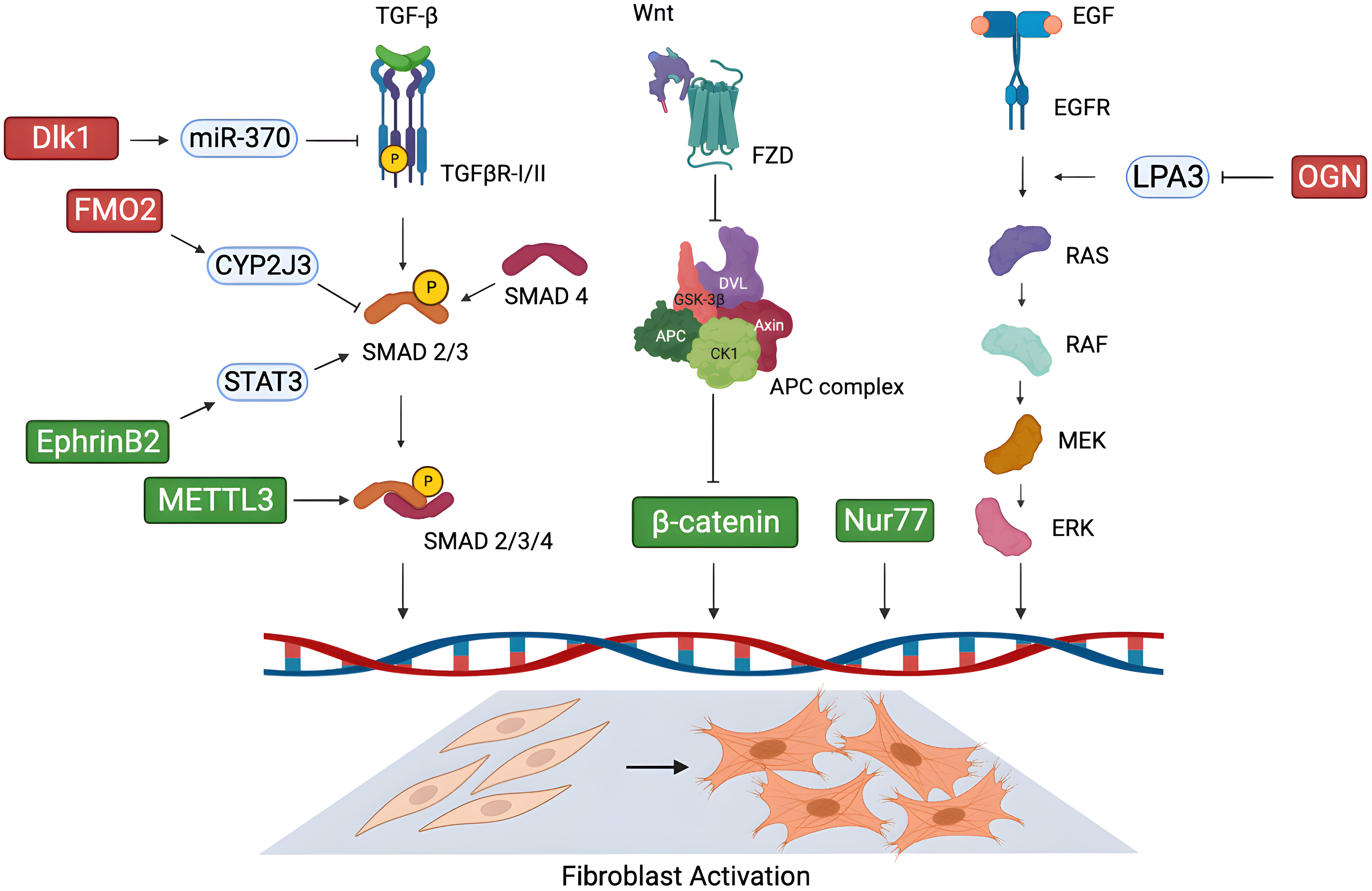
Fig. 1.

Novel possible molecular targets for cardiac fibrosis. New potential targets proposed in the literature in recent years for the treatment of cardiac fibrosis, and their mechanism of action in modulating cardiac fibroblast activation through key signalling pathways. (Green: pro-fibrotic factors; red: anti-fibrotic factors). Figure was created with the Biorender Software (https://www.biorender.com/). Dlk1, delta-like homolog 1; FMO2, flavin-containing monooxygenase 2; CYP2J3, cytochrome P450 superfamily 2J3; STAT3, signal transducer and activator of transcription 3; EphrinB2, erythropoietin-producing hepatoma interactor B2; METTL3, methyltransferase-like 3; TGF-β, transforming growth factor-beta; TGFβR, transforming growth factor-beta receptor; SMAD, small mothers against decapentaplegic; Wnt, wingless-INT; FZD, Freezled; APC, anaphase-promoting complex; Nur77, nuclear receptor 77; EGF, epidermal growth factor; EGFR, epidermal growth factor receptor; LPA3, lysophosphatidic acid 3; OGN, osteoglycin; RAS, rat sarcoma; RAF, rapidly accelerated fibrosarcoma; MEK, mitogen-activated protein kinase kinase; ERK, extracellular signal-regulated kinase; DVL, dishevelled; CK1, casein kinase; GSK-3β, glycogen synthase kinase-3 beta.

Osteoglycin (OGN) is a small leucine-rich proteoglycan secreted in the ECM, whose role in the fibrotic process is still debated. OGN was found to be overexpressed in the heart of patients with hypertensive [96] and ischemic disease [97]. In 2018, Zuo et al. [98] identified OGN as a negative regulator of cardiac fibrosis in response to chronic hypertension-induced injury. Angiotensin II infusion in OGN deficient (OGN-/-) mice revealed increased interstitial fibrosis and diastolic dysfunction. Accordingly, in vitro angiotensin II treatment on OGN-/- CFs led to enhanced cell proliferation and migration via activation of the epidermal growth factor receptor (EGFR) signalling, while OGN overexpression had the opposite effect. Specifically, the authors demonstrated that in physiological conditions OGN attenuates cardiac fibrosis binding to lysophosphatidic acid 3 (LPA3), thus preventing the activation of the Rho-dependent EGFR/ERK signalling, which induces fibroblast proliferation and migration [98].

Another interesting modulator of cardiac fibrosis is the nuclear receptor Nur77. It is involved in stress responses in different cell types [99, 100, 101], and it seems to play a dual role in the fibrotic process in heart tissue. A study by Medzikovic et al. [102] highlighted how Nur77 promoted CF differentiation into myofibroblasts. Conversely, its knockdown led to a reduction of myofibroblast marker expression and ECM proteins production, as well as decreased proliferation and wound healing capacity in rat cells stimulated with isoproterenol or TGF-β. On the other hand, authors showed that Nur77 in cardiomyocytes prevented these cells from releasing paracrine factors involved in the fibroblast-to-myofibroblast transition, thus counteracting also indirectly the fibrotic response in the myocardium through intercellular communication [102].

Among the factors that take part in the fibrotic process, it is worth mentioning fine regulation of fibrosis-related gene expression carried out by the N6-methyladenosine transferase methyltransferase-like 3 (METTL3) at the post-transcriptional level. METTL3 is the catalytic subunit of the methyltransferase complex which mediates mRNA methylation [103, 104, 105]. Recently, Li et al. [106] discovered the role of METTL3 as a promoter of CF activation and ECM remodelling in the process of MI-induced fibrosis. Specifically, METTL3 exerted its pro-fibrotic properties via tuning the m6A modification of mRNAs encoding collagens and factors of the TGFβ/SMAD pathway [106]. METTL3 silencing significantly reduced the levels of collagens in a mouse model of coronary artery ligation, as well as proliferation and myofibroblast transition of TGFβ-stimulated CFs in vitro.

In 2019, delta-like homolog 1 (Dlk1), a member of the epidermal growth factor (EGF)-like family involved in the control of cell differentiation [107], has been identified as a key anti-fibrotic factor in the heart. In detail, authors found that Dlk1 negatively regulated fibroblast-to-myofibroblast differentiation through the activation of miR-370, which in turn inhibited the TGF-β receptor 2 signalling pathway in these cells. Indeed, Dlk1-null mice exhibited alteration of ECM composition, with increased collagen deposition, as well as cardiac dysfunction. In addition, Dlk1-null CFs showed hyperactivation of TGFβ/SMAD3 signalling and myofibroblast differentiation. Since Dlk1 expression was significantly reduced in fibrotic tissues from both human ischemic patients and infarcted pigs, Rodriguez and colleagues [108] hypothesized the potential positive targeting of this molecule for the treatment of cardiac fibrosis characterized by aberrant TGFβ-signalling activation.

Another factor whose pro-fibrotic activity has been recently uncovered is erythropoietin-producing hepatoma interactor B2 (EphrinB2), a cell surface transmembrane ligand ubiquitously expressed in mammals, mediating organ-specific regulation of the fibrotic process [109, 110]. Increased expression of EphrinB2 has been detected in human and mouse hearts with severe cardiac fibrosis. In vivo silencing of EphrinB2 led to the attenuation of cardiac fibrosis and improvement of cardiac function, while its overexpression in CFs mediated myofibroblast differentiation and activation in vitro. In detail, EphrinB2 exerted its pro-fibrotic effects mainly via activating the signal transducer and activator of transcription 3 (STAT3) signalling in CFs, and promoting the interaction between STAT3 and SMAD3, thus inducing the fibrotic response [111]. Given this evidence, EphrinB2 could be considered as another promising candidate target against cardiac fibrosis.

Recently, a previously unknown anti-fibrotic activity of flavin-containing monooxygenase 2 (FMO2) has been characterized by Ni et al. [112]. FMO2 is an enzyme with nicotinamide adenine dinucleotide hydrogen/flavin adenine dinucleotide (NADH/FAD)-dependent activity, whose expression was found specifically enriched in CFs, but not in cardiomyocytes, and significantly down-regulated upon cardiac injury in rats, mice, and non-human primates. FMO2 silencing in rat hearts induced spontaneous fibrosis and altered cardiac function; in contrast, induction of FMO2 expression in infarcted rat and monkey hearts attenuated pathological remodelling and cardiac dysfunction. Authors demonstrated that FMO2 inhibited SMAD2/3 activation through an enzymatic activity-independent way: mechanistically, FMO2 was found to bind to cytochrome P450 superfamily 2J3 (CYP2J3), thus preventing the interaction of the latter with SMURF2, an E3 ubiquitin ligase, which selectively mediates the proteasome-dependent degradation of phosphorylated SMAD2/3 in the nucleus [113]. Thus, FMO2 inhibits TGFβ-signalling in CFs through a conserved mechanism in human cells, paving the way for new therapeutic opportunities for cardiac fibrosis [112].

The Wnt/β-catenin signalling pathway plays a consolidated role in the fibrotic process by promoting the activation of CFs [114, 115, 116]. Xiang and colleagues [117] have investigated the effect of inducible β-catenin loss in CFs in mice undergoing TAC. Fibroblast-specific β-catenin depletion in this system revealed reduced interstitial fibrosis, improved cardiac function, as well as lower levels of cardiomyocyte hypertrophy. Although β-catenin loss did not affect the number of activated and differentiated CFs, the expression of many pro-fibrotic genes, such as COL1A1, collagen type III alpha 1 (COL3A1), periostin (POSTN), was significantly reduced [117]. Given its many roles in physiology and pathology, though, also related to epithelial-to-mesenchymal transition, targeting of this pathway for cardiac fibrosis will have to be highly organ-specific to avoid potential side effects.

Vainio et al. [118] has explored the effects of monoclonal antibody (mAb) therapy against connective tissue growth factor (CTGF), whose increased expression has been correlated with fibrotic diseases of virtually every human organ [119], including the heart [120]. CTGF-mAb administration in a mouse model of MI resulted in reduced cardiac fibrosis and hypertrophy, as well as increased survival and left ventricular function. Heart tissue also showed increased expression of genes associated with developmental and repair processes, and reduction of inflammatory and fibrotic genes. Mechanistically, CTGF-mAb treatment on human CFs in vitro attenuated the expression and production of α-SMA and collagen I following TGF-β stimulation, through the activation of the JNK2 pathway [118].

A new interesting potential target for the treatment of cardiac fibrosis has been identified by Schafer and colleagues [121]. In this study, RNA-sequencing analysis of human CFs stimulated in vitro with TGF-β identified interleukin 11 (IL-11) expression as the most positively correlated response during myofibroblast differentiation, while IL-11 levels in healthy tissues and cells were found to be unaffected [122, 123]. Authors found that IL-11 and its receptor IL11RA were expressed specifically in CFs in a mouse model of fibrotic heart. They demonstrated that IL-11 binds to soluble receptor IL11RA and mediates trans-signalling on other CFs. This promotes an autocrine effect which induces the production of fibrogenic proteins in an ERK-dependent way. In vivo overexpression of IL-11 was shown to induce cardiac fibrosis, while its depletion had a protective effect. Given this evidence, IL-11 could represent a promising target for new anti-fibrotic therapies [121].

5. Epigenetic Approaches

In the search for new potential therapeutic targets against cardiac fibrosis, the field of epigenetic approaches opens the possibility of fine tuning the activation and the progression of the fibrotic process. In the wide scenario of epigenetic regulators and non-coding RNAs, we briefly present examples of potential targets recently identified for therapeutic intervention in this pathological condition, focusing on those with proven specific activity in CFs.

5.1 Histone Modifications

In recent years, numerous studies have suggested that epigenetic regulation, including histone methylation, has extensive roles in fibrosis [124]. Wang et al. [125] studied how the lysine demethylase 5B (KDM5B), a histone H3K4me2/me3 demethylase, can affect the pathophysiological events in cardiac fibrosis. Initially, they demonstrated KDM5B upregulation in CFs in response to pathological stress. Secondly, it was proposed the molecular mechanism through which KDM5B exerts its action: it demethylates the activated H3K4me2/3 modification bounding the activating transcription factor 3 (Atf3) promoter, inhibiting its expression. In the literature [126] Atf3 is considered an antifibrotic regulator of cardiac fibrosis, and its suppression is correlated with the activation of TGF-β signalling. Finally, the authors prevented the fibroblast transition into myofibroblast by inhibiting KDM5B in vitro and in vivo, thus proposing KDM5B as possible candidate target to ameliorate cardiac fibrosis and remodelling [125].

Another fundamental epigenetic modulation is histone acetylation/deacetylation, that enhances or opposes the accessibility of transcriptional factors, respectively. It is known that small molecules acting as histone deacetylase (HDAC) inhibitors play a role in the attenuation of cardiac remodelling and fibrosis [127]. Nural-Guvener et al. [128] proposed a strategy through selective class I-HDAC inhibition against CFs activation. In particular, they treated cluster of differentiation 90-positive (CD90+) CFs in vitro with mocetinostat (an HDAC inhibitor), showing reduction of collagen III and α-SMA. Moreover, they demonstrated the efficacy of selective class I-HDACs inhibition in reducing fibrosis in a congestive heart failure animal model, as well as its effect in reversing interstitial fibrosis and improving cardiac function after MI [128].

In a previous study, it was demonstrated that a group of epigenetic reader molecules called bromodomain and extra-terminal (BET) acetyl-lysine binding proteins, play a crucial role in regulating pathological fibrosis [129]. In particular, Stratton and Bagchi et al. [130] identified by RNA-sequencing the bromodomain-containing protein 4 (BRD4) involvement in cardiac fibrosis promoting the activation of quiescent CFs into Periostin (Postn)-positive cells. The authors validated the pro-fibrotic role of BRD4 using the BET bromodomains inhibitor JQ1. Specifically, cultured primary adult rat ventricular fibroblasts stimulated with TGF-β in the presence of JQ1 showed reduced expression of fibrotic markers, such as α-SMA [130]. Moreover, mice were treated with the JQ1 inhibitor after transverse aortic constriction, and the transcriptomic profiling of the CFs isolated from the hearts suggested a significant downregulation of Postn mRNA expression.

5.2 Long Non-Coding RNAs

The pivotal regulatory role of non-coding RNAs (ncRNAs) in every biological process is well established and consolidated, and so it is for the process of cardiac fibrosis. In the last years, many ncRNAs, mainly microRNAs (miRNAs), long non-coding RNAs (lncRNAs) and more recently also circular RNAs (circRNAs), have been discovered as fine tuners of cardiac fibrosis, paving the way to their therapeutic targeting. Indeed RNA-targeting therapeutics have a high potential for the treatment of several diseases.

LncRNAs contribute to cardiac diseases, including fibrosis. One of the first lncRNAs reported to be dysregulated during chronic cardiac remodelling is the Maternally Expressed Gene 3 (Meg3), which was found specifically expressed in CFs and downregulated during late cardiac remodelling in mouse hearts after TAC [131]. Meg3 was known to act as a transcriptional regulator of gene expression, recruiting and guiding chromatin remodelling complexes to target sites [132, 133]. In 2017, it was identified as a regulator of the expression of the fibrosis-related gene matrix metalloproteinase-2 (MMP2) in CFs through the recruitment of p53, this latter induced by TGF-β signalling; moreover, Meg3 silencing in vivo after TAC decreased myocardial fibrosis and improved diastolic function, thus revealing a new potential target for the prevention of pathological remodelling in heart diseases [131].

In 2019 Hao et al. [134] identified the lncRNA AK137033, named Safe (Sfrp2 antisense as fibrosis enhancer), specifically enriched in the nuclei of CFs. Its expression resulted up-regulated upon both MI induction in mouse hearts and TGF-β treatment of CFs in vitro. Safe silencing in CFs also prevented cell proliferation, myofibroblast differentiation, and ECM proteins secretion. In addition, in vivo knockdown of this lncRNA in a mouse model of MI reduced the infarcted area and cardiac fibrosis, leading to the improvement of cardiac function. Safe was found to exert its pro-fibrotic effect mainly via inducing the expression of its neighbouring protein-coding gene secreted freezled related protein 2 (Sfrp2), which was proven to synergically act with Safe and the RNA-binding protein HuR in the induction of fibrosis-related genes in CFs [134].

The Nuclear Enriched Abundant Transcript 1 (NEAT1) is a lncRNA involved in cancer and some fibrosis-related diseases. Its expression was found elevated in patients with heart failure. Neat1 acts as a pro-fibrotic lncRNA, by inducing Smad7 promoter methylation through recruitment of enhancer of zeste homologue 2 (EZH2). Silencing of NEAT11 significantly alleviated the progression of cardiac fibrosis and dysfunction in a mouse model of heart failure, while overexpression of Neat1 had the opposite effect. This suggests that NEAT11 could be a good candidate for targeted antisense therapy [135].

The scaffold attachment factor B interacting lncRNA (SAIL) was shown to be reduced in cardiac fibrotic tissue and activated CFs. In vitro overexpression of SAIL decreased proliferation and collagen production of CFs, and could alleviate cardiac fibrosis in a mouse model of cardiac fibrosis due to MI. SAIL blocks the access of the scaffold attachment factor B (SAFB) to RNA pol II and reduces the transcription of fibrosis-related genes. The human conserved fragment of SAIL (hSAIL) could also suppress the proliferation and collagen production of human CFs [136].

The lncRNA taurine upregulation gene 1 (TUG1) was found to be overexpressed after MI, and this correlated with reduced levels of miiR-133b in rat models of myocardial fibrosis. Also, angiotensin-treated cardiac myofibroblasts revealed increased expression of TUG1. In addition, TUG1 knockdown prevented myofibroblast activation, while its overexpression increased cell proliferation and collagen production. Authors proved TUG1 could act as a sponge against miR133b in CFs, thus promoting the expression of CTGF, a target of miR133b. On the basis of this evidence, the axis TUG1/miR133b/CTGF could be a new potential target for the treatment of cardiac fibrosis [137].

5.3 Micro-RNAs

Several miRNAs play a central role in the regulation of cardiac fibrosis. MiR-21 is highly expressed in cardiac cells, preferentially in non-myocytes, and it is known to be up-regulated and involved in the development of cardiac fibrosis, mainly promoting the activation of the ERK-MAPK pathway in CFs [138]. MiR-21 inhibition has been already proven effective for the prevention of cardiac fibrosis and dysfunction in small animal models of heart injury [139, 140]. Recently, Hinkel et al. [141] have tested the feasibility and anti-fibrotic efficacy of miR-21 silencing also in large animal models of heart failure, laying the foundation for potential applications in humans. In detail, the local, intracoronary delivery of locked nucleic acid (LNA)-antimiR-21 in pig hearts after I/R injury led to a reduction of adverse myocardial remodelling, associated with decreased fibroblast proliferation and macrophage infiltration, as well as improvement of cardiac function [141].

A critical miRNA in cardiac fibrosis is miR-125b, which has been proven to be necessary and sufficient for fibroblast-to-myofibroblast transition. MiR-125b expression was found to be enhanced in failing human hearts and in various animal models of cardiac fibrosis, as well as in CFs stimulated with TGF-β. Mechanistically, miR-125b targets the 3′-UTR of Apelin, a repressor of the fibrogenic pathway acting on the angiotensin II/TGF-β axis, thus promoting myofibroblast differentiation and activation. Indeed, in vivo miR-125b silencing through LNA delivery caused the attenuation of angiotensin-induced interstitial fibrosis in murine hearts [142]. In addition, also p53 was found to be a target of miR-125b, which induced in this way fibroblast proliferation. An in-depth analysis also revealed that miR-125b was at the centre stage of the fibrosis signalling, regulating many other related pathways, therefore representing a valid new potential target for anti-fibrotic therapies.

Another regulator of fibroblast-to-myofibroblast transition was identified in miR-146b-5p, which is overexpressed in the blood of patients with myocardial ischemia, and in the heart of mice with MI. Interestingly, miR-146b-5p is up-regulated in CFs, endothelial cells, and macrophages by hypoxia-induced NF-κB, independently from the classical TGF-β pathway, thus driving the switch toward a myofibroblast phenotype. In vivo treatment with miR-146b-5p antagomir markedly reduced cardiac fibrosis and improved myocardial function in both murine and porcine models of MI [143].

In contrast, a miRNA with anti-fibrotic properties with a potential therapeutic application is miR-101. This miRNA was found to be down-regulated in the peri-infarct area of rats undergoing coronary artery ligation, as well as in CFs stimulated with angiotensin II. On the contrary, in vitro overexpression of miR-101 suppressed fibroblast proliferation and ECM remodelling protein production, mainly targeting c-Fos, a downstream factor of the TGF-β signalling. The induction of miR-101 overexpression in vivo in post-infarct rat hearts remarkably alleviated interstitial fibrosis and improved cardiac performance [144].

5.4 Circular RNAs

Circular RNAs have been involved in the progression of both cancer and cardiovascular diseases. Given their peculiar secondary structure, with a covalent link bridging each end of the linear RNA which is converted into its circular form, circular RNAs are the most stable RNAs. This feature makes them optimal targets in RNA therapeutics.

In a rat model of post-ischemic myocardial fibrosis, circHNRNPH1 expression was found increased specifically in CFs. The expression of circHNRNPH1 could restrict the differentiation of CFs into myofibroblasts by de-repressing TGF-β signalling. The anti-fibrotic action of circHNRNPH1 is exerted through sponging miR-216-5p. This allows SMAD7 de-repression and the degradation of TGF-β receptor 1. Given the role of circHNRNPH1 in the TGF-β-mediated CF activation, this circRNA could be used to design new therapies for cardiac fibrosis linked to all cardiovascular diseases [145].

Similarly, circNFIB (which is conserved un humans) was found to be decreased in mouse cardiac samples post-MI, and its overexpression could attenuate CF proliferation [146]. Its action is dependent on TGF-β stimulation, and is mediated by the ability to sponge miR-433. circNFIB showed an anti-fibrotic effect both in vivo and in vitro on CFs, thus making it another good candidate for RNA-based anti-fibrotic therapies.

The circHIPK3 expression markedly increased in CFs and heart tissues in a mechanism mediated by TGF-β, and initiated by treatment with angiotensin II. Homeodomain-Interacting Protein Kinase 3 (CircHIPK3) was shown to promote CF proliferation, migration, and subsequent tissue fibrosis by sponging miR-29b-3p. This determined upregulating of its target genes actin alpha 2 (ACTA2), COL1A1 and COL3A1. The silencing of circHIPK3 attenuates CFs activation by inhibiting proliferation, migration, and α-SMA expression. Interestingly, a synergistic action of miR-29 overexpression and circHIPK3 silencing showed anti-fibrotic effects in vivo [147].

A recent study on patients with cardiac hypertrophy showed a significant decrease in yet another circRNA, that is the circYap isoform [148]. In a mouse model of hypertrophy, the expression of circYap was negatively correlated with the expression of fibrosis genes. The restoration of circYap via ectopic expression improved heart function and cardiac fibrosis in a pressure-overload mouse model of hypertrophy. CircYap expression could directly affect both survival and cell morphology of fibroblasts in vitro [148].

Finally, the circ_LAS1L was found to be down-regulated in acute MI patients and CFs in another clinical study. The circ_LAS1L functional role is to sponge miR-125b, thereby de-repressing this miRNA target genes involved in activation, proliferation, and migration of CFs [149].

6. Biotechnological Strategies

The continuous advancements in medical and biotechnological research have led to the development of innovative approaches based on biological and biotechnological therapies designed to counteract cardiac fibrosis. They include the use of biological non-cellular products, in situ reprogramming strategies, and immunotherapy approaches.

6.1 Exosomes

Several adult cell populations have been studied as therapeutic and/or regenerative products for the injured heart, including selected resident cardiac mesenchymal populations. Cardiosphere-Derived Cells (CDCs) have been shown to have beneficial effects, mainly through a protective paracrine action [150, 151], mediated in part by the release of extracellular membrane vesicles (EMVs), such as exosomes [152]. Human and rat dermal fibroblasts were treated with cardiosphere-derived EMVs (CSp-EMV) and then injected into rat hearts subjected to left anterior descending artery ligation [153]. In vitro treated fibroblasts displayed a less fibrotic phenotype, with significantly reduced levels of fibroblast specific protein 1 (FSP1) and discoidin domain receptor 2 (DDR2), and showed increased secretion of pro-angiogenic factors, such as stromal-cell derived factor 1 (SDF-1) and vascular endothelial growth factor (VEGF). Moreover, they exerted pro-angiogenic and anti-apoptotic effects in vitro. The in vivo administration of exosome-primed fibroblasts attenuated adverse remodelling, reduced the infarct area, and enhanced cardiac function. Authors speculated this therapeutic activity could be due to the specific profile of miRNAs carried by the EMV, since a similar effect on cardiomyocytes was previously proven for exosomes from CDCs [152]. This is an interesting example of a biological product directly influencing the functional phenotype of fibroblasts, with a relevant therapeutic outcome.

Recently, also the curative effect of exosomes derived from cortical bone stem cells (CBSCs) has been tested in murine models of I/R injury. CBSCs were already proven to exert beneficial effects, in terms of decreased scar size and improved cardiac function, when injected in mouse and pig models of MI [154]. The same effects were recapitulated in I/R mouse hearts where CBSC-derived exosomes (CBSC-dEXOs) were administered, highlighting their key role in mediating cardio-protection [155]. Mechanistically, in vitro treatment of CFs with CBSC-dEXOs decreased the expression of pro-fibrotic genes, such as collagen I, collagen III, and MMP2, even to a greater extent than whole CBSC-conditioned medium from which the exosomes were isolated. In addition, CBSC-dEXOs were also proven to significantly reduce CF activation induced by TGF-β. After analyzing the RNA content of CBSC-dEXOs, the authors suggested that this anti-fibrotic role could be exerted by small non-coding RNAs, mainly miRNAs: in fact, they caused the downregulation of ncRNAs implicated in ribosome stability and protein translation of factors necessary for the fibroblast-to-myofibroblast transition.

6.2 Direct Reprogramming

Cardiac regeneration aims at restoring functional cells that have been lost in the damaged heart. A therapeutic option to recover lost cardiomyocytes is the direct reprogramming of fibroblasts into cardiomyocytes through genetic tools or small molecules. This strategy could be also viewed as a way of simultaneously replacing potentially fibrotic cells with new parenchymal cells, thus representing also an anti-fibrotic approach. Several strategies have been proposed for the delivery of direct reprogramming factors in recent years, and we briefly summarize a selection based on artificial vectors/carriers.

Mónica P. A. Ferreira et al. [156], developed a functionalized spermine-modified dextran-based (AcDXSp) nanoparticle that allows the release of two molecules—CHIR99021 and SB431542—capable of inducing fibroblast reprogramming. Further functionalization of the nanoparticles with atrial natriuretic peptide (ANP) allowed tropism towards cardiac cells [157]. Specifically, AcDXSP nanoparticles were incubated with non-myocytes, and the effect of the release of the active molecules was confirmed. In fact, the administration of CHIR99021 significantly enhanced β-catenin stabilization, increasing its levels in both cytoplasm and nucleus. Instead, the release of SB431542 (a TGF-β inhibitor) prevented the intracellular translocation of SMAD3 to the nucleus, and inhibited its phosphorylation by anaplastic lymphoma kinase (ALK) [158]. This evidence thus supported the efficacy of this nanoparticle-based system for the direct reprogramming of fibroblasts into cardiomyocytes as a potential cardiac therapy [156].

Poly (lactic-co-glycolic acid) (PLGA)-polyethyleneimine (PEI) nanocarriers encapsulated with microRNAs represents a low-toxicity system to induce the direct reprogramming of cardiac human fibroblasts into cardiomyocyte-like cells. These nanoparticles were loaded with miR-1 and miR-133a, that initiate and drive muscle development and differentiation [159, 160]. Direct fibroblasts reprogramming was evaluated by the expression of markers such as cardiac troponins and α-actinin, confirming this strategy as a valid method for in vivo delivery and fibroblast targeting [161].

Qiaozi Wang et al. [162] have developed another non-viral system to reprogram CFs in situ, by coating neutrophil-mimicking membranes on mesoporous silicon nanoparticles (MSNs) with the FH peptide, and loading them with miR-1, -133, -208, and -499 (miR-Combo). Homing of the nanoparticle to the injured heart was achieved by the natural inflammation-homing capacity brought by neutrophil membranes, while the FH peptide’s high affinity to tenascin-C (TN-C), specifically expressed by CFs in the injured hearts, granted cell tropism. This study reported the intravenous injection of the nanoparticles loaded with the miR-combo in a mouse model of myocardial I/R, and evaluated the efficient in situ reprogramming of CFs into cardiomyocyte-like cells. Despite low rates of reprogramming, this strategy of delivering showed nonetheless an improvement in cardiac function, and attenuation of fibrosis in vivo [162].

6.3 CAR-T

Recently an innovative strategy based on chimeric antigen receptor T-cells (CAR-T) was proposed as an additional method to reduce cardiac fibrosis. The gene signature of CFs derived from healthy versus diseased human hearts were analysed, and fibroblast activation protein (FAP) was found as a specific marker of activated fibroblasts which is not expressed in cardiomyocytes. The study suggested that the adoptive transfer of antigen specific CD8+ T cells targeting the FAP protein can lead to partial removal of activated CFs in vivo, with significant reduction of cardiac fibrosis and restoration of function after injury in mice [163]. Joel G Rurik et al. [164] developed a therapeutic approach to generate transient CAR-T cells directly in vivo by delivering modified messenger RNA (mRNA) in T cell–targeted lipid nanoparticles (LNPs). Specifically, they injected CD5-targeted LNPs in a mouse model of heart failure, favouring the delivery of modified mRNA encoding for CAR in T lymphocytes, thus producing CAR-T cells in vivo. The transient CAR-T cells generated were specific for the FAP protein, so their action reduced the abundance of activated fibroblasts and consequently fibrosis, also restoring cardiac function after injury [164].

7. Conclusions

The quest for novel therapeutic strategies against cardiac fibrosis has taken multiple promising directions, that have provided encouraging preclinical data in recent years. A key obstacle in finding one good therapeutic option for treatment of myocardial fibrosis is represented by the complexity of the signalling network involved in the activation of repair mechanisms, which includes responses to surrounding stress and cell death signals, as well as the powerful crosstalk with the immune cells compartment. Therefore, any novel therapeutic approach will have to deal with, and possibly overcome, all the opposing signals in the microenvironment of the injured myocardium, making the quest quite complex. Moreover, different aetiologies and/or pathogenetic stimuli may be differentially sensitive to the same strategy, therefore future studies will have to define also the specifics of the population of potential target patients. Finally, given these many levels of complexity, future developments of therapies might work best with a synergistic approach, where multiple pathways are targeted by the same molecule or by combined treatments.

Author Contributions

EF, CC, SM, FT performed bibliographic search and manuscript drafting. EF and CC prepared the figures. SM and FT prepared the table. VP, FP and IC participated conception and design, drafted the manuscript, and revised it critically for important intellectual content. All authors have read and agreed to the published version of the manuscript. All authors have participated sufficiently in the work and agreed to be accountable for all aspects of the work.

Ethics Approval and Consent to Participate

Not applicable.

Acknowledgment

Not applicable.

Funding

IC is supported by grants # RM12117A805ED2FD and # RG11916B85CDBF76 from Sapienza University of Rome, and by grant # RG1221816BC8E766 from the Italian Health Ministry. FP is supported by Grant # A0375-2020-36621 from Regione Lazio (POR-FESR 2014-2021). EF is supported by grant # AR12218167E8FA71 from Sapienza University of Rome.

Conflict of Interest

The authors declare no conflict of interest.

References
[1]
Heron M. National Vital Statistics Reports Volume 70, Number 9 July 26, 2021 Deaths: Leading Causes for 2019. 2021. Available at: https://www.cdc.gov/nchs/products/index.htm (Accessed: 21 February 2023).
[2]
Virani SS, Alonso A, Aparicio HJ, Benjamin EJ, Bittencourt MS, Callaway CW, et al. Heart Disease and Stroke Statistics-2021 Update: A Report From the American Heart Association. Circulation. 2021; 143: e254–e743.
[3]
Townsend N, Wilson L, Bhatnagar P, Wickramasinghe K, Rayner M, Nichols M. Cardiovascular disease in Europe: epidemiological update 2016. European Heart Journal. 2016; 37: 3232–3245.
[4]
Díez J, de Boer RA. Management of cardiac fibrosis is the largest unmet medical need in heart failure. Cardiovascular Research. 2022; 118: e20–e22.
[5]
Zeisberg EM, Kalluri R. Origins of cardiac fibroblasts. Circulation Research. 2010; 107: 1304–1312.
[6]
Schirone L, Forte M, Palmerio S, Yee D, Nocella C, Angelini F, et al. A Review of the Molecular Mechanisms Underlying the Development and Progression of Cardiac Remodeling. Oxidative Medicine and Cellular Longevity. 2017; 2017: 3920195.
[7]
Pesce M, Duda GN, Forte G, Girao H, Raya A, Roca-Cusachs P, et al. Cardiac fibroblasts and mechanosensation in heart development, health and disease. Nature Reviews: Cardiology. 2023; 20: 309–324.
[8]
Schnee JM, Hsueh WA. Angiotensin II, adhesion, and cardiac fibrosis. Cardiovascular Research. 2000; 46: 264–268.
[9]
Sullivan RD, Mehta RM, Tripathi R, Reed GL, Gladysheva IP. Renin Activity in Heart Failure with Reduced Systolic Function-New Insights. International Journal of Molecular Sciences. 2019; 20: 3182.
[10]
Travers JG, Kamal FA, Robbins J, Yutzey KE, Blaxall BC. Cardiac Fibrosis: The Fibroblast Awakens. Circulation Research. 2016; 118: 1021–1040.
[11]
Gao X, He X, Luo B, Peng L, Lin J, Zuo Z. Angiotensin II increases collagen I expression via transforming growth factor-beta1 and extracellular signal-regulated kinase in cardiac fibroblasts. European Journal of Pharmacology. 2009; 606: 115–120.
[12]
Schorb W, Booz GW, Dostal DE, Conrad KM, Chang KC, Baker KM. Angiotensin II is mitogenic in neonatal rat cardiac fibroblasts. Circulation Research. 1993; 72: 1245–1254.
[13]
Villarreal FJ, Kim NN, Ungab GD, Printz MP, Dillmann WH. Identification of functional angiotensin II receptors on rat cardiac fibroblasts. Circulation. 1993; 88: 2849–2861.
[14]
Lee AA, Dillmann WH, McCulloch AD, Villarreal FJ. Angiotensin II stimulates the autocrine production of transforming growth factor-beta 1 in adult rat cardiac fibroblasts. Journal of Molecular and Cellular Cardiology. 1995; 27: 2347–2357.
[15]
Sun Y, Ramires FJ, Weber KT. Fibrosis of atria and great vessels in response to angiotensin II or aldosterone infusion. Cardiovascular Research. 1997; 35: 138–147.
[16]
Pahor M, Bernabei R, Sgadari A, Gambassi G, Jr, Lo Giudice P, Pacifici L, et al. Enalapril prevents cardiac fibrosis and arrhythmias in hypertensive rats. Hypertension. 1991; 18: 148–157.
[17]
Ham O, Jin W, Lei L, Huang HH, Tsuji K, Huang M, et al. Pathological cardiac remodeling occurs early in CKD mice from unilateral urinary obstruction, and is attenuated by Enalapril. Scientific Reports. 2018; 8: 16087.
[18]
Brilla CG, Zhou G, Matsubara L, Weber KT. Collagen metabolism in cultured adult rat cardiac fibroblasts: response to angiotensin II and aldosterone. Journal of Molecular and Cellular Cardiology. 1994; 26: 809–820.
[19]
Sparks MA, Rianto F, Diaz E, Revoori R, Hoang T, Bouknight L, et al. Direct Actions of AT1 (Type 1 Angiotensin) Receptors in Cardiomyocytes Do Not Contribute to Cardiac Hypertrophy. Hypertension. 2021; 77: 393–404.
[20]
Hartupee J, Mann DL. Neurohormonal activation in heart failure with reduced ejection fraction. Nature Reviews: Cardiology. 2017; 14: 30–38.
[21]
Tsai CF, Yang SF, Chu HJ, Ueng KC. Cross-talk between mineralocorticoid receptor/angiotensin II type 1 receptor and mitogen-activated protein kinase pathways underlies aldosterone-induced atrial fibrotic responses in HL-1 cardiomyocytes. International Journal of Cardiology. 2013; 169: 17–28.
[22]
Somanna NK, Yariswamy M, Garagliano JM, Siebenlist U, Mummidi S, Valente AJ, et al. Aldosterone-induced cardiomyocyte growth, and fibroblast migration and proliferation are mediated by TRAF3IP2. Cellular Signalling. 2015; 27: 1928–1938.
[23]
Stockand JD, Meszaros JG. Aldosterone stimulates proliferation of cardiac fibroblasts by activating Ki-RasA and MAPK1/2 signaling. American Journal of Physiology. Heart and Circulatory Physiology. 2003; 284: H176–H184.
[24]
Lemarié CA, Simeone SMC, Nikonova A, Ebrahimian T, Deschênes ME, Coffman TM, et al. Aldosterone-induced activation of signaling pathways requires activity of angiotensin type 1a receptors. Circulation Research. 2009; 105: 852–859.
[25]
Shafiq MM, Miller AB. Blocking aldosterone in heart failure. Therapeutic Advances in Cardiovascular Disease. 2009; 3: 379–385.
[26]
Zannad F, Alla F, Dousset B, Perez A, Pitt B. Limitation of excessive extracellular matrix turnover may contribute to survival benefit of spironolactone therapy in patients with congestive heart failure: insights from the randomized aldactone evaluation study (RALES). Rales Investigators. Circulation. 2000; 102: 2700–2706.
[27]
Hjalmarson A, Goldstein S, Fagerberg B, Wedel H, Waagstein F, Kjekshus J, et al. Effect of metoprolol CR/XL in chronic heart failure: Metoprolol CR/XL Randomised Intervention Trial in Congestive Heart Failure (MERIT-HF). The Lancet. 1999; 353: 2001–2007.
[28]
Dargie HJ, Lechat P. The Cardiac Insufficiency Bisoprolol Study II (CIBIS-II): a randomised trial. The Lancet. 1999; 353: 9–13.
[29]
Packer M, Fowler MB, Roecker EB, Coats AJS, Katus HA, Krum H, et al. Effect of carvedilol on the morbidity of patients with severe chronic heart failure: results of the carvedilol prospective randomized cumulative survival (COPERNICUS) study. Circulation. 2002; 106: 2194–2199.
[30]
Chimenti I, Pagano F, Cavarretta E, Angelini F, Peruzzi M, Barretta A, et al. Β-blockers treatment of cardiac surgery patients enhances isolation and improves phenotype of cardiosphere-derived cells. Scientific Reports. 2016; 6: 36774.
[31]
Delaunay M, Osman H, Kaiser S, Diviani D. The Role of Cyclic AMP Signaling in Cardiac Fibrosis. Cells. 2019; 9: 69.
[32]
Swaney JS, Roth DM, Olson ER, Naugle JE, Meszaros JG, Insel PA. Inhibition of cardiac myofibroblast formation and collagen synthesis by activation and overexpression of adenylyl cyclase. Proceedings of the National Academy of Sciences of the United States of America. 2005; 102: 437–442.
[33]
Liu X, Sun SQ, Hassid A, Ostrom RS. cAMP inhibits transforming growth factor-beta-stimulated collagen synthesis via inhibition of extracellular signal-regulated kinase 1/2 and Smad signaling in cardiac fibroblasts. Molecular Pharmacology. 2006; 70: 1992–2003.
[34]
Li J, Philip JL, Xu X, Theccanat T, Abdur Razzaque M, Akhter SA. β-Arrestins regulate human cardiac fibroblast transformation and collagen synthesis in adverse ventricular remodeling. Journal of Molecular and Cellular Cardiology. 2014; 76: 73–83.
[35]
Schumacher SM, Gao E, Zhu W, Chen X, Chuprun JK, Feldman AM, et al. Paroxetine-mediated GRK2 inhibition reverses cardiac dysfunction and remodeling after myocardial infarction. Science Translational Medicine. 2015; 7: 277ra31.
[36]
Woodall MC, Woodall BP, Gao E, Yuan A, Koch WJ. Cardiac Fibroblast GRK2 Deletion Enhances Contractility and Remodeling Following Ischemia/Reperfusion Injury. Circulation Research. 2016; 119: 1116–1127.
[37]
Travers JG, Kamal FA, Valiente-Alandi I, Nieman ML, Sargent MA, Lorenz JN, et al. Pharmacological and Activated Fibroblast Targeting of Gβγ-GRK2 After Myocardial Ischemia Attenuates Heart Failure Progression. Journal of the American College of Cardiology. 2017; 70: 958–971.
[38]
Turner NA, Porter KE, Smith WHT, White HL, Ball SG, Balmforth AJ. Chronic beta2-adrenergic receptor stimulation increases proliferation of human cardiac fibroblasts via an autocrine mechanism. Cardiovascular Research. 2003; 57: 784–792.
[39]
Yin F, Wang YY, Du JH, Li C, Lu ZZ, Han C, et al. Noncanonical cAMP pathway and p38 MAPK mediate beta2-adrenergic receptor-induced IL-6 production in neonatal mouse cardiac fibroblasts. Journal of Molecular and Cellular Cardiology. 2006; 40: 384–393.
[40]
Aránguiz-Urroz P, Canales J, Copaja M, Troncoso R, Vicencio JM, Carrillo C, et al. Beta(2)-adrenergic receptor regulates cardiac fibroblast autophagy and collagen degradation. Biochimica et Biophysica Acta. 2011; 1812: 23–31.
[41]
Pagano F, Angelini F, Siciliano C, Tasciotti J, Mangino G, De Falco E, et al. Beta2-adrenergic signaling affects the phenotype of human cardiac progenitor cells through EMT modulation. Pharmacological Research. 2018; 127: 41–48.
[42]
Michel LYM, Farah C, Balligand JL. The Beta3 Adrenergic Receptor in Healthy and Pathological Cardiovascular Tissues. Cells. 2020; 9: 2584.
[43]
Nantel F, Bonin H, Emorine LJ, Zilberfarb V, Strosberg AD, Bouvier M, et al. The human beta 3-adrenergic receptor is resistant to short term agonist-promoted desensitization. Molecular Pharmacology. 1993; 43: 548–555.
[44]
Kamiya M, Asai K, Maejima Y, Shirakabe A, Murai K, Noma S, et al. β3-Adrenergic Receptor Agonist Prevents Diastolic Dysfunction in an Angiotensin II-Induced Cardiomyopathy Mouse Model. The Journal of Pharmacology and Experimental Therapeutics. 2021; 376: 473–481.
[45]
Niu X, Zhao L, Li X, Xue Y, Wang B, Lv Z, et al. β3-Adrenoreceptor stimulation protects against myocardial infarction injury via eNOS and nNOS activation. PLoS ONE. 2014; 9: e98713.
[46]
Pouleur AC, Anker S, Brito D, Brosteanu O, Hasenclever D, Casadei B, et al. Rationale and design of a multicentre, randomized, placebo-controlled trial of mirabegron, a Beta3-adrenergic receptor agonist on left ventricular mass and diastolic function in patients with structural heart disease Beta3-left ventricular hypertrophy (Beta3-LVH). ESC Heart Failure. 2018; 5: 830–841.
[47]
Jeon KI, Xu X, Aizawa T, Lim JH, Jono H, Kwon DS, et al. Vinpocetine inhibits NF-kappaB-dependent inflammation via an IKK-dependent but PDE-independent mechanism. Proceedings of the National Academy of Sciences of the United States of America. 2010; 107: 9795–9800.
[48]
Miller CL, Cai Y, Oikawa M, Thomas T, Dostmann WR, Zaccolo M, et al. Cyclic nucleotide phosphodiesterase 1A: a key regulator of cardiac fibroblast activation and extracellular matrix remodeling in the heart. Basic Research in Cardiology. 2011; 106: 1023–1039.
[49]
Chen S, Zhang Y, Lighthouse JK, Mickelsen DM, Wu J, Yao P, et al. A Novel Role of Cyclic Nucleotide Phosphodiesterase 10A in Pathological Cardiac Remodeling and Dysfunction. Circulation. 2020; 141: 217–233.
[50]
Li G, Xing W, Zhang M, Geng F, Yang H, Zhang H, et al. Antifibrotic cardioprotection of berberine via downregulating myocardial IGF-1 receptor-regulated MMP-2/MMP-9 expression in diabetic rats. American Journal of Physiology. Heart and Circulatory Physiology. 2018; 315: H802–H813.
[51]
Liu M, Xu X, Zhao J, Tang Y. Naringenin inhibits transforming growth factor-β1-induced cardiac fibroblast proliferation and collagen synthesis via G0/G1 arrest. Experimental and Therapeutic Medicine. 2017; 14: 4425–4430.
[52]
Wei W, Rao F, Liu F, Xue Y, Deng C, Wang Z, et al. Involvement of Smad3 pathway in atrial fibrosis induced by elevated hydrostatic pressure. Journal of Cellular Physiology. 2018; 233: 4981–4989.
[53]
Gu J, Qiu M, Lu Y, Ji Y, Qian Z, Sun W. Piperlongumine attenuates angiotensin-II-induced cardiac hypertrophy and fibrosis by inhibiting Akt-FoxO1 signalling. Phytomedicine: International Journal of Phytotherapy and Phytopharmacology. 2021; 82: 153461.
[54]
Xiao J, Sheng X, Zhang X, Guo M, Ji X. Curcumin protects against myocardial infarction-induced cardiac fibrosis via SIRT1 activation in vivo and in vitro. Drug Design, Development and Therapy. 2016; 10: 1267–1277.
[55]
Wang NP, Wang ZF, Tootle S, Philip T, Zhao ZQ. Curcumin promotes cardiac repair and ameliorates cardiac dysfunction following myocardial infarction. British Journal of Pharmacology. 2012; 167: 1550–1562.
[56]
Liu R, Zhang HB, Yang J, Wang JR, Liu JX, Li CL. Curcumin alleviates isoproterenol-induced cardiac hypertrophy and fibrosis through inhibition of autophagy and activation of mTOR. European Review for Medical and Pharmacological Sciences. 2018; 22: 7500–7508.
[57]
Frati G, Schirone L, Chimenti I, Yee D, Biondi-Zoccai G, Volpe M, et al. An overview of the inflammatory signalling mechanisms in the myocardium underlying the development of diabetic cardiomyopathy. Cardiovascular Research. 2017; 113: 378–388.
[58]
Schimmel K, Jung M, Foinquinos A, José GS, Beaumont J, Bock K, et al. Natural Compound Library Screening Identifies New Molecules for the Treatment of Cardiac Fibrosis and Diastolic Dysfunction. Circulation. 2020; 141: 751–767.
[59]
Tian J, Zhang M, Suo M, Liu D, Wang X, Liu M, et al. Dapagliflozin alleviates cardiac fibrosis through suppressing EndMT and fibroblast activation via AMPKα/TGF-β/Smad signalling in type 2 diabetic rats. Journal of Cellular and Molecular Medicine. 2021; 25: 7642–7659.
[60]
Zhao M, Zhang J, Xu Y, Liu J, Ye J, Wang Z, et al. Selective Inhibition of NLRP3 Inflammasome Reverses Pressure Overload-Induced Pathological Cardiac Remodeling by Attenuating Hypertrophy, Fibrosis, and Inflammation. International Immunopharmacology. 2021; 99: 108046.
[61]
Valiente-Alandi I, Potter SJ, Salvador AM, Schafer AE, Schips T, Carrillo-Salinas F, et al. Inhibiting Fibronectin Attenuates Fibrosis and Improves Cardiac Function in a Model of Heart Failure. Circulation. 2018; 138: 1236–1252.
[62]
Garoffolo G, Casaburo M, Amadeo F, Salvi M, Bernava G, Piacentini L, et al. Reduction of Cardiac Fibrosis by Interference With YAP-Dependent Transactivation. Circulation Research. 2022; 131: 239–257.
[63]
Francisco J, Zhang Y, Jeong JI, Mizushima W, Ikeda S, Ivessa A, et al. Blockade of Fibroblast YAP Attenuates Cardiac Fibrosis and Dysfunction Through MRTF-A Inhibition. JACC: Basic to Translational Science. 2020; 5: 931–945.
[64]
Miller CL, Oikawa M, Cai Y, Wojtovich AP, Nagel DJ, Xu X, et al. Role of Ca2+/calmodulin-stimulated cyclic nucleotide phosphodiesterase 1 in mediating cardiomyocyte hypertrophy. Circulation Research. 2009; 105: 956–964.
[65]
Wu MP, Zhang YS, Xu X, Zhou Q, Li JD, Yan C. Vinpocetine Attenuates Pathological Cardiac Remodeling by Inhibiting Cardiac Hypertrophy and Fibrosis. Cardiovascular Drugs and Therapy. 2017; 31: 157–166.
[66]
Duinen MV, Reneerkens OAH, Lambrecht L, Sambeth A, Rutten BPF, Os JV, et al. Treatment of Cognitive Impairment in Schizophrenia: Potential Value of Phosphodiesterase Inhibitors in Prefrontal Dysfunction. Current Pharmaceutical Design. 2015; 21: 3813–3828.
[67]
Grauer SM, Pulito VL, Navarra RL, Kelly MP, Kelley C, Graf R, et al. Phosphodiesterase 10A inhibitor activity in preclinical models of the positive, cognitive, and negative symptoms of schizophrenia. The Journal of Pharmacology and Experimental Therapeutics. 2009; 331: 574–590.
[68]
Giampà C, Laurenti D, Anzilotti S, Bernardi G, Menniti FS, Fusco FR. Inhibition of the striatal specific phosphodiesterase PDE10A ameliorates striatal and cortical pathology in R6/2 mouse model of Huntington’s disease. PLoS ONE. 2010; 5: e13417.
[69]
Beaumont V, Zhong S, Lin H, Xu W, Bradaia A, Steidl E, et al. Phosphodiesterase 10A Inhibition Improves Cortico-Basal Ganglia Function in Huntington’s Disease Models. Neuron. 2016; 92: 1220–1237.
[70]
Delaughter MC, Taffet GE, Fiorotto ML, Entman ML, Schwartz RJ. Local insulin-like growth factor I expression induces physiologic, then pathologic, cardiac hypertrophy in transgenic mice. FASEB Journal. 1999; 13: 1923–1929.
[71]
Wang Z, Yu T, Huang P. Post-translational modifications of FOXO family proteins (Review). Molecular Medicine Reports. 2016; 14: 4931–4941.
[72]
Ni YG, Berenji K, Wang N, Oh M, Sachan N, Dey A, et al. Foxo transcription factors blunt cardiac hypertrophy by inhibiting calcineurin signaling. Circulation. 2006; 114: 1159–1168.
[73]
Jain SK, Rains J, Jones K. Effect of curcumin on protein glycosylation, lipid peroxidation, and oxygen radical generation in human red blood cells exposed to high glucose levels. Free Radical Biology & Medicine. 2006; 41: 92–96.
[74]
Aggarwal BB, Kumar A, Bharti AC. Anticancer potential of curcumin: preclinical and clinical studies. Anticancer Research. 2003; 23: 363–398.
[75]
Hsu CP, Zhai P, Yamamoto T, Maejima Y, Matsushima S, Hariharan N, et al. Silent information regulator 1 protects the heart from ischemia/reperfusion. Circulation. 2010; 122: 2170–2182.
[76]
Rizk SM, El-Maraghy SA, Nassar NN. A novel role for SIRT-1 in L-arginine protection against STZ induced myocardial fibrosis in rats. PLoS ONE. 2014; 9: e114560.
[77]
Zhao J, Chen Y, Chen Q, Hong T, Zhong Z, He J, et al. Curcumin Ameliorates Cardiac Fibrosis by Regulating Macrophage-Fibroblast Crosstalk via IL18-P-SMAD2/3 Signaling Pathway Inhibition. Frontiers in Pharmacology. 2022; 12: 784041.
[78]
Euler-Taimor G, Heger J. The complex pattern of SMAD signaling in the cardiovascular system. Cardiovascular Research. 2006; 69: 15–25.
[79]
Sciarretta S, Yee D, Nagarajan N, Bianchi F, Saito T, Valenti V, et al. Trehalose-Induced Activation of Autophagy Improves Cardiac Remodeling After Myocardial Infarction. Journal of the American College of Cardiology. 2018; 71: 1999–2010.
[80]
Chimenti I, Picchio V, Pagano F, Schirone L, Schiavon S, D’Ambrosio L, et al. The impact of autophagy modulation on phenotype and survival of cardiac stromal cells under metabolic stress. Cell Death Discovery. 2022; 8: 149.
[81]
Pagano F, Picchio V, Bordin A, Cavarretta E, Nocella C, Cozzolino C, et al. Progressive stages of dysmetabolism are associated with impaired biological features of human cardiac stromal cells mediated by the oxidative state and autophagy. The Journal of Pathology. 2022; 258: 136–148.
[82]
Emerging Risk Factors Collaboration, Di Angelantonio E, Kaptoge S, Wormser D, Willeit P, Butterworth AS, et al. Association of Cardiometabolic Multimorbidity With Mortality. The Journal of the American Medical Association. 2015; 314: 52–60.
[83]
Wiviott SD, Raz I, Bonaca MP, Mosenzon O, Kato ET, Cahn A, et al. Dapagliflozin and Cardiovascular Outcomes in Type 2 Diabetes. The New England Journal of Medicine. 2019; 380: 347–357.
[84]
Han S, Hagan DL, Taylor JR, Xin L, Meng W, Biller SA, et al. Dapagliflozin, a selective SGLT2 inhibitor, improves glucose homeostasis in normal and diabetic rats. Diabetes. 2008; 57: 1723–1729.
[85]
Komoroski B, Vachharajani N, Feng Y, Li L, Kornhauser D, Pfister M. Dapagliflozin, a novel, selective SGLT2 inhibitor, improved glycemic control over 2 weeks in patients with type 2 diabetes mellitus. Clinical Pharmacology and Therapeutics. 2009; 85: 513–519.
[86]
Plosker GL. Dapagliflozin: a review of its use in patients with type 2 diabetes. Drugs. 2014; 74: 2191–2209.
[87]
Li X, Liu J, Lu Q, Ren D, Sun X, Rousselle T, et al. AMPK: a therapeutic target of heart failure-not only metabolism regulation. Bioscience Reports. 2019; 39: BSR20181767.
[88]
Qi H, Liu Y, Li S, Chen Y, Li L, Cao Y, et al. Activation of AMPK Attenuated Cardiac Fibrosis by Inhibiting CDK2 via p21/p27 and miR-29 Family Pathways in Rats. Molecular Therapy. Nucleic Acids. 2017; 8: 277–290.
[89]
Suetomi T, Willeford A, Brand CS, Cho Y, Ross RS, Miyamoto S, et al. Inflammation and NLRP3 Inflammasome Activation Initiated in Response to Pressure Overload by Ca2+/Calmodulin-Dependent Protein Kinase II δ Signaling in Cardiomyocytes Are Essential for Adverse Cardiac Remodeling. Circulation. 2018; 138: 2530–2544.
[90]
Gao R, Shi H, Chang S, Gao Y, Li X, Lv C, et al. The selective NLRP3-inflammasome inhibitor MCC950 reduces myocardial fibrosis and improves cardiac remodeling in a mouse model of myocardial infarction. International Immunopharmacology. 2019; 74: 105575.
[91]
Panciera T, Azzolin L, Cordenonsi M, Piccolo S. Mechanobiology of YAP and TAZ in physiology and disease. Nature Reviews. Molecular Cell Biology. 2017; 18: 758–770.
[92]
Johansen AKZ, Molkentin JD. Hippo signaling does it again: arbitrating cardiac fibroblast identity and activation. Genes & Development. 2019; 33: 1457–1459.
[93]
Hernández-Perera O, Pérez-Sala D, Navarro-Antolín J, Sánchez-Pascuala R, Hernández G, Díaz C, et al. Effects of the 3-hydroxy-3-methylglutaryl-CoA reductase inhibitors, atorvastatin and simvastatin, on the expression of endothelin-1 and endothelial nitric oxide synthase in vascular endothelial cells. The Journal of Clinical Investigation. 1998; 101: 2711–2719.
[94]
Wassmann S, Laufs U, Müller K, Konkol C, Ahlbory K, Bäumer AT, et al. Cellular antioxidant effects of atorvastatin in vitro and in vivo. Arteriosclerosis, Thrombosis, and Vascular Biology. 2002; 22: 300–305.
[95]
Akahori H, Tsujino T, Naito Y, Matsumoto M, Sasaki N, Iwasaku T, et al. Atorvastatin ameliorates cardiac fibrosis and improves left ventricular diastolic function in hypertensive diastolic heart failure model rats. Journal of Hypertension. 2014; 32: 1534–1541; discussion 1541.
[96]
Petretto E, Sarwar R, Grieve I, Lu H, Kumaran MK, Muckett PJ, et al. Integrated genomic approaches implicate osteoglycin (Ogn) in the regulation of left ventricular mass. Nature Genetics. 2008; 40: 546–552.
[97]
Van Aelst LNL, Voss S, Carai P, Van Leeuwen R, Vanhoutte D, Sanders-van Wijk S, et al. Osteoglycin prevents cardiac dilatation and dysfunction after myocardial infarction through infarct collagen strengthening. Circulation Research. 2015; 116: 425–436.
[98]
Zuo C, Li X, Huang J, Chen D, Ji K, Yang Y, et al. Osteoglycin attenuates cardiac fibrosis by suppressing cardiac myofibroblast proliferation and migration through antagonizing lysophosphatidic acid 3/matrix metalloproteinase 2/epidermal growth factor receptor signalling. Cardiovascular Research. 2018; 114: 703–712.
[99]
Chao LC, Wroblewski K, Ilkayeva OR, Stevens RD, Bain J, Meyer GA, et al. Skeletal muscle Nur77 expression enhances oxidative metabolism and substrate utilization. Journal of Lipid Research. 2012; 53 :2610–2619.
[100]
Hanna RN, Carlin LM, Hubbeling HG, Nackiewicz D, Green AM, Punt JA, et al. The transcription factor NR4A1 (Nur77) controls bone marrow differentiation and the survival of Ly6C- monocytes. Nature Immunology. 2011; 12: 778–785.
[101]
Fernandez PM, Brunel F, Jimenez MA, Saez JM, Cereghini S, Zakin MM. Nuclear receptors Nor1 and NGFI-B/Nur77 play similar, albeit distinct, roles in the hypothalamo-pituitary-adrenal axis. Endocrinology. 2000; 141: 2392–2400.
[102]
Medzikovic L, Heese H, van Loenen PB, van Roomen CPAA, Hooijkaas IB, Christoffels VM, et al. Nuclear Receptor Nur77 Controls Cardiac Fibrosis through Distinct Actions on Fibroblasts and Cardiomyocytes. International Journal of Molecular Sciences. 2021; 22: 1600.
[103]
Meyer KD, Saletore Y, Zumbo P, Elemento O, Mason CE, Jaffrey SR. Comprehensive analysis of mRNA methylation reveals enrichment in 3’ UTRs and near stop codons. Cell. 2012; 149: 1635–1646.
[104]
Dominissini D, Moshitch-Moshkovitz S, Schwartz S, Salmon-Divon M, Ungar L, Osenberg S, et al. Topology of the human and mouse m6A RNA methylomes revealed by m6A-seq. Nature. 2012; 485: 201–206.
[105]
Roignant JY, Soller M. m6A in mRNA: An Ancient Mechanism for Fine-Tuning Gene Expression. Trends in Genetics. 2017; 33: 380–390.
[106]
Li T, Zhuang Y, Yang W, Xie Y, Shang W, Su S, et al. Silencing of METTL3 attenuates cardiac fibrosis induced by myocardial infarction via inhibiting the activation of cardiac fibroblasts. FASEB Journal. 2021; 35: e21162.
[107]
Laborda J. The role of the epidermal growth factor-like protein dlk in cell differentiation. Histology and Histopathology. 2000; 15: 119–129.
[108]
Rodriguez P, Sassi Y, Troncone L, Benard L, Ishikawa K, Gordon RE, et al. Deletion of delta-like 1 homologue accelerates fibroblast-myofibroblast differentiation and induces myocardial fibrosis. European Heart Journal. 2019; 40: 967–978.
[109]
Kida Y, Ieronimakis N, Schrimpf C, Reyes M, Duffield JS. EphrinB2 reverse signaling protects against capillary rarefaction and fibrosis after kidney injury. Journal of the American Society of Nephrology. 2013; 24: 559–572.
[110]
Lagares D, Ghassemi-Kakroodi P, Tremblay C, Santos A, Probst CK, Franklin A, et al. ADAM10-mediated ephrin-B2 shedding promotes myofibroblast activation and organ fibrosis. Nature Medicine. 2017; 23: 1405–1415.
[111]
Su SA, Yang D, Wu Y, Xie Y, Zhu W, Cai Z, et al. EphrinB2 Regulates Cardiac Fibrosis Through Modulating the Interaction of Stat3 and TGF-β/Smad3 Signaling. Circulation Research. 2017; 121: 617–627.
[112]
Ni C, Chen Y, Xu Y, Zhao J, Li Q, Xiao C, et al. Flavin Containing Monooxygenase 2 Prevents Cardiac Fibrosis via CYP2J3-SMURF2 Axis. Circulation Research. 2022; 131: 189–206.
[113]
Lin X, Liang M, Feng XH. Smurf2 is a ubiquitin E3 ligase mediating proteasome-dependent degradation of Smad2 in transforming growth factor-beta signaling. The Journal of Biological Chemistry. 2000; 275: 36818–36822.
[114]
Aisagbonhi O, Rai M, Ryzhov S, Atria N, Feoktistov I, Hatzopoulos AK. Experimental myocardial infarction triggers canonical Wnt signaling and endothelial-to-mesenchymal transition. Disease Models & Mechanisms. 2011; 4: 469–483.
[115]
Duan J, Gherghe C, Liu D, Hamlett E, Srikantha L, Rodgers L, et al. Wnt1/βcatenin injury response activates the epicardium and cardiac fibroblasts to promote cardiac repair. The EMBO Journal. 2012; 31: 429–442.
[116]
Blyszczuk P, Müller-Edenborn B, Valenta T, Osto E, Stellato M, Behnke S, et al. Transforming growth factor-β-dependent Wnt secretion controls myofibroblast formation and myocardial fibrosis progression in experimental autoimmune myocarditis. European Heart Journal. 2017; 38: 1413–1425.
[117]
Xiang FL, Fang M, Yutzey KE. Loss of β-catenin in resident cardiac fibroblasts attenuates fibrosis induced by pressure overload in mice. Nature Communications. 2017; 8: 712.
[118]
Vainio LE, Szabó Z, Lin R, Ulvila J, Yrjölä R, Alakoski T, et al. Connective Tissue Growth Factor Inhibition Enhances Cardiac Repair and Limits Fibrosis After Myocardial Infarction. JACC: Basic to Translational Science. 2019; 4: 83–94.
[119]
Jun JI, Lau LF. Taking aim at the extracellular matrix: CCN proteins as emerging therapeutic targets. Nature Reviews. Drug Discovery. 2011; 10: 945–963.
[120]
Daniels A, van Bilsen M, Goldschmeding R, van der Vusse GJ, van Nieuwenhoven FA. Connective tissue growth factor and cardiac fibrosis. Acta Physiologica. 2009; 195: 321–338.
[121]
Schafer S, Viswanathan S, Widjaja AA, Lim WW, Moreno-Moral A, DeLaughter DM, et al. IL-11 is a crucial determinant of cardiovascular fibrosis. Nature. 2017; 552: 110–115.
[122]
Lonsdale J, Thomas J, Salvatore M, Phillips R, Lo E, Shad S, et al. The Genotype-Tissue Expression (GTEx) project. Nature Genetics. 2013; 45: 580–585.
[123]
Forrest ARR, Kawaji H, Rehli M, Baillie JK, de Hoon MJL, Haberle V, et al. A promoter-level mammalian expression atlas. Nature. 2014; 507: 462–470.
[124]
Markouli M, Strepkos D, Chlamydas S, Piperi C. Histone lysine methyltransferase SETDB1 as a novel target for central nervous system diseases. Progress in Neurobiology. 2021; 200: 101968.
[125]
Wang B, Tan Y, Zhang Y, Zhang S, Duan X, Jiang Y, et al. Loss of KDM5B ameliorates pathological cardiac fibrosis and dysfunction by epigenetically enhancing ATF3 expression. Experimental & Molecular Medicine. 2022; 54: 2175–2187.
[126]
Li YL, Hao WJ, Chen BY, Chen J, Li GQ. Cardiac Fibroblast-Specific Activating Transcription Factor 3 Promotes Myocardial Repair after Myocardial Infarction. Chinese Medical Journal. 2018; 131: 2302–2309.
[127]
Kee HJ, Sohn IS, Nam KI, Park JE, Qian YR, Yin Z, et al. Inhibition of histone deacetylation blocks cardiac hypertrophy induced by angiotensin II infusion and aortic banding. Circulation. 2006; 113: 51–59.
[128]
Nural-Guvener HF, Zakharova L, Nimlos J, Popovic S, Mastroeni D, Gaballa MA. HDAC class I inhibitor, Mocetinostat, reverses cardiac fibrosis in heart failure and diminishes CD90+ cardiac myofibroblast activation. Fibrogenesis & Tissue Repair. 2014; 7: 10.
[129]
Stratton MS, Haldar SM, McKinsey TA. BRD4 inhibition for the treatment of pathological organ fibrosis. F1000Research. 2017; 6: F1000 Faculty Rev-1015.
[130]
Stratton MS, Bagchi RA, Felisbino MB, Hirsch RA, Smith HE, Riching AS, et al. Dynamic Chromatin Targeting of BRD4 Stimulates Cardiac Fibroblast Activation. Circulation Research. 2019; 125: 662–677.
[131]
Piccoli MT, Gupta SK, Viereck J, Foinquinos A, Samolovac S, Kramer FL, et al. Inhibition of the Cardiac Fibroblast-Enriched lncRNA Meg3 Prevents Cardiac Fibrosis and Diastolic Dysfunction. Circulation Research. 2017; 121: 575–583.
[132]
Mondal T, Subhash S, Vaid R, Enroth S, Uday S, Reinius B, et al. MEG3 long noncoding RNA regulates the TGF-β pathway genes through formation of RNA-DNA triplex structures. Nature Communications. 2015; 6: 7743.
[133]
Terashima M, Tange S, Ishimura A, Suzuki T. MEG3 Long Noncoding RNA Contributes to the Epigenetic Regulation of Epithelial-Mesenchymal Transition in Lung Cancer Cell Lines. The Journal of Biological Chemistry. 2017; 292: 82–99.
[134]
Hao K, Lei W, Wu H, Wu J, Yang Z, Yan S, et al. LncRNA-Safe contributes to cardiac fibrosis through Safe-Sfrp2-HuR complex in mouse myocardial infarction. Theranostics. 2019; 9: 7282–7297.
[135]
Ge Z, Yin C, Li Y, Tian D, Xiang Y, Li Q, et al. Long noncoding RNA NEAT1 promotes cardiac fibrosis in heart failure through increased recruitment of EZH2 to the Smad7 promoter region. Journal of Translational Medicine. 2022; 20: 7.
[136]
Luo S, Zhang M, Wu H, Ding X, Li D, Dong X, et al. SAIL: a new conserved anti-fibrotic lncRNA in the heart. Basic Research in Cardiology. 2021; 116: 15.
[137]
Zhang S, Wang N, Ma Q, Fan F, Ma X. LncRNA TUG1 acts as a competing endogenous RNA to mediate CTGF expression by sponging miR-133b in myocardial fibrosis after myocardial infarction. Cell Biology International. 2021; 45: 2534–2543.
[138]
Thum T, Gross C, Fiedler J, Fischer T, Kissler S, Bussen M, et al. MicroRNA-21 contributes to myocardial disease by stimulating MAP kinase signalling in fibroblasts. Nature. 2008; 456: 980–984.
[139]
Cardin S, Guasch E, Luo X, Naud P, Le Quang K, Shi Y, et al. Role for MicroRNA-21 in atrial profibrillatory fibrotic remodeling associated with experimental postinfarction heart failure. Circulation. Arrhythmia and Electrophysiology. 2012; 5: 1027–1035.
[140]
Ramanujam D, Sassi Y, Laggerbauer B, Engelhardt S. Viral Vector-Based Targeting of miR-21 in Cardiac Nonmyocyte Cells Reduces Pathologic Remodeling of the Heart. Molecular Therapy. 2016; 24: 1939–1948.
[141]
Hinkel R, Ramanujam D, Kaczmarek V, Howe A, Klett K, Beck C, et al. AntimiR-21 Prevents Myocardial Dysfunction in a Pig Model of Ischemia/Reperfusion Injury. Journal of the American College of Cardiology. 2020; 75: 1788–1800.
[142]
Nagpal V, Rai R, Place AT, Murphy SB, Verma SK, Ghosh AK, et al. MiR-125b Is Critical for Fibroblast-to-Myofibroblast Transition and Cardiac Fibrosis. Circulation. 2016; 133: 291–301.
[143]
Liao Y, Li H, Cao H, Dong Y, Gao L, Liu Z, et al. Therapeutic silencing miR-146b-5p improves cardiac remodeling in a porcine model of myocardial infarction by modulating the wound reparative phenotype. Protein & Cell. 2021; 12: 194–212.
[144]
Pan Z, Sun X, Shan H, Wang N, Wang J, Ren J, et al. MicroRNA-101 inhibited postinfarct cardiac fibrosis and improved left ventricular compliance via the FBJ osteosarcoma oncogene/transforming growth factor-β1 pathway. Circulation. 2012; 126: 840–850.
[145]
Li W, Wang Y, Deng Y, Ni H, Shen G, Liu X, et al. Epigenetic Control of circHNRNPH1 in Postischemic Myocardial Fibrosis through Targeting of TGF-β Receptor Type I. Molecular Therapy. Nucleic Acids. 2020; 25: 93–104.
[146]
Zhu Y, Pan W, Yang T, Meng X, Jiang Z, Tao L, et al. Upregulation of Circular RNA CircNFIB Attenuates Cardiac Fibrosis by Sponging miR-433. Frontiers in Genetics. 2019; 10: 564.
[147]
Ni H, Li W, Zhuge Y, Xu S, Wang Y, Chen Y, et al. Inhibition of circHIPK3 prevents angiotensin II-induced cardiac fibrosis by sponging miR-29b-3p. International Journal of Cardiology. 2019; 292: 188–196.
[148]
Wu N, Xu J, Du WW, Li X, Awan FM, Li F, et al. YAP Circular RNA, circYap, Attenuates Cardiac Fibrosis via Binding with Tropomyosin-4 and Gamma-Actin Decreasing Actin Polymerization. Molecular Therapy. 2021; 29: 1138–1150.
[149]
Sun LY, Zhao JC, Ge XM, Zhang H, Wang CM, Bie ZD. Circ_LAS1L regulates cardiac fibroblast activation, growth, and migration through miR-125b/SFRP5 pathway. Cell Biochemistry and Function. 2020; 38: 443–450.
[150]
Chimenti I, Smith RR, Li TS, Gerstenblith G, Messina E, Giacomello A, et al. Relative roles of direct regeneration versus paracrine effects of human cardiosphere-derived cells transplanted into infarcted mice. Circulation Research. 2010; 106: 971–980.
[151]
Tseliou E, Pollan S, Malliaras K, Terrovitis J, Sun B, Galang G, et al. Allogeneic cardiospheres safely boost cardiac function and attenuate adverse remodeling after myocardial infarction in immunologically mismatched rat strains. Journal of the American College of Cardiology. 2013; 61: 1108–1119.
[152]
Ibrahim AGE, Cheng K, Marbán E. Exosomes as critical agents of cardiac regeneration triggered by cell therapy. Stem Cell Reports. 2014; 2: 606–619.
[153]
Tseliou E, Fouad J, Reich H, Slipczuk L, de Couto G, Aminzadeh M, et al. Fibroblasts Rendered Antifibrotic, Antiapoptotic, and Angiogenic by Priming With Cardiosphere-Derived Extracellular Membrane Vesicles. Journal of the American College of Cardiology. 2015; 66: 599–611.
[154]
Mohsin S, Troupes CD, Starosta T, Sharp TE, Agra EJ, Smith S, et al. Unique Features of Cortical Bone Stem Cells Associated With Repair of the Injured Heart. Circulation Research. 2015; 117: 1024–1033.
[155]
Schena GJ, Murray EK, Hildebrand AN, Headrick AL, Yang Y, Koch KA, et al. Cortical bone stem cell-derived exosomes’ therapeutic effect on myocardial ischemia-reperfusion and cardiac remodeling. American Journal of Physiology Heart and Circulatory Physiology. 2021; 321: H1–H16.
[156]
Ferreira MPA, Talman V, Torrieri G, Liu D, Marques G, Moslova K, et al. Dual-Drug Delivery Using Dextran-Functionalized Nanoparticles Targeting Cardiac Fibroblasts for Cellular Reprogramming. Advanced Functional Materials. 2018; 28: 1705134.
[157]
Hystad ME, Oie E, Grøgaard HK, Kuusnemi K, Vuolteenaho O, Attramadal H, et al. Gene expression of natriuretic peptides and their receptors type-A and -C after myocardial infarction in rats. Scandinavian Journal of Clinical and Laboratory Investigation. 2001; 61: 139–150.
[158]
Inman GJ, Nicolás FJ, Callahan JF, Harling JD, Gaster LM, Reith AD, et al. SB-431542 is a potent and specific inhibitor of transforming growth factor-beta superfamily type I activin receptor-like kinase (ALK) receptors ALK4, ALK5, and ALK7. Molecular Pharmacology. 2002; 62: 65–74.
[159]
Zhao Y, Ransom JF, Li A, Vedantham V, von Drehle M, Muth AN, et al. Dysregulation of cardiogenesis, cardiac conduction, and cell cycle in mice lacking miRNA-1-2. Cell. 2007; 129: 303–317.
[160]
Liu N, Bezprozvannaya S, Williams AH, Qi X, Richardson JA, Bassel-Duby R, et al. microRNA-133a regulates cardiomyocyte proliferation and suppresses smooth muscle gene expression in the heart. Genes & Development. 2008; 22: 3242–3254.
[161]
Muniyandi P, Palaninathan V, Mizuki T, Maekawa T, Hanajiri T, Mohamed MS. Poly(lactic-co-glycolic acid)/Polyethylenimine Nanocarriers for Direct Genetic Reprogramming of MicroRNA Targeting Cardiac Fibroblasts. ACS Applied Nano Materials. 2020; 3: 2491–2505.
[162]
Wang Q, Song Y, Chen J, Li Q, Gao J, Tan H, et al. Direct in vivo reprogramming with non-viral sequential targeting nanoparticles promotes cardiac regeneration. Biomaterials. 2021; 276: 121028.
[163]
Aghajanian H, Kimura T, Rurik JG, Hancock AS, Leibowitz MS, Li L, et al. Targeting cardiac fibrosis with engineered T cells. Nature. 2019; 573: 430–433.
[164]
Rurik JG, Tombácz I, Yadegari A, Méndez Fernández PO, Shewale SV, Li L, et al. CAR T cells produced in vivo to treat cardiac injury. Science. 2022; 375: 91–96.

Publisher’s Note: IMR Press stays neutral with regard to jurisdictional claims in published maps and institutional affiliations.

Share
Back to top