IMR Press / RCM / Volume 24 / Issue 6 / DOI: 10.31083/j.rcm2406163
Open Access Review
A Narrative Review of Drug Therapy in Adult and Pediatric Cardiac Arrest
Show Less
1 Center for Resuscitation Medicine, University of Minnesota Medical School, Minneapolis, MN 55455, USA
2 INSERM U 1116, University of Lorraine, 54500 Vandœuvre-lès-Nancy, France
3 Division of Cardiology, Department of Medicine, University of Minnesota Medical School, Minneapolis, MN 55455, USA
4 Department of Cardiothoracic Surgery, Heart Centre, University of Cologne, 50937 Cologne, Germany
5 University of Minnesota Medical School, Minneapolis, MN 55455, USA
6 Emergency Department, University Hospital of Nancy, 54000 Nancy, France
*Correspondence: yanno001@umn.edu (Demetri Yannopoulos)
Rev. Cardiovasc. Med. 2023, 24(6), 163; https://doi.org/10.31083/j.rcm2406163
Submitted: 11 February 2023 | Revised: 5 May 2023 | Accepted: 10 May 2023 | Published: 6 June 2023
Copyright: © 2023 The Author(s). Published by IMR Press.
This is an open access article under the CC BY 4.0 license.
Abstract

Drugs are used during cardiopulmonary resuscitation (CPR) in association with chest compressions and ventilation. The main purpose of drugs during resuscitation is either to improve coronary perfusion pressure and myocardial perfusion in order to achieve return of spontaneous circulation (ROSC). The aim of this up-to-date review is to provide an overview of the main drugs used during cardiac arrest (CA), highlighting their historical context, pharmacology, and the data to support them. Epinephrine remains the only recommended vasopressor. Regardless of the controversy about optimal dosage and interval between doses in recent papers, epinephrine should be administered as early as possible to be the most effective in non-shockable rhythms. Despite inconsistent survival outcomes, amiodarone and lidocaine are the only two recommended antiarrhythmics to treat shockable rhythms after defibrillation. Beta-blockers have also been recently evaluated as antiarrhythmic drugs and show promising results but further evaluation is needed. Calcium, sodium bicarbonate, and magnesium are still widely used during resuscitation but have shown no benefit. Available data may even suggest a harmful effect and they are no longer recommended during routine CPR. In experimental studies, sodium nitroprusside showed an increase in survival and favorable neurological outcome when combined with enhanced CPR, but as of today, no clinical data is available. Finally, we review drug administration in pediatric CA. Epinephrine is recommended in pediatric CA and, although they have not shown any improvement in survival or neurological outcome, antiarrhythmic drugs have a 2b recommendation in the current guidelines for shockable rhythms.

Keywords
review
cardiac arrest
resuscitation
pharmacology
epinephrine
1. Introduction

This review aims to give an updated summary of the data available on the main drugs evaluated for cardiopulmonary resuscitation (CPR). Cardiac arrest (CA) is a frequent pathology with an incidence, for out-of-hospital cardiac arrest (OHCA) with attempted resuscitation, of 73 per 100,000 population in the United States and 56 per 100,000 in Europe [1, 2]. Although research on CA has been extensive, the survival rate remains low and stagnant, at approximately 9% [1]. For in-hospital cardiac arrest (IHCA), incidence was 17.16 for 1000 hospital admissions in the United States in 2020, and survival rates were as high as 22.4% [1]. While some interventions like bystander CPR or public defibrillation have had a real impact on outcomes, research on drugs used during CPR has not led to a significant breakthrough in patient care [3, 4, 5].

Over the years, the search for the perfect drug has led to the evaluation of a multitude of different molecules. Despite decades of research and some controversy regarding its benefit, epinephrine remains the only drug strongly recommended for CA since the 1960s. Although some concerns have been raised about possible harmful effects on the microcirculation, epinephrine increases return of spontaneous circulation (ROSC) rates and survival at thirty days when compared to placebo [6]. No other vasopressor drug (e.g., norepinephrine and vasopressin), has shown a significantly improved outcome after CA when compared to epinephrine.

Amiodarone and lidocaine remain the two drugs recommended for shockable rhythms, but in the most recent randomized trial neither of those drugs resulted in a higher survival rate [7]. Neuromodulation using beta-blockers as an adjunctive to antiarrhythmics has shown promising results in small cohorts when antiarrhythmics have failed [8].

Calcium and sodium bicarbonate are widely used in CA patients but are no longer recommended and should be used only in specific circumstances. In fact, there is evidence of harm, with the strongest data in the pediatric population [9, 10].

There is ongoing research exploring a number of investigational drugs for resuscitation. In particular, sodium nitroprusside, a vasodilator, has demonstrated very convincing results with improvement in hemodynamic parameters as well as survival in large animal studies [11].

This state-of-the-art paper on drug therapy in adult and pediatric cardiac arrest provides a comprehensive review of the most up-to-date data regarding vasopressors, antiarrhythmics, frequently used drugs (i.e., steroids, calcium, and bicarbonate), and emerging therapies.

2. Vasopressors

Vasopressors have been used for decades in CA research. The rationale for vasopressor use is to increase coronary perfusion pressure (CPP), which is the main determinant of myocardial perfusion and ROSC. We will review the most commonly used: epinephrine, norepinephrine, and vasopressin.

2.1 Epinephrine
2.1.1 Pharmacology and Rationale

Epinephrine is an adrenergic catecholamine secreted by the adrenal glands in response to a stress stimulus to maintain homeostasis [12]. Its half-life in physiological settings is around 2 to 3 minutes [13]. The key purpose of epinephrine is to achieve ROSC by significantly increasing CPP [14, 15, 16]. Its effect is mediated by
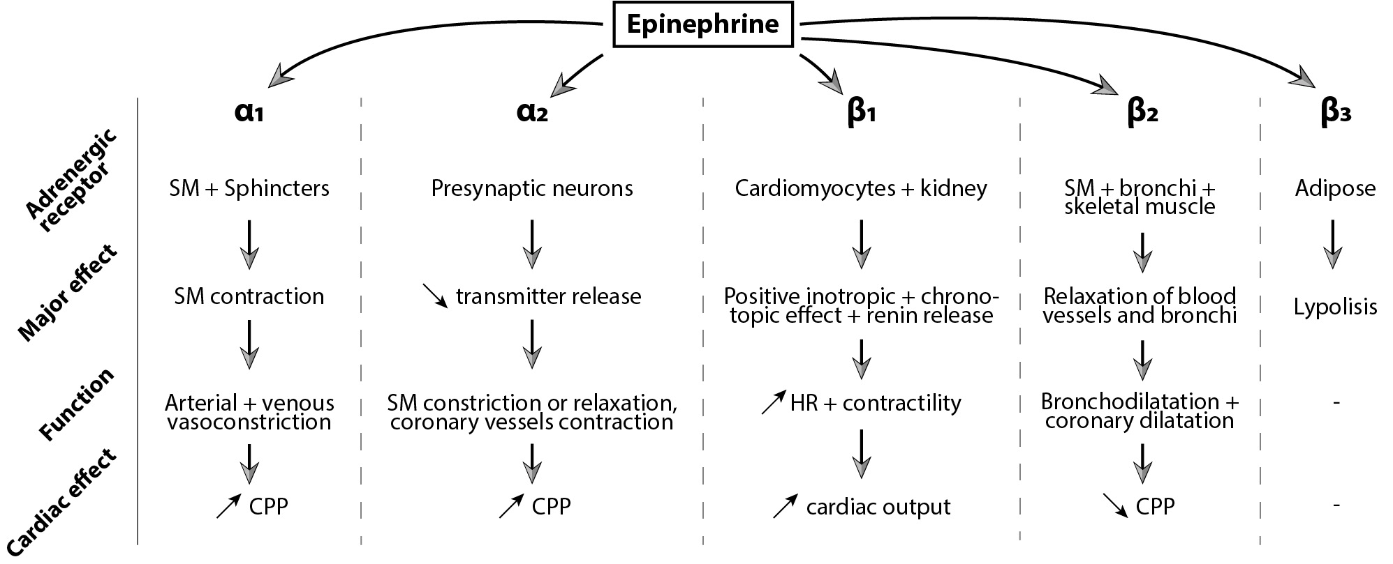
 α and ß adrenergic receptors. With 1 mg of epinephrine, α and ß adrenoreceptors will be activated, causing vasoconstriction, increasing aortic blood pressure and leading to higher CPP and cerebral perfusion pressure (CePP) [17, 18, 19]. Myocardial perfusion will improve, favoring the onset of ROSC. Epinephrine’s effects are summarized in Fig. 1.

Fig. 1.

Epinephrine’s effect mediated by α and ß adrenoreceptors. SM, smooth muscle; CPP, coronary perfusion pressure; HR, heart rate.

α1 receptors are mainly found in the smooth muscle cells of blood vessels and will induce smooth muscle contraction, resulting in peripheral arterial and venous vasoconstriction. α1 receptors are also located on cardiomyocytes, mediating contractility and vasoconstriction of the coronary arteries increasing CPP [20]. α2 receptors are predominantly present on presynaptic neurons. However, blood flow regulation through α2 adrenergic receptors is rather complex, as both pre and post-synaptic receptors play a role and either leads to vasoconstriction or vasodilatation [21]. In the heart, α2 receptors are mainly post synaptic on vascular smooth muscle cells and will induce coronary vasoconstriction [20].

ß adrenergic receptors have three subtypes, and their function varies according to the subtype and cell type. ß receptors are more sensitive to epinephrine than α receptors and are activated with lower doses. ß1 receptors are primarily located in cardiac tissue where they have a positive inotropic and chronotropic effect. ß2 receptors are predominant in smooth muscle cells of the bronchi and throughout the body. They induce bronchodilatation and smooth muscle relaxation [20, 22, 23]. ß1 and ß2 receptors make up 80% and 20% of ß adrenergic receptors expressed in a human heart, respectively. ß3 receptors have mainly metabolic effects, increasing fat oxidation and improving insulin-mediated glucose uptake but can also have a negative inotrope effect by antagonizing ß1 and ß2 effects [24, 25]. Thus, low-dose epinephrine can induce hypotension by activating ß2 receptors in smooth muscle cells of peripheral arteries. Higher doses of epinephrine will lead to peripheral vasoconstriction and positive inotropic and chronotropic effects.

2.1.2 Epinephrine Dose in Cardiac Arrest

Since the first CA guidelines in 1974, the American Heart Association (AHA) has recommended a 1 mg dose of epinephrine. This recommendation has persisted for decades, though the grounding evidence is based on a handful of animal studies from the early and mid-1900s. In 1906, Crile and Dolley [26] compared different resuscitation techniques and described the use of 1 to 2 mg of epinephrine in dogs. They found that cardiac compressions associated with ventilation and epinephrine improved survival rates. Later, Pearson and Redding [27, 28] concluded that intracardiac use of 1 mg of epinephrine on an asphyxia dog model of CA improved survival when 9 out of 10 animals survived in the group treated with epinephrine compared to 1 out of 10 in the group treated without epinephrine. Based on these data, 1mg became the recommended dose.

Epinephrine’s α1 effects may be dose-dependent, meaning that higher doses of epinephrine may improve CPP and therefore ROSC. Therefore, the idea of higher epinephrine doses during CPR was raised. Soon, the first few animal studies using high-dose epinephrine (HDE) (over 5 mg/bolus) were published and showed some encouraging results with higher myocardial blood flow, CePP, and ROSC [17, 29, 30]. During the 1990s, several double-blinded randomized controlled clinical trials (RCT) compared HDE (5 to 7 mg) to the standard 1 mg dose [31, 32, 33]. They found no significant difference between the HDE and standard-dose groups for survival at one hour (18% vs. 23% respectively, p = 0.12) and survival to discharge (3% vs. 5% respectively, p = 0.38) [31]. As summarized in a meta-analysis, HDE was significantly associated with a higher rate of ROSC but not with hospital discharge. Although the physiopathology remains unclear, HDE may worsen the neurological outcome of patients. In 2000, the International Liaison Committee on Resuscitation (ILCOR) formally recommended against HDE.

Clinical studies evaluating doses of epinephrine less than 1 mg are scarce. In 2018, Fisk et al. [34] published a before-after study with low (0.5 mg) vs. standard epinephrine (1 mg) in OHCA. In the “before” period, paramedics gave 1 mg of epinephrine at 4 min of CPR, followed by an additional 1 mg-dose every eight minutes to patients with a shockable rhythm and 1 mg-dose every two minutes to patients with a non-shockable rhythm. The “after” period used 0.5 mg of epinephrine instead of 1 mg [34]. Survival was not different between groups. This study modified the interval between epinephrine administrations as well as the dose, making it difficult to draw conclusions about the effect of lower doses.

A recent animal study compared low dose epinephrine to the standard 1 mg dose. CPP was significantly lower with a bolus of 0.25 mg compared to 1 mg, whereas with a 0.5 mg bolus CPP was not significantly different [35]. Cerebral near-infrared spectroscopy (NIRS) was used to monitor brain oxygenation and decreased rapidly with higher epinephrine doses. After 32 minutes of CPR, NIRS was 42% (39.5; 59.5) for 0.25 mg; 36% (32; 49) for 0.5 mg, and 32% (29.5; 43.5) for 1 mg (p = non-significant). Further studies are needed to assess the effect on cerebral perfusion and neurological outcome comparing low vs. standard dose epinephrine.

2.1.3 Time to First Epinephrine Injection

For OHCA, early administration of epinephrine less than 10 minutes after the 911 call may be associated with improved outcomes, and any delay may be associated with harm [36]. In one study, there was a 10% decrease in the odds of hospital discharge with Cerebral Performance Category (CPC) of 1–2 for every one-minute delay between 911 call and vasopressor administration in OHCA [37]. A meta-analysis showed that early pre-hospital administration might increase the rate of ROSC, survival to discharge and favorable neurologic outcomes [36].

For IHCA, Donnino et al. [38] identified a stepwise decrease in survival with increasing time to epinephrine and non-shockable rhythms over 4 minutes. Another study identified that for shockable rhythms, early administration of epinephrine (2 minutes after first defibrillation) was associated with decreased odds of ROSC (p < 0.001) and survival with good functional outcome (p < 0.001) [39]. It is worth noting that over half of the patients received epinephrine within 2 minutes after the first defibrillation, which goes against current AHA guidelines. In shockable rhythms, use of epinephrine should not delay defibrillation and should be administered after the second electric shock [40]. Non-shockable rhythms have very limited treatment resources outside of epinephrine and benefit more from early administration, especially for OHCA [41, 42, 43].

2.1.4 Intervals for Epinephrine Administration

Epinephrine administration is recommended every 3 to 5 minutes. However, this recommendation is not based on studies with strong experimental designs according to the AHA [40]. In physiological settings, epinephrine’s half-life is 2–3 minutes with a peak plasma concentration of approximately 90 seconds [44]. Observational studies of OHCA have evaluated intervals between the administration of 1 mg of epinephrine, and the results have been controversial. Grunau et al. [45] found improved survival with favorable neurologic outcome when the average epinephrine interval was <3 minutes compared to longer intervals of 5 minutes or more. On the other hand, a recent Japanese observational study enrolling over 10,000 patients with OHCA showed no difference in 1-month neurologically favorable survival when comparing shorter vs. longer intervals [46].

2.1.5 Epinephrine’s Effect over Time

Epinephrine may lose effectiveness with repeated doses over time. During CPR, epinephrine may not have the same metabolism as in a physiological state and could have a longer half-life. A 1 mg dose of epinephrine is high compared to endogenous concentrations of epinephrine, and receptors may be quickly saturated, making subsequent boluses less effective.

In porcine models of CA, CPP increases significantly only after the first few epinephrine injections. In Wagner et al.’s study [47], animals were treated with either epinephrine or saline after inducing ventricular fibrillation (VF). Compared to the saline group, animals who received epinephrine had significantly higher CPP for the two first boluses. After the two first injections, CPP still increased but was not significantly different from the saline group [47]. Bar-Joseph et al. [48] showed a significant increase in CPP after the first injection of repeated doses of epinephrine (0.1 mg/kg) with an increase of more than 300% and no further increase for the three next boluses. In Jaeger et al.’s animal model [35] of VF CA, the CPP absolute increase observed after the first 1mg epinephrine dose was of 93% and decreased with each subsequent dose, down to 46% for the 5th dose.

2.1.6 Epinephrine vs. Placebo Studies

Some researchers have questioned the benefit of epinephrine during CA. In 2011, Jacobs et al. [49] published the first RCT comparing epinephrine vs. placebo. Although survival in both groups was not significantly different, ROSC rates were significantly higher with epinephrine compared to placebo. A few years later in 2018, Perkins et al. [6] published another RCT, the PARAMEDIC-2 study. Over 8000 patients were included to compare the effect of standard-dose epinephrine to placebo [6]. For their primary outcome of survival at 30 days, they found a small but significant improvement with epinephrine (3.2% vs. 2.4% respectively; unadjusted odds ratio (OR) = 1.39; 95% CI: 1.06–1.82). Once again, ROSC was higher in the epinephrine group than the placebo group (36.4% vs. 11.7%). However, for their secondary outcome of favorable neurologic outcome at 3 months, there was no significant difference (adjusted OR = 1.31; 95% CI: 0.94–1.82). Also, there were more patients in the epinephrine group who survived with severe neurological impairment (mRs score of 4 or 5): 31% (39/126) vs. 17.8% (16/90). But, 20 patients were lost to follow-up for neurologic analysis in the epinephrine group and 29 in the placebo group, representing respectively 16% and 32% of the patients surviving until hospital discharge. Moreover, it is important to highlight that the median time from 911 call to epinephrine administration was 21.5 minutes (16–27.3 minutes). Such a duration between collapse and the first drug administration might explain the lack of difference in survival with good neurological outcome. A secondary analysis combining data from those two RCTs showed that the benefit of epinephrine for ROSC was greater for non-shockable rhythms compared to shockable rhythms (OR = 6.52; 95% CI: 5.56–7.63 vs. OR = 2.32; 95% CI: 1.86–2.89, p < 0.001). Survival with good neurological outcomes was not significantly different between these subgroups [50].

2.1.7 Epinephrine’s Deleterious Effects

Despite its benefits, epinephrine may also cause harm [50, 51, 52, 53]. Epinephrine improves CPP but also increases vascular resistance by its α1 effect, leading to lower coronary flow despite higher CPPs [47]. An identical effect has been identified for cerebral perfusion pressure: epinephrine-induced vasoconstriction may increase perfusion pressure but may not result in higher blood flow or improved oxygenation [14, 54, 55]. The microcirculation may be particularly impaired by epinephrine during CPR. An animal study showed that cerebral cortical microcirculatory blood flow was significantly decreased despite an increase in arterial pressure. Microphotographs in this study demonstrated the disappearance of microvessels of less than 20 μm in the epinephrine-treated group until 5 to 7 minutes after ROSC [54].

Recently, a pediatric CA rat model showed that following CPR with standard epinephrine use, penetrating arterioles showed significant constriction compared to animals resuscitated without epinephrine [56]. The placebo-treated group showed a progressive increase in capillary diameter post-ROSC, whereas capillary diameter remained stable in the epinephrine group [56]. Animal studies demonstrating persisting effects of epinephrine on the cerebral circulation may provide mechanistic evidence for randomized trials that have found decreased favorable neurological survival among patients treated with epinephrine compared to placebo. On the other hand, a pediatric IHCA swine model of asphyxia demonstrated an increase in cerebral blood flow and cerebral tissue oxygenation with the first two doses of epinephrine [57]. Cerebral autoregulation may differ between these pediatric IHCA models and adult OHCA or VF models.

Further epinephrine may increase myocardial oxygen consumption due to ß1 chronotropy. Animal studies have shown an increased heart rate associated with higher myocardial oxygen consumption, lower oxygen delivery, and impaired myocardial contractility with epinephrine [58, 59, 60]. A regression analysis has also shown that the total epinephrine dose may be associated with post-resuscitation myocardial dysfunction. A total epinephrine dose over 5 mg was significantly associated with a lower left ventricular ejection fraction and higher left ventricular end-systolic diameter [61]. Moreover, epinephrine use may have adverse metabolic effects with increased systemic inflammation and vascular endothelial injury, potentially aggravating reperfusion injury and inducing a systemic inflammatory response [62].

2.2 Norepinephrine
2.2.1 Pharmacology and Rationale

Norepinephrine (NE) is an endogenous catecholamine that functions as a neurotransmitter in the sympathetic nervous system. NE is mainly an α adrenergic agonist with very little action on ß2 receptors. However, it is slightly less potent than epinephrine on α receptors in most organs, and its ß1 agonistic effect is also lower [63]. Therefore, NE has less of a positive chronotropic and inotropic effect, leading to lower myocardial oxygen demand and fewer arrhythmias. Despite these potential advantages, experiments using NE during CPR are scarce.

2.2.2 Norepinephrine in Experimental Studies

Some experimental studies have shown promising findings when using NE during CPR. NE doses ranged from 0.045 mg/kg to 0.16 mg/kg in the different reported studies [64, 65, 66]. In a VF CA model, NE compared to epinephrine drastically reduced the resuscitation duration from 11.1 ± 3.6 to 1.4 ± 0.3 minutes [64]. Compared to HDE, NE improved myocardial oxygen extraction rate and decreased myocardial lactate [65, 66].

This may be related to the reduced chronotropic effect of NE by lack of ß1 agonist effect. Another study showed higher myocardial blood flow (118.9 ± 73.1 vs. 62.2 ± 45.4 mL/min/100g, p = 0.04) with NE compared to HDE but no difference in the oxygen extraction ratio or survival. However, the NE group did demonstrate an increase in oxygen consumption, potentially counterbalancing its positive effects on oxygen delivery [67]. Brown et al. [68] evaluated the effect of three different doses of NE (0.08 mg/kg, 0.12 mg/kg, and 0.16 mg/kg) and HDE (0.2 mg/kg) on cerebral blood flow in twenty swine in VF. The two higher doses of NE showed improved cerebral blood flow (CBF) compared to epinephrine (p < 0.05). In another swine model of CA in VF, NE significantly improved the cerebral perfusion gradient compared to saline (4.3 ± 1.2 vs. 2.5 ± 1.2 kPa respectively, p < 0.05) as well as the total brain blood flow (45 ± 21 vs. 21 ± 12 mL/min/100 g, p < 0.05) but no difference was found between NE and HDE [69]. NE may increase cerebral blood flow dose-dependently. With a stronger α-adrenergic effect compared to epinephrine, it could lead to higher myocardial blood flow and thereby increase CBF.

2.2.3 Norepinephrine in Clinical Trials

Higher resuscitation rates with NE were found in a small clinical trial of 50 OHCA patients with VF. After the 3rd shock, patients received a blinded treatment of either one dose of 1 mg of NE or 1 mg of epinephrine, followed by epinephrine in both groups if no ROSC was observed. 16/25 patients in the NE group were resuscitated compared to 8/25 in the epinephrine group (p < 0.05). Ventricular dysrhythmias were less frequent in the NE group than in the epinephrine group (8/16 vs. 6/8, respectively) [70].

Only one pre-hospital RCT has been published comparing high-dose NE to HDE and standard epinephrine [71]. 816 patients were enrolled between 1990 and 1992. Paramedics treated nontraumatic OHCA with up to three doses of either 1 mg or 15 mg of epinephrine or 11 mg of NE. ROSC rates were not significantly different between high-dose NE and standard dose epinephrine (13% vs. 8%, p = 0.19). Survival rates at hospital discharge were not significantly different between HDE, standard dose epinephrine, and high-dose NE groups (1.7% vs. 1.2% vs. 2.6%, p = 0.83 and p = 0.37 respectively). Drawing conclusions about NE from this RCT is challenging and this study should be cautiously interpreted as it is over three decades old, and CA care has changed over time. Moreover, the NE dosage was extremely high (11 mg) compared to HDE.

Taken together, the evidence does not support routine NE use during CPR and requires further investigation. It is not recommended to use NE instead of epinephrine during CPR.

2.3 Vasopressin
2.3.1 Pharmacology and Rationale

Vasopressin is an antidiuretic hormone secreted by the neurohypophysis. Vasopressin binds two types of receptors, V1 and V2. V1 receptors have a non-adrenergic vasoconstrictor effect in the smooth muscle. This vasoconstrictor effect mainly concerns the renal, musculocutaneous and splanchnic vascular territories. However, its vasoconstrictive effects on coronary and cerebral circulation are theoretically limited and may result in improved myocardial and cerebral perfusion. V2 receptor activation induces an antidiuretic effect by increasing the medullary and cortical permeability of the kidney’s collecting tubule to water. Higher endogenous concentrations of vasopressin have been found in successfully resuscitated patients compared to those who died, arguing that vasopressin may play an important role in achieving ROSC and survival [72]. The effects of vasopressin during CA include increased inotropy and systemic vasoconstriction, and potentiation of catecholaminergic effects [73].

2.3.2 Vasopressin in Experimental Studies

Vasopressin has been used for decades in animal models of CA, alone or in combination with epinephrine. In both VF and pulseless electrical activity (PEA) models of CA, 0.4 IU/kg of vasopressin significantly improved left ventricular myocardial blood flow (52 ± 8 vs. 43 ± 5 mL/min/kg, p < 0.01), cerebral blood flow (51 (48–70) vs. 18 (10–23) mL/min/100 g, p < 0.05) and cerebral oxygenation extraction ratio (0.38 (0.25–0.44) vs. 0.47 (0.41–0.57), p < 0.05) when compared to HDE [74, 75, 76]. Another VF model also showed improved CPP with a high dose of vasopressin (0.8 U/kg) when compared to standard doses of epinephrine at 2 and 4 minutes after drug administration [77]. Vasopressin’s vasoconstrictor effect lasted longer than epinephrine’s during CPR, up to 30 minutes after drug administration [75, 78]. ROSC rate tended to be more frequent in the vasopressin group compared to placebo, but the difference was non-significant [78].

The effects of vasopressin on survival are more controversial. While some studies showed an increase in survival [74, 79, 80], others found that vasopressin use was not associated with ROSC or survival [77, 81]. As a potent vasopressor, vasopressin may be responsible for lower cardiac index and myocardial contractility during the post resuscitative phase. Indeed, Prengel et al. [82] have demonstrated that cardiac index and myocardial contractility were significantly lower during the first 15 minutes after defibrillation in animals treated with vasopressin vs. epinephrine.

2.3.3 Vasopressin in Clinical Trials

None of the major RCTs demonstrated a benefit from the administration of vasopressin alone. A trial including 200 patients with IHCA failed to show any difference between the group treated with an initial first dose of vasopressin (40 IU) and the group treated with epinephrine. In this study, the vasopressin group received epinephrine as a second drug if ROSC did not occur. Rates of ROSC, hospital discharge, and thirty-day survival were similar in both groups [83]. In 2004, an RCT with OHCA found an increased hospital discharge rate among patients with asystole treated with vasopressin instead of epinephrine (4.7% vs. 1.5%, p = 0.04). Patients were randomized to receive either two doses of 40 IU of vasopressin or two doses of 1 mg of epinephrine, followed by additional epinephrine if no ROSC occurred. Comparing patients who needed additional treatment with epinephrine (after the two first doses of vasopressin or epinephrine), survival to hospital discharge was significantly higher for patients in the vasopressin group regardless of initial rhythm (6.2% vs. 1.7%, p = 0.002) [84]. This suggested that vasopressin may be more effective if given with epinephrine rather than alone. In 2008, 2894 OHCA patients were randomized in a clinical trial to receive a combination of epinephrine and vasopressin (40 IU) or epinephrine with placebo. There were no significant differences between the combination therapy and the epinephrine-only group in ROSC, survival to hospital admission, or 1-year survival [85]. Finally, a meta-analysis of six RCTs determined that vasopressin did not improve overall rates of ROSC or long-term survival. However, in a subgroup analysis of patients with asystole, vasopressin was associated with higher long-term survival [86].

Unfortunately, the encouraging effects of vasopressin on cardiac and cerebral blood flow shown in experimental studies have not translated to survival or neurologic outcomes in clinical trials. Further research is needed on the mechanisms of vasopressin’s action during CA. The use of vasopressin, whether in place of or in combination with epinephrine, is not recommended during CPR by international resuscitation guidelines.

2.4 In Summary

Epinephrine remains the only recommended vasopressor in CA, with a class 1 recommendation. A dose of 1 mg for adults should be administered every 3 to 5 minutes after an intra-venous or intra-osseous line is available in patients with non-shockable rhythms and after defibrillation in those with shockable rhythms [40].

3. Antiarrhythmics

Pulseless ventricular tachycardia (VT) and VF are the most treatable rhythms in OHCA and portend a better prognosis than PEA or asystole [1, 87]. However, a quarter of VT/VF patients have an arrhythmia that is refractory to defibrillation [88]. Antiarrhythmic drugs aim to terminate VT/VF, restore a perfusing rhythm, and achieve sustained ROSC by increasing defibrillation’s rate of success. Although amiodarone and lidocaine have shown an improvement in rate of ROSC and survival to hospital admission, this has not translated to higher neurologically intact survival [7, 89, 90, 91, 92, 93].

3.1 Amiodarone and Lidocaine
3.1.1 Pharmacology and Rationale

Amiodarone is a class III antiarrhythmic agent. Its main effects are α and ß blocking properties as well as blocking potassium, sodium, and calcium channels [94]. Lidocaine is a class Ib antiarrhythmic agent. It will prolong conduction velocity and shortens the duration of the action potential and the effective refractory period [95].

3.1.2 Amiodarone and Lidocaine in Clinical Trials

Kudenchuk et al. [93] randomized 504 patients with refractory VT/VF arrest to amiodarone vs. placebo, finding a higher survival to hospital admission with amiodarone as compared to placebo (44 vs. 34%; p = 0.03). The Amiodarone vs. Lidocaine in Prehospital Ventricular Fibrillation Evaluation (ALIVE) trial compared lidocaine to amiodarone administration in refractory VF arrest in 347 patients [96]. Concordant to the study by Kundenchuk et al. [93], amiodarone led to a higher survival to hospital admission (22.8 vs. 12.0%; p = 0.009) [96]. This study was not powered to detect differences in hospital discharge. Therefore, the Amiodarone Lidocaine or Placebo Study (ALPS) trial was published in 2016. The ALPS trial randomized 3026 patients with OHCA to amiodarone, lidocaine, or placebo and did not find a higher survival to discharge or neurologically intact survival with the antiarrhythmic drugs as compared to placebo [7]. This has been consistent across several systematic reviews and meta-analyses that include the data from the trials just mentioned [89, 90, 91, 92].

Time to treatment is an important determinant of outcomes and may help explain why there has been a lack of association between antiarrhythmic drugs and survival in OHCA [97]. In the ALPS study, there was a significant interaction between witnessed OHCA and survival benefit from antiarrhythmic drugs, with a 5% survival benefit over placebo for both lidocaine and amiodarone in the witnessed OHCA group but no benefit in the unwitnessed arrest group [7]. Time to drug administration is hard to quantify in unwitnessed arrests, and prompt drug administration is difficult in CA trials, with a mean time to medication administration of approximately 19 minutes after emergency medical services activation [7]. A more recent study utilized a Bayesian approach to reanalyze the ALPS data and suggested a 2.9% overall survival benefit for amiodarone (interquartile range (IQR) 1.4–3.8%) and 1.7% for lidocaine (IQR 0.8–3.2%) as compared to placebo [98]. This study also showed that amiodarone offered a strong probability (96%–99%) to improve neurological outcomes in refractory VT/VF patients, higher than for lidocaine or placebo (96%) [98] and as such amiodarone should be strongly considered till further data emerge for clinical use. There is a positive effect with the use of antiarrhythmic drugs as suggested by the consistent trend towards improved survival which may justify its use [92].

Given the potential benefit, current guidelines give amiodarone and lidocaine a 2b indication for refractory VT/VF arrest [43].

3.2 Magnesium

Magnesium does not increase ROSC or survival in VT/VF arrest [90, 99, 100, 101] or undifferentiated rhythms [102, 103]. However, case series have shown its utility in suppressing and preventing Torsades de Pointes [104]. Torsades de Pointes occurs in cases where there is a prolonged QT and often bradycardia leading to, a longer QTc reflective of a prolonged refractory period, in which an early after depolarization occurs, initiating the tachycardia. Magnesium leads to suppression of early afterdepolarizations [105, 106]. Magnesium is not recommended routinely in CA management.

3.3 Beta-Blockers

Patients in CA have high levels of catecholamines, and the activation of ß1 and ß2 receptors increases myocardial oxygen demand and potentially worsens ischemic injury. Given the main cause of polymorphic VT is ischemia, ß-blockers are recommended to treat ischemia and have shown that in this setting, they reduce the risk of ventricular arrhythmias [107]. The use of esmolol in combination with epinephrine highlights the role of ß1 adrenergic effects in myocardial dysfunction. When blocked with esmolol, post-resuscitation contractile function and left ventricular diastolic function improved, and recurrence of VF was reduced compared to epinephrine alone [60, 108, 109]. Esmolol has a protective effect on ischemia and reperfusion injury induced by epinephrine [110]. In patients with CA, the use of esmolol was explored in a small study (N = 25). When compared to placebo, esmolol had a trend towards improved survival though no specific benefit in terms of neurological outcomes [8]. The use of non-selective ß-blockers was found to be superior in terminating electrical storms and recurrence of ventricular arrhythmia, with patients treated with metoprolol and amiodarone being 77% less likely to have a termination of arrhythmic events when compared to those treated with propranolol and amiodarone [111]. More research is needed, and guidelines only make recommendations regarding the use of ß-blockers in the setting of cardiac ischemia [40].

Other antiarrhythmic medications including bretylium tosylate, and procainamide will not be discussed in this review.

4. Other Cardiac Arrest Drugs

We will first present here different drugs evaluated in CA. First, calcium and sodium bicarbonate have been drugs originally recommended in the first edition of the AHA recommendations of 1974. Multiple RCT’s have since shown their lack of benefit. Corticosteroids have been investigated more recently and more specifically in association with vasopressin.

Finally, we will also highlight the promising results of an investigational drug, sodium nitroprusside, over the last few years.

4.1 Calcium
4.1.1 Pharmacology and Rationale

The use of calcium in CA was recommended by the AHA in their first edition in 1974 [112]. The rationale behind this recommendation was that by increasing the calcium concentration, an ion involved in all muscle cells, contractility of the heart would be increased and defibrillation would be more successful. In healthy hearts, calcium has been shown to increase cardiac index and left ventricular stroke work [113]. During the cardiac cycle, an increase in intracellular calcium concentrations released mainly by the sarcoplasmic reticulum, allow for myocardial contraction.

4.1.2 Calcium in Clinical Trials

The use of calcium in CA was first described in a case series from 1951 in pediatric cardiac surgery when intracardiac calcium administration resulted in ROSC in four patients [114]. In 1985, two small randomized trials in OHCA showed a higher ROSC rate in the calcium-treated group, although the difference was non-significant [115, 116]. After controversial results from several observational studies [117, 118, 119, 120], a recent multicentric RCT was stopped early due to concern about harm in the calcium group. 383 patients with OHCA were randomized to receive either calcium chloride or placebo after the first dose of epinephrine [121]. 19% had ROSC in the calcium group compared to 27 in the placebo group (p = 0.9). At 30 days, 5.2% in the calcium group and 9.1% in the placebo group were alive (p = 0.17). Also, among patients with ROSC, 74% in the calcium group had hypercalcemia vs. 2% in the saline group. Forty percent of the patients receiving calcium had a calcium lever after ROSC between 1.47 and 2.00 mmol/L vs none in the placebo group. A secondary analysis assessed the long-term outcome of these patients. After one year, 3.6% of patients were alive with a favorable neurological outcome in the calcium group vs. 8.6% in the saline group (Relative Risk (RR): 0.42; 95% CI: 0.18–0.97) [122]. A systematic review published in December 2022 including 3 RCTs showed no benefit with calcium administration and possible harm with unfavorable neurologic outcome at 90 days [123]. The possible mechanism behind this finding may be that the anaerobic state of CPR triggers an influx of calcium by the Na/Ca exchanger [124]. Intracellular calcium level rises even more when reperfusion occurs and calcium accumulates in the mitochondria contributing to myocardial cell death [125]. Administering supplemental calcium may induce a calcium overload and hasten cell death.

Calcium is no longer recommended during CPR outside of special circumstances such as hypocalcemia and hyperkalemia, and may be considered in β-blocker or calcium channel blocker overdose [126].

4.2 Sodium Bicarbonate
4.2.1 Pharmacology and Rationale

Sodium bicarbonate (SB) (NaHCO3) is a salt that increases pH and corrects metabolic acidosis by utilizing the bicarbonate buffer system [127]. First, it dissociates into sodium and bicarbonate (HCO3-) ions. The bicarbonate ion then binds free hydrogen ions, becoming carbonic acid (H2CO3), which then converts into water and carbon dioxide, which is removed from the body via the respiratory system. The more HCO3- ions available, the more H+ ions can be turned into H2O, increasing pH [127].

During CA and CPR, no or low systemic blood flow result in a build-up of byproducts of anaerobic metabolism and ineffective removal of carbon dioxide which together lead to systemic acidosis. The harmful effects of severe acidosis include vasodilatation, protein denaturation, impaired ATP production, predisposition to arrhythmias, and a depressed response to vasopressors [128, 129]. The hypothesis of using an alkalizing agent, such as SB, is to buffer hydrogen ions and thus increase the chances of successful resuscitation [130].

The administration of SB during CA was listed as the first-line management in the original 1974 published guideline of Cardiac Care by the AHA [131]. Until the early 1980s, SB was used in about 85% of IHCA [132]. During the following years, evidence arose questioning the beneficial effects of SB.

Adverse effects of SB administration are mainly metabolic alkalosis. There are also concerns for exacerbating hypernatremia and therefore hyperosmolarity and reduced systemic vascular resistance compromising CPP. Also the excessive CO2 production could contribute to and increase intracellular acidosis [133, 134].

4.2.2 Sodium Bicarbonate in Clinical Trials

Several clinical studies have evaluated the use of SB in CA but have reported mixed results [135, 136, 137, 138, 139]. In a prospective, randomized controlled study from 2006, Vukmir et al. [140] found a trend toward improved survival rates in patients with prolonged CA (>15 minutes) that did not reach statistical significance and no difference overall in survival rates to hospital admission in patients receiving SB vs. placebo. More recently, Kim et al. [135] published a retrospective, observational, case-control study and found an association between SB and ROSC within 20 minutes after hospital admission. A smaller study by Ahn et al. [136] showed that the administration of SB in prolonged OHCA was not associated with any improvement in survival nor increased neurological outcomes at 1 or 6 months.

Other retrospective studies and meta-analyses have not demonstrated benefit with sodium bicarbonate and have found evidence of possible harm. A study analyzed patients from France and North America. In the North American dataset, 20.6% of patients were treated with SB and the use of SB was associated with a lower likelihood of favorable functional outcomes at hospital discharge. In the French dataset, only 2.2% of patients received SB and SB was not associated with higher survival with favorable neurological outcome [139]. Recently, Alshahrani et al. [141] published a meta-analysis investigating the effects of SB in CA, including 14 studies (4 RCT and 10 observational studies) with over 28,000 patients. They showed that SB was associated with poorer rates of ROSC and good neurological outcome at discharge [141].

Since 2010, AHA officially advised against SB use during routine CA except in cases of hyperkalemia or tricyclic antidepressant overdose due to evidence consistently showing outcomes were either the same as or worse than without SB administration [142].

4.3 Corticosteroids
4.3.1 Pharmacology and Rationale

It is well known that reperfusion after ROSC induces a pro-inflammatory response with a rise in inflammatory biomarkers [143]. A cytokine storm with activation of platelets and leukocytes and vasodilatory shock are common in the post-cardiac arrest syndrome [144, 145]. In other situations, corticosteroids have been shown to reduce the pro-inflammatory response, restore effective blood volume by increasing mineralocorticoid activity, increase systemic vascular resistance, and even improve survival in septic shock [145, 146, 147, 148]. Also, post-resuscitation cortisol production may be compromised because of reperfusion injury to adrenal tissues.

4.3.2 Corticosteroids in Clinical Trials

For these reasons, it may be logical to consider corticosteroids in the management of CA. However, data from clinical trials have not been convincing. In a recent study, the use of corticosteroids during IHCA CPR was not associated with hemodynamic improvement or lower inflammatory biomarkers as compared to placebo [149]. Very few papers have compared the use of steroids to epinephrine. Apart from an increase in ROSC rate in a nonrandomized, non-blinded study, there was no benefit on outcomes with the use of corticosteroids during CPR [150, 151, 152].

A few RCTs have investigated the use of corticosteroids (40 mg of methylprednisolone) in combination with vasopressin (20 IU) and epinephrine (1 mg) compared to epinephrine alone. Three of them showed an increase in ROSC rate with that combination of drugs [153, 154, 155]. Two also showed an increase in survival to hospital discharge for IHCA, with the most recent study demonstrating a higher survival with favorable neurological outcome [153, 154, 155]. A meta-analysis including those three studies found an OR of 2.09 (95% CI: 1.54–2.84) for ROSC and of 1.64 (95% CI: 0.99–2.72) for favorable neurological outcomes when administering vasopressin with steroids and epinephrine vs. epinephrine and placebo [156]. A more recent study published in 2022 confirmed these findings that the addition of corticosteroid and vasopressin after the first dose of epinephrine improved rate of ROSC but had no effect on long-term survival when compared to epinephrine alone [157].

In summary, the use of corticosteroids during CPR may be considered (recommendation 2b) but has unsure benefits.

4.4 Sodium Nitroprusside
4.4.1 Pharmacology and Rationale

Sodium nitroprusside (SNP) has emerged as a potential paradigm shift in the medical management of CA. While traditional pharmacologic approaches with vasopressors rely on improving systemic pressures in order to improve the CPP, SNP is a potent vasodilator that results in both large and small vessel relaxation through the release of nitric oxide [158]. Indeed, the ischemia-reperfusion injury resulting from the low-flow state of CA and CPR is linked to marked endothelial dysfunction. Troelsen et al. [159] have demonstrated that coronary arteries in rats suffering from CA have reduced vasodilatory capacity after being challenged with acetylcholine when compared to controls. Moreover, the concentration of vascular adhesion molecules like P-selectin and von-Willebrand factor is increased after CA [159]. Animals suffering from CA experience marked decreases in the neuronal nitric oxide synthase levels [160, 161] and thus a limited capacity for nitric oxide-mediated vasodilation and blood flow regulation. Knock-out of the nitric oxide synthase gene before the induction of CA is also associated with worse outcomes, including a decreased ROSC rate and worse left-ventricular function [162]. These findings suggest a potential benefit in CA from medications targeting nitric oxide like SNP.

4.4.2 Sodium Nitroprusside in Experimental Studies

This hypothesis has been successfully validated and replicated in multiple animal studies using the porcine model of CA. SNP-enhanced CPR has been shown to markedly improve carotid blood flow in a porcine model of CA when compared to epinephrine. In addition, SNP treatment improved the metabolic derangement observed in CA, as these animals had improved arterial pH [163, 164]. Left ventricular diastolic function after ROSC appears to also be improved as animals treated with SNP was shown to have decreased left ventricular septal wall thickness and increased cavity diameter [165]. Cerebral histology from SNP-treated CA animals demonstrated decreased ischemic brain injury [166]. These findings were translated to increased 24-hour survival and improved neurologic status and cerebral performance capacity [11, 163, 164, 165, 167].

While SNP is generally considered to be anti-hypertensive, systemic blood pressures were maintained in these experiments. SNP exerts its effects on both the systemic and pulmonary vasculature as it has been demonstrated to reduce hypoxic pulmonary vasoconstriction and thus augment the left ventricular preload and CPR cardiac output. Hypoxic vasoconstriction reversal leads to intrapulmonary shunts and concomitant increases in the alveolar-arterial oxygen gradient. The resulting hypoxia was shown to respond to supplemental oxygen [168]. Vasodilation is also observed in the cerebral microvasculature, as SNP-treated animals had an increased number of perfused microvessels, greater arteriolar diameter, and a higher microvascular flow index [169]. The hemodynamic effects of SNP are observed in animals receiving either conventional [11, 166, 167] or extracorporeal CPR [168]. These findings lay the framework for a clinical trial design testing SNP in human CA patients.

Recommendations of drugs for adult CA are summarized in Table 1 with their respective class of strength and level of evidence (Table 2).

Table 1.Drug administration for adults during CPR.
Drug COR LOE Recommendation
Epinephrine 1 B-R Use is recommended in CA
- 1 mg every 3–5 min 2a B-R Reasonable to administer 1 mg every 3–5 minutes
Norepinephrine No recommendations
Vasopressin 2b C-LD May be considered alone or with epinephrine but offers no advantage as substitute for epinephrine
Amiodarone 2b B-R May be considered for VF or pVT if unresponsive to 3 defibrillations
Lidocaine 2b B-R
Magnesium 3 B-R Routine use not recommended-no benefit
Beta-blockers No recommendations
Calcium 3 B-NR Routine use not recommended-no benefit
Sodium Bicarbonate 3 B-R Routine use not recommended-no benefit
Sodium Nitroprusside No recommendations
Corticosteroids 2b C-LD Use during CPR is of uncertain benefit

COR, Class of recommendation and LOE, level of evidence (adapted from 2020 AHA guidelines for cardiopulmonary resuscitation and emergency cardiovascular care, Circulation, 2020). The colors of the drug correspond to the class of recommendation (Please see Table 2). CA, cardiac arrest; VF, ventricular fibrillation; pVT, pulseless ventricular tachycardia; CPR, cardiopulmonary resuscitation; AHA, the American Heart Association.

Table 2.Class of recommendation and level of evidence.
Class of recommendation: Level of evidence:
1: Strong recommendation, benefit >>> risk B-R: randomized, moderate quality evidence from 1 or more randomized controlled trial
2a: Moderate recommendation, benefit >> risk
2b: Weak recommendation, benefit risk B-NR: nonrandomized, moderate quality evidence from 1 or more well-designed, well executed nonrandomized, observational or registry studies
3: No benefit, benefit = risk or harm C-LD: limited data, randomized or nonrandomized observational or registry studies with limitations of design or execution
5. Pediatric Cardiac Arrest

Drugs are used during pediatric CA to improve CPR hemodynamics, restore a perfusing rhythm, and address reversible causes. Because children have distinct differences in physiology and reasons for arrest compared to adults, it is worthwhile to review pediatric-specific literature. It is important to note that the evidence in pediatrics is primarily derived from IHCA. Guidelines are summarized in Table 3.

Table 3.Drug administration during pediatric cardiac arrest during CPR.
Drug COR LOE Recommendation
Epinephrine 2a C-LD It is reasonable to administer epinephrine. IV/IO route preferred to ETT
It is reasonable to administer initial dose within 5 minutes
It is reasonable to administer epinephrine every 3–5 minutes
Amiodarone 2b C-LD Amiodarone or lidocaine may be used for shock resistant VF or pulseless VT
Lidocaine
Sodium Bicarbonate 3: HARM B-NR Routine administration is not recommended (in the absence of hyperkalemia or sodium channel blocker toxicity)
Calcium 3: HARM B-NR Routine administration is not recommended (in the absence of hypocalcemia, hyperkalemia, hypermagnesemia, or calcium channel blocker overdose)

COR, Class of recommendation and LOE, level of evidence (adapted from 2020 AHA guidelines for cardiopulmonary resuscitation and emergency Cardiovascular care; Circulation; 2020). The colors of the drug correspond to the class of recommendation (Please see Table 2). IV, intra-venous; IO, intraosseous; ETT, endo-tracheal tube; VF, ventricular fibrillation; VT ventricular tachycardia; AHA, the American Heart Association.

5.1 Epinephrine

Epinephrine is the pharmacologic cornerstone of pediatric CPR and has been implemented in resuscitation for over 100 years [26]. However, there are controversies and knowledge gaps regarding timing, dose and dosing intervals, and long-term outcomes [170].

The strongest evidence for timing of the initial dose comes from a series of “Time to Epinephrine” studies using the AHA Get With the Guidelines registry. For pediatric patients IHCA with initially non-shockable rhythms, Andersen et al. [171] showed that earlier epinephrine (within the first 2 minutes) was associated with ROSC, higher survival to discharge, and neurologically favorable survival. Delays to first epinephrine linearly decreased the chance of a good outcome.

What dose should be given for pediatric CA? Perondi and colleagues [172] conducted a randomized, double-blinded trial of standard (0.01 mg/kg) vs. high dose (0.1 mg/kg) epinephrine, administered as a rescue after a single failed dose of standard epinephrine. The trial found no benefit and possible harm with high doses [172, 173]. Lower doses of epinephrine are often used and may be beneficial during the peri-arrest period, but there is no data to support lowdose epinephrine during CPR [174, 175].

How often should epinephrine be given? Current guidelines are grounded by a practical approach that allows providers to sync epinephrine doses with pulse checks, compressor changes, and defibrillation. There are no randomized trials, and observational data are conflicting regarding epinephrine dosing intervals [176, 177]. Kienzle et al. [176] used documented epinephrine times taken directly from code sheets. In her study, a quarter of patients received “frequent” epinephrine (given every 2 minutes), and these patients had higher ROSC and favorable neurologic survival [176]. This was a time-mediated phenomenon whereby the beneficial effect of frequent epinephrine was largely mediated by shortening the duration of CPR. Further supporting this finding, the ICU-RESUS trial, a hybrid cluster-randomized interventional trial across 18 US pediatric intensive care units (ICUs) and cardiovascular intensive care units (CVICUs) comprising a novel patient-centric CPR improvement bundle, also found that frequent epinephrine use was associated with improved outcomes in children with IHCA [178].

For now, guidelines for epinephrine use in pediatric patients with CA are weak to moderate recommendations based on limited data [170, 179, 180] (Table 3). To summarize, “it is reasonable” to administer 0.01 mg/kg of epinephrine during CA, with the first dose given within 5 minutes or as early as possible and subsequent doses in 3–5 minutes intervals. Further study is needed to understand the impact of epinephrine on long-term outcomes.

5.2 Anti-Arhythmics

The use of amiodarone and lidocaine for shock-resistant VF and pVT in children with CA should not be extrapolated from adult data, because the etiologies of pediatric dysrhythmias (i.e., congenital heart disease, inherited conduction abnormalities, myocarditis, and cardiomyopathies) differ from those in adults (primarily coronary artery disease) [181]. The evidence in pediatrics is limited to two observational cohorts of IHCA from the Get With the Guidelines registry [182, 183]. In brief, there were no differences in survival to hospital discharge or favorable neurologic outcomes among children receiving amiodarone or lidocaine for a shockable rhythm.

5.3 Calcium and Sodium Bicarbonate

Calcium and sodium bicarbonate are commonly used during pediatric CA but are not recommended outside of specific scenarios due to their association with harm [170, 179]. Secondary analyses of the recent multicenter ICU-RESUS trial found that sodium bicarbonate and calcium were used in 40–50% of all CA and were each independently associated with mortality and poor neurologic outcome [184, 9]. Registry data similarly found that calcium and bicarbonate were each associated with worse outcomes, longer duration of CPR, greater illness severity, and use of other advanced life support interventions [10, 185]. Although there is biological plausibility that calcium and bicarbonate could cause harm, it remains unclear whether these drugs have a true treatment effect or are simply associated with “last ditch” efforts in challenging resuscitations.

6. Conclusions

Despite many remaining gaps, epinephrine, and to a lesser extent amiodarone and lidocaine, stand as the only drugs recommended during resuscitation by international guidelines. Epinephrine administration remains recommended every 3 to 5 minutes at a standard dose of 1mg for adults and 0.01 mg/kg for pediatric CA. Although routinely used, calcium and sodium bicarbonate are not recommended except for specific circumstances and may cause harm. Further investigations are needed regarding the use of other vasopressors, beta-blockers, or sodium nitroprusside during CPR in clinical settings.

Author Contributions

DJ: Conceptualization, Data curation, Resources, Writing – original draft, and Writing – review & editing. AM, MK, AG, CG: Conceptualization, Writing – original draft, and Writing – review & editing. DO: Data curation, Writing – original draft and Writing – review & editing. TC: Conceptualization, Supervision, Writing – review & editing. DY: Conceptualization, Project administration, Supervision, Writing – review & editing. All authors read and approved the final manuscript. All authors have participated sufficiently in the work and agreed to be accountable for all aspects of the work.

Ethics Approval and Consent to Participate

Not applicable.

Acknowledgment

Not applicable.

Funding

This project was supported by the German Research Foundation (GA 3379/1-1).

Conflict of Interest

The authors declare no conflict of interest.

References
[1]
Tsao CW, Aday AW, Almarzooq ZI, Alonso A, Beaton AZ, Bittencourt MS, et al. Heart Disease and Stroke Statistics-2022 Update: A Report From the American Heart Association. Circulation. 2022; 145: e153–e639.
[2]
Gräsner JT, Wnent J, Herlitz J, Perkins GD, Lefering R, Tjelmeland I, et al. Survival after out-of-hospital cardiac arrest in Europe - Results of the EuReCa TWO study. Resuscitation. 2020; 148: 218–226.
[3]
Holmberg MJ, Vognsen M, Andersen MS, Donnino MW, Andersen LW. Bystander automated external defibrillator use and clinical outcomes after out-of-hospital cardiac arrest: A systematic review and meta-analysis. Resuscitation. 2017; 120: 77–87.
[4]
Ruan Y, Sun G, Li C, An Y, Yue L, Zhu M, et al. Accessibility of automatic external defibrillators and survival rate of people with out-of-hospital cardiac arrest: A systematic review of real-world studies. Resuscitation. 2021; 167: 200–208.
[5]
Downing J, Al Falasi R, Cardona S, Fairchild M, Lowie B, Chan C, et al. How effective is extracorporeal cardiopulmonary resuscitation (ECPR) for out-of-hospital cardiac arrest? A systematic review and meta-analysis. The American Journal of Emergency Medicine. 2022; 51: 127–138.
[6]
Perkins GD, Ji C, Deakin CD, Quinn T, Nolan JP, Scomparin C, et al. A Randomized Trial of Epinephrine in Out-of-Hospital Cardiac Arrest. The New England Journal of Medicine. 2018; 379: 711–721.
[7]
Kudenchuk PJ, Brown SP, Daya M, Rea T, Nichol G, Morrison LJ, et al. Amiodarone, Lidocaine, or Placebo in Out-of-Hospital Cardiac Arrest. The New England Journal of Medicine. 2016; 374: 1711–1722.
[8]
Driver BE, Debaty G, Plummer DW, Smith SW. Use of esmolol after failure of standard cardiopulmonary resuscitation to treat patients with refractory ventricular fibrillation. Resuscitation. 2014; 85: 1337–1341.
[9]
Cashen K, Sutton RM, Reeder RW, Ahmed T, Bell MJ, Berg RA, et al. Calcium use during paediatric in-hospital cardiac arrest is associated with worse outcomes. Resuscitation. 2023; 185: 109673.
[10]
Raymond TT, Stromberg D, Stigall W, Burton G, Zaritsky A, American Heart Association’s Get With The Guidelines-Resuscitation Investigators. Sodium bicarbonate use during in-hospital pediatric pulseless cardiac arrest - a report from the American Heart Association Get With The Guidelines(®)-Resuscitation. Resuscitation. 2015; 89: 106–113.
[11]
Schultz J, Segal N, Kolbeck J, Caldwell E, Thorsgard M, McKnite S, et al. Sodium nitroprusside enhanced cardiopulmonary resuscitation prevents post-resuscitation left ventricular dysfunction and improves 24-hour survival and neurological function in a porcine model of prolonged untreated ventricular fibrillation. Resuscitation. 2011; 82: S35–S40.
[12]
Paravati S, Rosani A, Warrington SJ. Physiology, Catecholamines. 2022. Available at: https://www.ncbi.nlm.nih.gov/books/NBK507716/ (Accessed: 14 January 2023)
[13]
Clutter WE, Bier DM, Shah SD, Cryer PE. Epinephrine plasma metabolic clearance rates and physiologic thresholds for metabolic and hemodynamic actions in man. The Journal of Clinical Investigation. 1980; 66: 94–101.
[14]
Hardig BM, Götberg M, Rundgren M, Götberg M, Zughaft D, Kopotic R, et al. Physiologic effect of repeated adrenaline (epinephrine) doses during cardiopulmonary resuscitation in the cath lab setting: A randomised porcine study. Resuscitation. 2016; 101: 77–83.
[15]
Burnett AM, Segal N, Salzman JG, McKnite MS, Frascone RJ. Potential negative effects of epinephrine on carotid blood flow and ETCO2 during active compression-decompression CPR utilizing an impedance threshold device. Resuscitation. 2012; 83: 1021–1024.
[16]
Chase PB, Kern KB, Sanders AB, Otto CW, Ewy GA. Effects of graded doses of epinephrine on both noninvasive and invasive measures of myocardial perfusion and blood flow during cardiopulmonary resuscitation. Critical Care Medicine. 1993; 21: 413–419.
[17]
Brown CG, Werman HA, Davis EA, Hobson J, Hamlin RL. The effects of graded doses of epinephrine on regional myocardial blood flow during cardiopulmonary resuscitation in swine. Circulation. 1987; 75: 491–497.
[18]
Brown CG, Werman HA. Adrenergic agonists during cardiopulmonary resuscitation. Resuscitation. 1990; 19: 1–16.
[19]
Berkowitz ID, Gervais H, Schleien CL, Koehler RC, Dean JM, Traystman RJ. Epinephrine dosage effects on cerebral and myocardial blood flow in an infant swine model of cardiopulmonary resuscitation. Anesthesiology. 1991; 75: 1041–1050.
[20]
Motiejunaite J, Amar L, Vidal-Petiot E. Adrenergic receptors and cardiovascular effects of catecholamines. Annales D’endocrinologie. 2021; 82: 193–197.
[21]
Seyrek M, Halici Z, Yildiz O, Ulusoy HB. Interaction between dexmedetomidine and α-adrenergic receptors: emphasis on vascular actions. Journal of Cardiothoracic and Vascular Anesthesia. 2011; 25: 856–862.
[22]
Ippolito M, Benovic JL. Biased agonism at β-adrenergic receptors. Cellular Signalling. 2021; 80: 109905.
[23]
Chen H, Zhang S, Hou R, Liu H. Gi-protein-coupled β1-adrenergic receptor: re-understanding the selectivity of β1-adrenergic receptor to G protein. Acta Biochimica et Biophysica Sinica. 2022; 54: 1043–1048.
[24]
Gauthier C, Tavernier G, Charpentier F, Langin D, Le Marec H. Functional beta3-adrenoceptor in the human heart. The Journal of Clinical Investigation. 1996; 98: 556–562.
[25]
Wachter SB, Gilbert EM. Beta-adrenergic receptors, from their discovery and characterization through their manipulation to beneficial clinical application. Cardiology. 2012; 122: 104–112.
[26]
Crile G, Dolley DH. An experimental research into the resuscitation of dogs killed by anesthetics and asphyxia. The Journal of Experimental Medicine. 1906; 8: 713–725
[27]
Redding JS, Pearson JW. Resuscitation from asphyxia. JAMA. 1962; 182: 283–286.
[28]
Pearson JW, Redding JS. Epinephrine in Cardiac Resuscitation. American Heart Journal. 1963; 66: 210–214.
[29]
Lindner KH, Ahnefeld FW, Bowdler IM. Comparison of different doses of epinephrine on myocardial perfusion and resuscitation success during cardiopulmonary resuscitation in a pig model. The American Journal of Emergency Medicine. 1991; 9: 27–31.
[30]
Brunette DD, Jameson SJ. Comparison of standard versus high-dose epinephrine in the resuscitation of cardiac arrest in dogs. Annals of Emergency Medicine. 1990; 19: 8–11.
[31]
Stiell IG, Hebert PC, Weitzman BN, Wells GA, Raman S, Stark RM, et al. High-dose epinephrine in adult cardiac arrest. The New England Journal of Medicine. 1992; 327: 1045–1050.
[32]
Gueugniaud PY, Mols P, Goldstein P, Pham E, Dubien PY, Deweerdt C, et al. A comparison of repeated high doses and repeated standard doses of epinephrine for cardiac arrest outside the hospital. European Epinephrine Study Group. The New England Journal of Medicine. 1998; 339: 1595–1601.
[33]
Choux C, Gueugniaud PY, Barbieux A, Pham E, Lae C, Dubien PY, et al. Standard doses versus repeated high doses of epinephrine in cardiac arrest outside the hospital. Resuscitation. 1995; 29: 3–9.
[34]
Fisk CA, Olsufka M, Yin L, McCoy AM, Latimer AJ, Maynard C, et al. Lower-dose epinephrine administration and out-of-hospital cardiac arrest outcomes. Resuscitation. 2018; 124: 43–48.
[35]
Jaeger D, Koger J, Duhem H, Fritz C, Jeangeorges V, Duarte K, et al. Mildly Reduced Doses of Adrenaline Do Not Affect Key Hemodynamic Parameters during Cardio-Pulmonary Resuscitation in a Pig Model of Cardiac Arrest. Journal of Clinical Medicine. 2021; 10: 4674.
[36]
Ran L, Liu J, Tanaka H, Hubble MW, Hiroshi T, Huang W. Early Administration of Adrenaline for Out-of-Hospital Cardiac Arrest: A Systematic Review and Meta-Analysis. Journal of the American Heart Association. 2020; 9: e014330.
[37]
Hubble MW, Tyson C. Impact of Early Vasopressor Administration on Neurological Outcomes after Prolonged Out-of-Hospital Cardiac Arrest. Prehospital and Disaster Medicine. 2017; 32: 297–304.
[38]
Donnino MW, Salciccioli JD, Howell MD, Cocchi MN, Giberson B, Berg K, et al. Time to administration of epinephrine and outcome after in-hospital cardiac arrest with non-shockable rhythms: retrospective analysis of large in-hospital data registry. British Medical Journal. 2014; 348: g3028.
[39]
Andersen LW, Kurth T, Chase M, Berg KM, Cocchi MN, Callaway C, et al. Early administration of epinephrine (adrenaline) in patients with cardiac arrest with initial shockable rhythm in hospital: propensity score matched analysis. British Medical Journal. 2016; 353: i1577.
[40]
Panchal AR, Bartos JA, Cabañas JG, Donnino MW, Drennan IR, Hirsch KG, et al. Part 3: Adult Basic and Advanced Life Support: 2020 American Heart Association Guidelines for Cardiopulmonary Resuscitation and Emergency Cardiovascular Care. Circulation. 2020; 142: S366–S468.
[41]
Ewy GA, Bobrow BJ, Chikani V, Sanders AB, Otto CW, Spaite DW, et al. The time dependent association of adrenaline administration and survival from out-of-hospital cardiac arrest. Resuscitation. 2015; 96: 180–185.
[42]
Fukuda T, Ohashi-Fukuda N, Inokuchi R, Kondo Y, Taira T, Kukita I. Timing of Intravenous Epinephrine Administration During Out-of-Hospital Cardiac Arrest. Shock: Injury, Inflammation, and Sepsis: Laboratory and Clinical Approaches. 2021; 56: 709–717.
[43]
Hansen M, Schmicker RH, Newgard CD, Grunau B, Scheuermeyer F, Cheskes S, et al. Time to Epinephrine Administration and Survival From Nonshockable Out-of-Hospital Cardiac Arrest Among Children and Adults. Circulation. 2018; 137: 2032–2040.
[44]
PubChem. National Center for Biotechnology Information (2022). PubChem Compound Summary for CID 5816, Epinephrine. Available at: https://pubchem.ncbi.nlm.nih.gov/compound/Epinephrine (Accessed: 13 January 2023).
[45]
Grunau B, Kawano T, Scheuermeyer FX, Drennan I, Fordyce CB, van Diepen S, et al. The Association of the Average Epinephrine Dosing Interval and Survival With Favorable Neurologic Status at Hospital Discharge in Out-of-Hospital Cardiac Arrest. Annals of Emergency Medicine. 2019; 74: 797–806.
[46]
Fukuda T, Kaneshima H, Matsudaira A, Chinen T, Sekiguchi H, Ohashi-Fukuda N, et al. Epinephrine dosing interval and neurological outcome in out-of-hospital cardiac arrest. Perfusion. 2022; 37: 835–846.
[47]
Wagner H, Götberg M, Madsen Hardig B, Rundgren M, Carlson J, Götberg M, et al. Repeated epinephrine doses during prolonged cardiopulmonary resuscitation have limited effects on myocardial blood flow: a randomized porcine study. BMC Cardiovascular Disorders. 2014; 14: 199.
[48]
Bar-Joseph G, Weinberger T, Ben-Haim S. Response to repeated equal doses of epinephrine during cardiopulmonary resuscitation in dogs. Annals of Emergency Medicine. 2000; 35: 3–10.
[49]
Jacobs IG, Finn JC, Jelinek GA, Oxer HF, Thompson PL. Effect of adrenaline on survival in out-of-hospital cardiac arrest: A randomised double-blind placebo-controlled trial. Resuscitation. 2011; 82: 1138–1143.
[50]
Perkins GD, Kenna C, Ji C, Deakin CD, Nolan JP, Quinn T, et al. The effects of adrenaline in out of hospital cardiac arrest with shockable and non-shockable rhythms: Findings from the PACA and PARAMEDIC-2 randomised controlled trials. Resuscitation. 2019; 140: 55–63.
[51]
Huan L, Qin F, Wu Y. Effects of epinephrine for out-of-hospital cardiac arrest: A systematic review and meta-analysis of randomized controlled trials. Medicine. 2019; 98: e17502.
[52]
Aves T, Chopra A, Patel M, Lin S. Epinephrine for Out-of-Hospital Cardiac Arrest: An Updated Systematic Review and Meta-Analysis. Critical Care Medicine. 2020; 48: 225–229.
[53]
Ludwin K, Safiejko K, Smereka J, Nadolny K, Cyran M, Yakubtsevich R, et al. Systematic review and meta-analysis appraising efficacy and safety of adrenaline for adult cardiopulmonary resuscitation. Cardiology Journal. 2021; 28: 279–292.
[54]
Ristagno G, Tang W, Huang L, Fymat A, Chang YT, Sun S, et al. Epinephrine reduces cerebral perfusion during cardiopulmonary resuscitation. Critical Care Medicine. 2009; 37: 1408–1415.
[55]
Ristagno G, Tang W, Sun S, Weil MH. Cerebral cortical microvascular flow during and following cardiopulmonary resuscitation after short duration of cardiac arrest. Resuscitation. 2008; 77: 229–234.
[56]
Oghifobibi OA, Toader AE, Nicholas MA, Nelson BP, Alindogan NG, Wolf MS, et al. Resuscitation with epinephrine worsens cerebral capillary no-reflow after experimental pediatric cardiac arrest: An in vivo multiphoton microscopy evaluation. Journal of Cerebral Blood Flow and Metabolism. 2022; 42: 2255–2269.
[57]
Mavroudis CD, Ko TS, Morgan RW, Volk LE, Landis WP, Smood B, et al. Epinephrine’s effects on cerebrovascular and systemic hemodynamics during cardiopulmonary resuscitation. Critical Care. 2020; 24: 1–13.
[58]
Ditchey RV, Lindenfeld J. Failure of epinephrine to improve the balance between myocardial oxygen supply and demand during closed-chest resuscitation in dogs. Circulation. 1988; 78: 382–389.
[59]
Tang W, Weil MH, Sun S, Noc M, Yang L, Gazmuri RJ. Epinephrine increases the severity of postresuscitation myocardial dysfunction. Circulation. 1995; 92: 3089–3093.
[60]
Zhang Q, Li C. Combination of epinephrine with esmolol attenuates post-resuscitation myocardial dysfunction in a porcine model of cardiac arrest. PloS One. 2013; 8: e82677.
[61]
Chang WT, Ma MHM, Chien KL, Huang CH, Tsai MS, Shih FY, et al. Postresuscitation myocardial dysfunction: correlated factors and prognostic implications. Intensive Care Medicine. 2007; 33: 88–95.
[62]
Bro-Jeppesen J, Johansson PI, Kjaergaard J, Wanscher M, Ostrowski SR, Bjerre M, et al. Level of systemic inflammation and endothelial injury is associated with cardiovascular dysfunction and vasopressor support in post-cardiac arrest patients. Resuscitation. 2017; 121: 179–186.
[63]
Proudman RGW, Baker JG. The selectivity of α-adrenoceptor agonists for the human α1A, α1B, and α1D-adrenoceptors. Pharmacology Research & Perspectives. 2021; 9: e00799.
[64]
Lindner KH, Ahnefeld FW. Comparison of epinephrine and norepinephrine in the treatment of asphyxial or fibrillatory cardiac arrest in a porcine model. Critical Care Medicine. 1989; 17: 437–441.
[65]
Lindner KH, Ahnefeld FW, Schuermann W, Bowdler IM. Epinephrine and norepinephrine in cardiopulmonary resuscitation: effects on myocardial oxygen delivery and consumption. Chest. 1990; 97: 1458–1462.
[66]
Robinson LA, Brown CG, Jenkins J, Van Ligten PF, Werman H, Ashton J, et al. The effect of norepinephrine versus epinephrine on myocardial hemodynamics during CPR. Annals of Emergency Medicine. 1989; 18: 336–340.
[67]
Hoekstra JW, Van Ligten P, Neumar R, Werman HA, Anderson J, Brown CG. Effect of high dose norepinephrine versus epinephrine on cerebral and myocardial blood flow during CPR. Resuscitation. 1990; 19: 227–240.
[68]
Brown CG, Robinson LA, Jenkins J, Bowman K, Schlaifer J, Werman H, et al. The effect of norepinephrine versus epinephrine on regional cerebral blood flow during cardiopulmonary resuscitation. The American Journal of Emergency Medicine. 1989; 7: 278–282.
[69]
Lindner KH, Ahnefeld FW, Pfenninger EG, Schuermann W, Bowdler IM. Effects of epinephrine and norepinephrine on cerebral oxygen delivery and consumption during open-chest CPR. Annals of Emergency Medicine. 1990; 19: 249–254.
[70]
Lindner KH, Ahnefeld FW, Grünert A. Epinephrine versus norepinephrine in prehospital ventricular fibrillation. The American Journal of Cardiology. 1991; 67: 427–428.
[71]
Callaham M, Madsen CD, Barton CW, Saunders CE, Pointer J. A randomized clinical trial of high-dose epinephrine and norepinephrine vs standard-dose epinephrine in prehospital cardiac arrest. JAMA. 1992; 268: 2667–2672.
[72]
Lindner KH, Strohmenger HU, Ensinger H, Hetzel WD, Ahnefeld FW, Georgieff M. Stress hormone response during and after cardiopulmonary resuscitation. Anesthesiology. 1992; 77: 662–668.
[73]
Mulligan KA, McKnite SH, Lindner KH, Lindstrom PJ, Detloff B, Lurie KG. Synergistic effects of vasopressin plus epinephrine during cardiopulmonary resuscitation. Resuscitation. 1997; 35: 265–271.
[74]
Wenzel V, Lindner KH, Prengel AW, Maier C, Voelckel W, Lurie KG, et al. Vasopressin improves vital organ blood flow after prolonged cardiac arrest with postcountershock pulseless electrical activity in pigs. Critical Care Medicine. 1999; 27: 486–492.
[75]
Lindner KH, Prengel AW, Pfenninger EG, Lindner IM, Strohmenger HU, Georgieff M, et al. Vasopressin improves vital organ blood flow during closed-chest cardiopulmonary resuscitation in pigs. Circulation. 1995; 91: 215–221.
[76]
Prengel AW, Lindner KH, Keller A. Cerebral oxygenation during cardiopulmonary resuscitation with epinephrine and vasopressin in pigs. Stroke. 1996; 27: 1241–1248.
[77]
Babar SI, Berg RA, Hilwig RW, Kern KB, Ewy GA. Vasopressin versus epinephrine during cardiopulmonary resuscitation: a randomized swine outcome study. Resuscitation. 1999; 41: 185–192.
[78]
Palácio MÂG, Paiva EFD, Azevedo LCPD, Timerman A. Experimental cardiac arrest treatment with adrenaline, vasopressin, or placebo. Arquivos Brasileiros De Cardiologia. 2013; 101: 536–544.
[79]
McNamara PJ, Engelberts D, Finelli M, Adeli K, Kavanagh BP. Vasopressin improves survival compared with epinephrine in a neonatal piglet model of asphyxial cardiac arrest. Pediatric Research. 2014; 75: 738–748.
[80]
Little CM, Marietta MH, Peng K, Heard K, Fragoso M, Severyn FA, et al. Vasopressin alone or with epinephrine may be superior to epinephrine in a clinically relevant porcine model of pulseless electrical activity cardiac arrest. The American Journal of Emergency Medicine. 2006; 24: 810–814.
[81]
Voelckel WG, Lurie KG, McKnite S, Zielinski T, Lindstrom P, Peterson C, et al. Comparison of epinephrine and vasopressin in a pediatric porcine model of asphyxial cardiac arrest. Critical Care Medicine. 2000; 28: 3777–3783.
[82]
Prengel AW, Lindner KH, Keller A, Lurie KG. Cardiovascular function during the postresuscitation phase after cardiac arrest in pigs: a comparison of epinephrine versus vasopressin. Critical Care Medicine. 1996; 24: 2014–2019.
[83]
Stiell IG, Hébert PC, Wells GA, Vandemheen KL, Tang AS, Higginson LA, et al. Vasopressin versus epinephrine for inhospital cardiac arrest: a randomised controlled trial. The Lancet. 2001; 358: 105–109.
[84]
Wenzel V, Krismer AC, Arntz HR, Sitter H, Stadlbauer KH, Lindner KH, et al. A comparison of vasopressin and epinephrine for out-of-hospital cardiopulmonary resuscitation. The New England Journal of Medicine. 2004; 350: 105–113.
[85]
Gueugniaud PY, David JS, Chanzy E, Hubert H, Dubien PY, Mauriaucourt P, et al. Vasopressin and epinephrine vs. epinephrine alone in cardiopulmonary resuscitation. The New England Journal of Medicine. 2008; 359: 21–30.
[86]
Mentzelopoulos SD, Zakynthinos SG, Siempos I, Malachias S, Ulmer H, Wenzel V. Vasopressin for cardiac arrest: meta-analysis of randomized controlled trials. Resuscitation. 2012; 83: 32–39.
[87]
Hawkes C, Booth S, Ji C, Brace-McDonnell SJ, Whittington A, Mapstone J, et al. Epidemiology and outcomes from out-of-hospital cardiac arrests in England. Resuscitation. 2017; 110: 133–140.
[88]
Sakai T, Iwami T, Tasaki O, Kawamura T, Hayashi Y, Rinka H, et al. Incidence and outcomes of out-of-hospital cardiac arrest with shock-resistant ventricular fibrillation: Data from a large population-based cohort. Resuscitation. 2010; 81: 956–961.
[89]
Ali MU, Fitzpatrick-Lewis D, Kenny M, Raina P, Atkins DL, Soar J, et al. Effectiveness of antiarrhythmic drugs for shockable cardiac arrest: A systematic review. Resuscitation. 2018; 132: 63–72.
[90]
Chowdhury A, Fernandes B, Melhuish TM, White LD. Antiarrhythmics in Cardiac Arrest: A Systematic Review and Meta-Analysis. Heart, Lung & Circulation. 2018; 27: 280–290.
[91]
McLeod SL, Brignardello-Petersen R, Worster A, You J, Iansavichene A, Guyatt G, et al. Comparative effectiveness of antiarrhythmics for out-of-hospital cardiac arrest: A systematic review and network meta-analysis. Resuscitation. 2017; 121: 90–97.
[92]
Karlis G, Afantenou S. Drugs in cardiac arrest: the rise and fall of antiarrhythmics. Acta Cardiologica. 2018; 73: 325–327.
[93]
Kudenchuk PJ, Cobb LA, Copass MK, Cummins RO, Doherty AM, Fahrenbruch CE, et al. Amiodarone for resuscitation after out-of-hospital cardiac arrest due to ventricular fibrillation. The New England Journal of Medicine. 1999; 341: 871–878.
[94]
Roden DM. Antiarrhythmic drugs: from mechanisms to clinical practice. Heart. 2000; 84: 339–346.
[95]
Lundin A, Djärv T, Engdahl J, Hollenberg J, Nordberg P, Ravn-Fischer A, et al. Drug therapy in cardiac arrest: a review of the literature. European Heart Journal-Cardiovascular Pharmacotherapy. 2016; 2: 54–75.
[96]
Dorian P, Cass D, Schwartz B, Cooper R, Gelaznikas R, Barr A. Amiodarone as compared with lidocaine for shock-resistant ventricular fibrillation. The New England Journal of Medicine. 2002; 346: 884–890.
[97]
Rahimi M, Dorian P, Cheskes S, Lebovic G, Lin S. Effect of Time to Treatment With Antiarrhythmic Drugs on Return of Spontaneous Circulation in Shock-Refractory Out-of-Hospital Cardiac Arrest. Journal of the American Heart Association. 2022; 11: e023958.
[98]
Lane DJ, Grunau B, Kudenchuk P, Dorian P, Wang HE, Daya MR, et al. Bayesian analysis of amiodarone or lidocaine versus placebo for out-of-hospital cardiac arrest. Heart. 2022; 108: 1777–1783.
[99]
Manz M, Jung W, Lüderitz B. Einfluß von Magnesium auf anhaltende ventrikuläre Tachykardien. Herz. 1997; 22: 51–55.
[100]
Hassan TB, Jagger C, Barnett DB. A randomised trial to investigate the efficacy of magnesium sulphate for refractory ventricular fibrillation. Emergency Medicine Journal. 2002; 19: 57–62.
[101]
Allegra J, Lavery R, Cody R, Birnbaum G, Brennan J, Hartman A, et al. Magnesium sulfate in the treatment of refractory ventricular fibrillation in the prehospital setting. Resuscitation. 2001; 49: 245–249.
[102]
Thel MC, Armstrong AL, McNulty SE, Califf RM, O’Connor CM. Randomised trial of magnesium in in-hospital cardiac arrest. Duke Internal Medicine Housestaff. The Lancet. 1997; 350: 1272–1276.
[103]
Fatovich DM, Prentice DA, Dobb GJ. Magnesium in cardiac arrest (the magic trial). Resuscitation. 1997; 35: 237–241.
[104]
Tzivoni D, Keren A, Cohen AM, Loebel H, Zahavi I, Chenzbraun A, et al. Magnesium therapy for torsades de pointes. The American Journal of Cardiology. 1984; 53: 528–530.
[105]
Bailie DS, Inoue H, Kaseda S, Ben-David J, Zipes DP. Magnesium suppression of early afterdepolarizations and ventricular tachyarrhythmias induced by cesium in dogs. Circulation. 1988; 77: 1395–1402.
[106]
Tzivoni D, Banai S, Schuger C, Benhorin J, Keren A, Gottlieb S, et al. Treatment of torsade de pointes with magnesium sulfate. Circulation. 1988; 77: 392–397.
[107]
Chatterjee S, Chaudhuri D, Vedanthan R, Fuster V, Ibanez B, Bangalore S, et al. Early intravenous beta-blockers in patients with acute coronary syndrome–a meta-analysis of randomized trials. International Journal of Cardiology. 2013; 168: 915–921.
[108]
Jingjun L, Yan Z, Weijie, Dongdong Z, Guosheng L, Mingwei B. Effect and mechanism of esmolol given during cardiopulmonary resuscitation in a porcine ventricular fibrillation model. Resuscitation. 2009; 80: 1052–1059.
[109]
Cammarata G, Weil MH, Sun S, Tang W, Wang J, Huang L. Beta1-adrenergic blockade during cardiopulmonary resuscitation improves survival. Critical Care Medicine. 2004; 32: S440–S443.
[110]
Oyama Y, Blaskowsky J, Eckle T. Dose-dependent Effects of Esmolol-epinephrine Combination Therapy in Myocardial Ischemia and Reperfusion Injury. Current Pharmaceutical Design. 2019; 25: 2199–2206.
[111]
Chatzidou S, Kontogiannis C, Tsilimigras DI, Georgiopoulos G, Kosmopoulos M, Papadopoulou E, et al. Propranolol Versus Metoprolol for Treatment of Electrical Storm in Patients With Implantable Cardioverter-Defibrillator. Journal of the American College of Cardiology. 2018; 71: 1897–1906.
[112]
American Heart Association. Standards for Cardiopulmonary Resuscitation (CPR) and Emergency Cardiac Care (ECC). JAMA. 1974; 227: 833–868.
[113]
Denlinger JK, Kaplan JA, Lecky JH, Wollman H. Cardiovascular responses to calcium administered intravenously to man during halothane anesthesia. Anesthesiology. 1975; 42: 390–397.
[114]
KAY JH, BLALOCK A. The use of calcium chloride in the treatment of cardiac arrest in patients. Surgery, Gynecology & Obstetrics. 1951; 93: 97–102.
[115]
Stueven HA, Thompson B, Aprahamian C, Tonsfeldt DJ, Kastenson EH. The effectiveness of calcium chloride in refractory electromechanical dissociation. Annals of Emergency Medicine. 1985; 14: 626–629.
[116]
Stueven HA, Thompson B, Aprahamian C, Tonsfeldt DJ, Kastenson EH. Lack of effectiveness of calcium chloride in refractory asystole. Annals of Emergency Medicine. 1985; 14: 630–632.
[117]
George AL, Jr, Folk BP, 3rd, Crecelius PL, Campbell WB. Calcium and cardiac arrest. Annals of Internal Medicine. 1987; 106: 472–473.
[118]
Stiell IG, Wells GA, Hebert PC, Laupacis A, Weitzman BN. Association of drug therapy with survival in cardiac arrest: limited role of advanced cardiac life support drugs. Academic Emergency Medicine. 1995; 2: 264–273.
[119]
van Walraven C, Stiell IG, Wells GA, Hébert PC, Vandemheen K. Do advanced cardiac life support drugs increase resuscitation rates from in-hospital cardiac arrest? The OTAC Study Group. Annals of Emergency Medicine. 1998; 32: 544–553.
[120]
Wang CH, Huang CH, Chang WT, Tsai MS, Yu PH, Wu YW, et al. The effects of calcium and sodium bicarbonate on severe hyperkalaemia during cardiopulmonary resuscitation: A retrospective cohort study of adult in-hospital cardiac arrest. Resuscitation. 2016; 98: 105–111.
[121]
Vallentin MF, Granfeldt A, Meilandt C, Povlsen AL, Sindberg B, Holmberg MJ, et al. Effect of Intravenous or Intraosseous Calcium vs Saline on Return of Spontaneous Circulation in Adults With Out-of-Hospital Cardiac Arrest: A Randomized Clinical Trial. JAMA. 2021; 326: 2268–2276.
[122]
Vallentin MF, Granfeldt A, Meilandt C, Povlsen AL, Sindberg B, Holmberg MJ, et al. Effect of calcium vs. placebo on long-term outcomes in patients with out-of-hospital cardiac arrest. Resuscitation. 2022; 179: 21–24.
[123]
Messias Hirano Padrao E, Bustos B, Mahesh A, de Almeida Castro M, Randhawa R, John Dipollina C, et al. Calcium use during cardiac arrest: A systematic review. Resuscitation Plus. 2022; 12: 100315.
[124]
Allen DG, Cairns SP, Turvey SE, Lee JA. Intracellular calcium and myocardial function during ischemia. Advances in Experimental Medicine and Biology. 1993; 346: 19–29.
[125]
Donovan PJ, Propp DA. Calcium and its role in cardiac arrest: understanding the controversy. The Journal of Emergency Medicine. 1985; 3: 105–116.
[126]
Vanden Hoek TL, Morrison LJ, Shuster M, Donnino M, Sinz E, Lavonas EJ, et al. Part 12: cardiac arrest in special situations: 2010 American Heart Association Guidelines for Cardiopulmonary Resuscitation and Emergency Cardiovascular Care. Circulation. 2010; 122: S829–S8261.
[127]
Hamm LL, Nakhoul N, Hering-Smith KS. Acid-Base Homeostasis. Clinical Journal of the American Society of Nephrology. 2015; 10: 2232–2242.
[128]
Kraut JA, Madias NE. Metabolic acidosis: pathophysiology, diagnosis and management. Nature Reviews Nephrology. 2010; 6: 274–285.
[129]
Weil MH, Houle DB, Brown EB, Jr, Campbell GS, Heath C. Vasopressor agents-influence of acidosis on cardiac and vascular responsiveness. California Medicine. 1958; 88: 437–440.
[130]
Gazmuri RJ. Buffer treatment for cardiac resuscitation: putting the cart before the horse? Critical Care Medicine. 1999; 27: 875–876.
[131]
Carveth S. Standards for cardiopulmonary resuscitation and emergency cardiac care. JAMA. 1974; 227: 796–797.
[132]
Batenhorst RL, Clifton GD, Booth DC, Hendrickson NM, Ryberg ML. Evaluation of 516 cardiopulmonary resuscitation attempts. American Journal of Hospital Pharmacy. 1985; 42: 2478–2483.
[133]
Graf H, Leach W, Arieff AI. Evidence for a detrimental effect of bicarbonate therapy in hypoxic lactic acidosis. Science. 1985; 227: 754–756.
[134]
Kette F, Weil MH, Gazmuri RJ. Buffer solutions may compromise cardiac resuscitation by reducing coronary perfusion presssure. JAMA. 1991; 266: 2121–2126.
[135]
Kim J, Kim K, Park J, Jo YH, Lee JH, Hwang JE, et al. Sodium bicarbonate administration during ongoing resuscitation is associated with increased return of spontaneous circulation. The American Journal of Emergency Medicine. 2016; 34: 225–229.
[136]
Ahn S, Kim YJ, Sohn CH, Seo DW, Lim KS, Donnino MW, et al. Sodium bicarbonate on severe metabolic acidosis during prolonged cardiopulmonary resuscitation: a double-blind, randomized, placebo-controlled pilot study. Journal of Thoracic Disease. 2018; 10: 2295–2302.
[137]
Chen YC, Hung MS, Liu CY, Hsiao CT, Yang YH. The association of emergency department administration of sodium bicarbonate after out of hospital cardiac arrest with outcomes. The American Journal of Emergency Medicine. 2018; 36: 1998–2004.
[138]
Kawano T, Grunau B, Scheuermeyer FX, Gibo K, Dick W, Fordyce CB, et al. Prehospital sodium bicarbonate use could worsen long term survival with favorable neurological recovery among patients with out-of-hospital cardiac arrest. Resuscitation. 2017; 119: 63–69.
[139]
Touron M, Javaudin F, Lebastard Q, Baert V, Heidet M, Hubert H, et al. Effect of sodium bicarbonate on functional outcome in patients with out-of-hospital cardiac arrest: a post-hoc analysis of a French and North-American dataset. European Journal of Emergency Medicine. 2022; 29: 210–220.
[140]
Vukmir RB, Katz L, Sodium Bicarbonate Study Group. Sodium bicarbonate improves outcome in prolonged prehospital cardiac arrest. The American Journal of Emergency Medicine. 2006; 24: 156–161.
[141]
Alshahrani MS, Aldandan HW. Use of sodium bicarbonate in out-of-hospital cardiac arrest: a systematic review and meta-analysis. International Journal of Emergency Medicine. 2021; 14: 21.
[142]
Neumar RW, Otto CW, Link MS, Kronick SL, Shuster M, Callaway CW, et al. Part 8: adult advanced cardiovascular life support: 2010 American Heart Association Guidelines for Cardiopulmonary Resuscitation and Emergency Cardiovascular Care. Circulation. 2010; 122: S729–67.
[143]
Langeland H, Damås JK, Mollnes TE, Ludviksen JK, Ueland T, Michelsen AE, et al. The inflammatory response is related to circulatory failure after out-of-hospital cardiac arrest: A prospective cohort study. Resuscitation. 2022; 170: 115–125.
[144]
Mentzelopoulos SD, Zakynthinos SG. Post-cardiac arrest syndrome: pathological processes, biomarkers and vasopressor support, and potential therapeutic targets. Resuscitation. 2017; 121: A12–A14.
[145]
Jozwiak M, Bougouin W, Geri G, Grimaldi D, Cariou A. Post-resuscitation shock: recent advances in pathophysiology and treatment. Annals of Intensive Care. 2020; 10: 170.
[146]
Barnes PJ, Adcock I. Anti-inflammatory actions of steroids: molecular mechanisms. Trends in Pharmacological Sciences. 1993; 14: 436–441.
[147]
Annane D, Sébille V, Charpentier C, Bollaert PE, François B, Korach JM, et al. Effect of treatment with low doses of hydrocortisone and fludrocortisone on mortality in patients with septic shock. JAMA. 2002; 288: 862–871.
[148]
Annane D, Renault A, Brun-Buisson C, Megarbane B, Quenot JP, Siami S, et al. Hydrocortisone plus Fludrocortisone for Adults with Septic Shock. The New England Journal of Medicine. 2018; 378: 809–818.
[149]
Mentzelopoulos SD, Pappa E, Malachias S, Vrettou CS, Giannopoulos A, Karlis G, et al. Physiologic effects of stress dose corticosteroids in in-hospital cardiac arrest (CORTICA): A randomized clinical trial. Resuscitation Plus. 2022; 10: 100252.
[150]
Tsai MS, Huang CH, Chang WT, Chen WJ, Hsu CY, Hsieh CC, et al. The effect of hydrocortisone on the outcome of out-of-hospital cardiac arrest patients: a pilot study. The American Journal of Emergency Medicine. 2007; 25: 318–325.
[151]
Paris PM, Stewart RD, Deggler F. Prehospital use of dexamethasone in pulseless idioventricular rhythm. Annals of Emergency Medicine. 1984; 13: 1008–1010.
[152]
Bolvardi E, Seyedi E, Seyedi M, Abbasi AA, Golmakani R, Ahmadi K. Studying the influence of epinephrine mixed with prednisolone on the neurologic side effects after recovery in patients suffering from cardiopulmonary arrest. Biomedical and Pharmacology Journal. 2016; 9: 209–214.
[153]
Andersen LW, Isbye D, Kjærgaard J, Kristensen CM, Darling S, Zwisler ST, et al. Effect of Vasopressin and Methylprednisolone vs Placebo on Return of Spontaneous Circulation in Patients With In-Hospital Cardiac Arrest: A Randomized Clinical Trial. JAMA. 2021; 326: 1586–1594.
[154]
Mentzelopoulos SD, Zakynthinos SG, Tzoufi M, Katsios N, Papastylianou A, Gkisioti S, et al. Vasopressin, epinephrine, and corticosteroids for in-hospital cardiac arrest. Archives of Internal Medicine. 2009; 169: 15–24.
[155]
Mentzelopoulos SD, Malachias S, Chamos C, Konstantopoulos D, Ntaidou T, Papastylianou A, et al. Vasopressin, steroids, and epinephrine and neurologically favorable survival after in-hospital cardiac arrest: a randomized clinical trial. JAMA. 2013; 310: 270–279.
[156]
Holmberg MJ, Granfeldt A, Mentzelopoulos SD, Andersen LW. Vasopressin and glucocorticoids for in-hospital cardiac arrest: A systematic review and meta-analysis of individual participant data. Resuscitation. 2022; 171: 48–56.
[157]
Granfeldt A, Sindberg B, Isbye D, Kjærgaard J, Kristensen CM, Darling S, et al. Effect of vasopressin and methylprednisolone vs. placebo on long-term outcomes in patients with in-hospital cardiac arrest a randomized clinical trial. Resuscitation. 2022; 175: 67–71.
[158]
Fumagalli F, Ristagno G. The patient is in cardiac arrest! Let’s be snappy: prepare a bolus of sodium nitroprusside, while I compress the chest. It’s not a joke! Critical Care Medicine. 2011; 39: 1548–1549.
[159]
Troelsen TT, Granfeldt A, Secher N, Tønnesen EK, Simonsen U. Impaired NO-mediated vasodilatation in rat coronary arteries after asphyxial cardiac arrest. Acta Anaesthesiologica Scandinavica. 2015; 59: 654–667.
[160]
Gulyaeva NV, Stepanichev MYu, Onufriev MV, Lazareva NA, Zarzhetsky YV, Gurvitch AM, et al. Cardiac arrest induces decrease of nitric oxide synthase activity and increase of free radical generation in rat brain regions. Neuroscience Letters. 1996; 220: 147–150.
[161]
Lin HW, Gresia VL, Stradecki HM, Alekseyenko A, Dezfulian C, Neumann JT, et al. Protein kinase C delta modulates endothelial nitric oxide synthase after cardiac arrest. Journal of Cerebral Blood Flow and Metabolism. 2014; 34: 613–620.
[162]
Beiser DG, Orbelyan GA, Inouye BT, Costakis JG, Hamann KJ, McNally EM, et al. Genetic deletion of NOS3 increases lethal cardiac dysfunction following mouse cardiac arrest. Resuscitation. 2011; 82: 115–121.
[163]
Yannopoulos D, Matsuura T, Schultz J, Rudser K, Halperin HR, Lurie KG. Sodium nitroprusside enhanced cardiopulmonary resuscitation improves survival with good neurological function in a porcine model of prolonged cardiac arrest. Critical Care Medicine. 2011; 39: 1269–1274.
[164]
Schultz J, Segal N, Kolbeck J, McKnite S, Caldwell E, Yannopoulos D. Sodium nitroprusside enhanced cardiopulmonary resuscitation (SNPeCPR) improves vital organ perfusion pressures and carotid blood flow in a porcine model of cardiac arrest. Resuscitation. 2012; 83: 374–377.
[165]
Schultz JC, Segal N, Caldwell E, Kolbeck J, McKnite S, Lebedoff N, et al. Sodium nitroprusside-enhanced cardiopulmonary resuscitation improves resuscitation rates after prolonged untreated cardiac arrest in two porcine models. Critical Care Medicine. 2011; 39: 2705–2710.
[166]
Yannopoulos D, Segal N, Matsuura T, Sarraf M, Thorsgard M, Caldwell E, et al. Ischemic post-conditioning and vasodilator therapy during standard cardiopulmonary resuscitation to reduce cardiac and brain injury after prolonged untreated ventricular fibrillation. Resuscitation. 2013; 84: 1143–1149.
[167]
Yannopoulos D, Segal N, McKnite S, Aufderheide TP, Lurie KG. Controlled pauses at the initiation of sodium nitroprusside-enhanced cardiopulmonary resuscitation facilitate neurological and cardiac recovery after 15 mins of untreated ventricular fibrillation. Critical Care Medicine. 2012; 40: 1562–1569.
[168]
Ripeckyj A, Kosmopoulos M, Shekar K, Carlson C, Kalra R, Rees J, et al. Sodium Nitroprusside-Enhanced Cardiopulmonary Resuscitation Improves Blood Flow by Pulmonary Vasodilation Leading to Higher Oxygen Requirements. Basic to Translational Science. 2020; 5: 183–192.
[169]
Lee HY, Jung YH, Mamadjonov N, Jeung KW, Kim MC, Lim KS, et al. Effects of Sodium Nitroprusside Administered Via a Subdural Intracranial Catheter on the Microcirculation, Oxygenation, and Electrocortical Activity of the Cerebral Cortex in a Pig Cardiac Arrest Model. Journal of the American Heart Association. 2022; 11: e025400.
[170]
Maconochie IK, Aickin R, Hazinski MF, Atkins DL, Bingham R, Couto TB, et al. Pediatric Life Support: 2020 International Consensus on Cardiopulmonary Resuscitation and Emergency Cardiovascular Care Science With Treatment Recommendations. Circulation. 2020; 142: S140–S184.
[171]
Andersen LW, Berg KM, Saindon BZ, Massaro JM, Raymond TT, Berg RA, et al. Time to Epinephrine and Survival After Pediatric In-Hospital Cardiac Arrest. JAMA. 2015; 314: 802–810.
[172]
Perondi MBM, Reis AG, Paiva EF, Nadkarni VM, Berg RA. A comparison of high-dose and standard-dose epinephrine in children with cardiac arrest. The New England Journal of Medicine. 2004; 350: 1722–1730.
[173]
Dieckmann RA, Vardis R. High-dose epinephrine in pediatric out-of-hospital cardiopulmonary arrest. Pediatrics. 1995; 95: 901–913.
[174]
Ross CE, Asaro LA, Wypij D, Holland CC, Donnino MW, Kleinman ME. Physiologic response to pre-arrest bolus dilute epinephrine in the pediatric intensive care unit. Resuscitation. 2018; 126: 137–142.
[175]
Ross CE, Hayes MM, Kleinman ME, Donnino MW, Sullivan AM. Peri-arrest bolus epinephrine practices amongst pediatric resuscitation experts. Resuscitation Plus. 2022; 9: 100200.
[176]
Kienzle MF, Morgan RW, Faerber JA, Graham K, Katcoff H, Landis WP, et al. The Effect of Epinephrine Dosing Intervals on Outcomes from Pediatric In-Hospital Cardiac Arrest. American Journal of Respiratory and Critical Care Medicine. 2021; 204: 977–985.
[177]
Hoyme DB, Patel SS, Samson RA, Raymond TT, Nadkarni VM, Gaies MG, et al. Epinephrine dosing interval and survival outcomes during pediatric in-hospital cardiac arrest. Resuscitation. 2017; 117: 18–23.
[178]
ICU-RESUS and Eunice Kennedy Shriver National Institute of Child Health, Human Development Collaborative Pediatric Critical Care Research Network Investigator Groups, Sutton RM, Wolfe HA, Reeder RW, Ahmed T, et al. Effect of Physiologic Point-of-Care Cardiopulmonary Resuscitation Training on Survival With Favorable Neurologic Outcome in Cardiac Arrest in Pediatric ICUs: A Randomized Clinical Trial. JAMA. 2022; 327: 934–945.
[179]
Topjian AA, Raymond TT, Atkins D, Chan M, Duff JP, Joyner BL, Jr, et al. Part 4: Pediatric Basic and Advanced Life Support: 2020 American Heart Association Guidelines for Cardiopulmonary Resuscitation and Emergency Cardiovascular Care. Circulation. 2020; 142: S469–S523.
[180]
Van de Voorde P, Turner NM, Djakow J, de Lucas N, Martinez-Mejias A, Biarent D, et al. European Resuscitation Council Guidelines 2021: Paediatric Life Support. Resuscitation. 2021; 161: 327–387.
[181]
Soar J, Donnino MW, Maconochie I, Aickin R, Atkins DL, Andersen LW, et al. 2018 International Consensus on Cardiopulmonary Resuscitation and Emergency Cardiovascular Care Science With Treatment Recommendations Summary. Circulation. 2018; 138: e714–e730.
[182]
Valdes SO, Donoghue AJ, Hoyme DB, Hammond R, Berg MD, Berg RA, et al. Outcomes associated with amiodarone and lidocaine in the treatment of in-hospital pediatric cardiac arrest with pulseless ventricular tachycardia or ventricular fibrillation. Resuscitation. 2014; 85: 381–386.
[183]
Holmberg MJ, Ross CE, Atkins DL, Valdes SO, Donnino MW, Andersen LW, et al. Lidocaine versus amiodarone for pediatric in-hospital cardiac arrest: An observational study. Resuscitation. 2020; 149: 191–201.
[184]
Cashen K, Reeder RW, Ahmed T, Bell MJ, Berg RA, Burns C, et al. Sodium Bicarbonate Use During Pediatric Cardiopulmonary Resuscitation: A Secondary Analysis of the ICU-RESUScitation Project Trial. Pediatric Critical Care Medicine. 2022; 23: 784–792.
[185]
Srinivasan V, Morris MC, Helfaer MA, Berg RA, Nadkarni VM, American Heart Association National Registry of CPR Investigators. Calcium use during in-hospital pediatric cardiopulmonary resuscitation: a report from the National Registry of Cardiopulmonary Resuscitation. Pediatrics. 2008; 121: e1144–e1151.

Publisher’s Note: IMR Press stays neutral with regard to jurisdictional claims in published maps and institutional affiliations.

Share
Back to top