IMR Press / RCM / Volume 24 / Issue 10 / DOI: 10.31083/j.rcm2410276
Open Access Review
Research Advances in Targeted Therapy for Heart Failure
Show Less
1 Department of Cardiology, Liuzhou People's Hospital, Affiliated of Guangxi Medical University, 545006 Liuzhou, Guangxi, China
*Correspondence: gxlyl@126.com (Yan-Li Liu)
Rev. Cardiovasc. Med. 2023, 24(10), 276; https://doi.org/10.31083/j.rcm2410276
Submitted: 21 February 2023 | Revised: 16 April 2023 | Accepted: 17 May 2023 | Published: 7 October 2023
(This article belongs to the Special Issue Congestive Heart Failure)
Copyright: © 2023 The Author(s). Published by IMR Press.
This is an open access article under the CC BY 4.0 license.
Abstract

Cardiovascular disease is one of the major diseases threatening the health of Chinese residents, and the death rate has long been the highest on the disease spectrum in China. With the progress of population aging, the prevalence and mortality of cardiovascular diseases remain on the rise, and the current treatment effect on and prognosis of heart failure (HF) are not satisfactory. It is particularly important to explore the potential pathogenic mechanisms of HF and identify new therapeutic targets.

Keywords
HF
targeted therapy
intestinal flora
signaling channels
mitochondrial autophagy
gene therapy

Studies have shown that the number of people suffering from cardiovascular diseases in China is currently approximately 330 million, and 8.9 million are heart failure patients [1]. Heart failure (abbreviated as HF) is a group of syndromes in which various structural or functional heart diseases lead to impaired ventricular filling and/or ejection function, and cardiac blood output cannot meet the metabolic needs of body tissues, with clinical manifestations of pulmonary and/or body circulation stasis and insufficient blood perfusion to organs and tissues. According to the ejection fraction, HF with reduced ejection fraction (HFrEF, left ventricular ejection fractions (LVEF) <40%), and HF with preserved ejection fraction (HFpEF, LVEF) can be classified, as well as fraction (HFpEF, LVEF 50%) and HF with midrange ejection fraction (HFmrEF, LVEF 40% to 49%). Drugs commonly used to treat HF include diuretics, angiotensin-converting enzyme inhibitors (ACEIs), β-blockers, aldosterone receptor antagonists, angiotensin domain receptor antagonists (ARBs), digitalis, ivabradine, vilisicam, sodium glucose cotransporter 2 (SGLT2) inhibitors, angiotensin receptor neprilysin inhibitor (ARNI), and Chinese herbal medicine. The current treatment effect and prognosis of HF are not very satisfactory. Therefore, it is particularly important to explore the potential pathogenic mechanisms of HF and discover new therapeutic targets.

1. Gut Flora and HF Targeted Therapy

The intestinal flora is a relatively complex ecosystem composed of a variety of intestinal microorganisms that reside in the human gut, they are large in number and diverse. Each microorganism has the capacity to produce hundreds of different known and unknown metabolites that act on the intestine itself [1]. Intestinal flora dysbiosis is a pathological lesion caused by the inhibition of sensitive intestinal bacteria due to age, diet, drug abuse, disease and other factors, following which uninhibited bacteria take the opportunity to multiply, thus causing flora dysbiosis and imbalance of intestinal flora metabolites, resulting in the disruption of normal physiological combinations. Studies have shown that intestinal flora dysbiosis is closely related to the development of HF, that metabolites of the intestinal flora, mainly short-chain fatty acids (SCFAs), trimethylamine-N-oxide (TMAO) and bile acid (BA), are involved in the development of HF through various metabolic pathways, and they affect the prognosis of HF [2]. Diet, drugs (antibiotics), probiotics and fecal microbiota transplantation (FMT) can alter the intestinal flora components and provide new therapeutic ideas for the treatment of HF.

Dietary interventions are the safest and most effective way to improve the intestinal flora. Marques et al. [3] administered a high-fiber diet or added acetate as a dietary intervention in mice using an HF model and observed that it led to an increase in SCFA levels, in turn resulting in blood pressure levels being effectively controlled and myocardial hypertrophy and myocardial fibrosis being reduced, thus improving cardiac function. The American College of Cardiology/American Heart Association guidelines formally adopted a strong recommendation, the Dietary Approaches to Stop Hypertension (DASH), which recommends a diet that is rich in fruits, vegetables, whole grains and low-fat dairy products, including meat, fish, poultry, nuts and legumes, and that limits sugary foods and beverages, red meat and legumes, and added fats. Several observational studies have been shown to reduce the incidence of HF [4, 5, 6]. The Mediterranean diet, mainly high in fruits, vegetables, legumes and whole grains and low in red/processed meats and refined carbohydrates, is beneficial in delaying cardiovascular disease (CVD) and HF. In fact, a systematic review and meta-analysis comparing randomized, controlled trials of the Mediterranean diet involving 10,950 people showed that the Mediterranean diet reduced the incidence of HF by 70% [7].

Antibiotic interventions, one of the most common and effective experimental interventions to regulate the intestinal flora in clinical practice. A variety of antibiotics have been shown to reduce the levels of inflammatory factors in the body, such as interleukin-1β (IL-1β), interleukin-6 (IL-6) and tumor necrosis factor-α (TNF-α). Studies have shown that rifaximin can promote the growth of beneficial intestinal bacteria, such as bifidobacteria and lactobacilli, through its bactericidal, antibacterial and anti-inflammatory effects [8]. Studies have reported that oral vancomycin reduces infarct size and improves postinfarct cardiac function in rats, and follow-up studies have shown that a mixture of streptomycin, neomycin, polymyxin B, and bacteriocin reduced myocardial infarct size and altered microbial-associated metabolites [9, 10]. However, their effectiveness in improving HF has yet to be further verified.

Probiotics are a group of intestinal physiological bacteria that live in mutually beneficial symbiosis with their hosts and contribute to their health [11]. Probiotics in clinical use include bacterial and fungal microorganisms, including Lactobacillus spp., Bifidobacterium spp. and Saccharomyces boulardii [12]. Probiotics can colonize the intestine, prevent the adherent colonization of pathogenic bacteria, regulate intestinal flora disorders, and improve the intestinal inflammatory response. It has been reported that treatment of rats with a drink containing Lactobacillus plantarum 299v 24 hours before coronary artery ligation reduced infarct size and improved left ventricular function [10, 13]. In another study, it was found that the administration of Lactobacillus rhamnosus GR-1 treatment significantly improved left ventricular hypertrophy and increased left ventricular ejection fraction in a rat model of acute myocardial infarction. In a randomized, double-blind, controlled study, Saccharomyces boulardii was beneficial in patients with HF, with short-term improvements in left ventricular ejection fraction and reductions in serum creatinine and inflammatory markers [14].

FMT is the transplantation of functional bacteria from healthy human feces into the patient’s gastrointestinal tract to improve the structure and composition of the intestinal flora, and it can reverse dysbiosis of the intestinal flora and re-establish its normal function in inflammatory bowel disease [15, 16], but the therapeutic role of FMT in other diseases is unclear. Standardization and optimization of FMT procedures are essential, including screening of suitable donors, development of noninvasive delivery methods (e.g., capsules), and identification of active ingredients to develop a rational and individualized therapeutic strategy. The ability of FMT to improve HF by restoring the diversity and function of the intestinal flora requires extensive experimental and clinical studies.

Chinese medicine interventions are rich in a variety of chemical components. In particular, Chinese medicine compounds not only contain alkaloids, polysaccharides, glycosides and other effective drug components, but they are also rich in vitamins, dietary fiber and other nutrients, with a variety of pharmacological effects. They can also provide intestinal nutrition, promote the repair of intestinal mucosal epithelial cells and the expression of tight junction proteins, regulate intestinal immunity, reduce intestinal inflammation, and facilitate the intestinal flora. Single Chinese medicines [17], Chinese medicine monomers and Chinese medicine compounds can take the intestinal flora as an effective target to play a pharmacological role in the prevention and treatment of chronic HF. For example, Bao Yuan Tang, which is a Proprietary Chinese Medicine, can improve the dysregulated intestinal flora in rats with isoproterenol-induced myocardial hypertrophy, thereby regulating the metabolism of short-chain fatty acids, bile acids, and amino acids involved in the intestinal flora so that the downstream pro-inflammatory, pro-oxidative, and pro-myocardial hypertrophy signaling pathways can be effectively inhibited, thus exerting its effects of lowering blood pressure and blood lipids, alleviating myocardial hypertrophy, and improving cardiac function [18].

2. Cytokines, Chemokines and Targeted Therapy for HF

A large body of evidence suggests that cytokines and chemokines are closely associated with HF. The proinflammatory cytokines TNF-α, IL-1, and IL-6 cytokine-induced growth factor synthesis could play a chronic fibrotic role, and it is an important factor in the pathogenesis of HFpEF. There is now experimental evidence that cytokine and chemokine targeting hold therapeutic promise for HF [19].

In the early 1990s, studies showed significantly elevated levels of circulating TNF-α in patients with HFrEF, along with increased TNF-α expression in the myocardium in experimental models of HF and in patients with cardiomyopathy [20, 21, 22]. There is evidence to support a pathogenic role of TNF-α in HF. Early clinical studies have shown attenuated cardiac dysfunction in patients receiving TNF-α antagonists [23]. The Randomized Enalapril Global Evaluation (RENEWAL) trial indicate that enalapril had no effect on the primary endpoint of death or hospitalization due to HF in patients with HFrEF [23]. The phase II anti-TNF-α trial in congestive HF examined the role of infliximab, a chimeric monoclonal anti-TNF-α antibody, in patients with HFrEF and showed adverse effects and increased all-cause mortality and HF hospitalization with infliximab compared with conventional therapy [24]. The above findings can be attributed to the role of TNF-α in promoting the development of heart failure and adverse myocardial remodeling. But TNF-α has been shown to have cardioprotective effects, so it could be a potential factor for targeting in HF, but more experimental studies are needed to explore and validate it.

There are 11 cytokine members and 10 receptors that make up the IL-1 family; of these, the most well-studied pathogenic mechanisms in the cardiovascular system are the IL-1α/IL-1β, IL-18 and IL-33/stromal cell 2 (ST2) axes. In mice, effective inhibition of the inflammatory response inhibits the myocardial remodeling process after myocardial infarction, which is entirely dependent on the genetic disruption of IL-1 signaling due to the deletion of the IL-1 signaling receptor interleukin-1 receptor 1 (IL1R1) [24]. The pharmacological targeting of the IL-1 cascade has also shown excellent protective effects in experimental animal models. Although the potential efficacy of IL-1 targeting in HF patients still needs to be confirmed in large clinical trials, there is considerable evidence that IL-1 may have a cardioprotective effect. In a study supporting the mechanistic role of IL-1β in the development of atherosclerotic thrombotic disease, a better prognosis was found in patients receiving the anti-IL-1β monoclonal antibody canakinumab (CANTOS study) [25] compared to those on standard therapy and with previous myocardial infarction and evidence of active inflammation (elevated high-sensitivity C-reactive protein (hsCRP)), compounded with a better prognosis. Risk of endpoint (non-fatal myocardial infarction, non-fatal stroke or death) by 15% compared to patients who did not option for. The pre-defined exploratory analysis of the CANTOS trial (an outcome study of post-anti-inflammatory thrombosis) data has increased our confidence in its use as increased IL-1β inhibition has significantly reduced HF hospitalization or HF-related mortality [26]. In treating a subgroup of HF patients with canakinumab, we also found the same positive results in both human and animal studies, even though the CANTOS trial was not designed solely to test the effectiveness of IL-1β-targeted therapy for HF. In patients with HF after myocardial infarction, another anti-Ll-1β antibody, gigocizumab, was found to have a protective effect on the myocardium in animal models, preventing death.

A number of cytokines have been implicated in the development of cardiovascular disease, including IL-11, leukemia inhibitory factor (LIF), cardiolipin-1 and tumor suppressor M. These are typical members of the gp130 family of cytokines, the most well-studied of which is IL-6. Tocilizumab significantly reduced circulating blood levels of N-terminal pro brain natriuretic peptide (NT-proBNP) in patients with rheumatoid arthritis without previous cardiovascular disease, indirectly demonstrating its protective effect in slowing the progression of HF [27]. In another trial, in patients with non-ST-segment elevation myocardial infarction (NSTEMI), a single effective dose of tocilizumab before coronary angiography was effective in suppressing not only systemic validation levels but also in reducing troponin T release, indirectly suggesting that suppression of inflammation is effective in protecting against myocardial necrosis. This experiment provides ample evidence of the protective effect of tocilizumab against ischemic myocardial injury in acute coronary syndromes [28]. The complexity of the effect of IL-6 on the inflammatory cascade is demonstrated by the opposite trend in serum C-reactive protein (CRP) and chemokine C-X-C motif chemokine ligand 10 and C-C motif chemokine ligand 4 (CXCL10 and CCL4) levels, which well illustrates the uncertainty of the classical and cross-signaling effects of cytokines in anti-IL-6 therapeutic agents. In this context, more studies and more definitive results are needed to confirm the use of IL-6 targeting for the treatment of HF, which remains a potential target for HF therapy and still holds good promise to be explored [29].

Chemotactic cytokines of 8–12 kDa, called chemokines, are responsible for regulating cellular localization as well as migration in development, homeostasis and inflammation in the body [30]. Of these, XC, CC, CXC and CX3C chemokines are the four most studied subfamilies. Among the inflammatory CC chemokines regarding HF, C-C motif chemokine ligand 4/major capsid protein-1 (CCL2/MCP-1) has the ability to inhibit cardiac remodeling and prevent persistent myocardial damage leading to ischemic necrosis, and has been suggested in numerous studies as a potential therapeutic target for HF [31, 32]. CCL2 in endothelial cells, vascular smooth muscle cells, monocytes and cardiomyocytes, whose expression is consistently and stably upregulated in animal models of HF with cardiac injury and cardiac remodeling, may be associated with Toll-like receptor (TLR)-mediated signaling activation, neurohumoral cascade responses or pro-inflammatory cytokine-mediated pathways [31, 33, 34]. As research continues to ascend, persistent high expression of CCL2 in HF animal experiments, with or without myocardial infarction, is closely associated with myocardial dysfunction, fibrosis, and ultimately cardiac remodeling. In contrast, in patients with HFrEF, peripheral blood CCL2 levels showed a significant positive correlation with heart failure symptoms and left heart systolic function [35]. In patients with advanced HF, peripheral blood CCL2 levels are also positively correlated with the occurrence of adverse cardiovascular events [36]. CCL2-driven pro-inflammatory signaling leads to a sustained increase in immune response in failing cardiomyocytes, resulting in further swelling, necrosis and thus an increased risk of death from heart failure; it has been reported that in animal models of myocardial infarction, blocking the CCL2/chemokine receptor 2 (CCR2) axis is effective in reduce infarct size and protect surviving myocardium [37, 38]. At the same time, CCL2 has a powerful fibrotic effect, causing dead myocardium to fill the gap left by myocardial death through fibrosis, which to some extent accelerates myocardial remodeling [39]. A number of researchers have also suggested that CCL2 has a direct myocardial damaging effect, rapidly leading to increased risk of death due to cardiac contractile dysfunction [40]. Therefore, effective inhibition of the CCL2/CCR2 axis is expected to be a target for the future treatment of HF.

3. Wnt Signaling Pathway and HF Targeted Therapy

Wnt is a cysteine-rich glycoprotein in the extracellular matrix (EMC) that plays a key role in a variety of pathological processes including neurodegeneration, osteoporosis, cancer, cardiac arrhythmias, and myocardial infarction. The Wnt (wingless/integrated) signaling pathway is a fundamental signaling pathway that regulates heart and vascular development and plays a critical role in the development of Frizzled (Fzd) receptors, low density lipoprotein (LDL) receptor related protein 5/6 (LRP5/6) receptors and the downstream signaling molecules glycogen synthase kinase 3β (GSK3β), and β-catenin, the dispersion protein Disheveled (Dsh), T-cell factor/lymph-like enhancer factor (Tcf/Lef), cell scaffold axis protein (Axin) and other receptor proteins involved in the Wnt signaling pathway [41].

Myocardial hypertrophy is a compensatory response of the heart. Some findings have confirmed that the Wnt/β-catenin signaling pathway plays an important role in the pathophysiology of myocardial hypertrophy. After cardiac injury, some conduction pathways that are active during the embryonic period, such as the Wnt/β-catenin signaling pathway, are reactivated to promote the progression of the myocardial remodeling process [42]. Malekar et al. [43] showed that the classical Wnt signaling pathway can be activated in Dsh-overexpressing transgenic mice and that Dsh-overexpressing mice develop a severe cardiac hypertrophy phenotype 3 months after birth. It has also been shown that the nonclassical Wnt pathway is also associated with cardiac hypertrophy [43, 44]. Current studies have clarified the role of the Wnt signaling pathway in pathological processes, such as myocardial hypertrophy, myocardial fibrosis, and wound healing after myocardial infarction, while its role in the pathological process of HF must still be elucidated by further studies. Myocardial hypertrophy and myocardial fibrosis, which are among the independent risk factors for HF, can therefore be considered potential targets for the treatment of HF.

Currently, some breakthrough research progress has been made in regulating the activity of the Wnt signaling pathway by targeted degradation techniques. Using targeted degradation technology to precisely modulate Wnt signaling pathway activity, which in turn can be used to treat heart diseases, such as HF, Yeguang Chen’s research team successfully synthesized axin-derived peptides based on binding to β-catenin using PROTAC (small molecule proteolytic targeting chimera) technology and found that two stapled peptides, SAHPA1 and xStAx, enhanced or weakened Wnt/β-catenin signaling, respectively, by coupling SAHPA1 or xStAx coupled to Von Hippel-Lindau (VHL) ligands to engineer PROTACs for efficient β-catenin protein degradation. The obtained xStAx-vhl maintained β-catenin degradation in vivo and strongly inhibited Wnt signaling in cancer cells and antigen presenting cell (APC)-/- like organs, and the results suggest that xStAxvhl highlights the potential of β-catenin degraders PROTACs as a novel class of anticancer drugs [45]. Further studies are needed to verify whether xStAx-vhl plays an important role in treating or delaying cardiac hypertrophy by degrading the β-catenin protein and downregulating the activity of the Wnt signaling pathway.

Other proteins in the Wnt signaling pathway can also be regulated by targeted degradation techniques. For example, Dsh proteins can regulate both classical and nonclassical Wnt signaling pathways, and to treat pathophysiological processes, such as cardiac hypertrophy, myocardial fibrosis, myocardial repair, and myocardial remodeling. In addition, the axin protein can be degraded by both the ubiquitin-proteasome pathway and the autophagy-lysosome pathway, and by designing specific PROTAC or autophagy-tethering compound (ATTEC) small molecule compounds, the function of the Wnt signaling pathway can also be precisely regulated (Fig. 1).
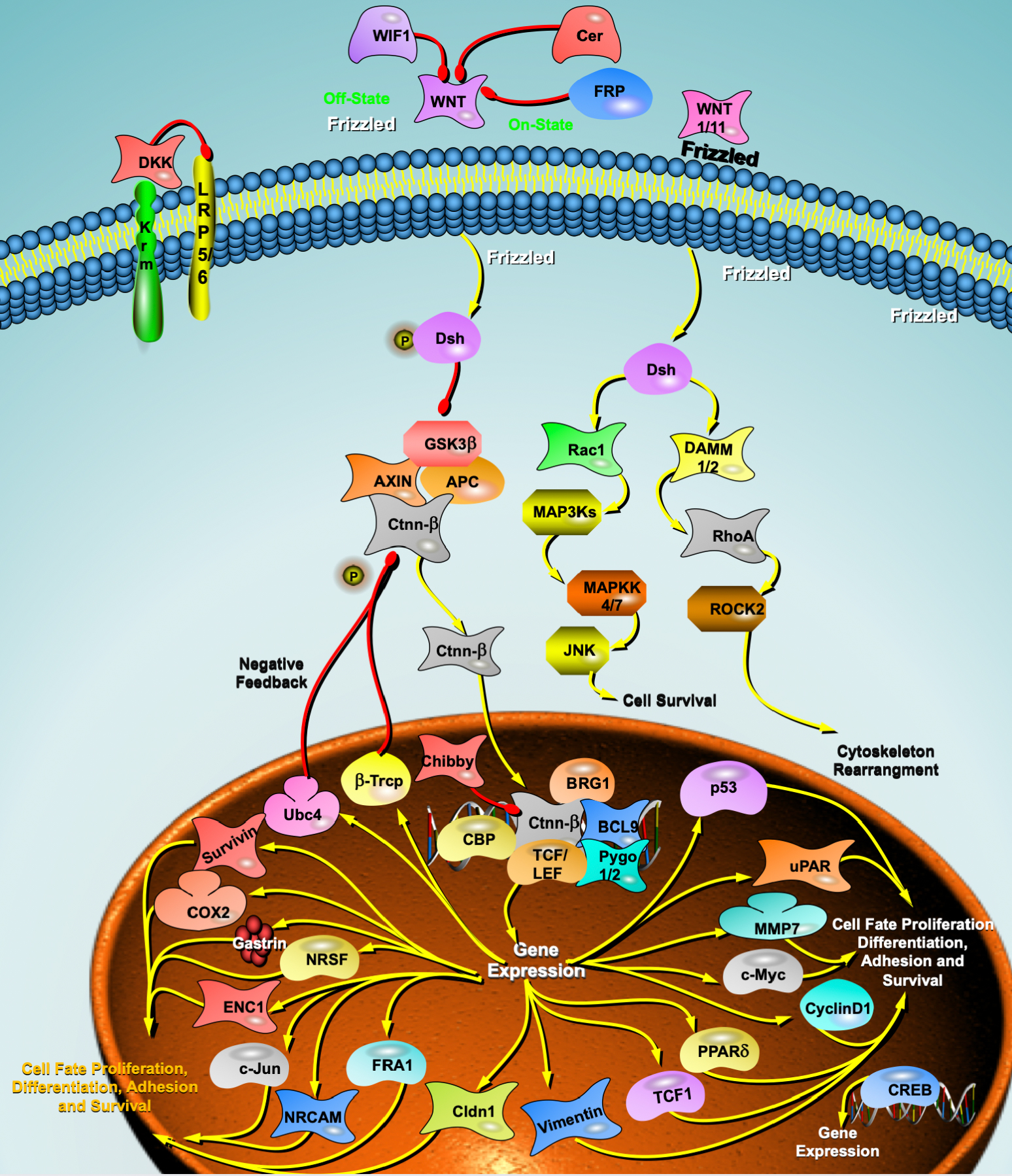
Fig. 1.

Wnt signaling pathway and heart failure. DKK, Dickkopf; WIF1, WNT inhibitory factor 1; BRG1, Brahma-related gene 1; Krm, Kremen; LRP5/6, lipoprotein receptor-related protein 5/6; FRP, Frizzled receptor proteins; Dsh, Dishevelled; GSK3β, glycogen synthase kinase 3β; APC, APC regulator of WNT signaling pathway; AXIN, axin protein; Ctnn-β, Catenin Beta; Rac1, Rac Family Small GTPase 1; DAMM1/2, Dishevelled Associated Activator Of Morphogenesis 1/2 MAPSKs, Mitogen‑activated protein kinase; MAPKK4/7, Mitogen-activated protein kinase kinase 4/7; JNK, c-Jun N-terminal kinase; ROCK2, Rho Associated Coiled-Coil Containing Protein Kinase 2; RhoA, Ras homolog family member A; cox2, Cyclooxygenase-2; ENC1, ectodermal-neural cortex 1; c-Jun, Transcription factor Jun; NRCAM, Neuronal Cell Adhesion Molecule; FRA1, Fos-related antigen1; Cldn1, Claudin 1; TCF1, T cell factor 1; PPARδ, peroxisome proliferator-activated receptors; CREB, cAMP response element binding protein; c-Myc, MYC proto-oncogene; MMP7, Matrix Metallopeptidase 7; uPAR, urokinase plasminogen activator surface receptor; p53, Tumor protein P53; Pygo1/2, Pygopus Family PHD Finger 1; BCL9, B cell lymphoma 9; TCF/LEF, T-cell factor/lymphoid enhancer factor; CBP, CREB binding protein; β-Tcrp, β-tranducin repeats containing protein; Ubc4, ubiquitin conjugating enzyme 4.

4. Mitochondrial Autophagy and Targeted Therapy for HF

Autophagy is the phagocytosis of eukaryotic cells by their own cytoplasmic proteins or organelles to achieve the metabolic needs of the cell itself and the renewal of some organelles through lysosomal degradation, the most important of which is mitochondrial autophagy in selective autophagy. Studies have shown that mitochondrial autophagy (mitophagy) is closely associated with HF [46, 47].

Mitochondrial autophagy is the process by which autophagic vesicles selectively wrap and transport damaged mitochondria to lysosomes for hydrolysis. This concept was first proposed by Lemasters in 2005 [48]. The current findings suggest two main types of mitochondrial autophagy mechanisms: ubiquitin dependent and non-ubiquitin dependent. Among them, ubiquitin-dependent mitochondrial autophagy includes PTEN-induced putative kinase 1 (PINK1)/parkin signaling pathway-mediated mitochondrial autophagy and non-parkin-dependent mitochondrial autophagy, while non-ubiquitin-dependent mitochondrial autophagy is directly mediated by mitochondrial autophagy receptors.

Song et al. [49] observed that parkin-mediated reduction in mitochondrial autophagy in cardiomyocytes from Mitofusin-2 (MFN2) gene mutant mice induced cardiac hypertrophy and HF. Chen et al. [50] found in a clinical study that PINK1 protein levels were significantly reduced and mitochondrial autophagy was inefficient in patients with advanced HF, whereas normal expression of PINK1 and parkin could attenuate myocardial cell injury, delay the progression of HF, and prolong patients’ lives.

It was found that the PINK1/parkin-mediated mitochondrial autophagy pathway is a potential target for the treatment of HF. In an experimental study of safranin, it was observed that parkin-mediated mitochondrial ubiquitination was significantly inhibited after knockdown of the PTEN-induced putative kinase 1 (PINK1) gene, counteracting the beneficial effects of safranin on HF, suggesting that safranin could promote PINK1/parkin signaling pathway-mediated mitochondrial autophagy for the treatment of HF [51]. Xiong et al. [52] found that overexpression of PINK1 promoted parkin translocation to the damaged mitochondrial outer membrane and phosphorylation, maintained cardiomyocyte homeostasis, and attenuated myocardial injury in a mouse model of angiotensin II-induced HF. In a tacrolimus (TAC)-induced mouse model of HF, Wang et al. [53] showed that high expression of AMP-activated protein kinase α2 (AMPKα2) in cardiomyocytes mediated by recombinant adeno-associated virus type 9 could activate the phosphorylation of Serrate284 and Serrate495 (Ser284 and Ser495) sites on PINK1, thereby increasing the role of the PINK1/parkin signaling pathway in mitochondrial autophagy and alleviating HF.

5. Noncoding RNA and HF Targeted Therapy
5.1 MicroRNAs

Noncoding RNAs (NCRs) are RNAs that do not encode proteins. MicroRNAs (miRNAs) are a class of single-stranded noncoding RNAs of approximately 22 nucleotides in length, and mature miRNAs contribute to the posttranscriptional degradation of target mRNAs by binding specifically to the 3 untranslated region (UTR) of target mRNAs or by inhibiting the posttranscriptional degradation of target mRNAs [54].

A growing number of studies have shown that microRNAs (miRNAs) play an important role in cardiovascular disease and are involved in several pathophysiological processes associated with HF, such as myocardial remodeling, cardiac hypertrophy, myocardial fibrosis, apoptosis, and hypoxia [55, 56]. An increasing number of miRNAs have exhibited a dysregulated and predominantly upregulated pattern in the later stages of end-stage HF [57]. The etiology of HF (e.g., ischemic, aortic stenosis, or idiopathic cardiomyopathy) is associated with differentially expressed miRNA patterns [58]. Thus, miRNAs play an active role in the development and progression of HF.

In the plasma of hypertrophic cardiomyopathy (HCM) patients diagnosed with no symptoms of HF, miR-29a, the only miRNA associated with left ventricular hypertrophy and fibrosis, was found to be significantly upregulated [59]. In addition, miRNAs can be used to determine the prognosis of HF patients. Qiang et al. [60] examined miRNAs in endothelial progenitor cells (circulating from monocytes) from 106 HF patients and found that low levels of miR-126 were associated with cardiovascular death in patients with ischemic HF, while high levels of miR-508a-5p were associated with nonischemic HF patients. Additional studies have shown that decreases in miR-18a-5p and miR-652-3p during hospitalization for HF predicted 180-day mortality [61]. One meta-analysis examining the expression of circulating miRNAs and patient prognosis [62], which included four relevant articles assessing 19 circulating miRNAs in 867 patients, showed that low expression of miR-1, miR-423-5p, miR-126, miR-21, miR-23, miR-30d, miR-18a-5p, miR-16-5p, miR-18b-5p, miR-36b-5p, miR-206a-3p, miR-2313-3a-3p, and miR-423-128 was associated with significantly poorer overall survival in HF patients (p < 0.05). Among these molecules, miR-18a-5p, miR-18b-5p, miR-30d, miR-30e-5p, and miR-423-5p were strong biomarkers of HF prognosis.

It has also been shown that microRNAs can be used as biomarkers of response to HF treatment. For example, one animal study found [63] that plasma levels of miR-16, miR-20b, miR-93, miR-106b, miR-223 and miR-423-5p were elevated in rats with hypertension-induced HF compared to controls. Nie et al. [64] showed that miR-217 was highly expressed in the myocardial tissue of patients with chronic HF and exacerbated pressure load-induced cardiac dysfunction. These findings suggest that miR-217 plays an important role in myocardial hypertrophy and dysfunction and could be a therapeutic target for HF.

5.2 LncRNA

Long noncoding RNAs (lncRNAs) are a set of functional RNA molecules greater than 200 nucleotides in length that do not encode proteins and are generally present within longer coding genes, between coding genes, or in antisense to coding sequences [65]. LncRNAs are involved in cell differentiation, proliferation, apoptosis and autophagy, affecting the development and progression of various cardiovascular diseases, such as hypertension, atherosclerosis and HF. Liu et al. [66] showed that lncRNA H19 is highly expressed in the embryonic period, and its expression gradually decreases as individuals mature. The expression of lncRNA H19 decreases as individuals mature but increases again when vascular damage or cardiac insufficiency occurs. The results of another study observed a significant increase in H19 expression in patients with HF and in a mouse model of myocardial hypertrophy [67]. Han et al. [68] showed that high expression of H19 could be associated with disorders of lipid metabolism, and these findings suggest that the extent of HF might be understood by measuring H19 expression levels. In addition, studies have shown that the lncRNA long intergenic noncoding RNA predicting cardiac remodeling (LIPCAR) is upregulated early and downregulated later in the plasma of HF patients, and it has also been suggested that LIPCAR might be used as an HF biomarker [69]. Although the mechanisms of action of only a few lncRNAs have been elucidated, further systematic and comprehensive studies can be conducted to shed more light on the complex regulatory mechanisms and functional targets of lncRNAs, providing new ideas for the diagnosis and treatment of cardiovascular diseases and new targets for the development of new drugs.

6. N6-Methyladenosine and HF Targeting Therapy

The types of RNA modifications include N6-methyladenosine (m6A), N1-methyladenosine, 5-methylcytosine, pseudouridine nucleoside, N6,2-O-dimethyladenosine, and N7-methylguanosine. m6A methylation is a methylation modification formed by the N at position 6 of adenine (A) catalyzed by methyltransferase, which occurs mainly in the highly conserved consensus motif of 5-RRRACU- 3 (R=A or G), in the beginning segment of the 3 untranslated region of the mRNA molecule and near the termination codon. m6A modifications are closely related to the development of cardiovascular diseases, including myocardial hypertrophy, HF, ischemic heart disease, aortic aneurysms, vascular calcification, and pulmonary hypertension.

Differentially m6A-modified transcripts in HF patients are mainly involved in cardiac metabolism and signaling. Calmodulin 1 (Calm1) mRNA, a member of the calcium/calmodulin-dependent protein kinase II (CamKII) signaling pathway, was not altered during m6A modification, whereas Calm1 protein expression was significantly reduced in failing heart tissue, suggesting that, in the development of HF, m6A methylation affects calm1 translation, but not transcription, during the development of HF. m6A-seq revealed differentially methylated transcripts of epigenetic proteins, transcription factors and upstream regulators of signaling pathways, suggesting that m6A methylation is involved in the regulation of gene expression in HF [70]. Dorn et al. [71] showed that m6A methylation levels are elevated by hypertrophic stimuli. The m6A RNA methylation enzyme methyltransferase-like 3 (METTL3) plays an important and positive role in cardiomyocyte hypertrophy, and in an in vitro model, the growth tendency of cardiomyocyte hypertrophy is completely lost when METTL3 is inhibited, and no hypertrophy occurs, whereas overexpression of METTL3 promotes spontaneous and compensatory hypertrophy. In an in vivo model, cardiac-specific METTL3 knockout mice exhibited cardiac remodeling and HF, followed by dysregulation of cardiac homeostasis, while elevated levels of the m6A RNA methyltransferase METTL3 led to cardiac hypertrophy.

Mathiyalagan et al. [72] found that obesity-associated protein (fat mass and obesity-association protein, FTO) is a demethylase that plays an important role in myocardial homeostasis and remodeling during cardiac contraction. FTO overexpression attenuates the ischemia-induced elevation of m6A modification. m6A modification is increased and FTO expression is significantly decreased in the infarct and peri-infarct regions of failing hearts compared to healthy heart tissue. FTO overexpression attenuated the ischemia-induced elevation of m6A modifications. In FTO knockout mice, HF progressed more rapidly, with a lower ejection fraction and more severe cardiac dilatation, suggesting an integral role for FTO in HF. However, the molecular mechanism of action remains unclear, and many key issues must be further investigated to more deeply explain the regulatory mechanism of m6A methylation in the development of the cardiovascular system, which could help to improve the diagnosis, treatment and prognostic judgment of the disease and provide a more scientific basis for the targeting of m6A in the treatment of HF and other cardiovascular diseases in the future.

7. Other Targeted Therapies
7.1 ACE2-Ang1-7 Axis

The renin angiotensin system (RAS) plays a very important role in regulating normal physiology and the mechanisms of cardiovascular disease development. Angiotensin converting enzyme (ACE) is an important key enzyme of the classical RAS pathway angiotension converting enzyme-angiotensin II-type 1 (ACE-Ang II-AT1), which converts angiotensin (Ang) I to Ang II, and the increase in Ang II is an important part of the RAS involved in the development of cardiovascular diseases. Another RAS pathway, ACE2-Ang (1-7) MAS, exerts action antagonistic to the classical RAS pathway, and the two pathways are functionally antagonistic to each other, giving the RAS a dual effect [73]. Activation of the ACE2/Ang-(1-7) pathway effectively delays the progression of HF, and changes in the balance between ACE/Ang II and ACE2/Ang (1-7) play an important pathophysiological role in HF failure. Ang (1-7) is a cardioprotective peptide in the RAS that counteracts the cardiotoxic effects of Ang II and has protective effects against pathological cardiac remodeling and HF [74]. The ACE2-Ang (1-7) pathway, known as the second metabolic axis of the RAS, plays a crucial role in cardiovascular disease. Ang (1-7) plays a cardioprotective role and has the potential to contribute to HF treatment.

7.2 Advanced Glycation End Products

Advanced glycation end products (AGEs) are compounds produced by the nonenzymatic reaction of free amino groups in proteins, lipids, and nucleic acids with reducing sugars (e.g., glucose, fructose, pentose, etc.), i.e., the Maillard reaction, and they are classified as endogenous or exogenous according to their sources [75]. The exogenous pathway from food is the main source of AGEs in humans. Protein-bound AGEs have been most extensively studied, and AGEs affect downstream HF-related signals, including the proto-oncogene protein p21 (ras), stress-activated protein kinase (SAPK), the activator of transcription (STAT) pathway, and several other pathways, by coupling to cell membrane proteins and altering their structures or directly activating cell surface receptors [76, 77], promotes ventricular remodeling and myocardial injury during heart failure, and also participates in inflammation, oxidative stress and apoptosis of myocardial tissue and autophagy of cardiomyocytes. Gao et al. [78] found that Receptor of Advanced Glycation Endproducts (RAGE) activation in a mouse model caused excessive autophagic activity through activation of the RAGE-NF-κB (nuclear factor kappa B) /BNIP3 (BCL2/adenovirus E1B 19 kDa protein-interacting protein 3)/beclin1 signaling pathway, contributing to the apoptosis of cardiomyocytes to promote HF. In addition, the binding of AGEs to RAGE in the progression of HF can cause impairment of calcium metabolism, atherosclerosis, vasoconstriction, and myocardial fibrosis, thereby causing myocardial systolic-diastolic dysfunction. Studies have shown that AGEs bind to their receptors to promote the development of various cardiovascular diseases; therefore, by reducing AGEs or RAGEs and thus blocking the activation of AGE-related downstream cellular pathways, therapeutic or preventive effects on cardiovascular diseases can be achieved.

The most important exogenous source of AGEs is food, and the way in which food is cooked is the most important factor affecting the content of AGEs. The formation of AGEs is accelerated in high-temperature, long and deeply dry cooking methods (grilling, frying, deep-frying, etc.), so changes in cooking habits and methods can help to reduce the accumulation of AGEs in the body [79]. Toprak et al. [80] observed that the mechanism of the diastolic effect of alagebrium (ALT-711) on carotid arteries in healthy animals is not only to reduce the cross-linking of AGEs with collagen but that ALT-711 has the ability to improve the uptake and release of Ca2+ from the sarcoplasmic reticulum of cardiac myocytes. Some studies have shown that the angiotensin II receptor blockers telmisartan and losartan inhibit endogenous AGE production in cultured cells in vitro [81]. It follows that drugs can also antagonize AGEs.

7.3 Genetic Therapy

Due to the difficulties in targeting drugs to receptors and intracellular pathways, many years ago, researchers proposed cardiac gene therapy as an alternative therapeutic approach, whereby cell-targeted delivery of exogenous genes (transgenes) would produce “therapeutic” proteins that could compensate for pathological downregulation or counteract harmful molecular processes. The selection of appropriate targets and the availability of effective gene vectors are the key requirements for the success of this therapeutic approach.

A study published by Leiden’s research team in 1990 provided a history of cardiac gene therapy. In a landmark study, rat cardiomyocytes were transfected in vivo by direct injection of plasmid DNA containing the β-galactosidase gene in the left ventricular wall, and then β-galactosidase activity was found in the myocardium for up to 4 weeks [82]. In 2012, the CUPID (Calcium Up-Regulation by Percutaneous Administration of Gene Therapy in Cardiac Disease) study was the first clinical trial of cardiac gene therapy for HF. Replication-deficient adeno-associated virus type 1 (AAV1) was the vector selected to carry the Sarco/endoplasmic reticulum Ca2+-ATPase (SERCA2a) transgene. While the initial phase demonstrated safety and some beneficial effects, the final phase IIb CUPID trial was not successful. It involved 250 patients treated with intracoronary AAV1/SERCA2a or placebo. AAV1/SERCA2a treatment failed to prolong the time to the first endpoint event. Compared with placebo, AAV1/SERCA2a treatment had no significant effect on any endpoint, including New York Heart Association (NYHA) functional class, 6-minute walk test distance, or NT-proBNP levels [83, 84]. Despite the failure of the CUPID phase IIb trial, decades of preclinical studies have finally made gene therapy “palpable” in clinical cardiology.

As research on cardiac gene therapy has continued, a number of directions have been identified according to which good translational potential could exist. AC6 (adenylate cyclase) catalyzes the conversion of ATP into cyclic adenosine monophosphate (cAMP), a molecule essential for cardiac function; S100 calcium binding protein A1 (S100A1) is a protein that regulates sarcoplasmic reticulum Ca2+ cycling and mitochondrial function through interaction with ryanodine receptors, SERCA2 and mitochondrial F1-ATPase activity, with antihypertrophic, inotropic and antiarrhythmic effects and attenuating energy expenditure in HF [85]; SDF-1 (stromal cell-derived factor-1) has been shown to be essential in cardiac stem cell therapy; and a new frontier has also been proposed: gene therapy to stimulate cardiac regeneration. Other researchers have proposed cardiac stem cell therapy, among other therapies, as a targeted direction for the treatment of HF, but more studies and trials are needed to validate the roles of these therapeutic targets.

7.4 Chinese Herbal Treatment

Chinese medicine refers to natural drugs and their processed substitutes that are guided by the theory of Chinese medicine, have a unique theoretical system and application form, used to prevent and treat diseases and have rehabilitation and health care effects, mainly including plant drugs, animal drugs and mineral drugs [17]. Chinese medicine is divided into proprietary Chinese medicine and Chinese herbal medicine according to the processing undergone (Table 1). Chinese herbal medicines are rich in various chemical components, especially compounds that not only contain alkaloids, polysaccharides, glycosides and other effective drug components but are also rich in vitamins, dietary fiber and other nutrients, with a variety of pharmacological effects. The multitarget therapeutic effect of Chinese medicine can effectively avoid the adverse therapeutic effect caused by the defective therapeutic target and the weakened efficacy caused by the defective drug metabolism in the process of Western medicine treatment, and Chinese medicine can also effectively alleviate the clinical symptoms of patients with chronic HF, improve the prognosis of patients and improve quality of life. Chinese medicine monomers, single Chinese medicines and Chinese medicine compounds all have the characteristics of multichannel and multitarget therapy, which can regulate the body as a whole and affect the physiological functions of multiple systems of the body, so the targeted treatment of HF with Chinese medicine is also worthy of exploration [17, 86].

Table 1.Proprietary Chinese medicines and herbal medicines available for the treatment of heart failure.
Proprietary Chinese medicine Herbal medicine
Baoyuan Tang, Danshen Yin, Shengmai San, Taohongsiwu Tang, Tinglidazaoxiefei Tang, Xuefuzhuyu Tang, Zhenwu Tang, Danqi Pill, Fufang danshen Dripping Pill, Shengmai Capsule, Qili Qiangxin Capsule, Qishen Yiqi Dripping Pill, Danhong Injection, Huangqi Injection, Shenmai Injection, Shenfu Injection, Shengmai Injection Baishao, Baizhu, Bingpian, Chaihu, Chenpi, Chishao, Chuanxiong, Danggui, Danshen, Dazao, Fuling, Fupian, Fuzi, Gancao, Guizhi, Honghua, Hongshen, Huangqi, Jiangxiang, Jiegeng, Maidong, Maimendong, Niuxi, Renshen, Rougui, Sanqi, Shaoyao, Sharen, Shengdihuang, Shengjiang, Shudi, Tanxiang, Taoren, Tingli, Tinglizi, Wuweizi, Xiangjiapi, Yuzhu, Zexie, Zhiqiao
8. Conclusions

HF is a complex, multifactorial clinical syndrome with heterogeneity. Despite the disappointing results of current targeted therapy studies and targeted therapy being influenced by many factors, targeted therapy remains a promising and attractive direction for the treatment of HF. The potential factors leading to negative outcomes should be carefully analyzed, and further research and optimization should be promoted to discover new molecular targets with more significant therapeutic potential to exert cardioprotective effects and become new approaches to HF treatment.

Author Contributions

LM conceived the study, participated in the design and drafted the manuscript. YLL reviewed relevant literature, collected data for the manuscript, drew diagrams and created tables. Both authors contributed to editorial changes in the manuscript. Both authors read and approved the final manuscript. Both authors have participated sufficiently in the work and agreed to be accountable for all aspects of the work.

Ethics Approval and Consent to Participate

Not applicable.

Acknowledgment

We thank all the staff in the laboratory.

Funding

The authors acknowledge the essential role of the funding of National Natural Science Foundation of Guangxi (2020GXNSFAA297003), National Natural Science Foundation of China (NSFC: 82060072 and 81760073), Project of Liuzhou Science and Technology (2020NBAB0818, 2021CBC0106 and 2021CBC0108), the project of Liuzhou people’s Hospital (LYRGCC202107 and LYRGCC202203), Guangxi Medical and health key discipline construction project and Guangxi Health Commission Key Laboratory of Clinical Biotechnology (Liuzhou People’s Hospital).

Conflict of Interest

The authors declare no conflict of interest.

References
[1]
Sonnenburg JL, Bäckhed F. Diet-microbiota interactions as moderators of human metabolism. Nature. 2016; 535: 56–64.
[2]
Ahmad AF, Ward NC, Dwivedi G. The gut microbiome and heart failure. Current Opinion in Cardiology. 2019; 34: 225–232.
[3]
Marques FZ, Nelson E, Chu PY, Horlock D, Fiedler A, Ziemann M, et al. High-Fiber Diet and Acetate Supplementation Change the Gut Microbiota and Prevent the Development of Hypertension and Heart Failure in Hypertensive Mice. Circulation. 2017; 135: 964–977.
[4]
Nguyen HT, Bertoni AG, Nettleton JA, Bluemke DA, Levitan EB, Burke GL. DASH eating pattern is associated with favorable left ventricular function in the multi-ethnic study of atherosclerosis. Journal of the American College of Nutrition. 2012; 31: 401–407.
[5]
Salehi-Abargouei A, Maghsoudi Z, Shirani F, Azadbakht L. Effects of Dietary Approaches to Stop Hypertension (DASH)-style diet on fatal or nonfatal cardiovascular diseases–incidence: a systematic review and meta-analysis on observational prospective studies. Nutrition. 2013; 29: 611–618.
[6]
Mathew AV, Seymour EM, Byun J, Pennathur S, Hummel SL. Altered Metabolic Profile With Sodium-Restricted Dietary Approaches to Stop Hypertension Diet in Hypertensive Heart Failure With Preserved Ejection Fraction. Journal of Cardiac Failure. 2015; 21: 963–967.
[7]
Liyanage T, Ninomiya T, Wang A, Neal B, Jun M, Wong MG, et al. Effects of the Mediterranean Diet on Cardiovascular Outcomes-A Systematic Review and Meta-Analysis. PLoS ONE. 2016; 11: e0159252.
[8]
Chen ML, Yi L, Zhang Y, Zhou X, Ran L, Yang J, et al. Resveratrol Attenuates Trimethylamine-N-Oxide (TMAO)-Induced Atherosclerosis by Regulating TMAO Synthesis and Bile Acid Metabolism via Remodeling of the Gut Microbiota. mBio. 2016; 7: e02210–e02215.
[9]
Andraws R, Berger JS, Brown DL. Effects of antibiotic therapy on outcomes of patients with coronary artery disease: a meta-analysis of randomized controlled trials. Journal of the American Medical Association. 2005; 293: 2641–2647.
[10]
Lam V, Su J, Koprowski S, Hsu A, Tweddell JS, Rafiee P, et al. Intestinal microbiota determine severity of myocardial infarction in rats. FASEB Journal. 2012; 26: 1727–1735.
[11]
Hill C, Guarner F, Reid G, Gibson GR, Merenstein DJ, Pot B, et al. Expert consensus document. The International Scientific Association for Probiotics and Prebiotics consensus statement on the scope and appropriate use of the term probiotic. Nature Reviews Gastroenterology & Hepatology. 2014; 11: 506–514.
[12]
Patel R, DuPont HL. New approaches for bacteriotherapy: prebiotics, new-generation probiotics, and synbiotics. Clinical Infectious Diseases. 2015; 60: S108–S121.
[13]
Lam V, Su J, Hsu A, Gross GJ, Salzman NH, Baker JE. Intestinal Microbial Metabolites Are Linked to Severity of Myocardial Infarction in Rats. PLoS ONE. 2016; 11: e0160840.
[14]
Gan XT, Ettinger G, Huang CX, Burton JP, Haist JV, Rajapurohitam V, et al. Probiotic administration attenuates myocardial hypertrophy and heart failure after myocardial infarction in the rat. Circulation: Heart Failure. 2014; 7: 491–499.
[15]
Kootte RS, Levin E, Salojärvi J, Smits LP, Hartstra AV, Udayappan SD, et al. Improvement of Insulin Sensitivity after Lean Donor Feces in Metabolic Syndrome Is Driven by Baseline Intestinal Microbiota Composition. Cell Metabolism. 2017; 26: 611–619.e6.
[16]
Vrieze A, Van Nood E, Holleman F, Salojärvi J, Kootte RS, Bartelsman JFWM, et al. Transfer of intestinal microbiota from lean donors increases insulin sensitivity in individuals with metabolic syndrome. Gastroenterology. 2012; 143: 913–916.e7.
[17]
Xu L, Chen L, Gu G, Wang Y, Xu Y, Zhong Y. Natural products from traditional Chinese medicine for the prevention and treatment of heart failure: progress and perspectives. Reviews in Cardiovascular Medicine. 2022; 23: 60.
[18]
Sanna S, van Zuydam NR, Mahajan A, Kurilshikov A, Vich Vila A, Võsa U, et al. Causal relationships among the gut microbiome, short-chain fatty acids and metabolic diseases. Nature Genetics. 2019; 51: 600–605.
[19]
Hanna A, Frangogiannis NG. Inflammatory Cytokines and Chemokines as Therapeutic Targets in Heart Failure. Cardiovascular Drugs and Therapy. 2020; 34: 849–863.
[20]
Kapadia S, Lee J, Torre-Amione G, Birdsall HH, Ma TS, Mann DL. Tumor necrosis factor-alpha gene and protein expression in adult feline myocardium after endotoxin administration. The Journal of Clinical Investigation. 1995; 96: 1042–1052.
[21]
Kapadia SR, Oral H, Lee J, Nakano M, Taffet GE, Mann DL. Hemodynamic regulation of tumor necrosis factor-alpha gene and protein expression in adult feline myocardium. Circulation Research. 1997; 81: 187–195.
[22]
Torre-Amione G, Kapadia S, Lee J, Durand JB, Bies RD, Young JB, et al. Tumor necrosis factor-alpha and tumor necrosis factor receptors in the failing human heart. Circulation. 1996; 93: 704–711.
[23]
Deswal A, Bozkurt B, Seta Y, Parilti-Eiswirth S, Hayes FA, Blosch C, et al. Safety and efficacy of a soluble P75 tumor necrosis factor receptor (Enbrel, etanercept) in patients with advanced heart failure. Circulation. 1999; 99: 3224–3226.
[24]
Chung ES, Packer M, Lo KH, Fasanmade AA, Willerson JT, Anti-TNF Therapy Against Congestive Heart Failure Investigators. Randomized, double-blind, placebo-controlled, pilot trial of infliximab, a chimeric monoclonal antibody to tumor necrosis factor-alpha, in patients with moderate-to-severe heart failure: results of the anti-TNF Therapy Against Congestive Heart Failure (ATTACH) trial. Circulation. 2003; 107: 3133–3140.
[25]
Ridker PM, Everett BM, Thuren T, MacFadyen JG, Chang WH, Ballantyne C, et al. Antiinflammatory Therapy with Canakinumab for Atherosclerotic Disease. The New England Journal of Medicine. 2017; 377: 1119–1131.
[26]
Everett BM, Cornel JH, Lainscak M, Anker SD, Abbate A, Thuren T, et al. Anti-Inflammatory Therapy With Canakinumab for the Prevention of Hospitalization for Heart Failure. Circulation. 2019; 139: 1289–1299.
[27]
Yokoe I, Kobayashi H, Kobayashi Y, Giles JT, Yoneyama K, Kitamura N, et al. Impact of tocilizumab on N-terminal pro-brain natriuretic peptide levels in patients with active rheumatoid arthritis without cardiac symptoms. Scandinavian Journal of Rheumatology. 2018; 47: 364–370.
[28]
Kleveland O, Kunszt G, Bratlie M, Ueland T, Broch K, Holte E, et al. Effect of a single dose of the interleukin-6 receptor antagonist tocilizumab on inflammation and troponin T release in patients with non-ST-elevation myocardial infarction: a double-blind, randomized, placebo-controlled phase 2 trial. European Heart Journal. 2016; 37: 2406–2413.
[29]
Kleveland O, Ueland T, Kunszt G, Bratlie M, Yndestad A, Broch K, et al. Interleukin-6 receptor inhibition with tocilizumab induces a selective and substantial increase in plasma IP-10 and MIP-1β in non-ST-elevation myocardial infarction. International Journal of Cardiology. 2018; 271: 1–7.
[30]
Sokol CL, Luster AD. The chemokine system in innate immunity. Cold Spring Harbor Perspectives in Biology. 2015; 7: a016303.
[31]
Chen B, Frangogiannis NG. Chemokines in Myocardial Infarction. Journal of Cardiovascular Translational Research. 2021; 14: 35–52.
[32]
Xia Y, Frangogiannis NG. MCP-1/CCL2 as a therapeutic target in myocardial infarction and ischemic cardiomyopathy. Inflammation & Allergy Drug Targets. 2007; 6: 101–107.
[33]
Behr TM, Wang X, Aiyar N, Coatney RW, Li X, Koster P, et al. Monocyte chemoattractant protein-1 is upregulated in rats with volume-overload congestive heart failure. Circulation. 2000; 102: 1315–1322.
[34]
Dewald O, Frangogiannis NG, Zoerlein M, Duerr GD, Klemm C, Knuefermann P, et al. Development of murine ischemic cardiomyopathy is associated with a transient inflammatory reaction and depends on reactive oxygen species. Proceedings of the National Academy of Sciences of the United States of America. 2003; 100: 2700–2705.
[35]
Stumpf C, Lehner C, Raaz D, Yilmaz A, Anger T, Daniel WG, et al. Platelets contribute to enhanced MCP-1 levels in patients with chronic heart failure. Heart. 2008; 94: 65–69.
[36]
Hohensinner PJ, Rychli K, Zorn G, Hülsmann M, Berger R, Mörtl D, et al. Macrophage-modulating cytokines predict adverse outcome in heart failure. Thrombosis and Haemostasis. 2010; 103: 435–441.
[37]
Wang J, Seo MJ, Deci MB, Weil BR, Canty JM, Nguyen J. Effect of CCR2 inhibitor-loaded lipid micelles on inflammatory cell migration and cardiac function after myocardial infarction. International Journal of Nanomedicine. 2018; 13: 6441–6451.
[38]
Lu W, Xie Z, Tang Y, Bai L, Yao Y, Fu C, et al. Photoluminescent Mesoporous Silicon Nanoparticles with siCCR2 Improve the Effects of Mesenchymal Stromal Cell Transplantation after Acute Myocardial Infarction. Theranostics. 2015; 5: 1068–1082.
[39]
Frangogiannis NG, Dewald O, Xia Y, Ren G, Haudek S, Leucker T, et al. Critical role of monocyte chemoattractant protein-1/CC chemokine ligand 2 in the pathogenesis of ischemic cardiomyopathy. Circulation. 2007; 115: 584–592.
[40]
Zhang W, Zhu T, Chen L, Luo W, Chao J. MCP-1 mediates ischemia-reperfusion-induced cardiomyocyte apoptosis via MCPIP1 and CaSR. American Journal of Physiology - Heart and Circulatory Physiology. 2020; 318: H59–H71.
[41]
Ramachandran I, Thavathiru E, Ramalingam S, Natarajan G, Mills WK, Benbrook DM, et al. Wnt inhibitory factor 1 induces apoptosis and inhibits cervical cancer growth, invasion and angiogenesis in vivo. Oncogene. 2012; 31: 2725–2737.
[42]
Abplanalp WT, John D, Cremer S, Assmus B, Dorsheimer L, Hoffmann J, et al. Single-cell RNA-sequencing reveals profound changes in circulating immune cells in patients with heart failure. Cardiovascular Research. 2021; 117: 484–494.
[43]
Malekar P, Hagenmueller M, Anyanwu A, Buss S, Streit MR, Weiss CS, et al. Wnt signaling is critical for maladaptive cardiac hypertrophy and accelerates myocardial remodeling. Hypertension. 2010; 55: 939–945.
[44]
Petrich BG, Eloff BC, Lerner DL, Kovacs A, Saffitz JE, Rosenbaum DS, et al. Targeted activation of c-Jun N-terminal kinase in vivo induces restrictive cardiomyopathy and conduction defects. The Journal of Biological Chemistry. 2004; 279: 15330–15338.
[45]
Liao H, Li X, Zhao L, Wang Y, Wang X, Wu Y, et al. A PROTAC peptide induces durable β-catenin degradation and suppresses Wnt-dependent intestinal cancer. Cell Discovery. 2020; 6: 35.
[46]
Saha S, Panigrahi DP, Patil S, Bhutia SK. Autophagy in health and disease: A comprehensive review. Biomedicine & Pharmacotherapy. 2018; 104: 485–495.
[47]
Hall AR, Burke N, Dongworth RK, Hausenloy DJ. Mitochondrial fusion and fission proteins: novel therapeutic targets for combating cardiovascular disease. British Journal of Pharmacology. 2014; 171: 1890–1906.
[48]
Lemasters JJ. Selective mitochondrial autophagy, or mitophagy, as a targeted defense against oxidative stress, mitochondrial dysfunction, and aging. Rejuvenation Research. 2005; 8: 3–5.
[49]
Song M, Chen Y, Gong G, Murphy E, Rabinovitch PS, Dorn GW II. Super-suppression of mitochondrial reactive oxygen species signaling impairs compensatory autophagy in primary mitophagic cardiomyopathy. Circulation Research. 2014; 115: 348–353.
[50]
Chen G, Kroemer G, Kepp O. Mitophagy: An Emerging Role in Aging and Age-Associated Diseases. Frontiers in Cell and Developmental Biology. 2020; 8: 200.
[51]
Abudureyimu M, Yu W, Cao RY, Zhang Y, Liu H, Zheng H. Berberine Promotes Cardiac Function by Upregulating PINK1/Parkin-Mediated Mitophagy in Heart Failure. Frontiers in Physiology. 2020; 11: 565751.
[52]
Xiong W, Hua J, Liu Z, Cai W, Bai Y, Zhan Q, et al. PTEN induced putative kinase 1 (PINK1) alleviates angiotensin II-induced cardiac injury by ameliorating mitochondrial dysfunction. International Journal of Cardiology. 2018; 266: 198–205.
[53]
Wang B, Nie J, Wu L, Hu Y, Wen Z, Dong L, et al. AMPKα2 Protects Against the Development of Heart Failure by Enhancing Mitophagy via PINK1 Phosphorylation. Circulation Research. 2018; 122: 712–729.
[54]
Simpson LJ, Ansel KM. MicroRNA regulation of lymphocyte tolerance and autoimmunity. The Journal of Clinical Investigation. 2015; 125: 2242–2249.
[55]
Tijsen AJ, Pinto YM, Creemers EE. Non-cardiomyocyte microRNAs in heart failure. Cardiovascular Research. 2012; 93: 573–582.
[56]
Melman YF, Shah R, Das S. MicroRNAs in heart failure: is the picture becoming less miRky? Circulation: Heart Failure. 2014; 7: 203–214.
[57]
Bagnall RD, Tsoutsman T, Shephard RE, Ritchie W, Semsarian C. Global microRNA profiling of the mouse ventricles during development of severe hypertrophic cardiomyopathy and heart failure. PLoS ONE. 2012; 7: e44744.
[58]
Ikeda S, Kong SW, Lu J, Bisping E, Zhang H, Allen PD, et al. Altered microRNA expression in human heart disease. Physiological Genomics. 2007; 31: 367–373.
[59]
Roncarati R, Viviani Anselmi C, Losi MA, Papa L, Cavarretta E, Da Costa Martins P, et al. Circulating miR-29a, among other up-regulated microRNAs, is the only biomarker for both hypertrophy and fibrosis in patients with hypertrophic cardiomyopathy. Journal of the American College of Cardiology. 2014; 63: 920–927.
[60]
Qiang L, Hong L, Ningfu W, Huaihong C, Jing W. Expression of miR-126 and miR-508-5p in endothelial progenitor cells is associated with the prognosis of chronic heart failure patients. International Journal of Cardiology. 2013; 168: 2082–2088.
[61]
Ovchinnikova ES, Schmitter D, Vegter EL, Ter Maaten JM, Valente MAE, Liu LCY, et al. Signature of circulating microRNAs in patients with acute heart failure. European Journal of Heart Failure. 2016; 18: 414–423.
[62]
Yang J, Yang XS, Fan SW, Zhao XY, Li C, Zhao ZY, et al. Prognostic value of microRNAs in heart failure: A meta-analysis. Medicine. 2021; 100: e27744.
[63]
Dickinson BA, Semus HM, Montgomery RL, Stack C, Latimer PA, Lewton SM, et al. Plasma microRNAs serve as biomarkers of therapeutic efficacy and disease progression in hypertension-induced heart failure. European Journal of Heart Failure. 2013; 15: 650–659.
[64]
Nie X, Fan J, Li H, Yin Z, Zhao Y, Dai B, et al. miR-217 Promotes Cardiac Hypertrophy and Dysfunction by Targeting PTEN. Molecular Therapy - Nucleic Acids. 2018; 12: 254–266.
[65]
Devaux Y, Zangrando J, Schroen B, Creemers EE, Pedrazzini T, Chang CP, et al. Long noncoding RNAs in cardiac development and ageing. Nature Reviews Cardiology. 2015; 12: 415–425.
[66]
Liu L, An X, Li Z, Song Y, Li L, Zuo S, et al. The H19 long noncoding RNA is a novel negative regulator of cardiomyocyte hypertrophy. Cardiovascular Research. 2016; 111: 56–65.
[67]
Greco S, Zaccagnini G, Perfetti A, Fuschi P, Valaperta R, Voellenkle C, et al. Long noncoding RNA dysregulation in ischemic heart failure. Journal of Translational Medicine. 2016; 14: 183.
[68]
Han P, Li W, Lin CH, Yang J, Shang C, Nuernberg ST, et al. A long noncoding RNA protects the heart from pathological hypertrophy. Nature. 2014; 514: 102–106.
[69]
Schulte C, Barwari T, Joshi A, Theofilatos K, Zampetaki A, Barallobre-Barreiro J, et al. Comparative Analysis of Circulating Noncoding RNAs Versus Protein Biomarkers in the Detection of Myocardial Injury. Circulation Research. 2019; 125: 328–340.
[70]
Berulava T, Buchholz E, Elerdashvili V, Pena T, Islam MR, Lbik D, et al. Changes in m6A RNA methylation contribute to heart failure progression by modulating translation. European Journal of Heart Failure. 2020; 22: 54–66.
[71]
Dorn LE, Lasman L, Chen J, Xu X, Hund TJ, Medvedovic M, et al. The N6-Methyladenosine mRNA Methylase METTL3 Controls Cardiac Homeostasis and Hypertrophy. Circulation. 2019; 139: 533–545.
[72]
Mathiyalagan P, Adamiak M, Mayourian J, Sassi Y, Liang Y, Agarwal N, et al. FTO-Dependent N6-Methyladenosine Regulates Cardiac Function During Remodeling and Repair. Circulation. 2019; 139: 518–532.
[73]
Echeverría-Rodríguez O, Del Valle-Mondragón L, Hong E. Angiotensin 1-7 improves insulin sensitivity by increasing skeletal muscle glucose uptake in vivo. Peptides. 2014; 51: 26–30.
[74]
Miller AJ, Arnold AC. The renin-angiotensin system in cardiovascular autonomic control: recent developments and clinical implications. Clinical Autonomic Research. 2019; 29: 231–243.
[75]
Singh R, Barden A, Mori T, Beilin L. Advanced glycation end-products: a review. Diabetologia. 2001; 44: 129–146.
[76]
Tamanna N, Mahmood N. Food Processing and Maillard Reaction Products: Effect on Human Health and Nutrition. International Journal of Food Science. 2015; 2015: 526762.
[77]
Li L, Hao J, Jiang X, Li P, Sen H. Cardioprotective effects of ulinastatin against isoproterenol-induced chronic heart failure through the PI3K Akt, p38 MAPK and NF-κB pathways. Molecular Medicine Reports. 2018; 17: 1354–1360.
[78]
Gao W, Zhou Z, Liang B, Huang Y, Yang Z, Chen Y, et al. Inhibiting Receptor of Advanced Glycation End Products Attenuates Pressure Overload-Induced Cardiac Dysfunction by Preventing Excessive Autophagy. Frontiers in Physiology. 2018; 9: 1333.
[79]
O’Brien J, Morrissey PA. Nutritional and toxicological aspects of the Maillard browning reaction in foods. Critical Reviews in Food Science and Nutrition. 1989; 28: 211–248.
[80]
Toprak C, Sirmagul B, Yigitaslan S. Functional Effects of Alagebrium (ALT-711)-Isolated Rat Carotid Artery. The Eurasian Journal of Medicine. 2017; 49: 188–192.
[81]
Komiya N, Hirose H, Saisho Y, Saito I, Itoh H. Effects of 12-month valsartan therapy on glycation and oxidative stress markers in type 2 diabetic subjects with hypertension. International Heart Journal. 2008; 49: 681–689.
[82]
Lin H, Parmacek MS, Morle G, Bolling S, Leiden JM. Expression of recombinant genes in myocardium in vivo after direct injection of DNA. Circulation. 1990; 82: 2217–2221.
[83]
Greenberg B, Butler J, Felker GM, Ponikowski P, Voors AA, Desai AS, et al. Calcium upregulation by percutaneous administration of gene therapy in patients with cardiac disease (CUPID 2): a randomised, multinational, double-blind, placebo-controlled, phase 2b trial. The Lancet. 2016; 387: 1178–1186.
[84]
Greenberg B, Yaroshinsky A, Zsebo KM, Butler J, Felker GM, Voors AA, et al. Design of a phase 2b trial of intracoronary administration of AAV1/SERCA2a in patients with advanced heart failure: the CUPID 2 trial (calcium up-regulation by percutaneous administration of gene therapy in cardiac disease phase 2b). JACC: Heart Failure. 2014; 2: 84–92.
[85]
Most P, Seifert H, Gao E, Funakoshi H, Völkers M, Heierhorst J, et al. Cardiac S100A1 protein levels determine contractile performance and propensity toward heart failure after myocardial infarction. Circulation. 2006; 114: 1258–1268.
[86]
Wang Y, Wang Q, Li C, Lu L, Zhang Q, Zhu R, et al. A Review of Chinese Herbal Medicine for the Treatment of Chronic Heart Failure. Current Pharmaceutical Design. 2017; 23: 5115–5124.

Publisher’s Note: IMR Press stays neutral with regard to jurisdictional claims in published maps and institutional affiliations.

Share
Back to top