
Academic Editor: Peter A. McCullough
Reduction in X-ray exposure during cardiac catheterization is important to
reduce radiation risks to operators and personnel. Reducing scattered radiation
from the patient can achieve this goal. The goal of this study was to evaluate
the reduction in radiation using simple partial shielding of patients undergoing
cardiac catheterization. By putting a lead-based apron on the lower extremities
of patients undergoing cardiac catheterization, we analyzed the reduction in
total radiation dose with and without this shielding. One hundred and twelve
patients were divided into two groups. In one group, the protective lead-based
apron was put on the lower extremities of patients. Another group did not have
any shielding. Total duration of angiography was 332 minutes and 45 seconds in
the first group and 269 minutes and 10 seconds in the second group. The total
radiation exposure was 33
Many Interventional procedures require x-ray radiation. Since introducing x-ray for medical proposes, protective strategies had developed for more safety, not only for patients but also for operators. Since then, many protocols were introduced to reduce radiation exposure. Scattered tradition is one of the major causes of radiation exposure. Definition of scatter radiation in the USA national cancer institute (NCI) is the radiation that spreads out in different directions from a radiation beam when the beam interacts with a substance, such as body tissue for example the patients’ body surface. There are some factors affecting quantity of scatter radiation such as volume of tissue, density of matter, field size and kilo voltage.
Recently, some radiation protection shields are introduced by companies to minimize scattered radiation beyond the usual protection used by medical staffs. These shields are expensive. Using simple lead-based apron as partial protective shield on patients during cardiac catheterization for reduction of radiation exposure has not been well studied. The goal of our study was to evaluate if using commonly available protective lead-based apron on patients before cardiac catheterization can reduce the exposure dose.
In this study, we assessed the radiation exposure in the
angiography room in the presence or absence of the apron partially covering the
patient in the area not needing fluoroscope (lower extremities). This study was
performed using a GE Fluoroscopy device (Innova 3100; GE Healthcare, Waukesha,
WI, USA) Travel distance of 30 cm). One hundred and twelve patients who were
candidate for angiography or angioplasty were randomly divided into two groups.
All procedures were done by trans-radial approach. All procedures were performed
during 6 weeks period by a single operator with different angiography personnel
as aid team. The procedural numbers were not matched in both groups as patients
were only matched base on their body mass index (BMI). For patient preparation in
our study, we used RAD board (Merit Medical Systems., South
Jordan, UT, USA) Randomization was performed manually week by week. In the first
group (n = 56), lead-based apron shield was put on the patient’s lower
extremities in the supine position as showed in Figs. 1,2. In the second
group (n = 56), the procedure was performed routinely without any shielding. Both
groups were matched based on their (BMI). During the procedure, all images were
acquired with fluoroscopy set to “low” (7.5 frames/sec,
4 rad/min, max 100 kV) with cinematic acquisition
imaging frame rates at 15 frames/sec and source to image distance
at 100 centimeters. Focal distance was set to 30 cm with table
height of 85 cm. Ionizing radiation was measured with a RadCal
Dosimeter (model 9010, RadCal Corp., Monrovia, CA, USA) placed on top of the
shield in the center of the apron in the first group and between both thighs in
the second group in the similar positions. In addition, a moveable large size
lead shield positioned just proximal to the femoral insertion site. We used
electronic dosimeter for this study. The radiation protection apron is skirt
composed of 97 wt% tungsten and 3 wt% polyethylene with a length of 100 cm,
width of 25 cm and thickness of 0.1 cm. It was folded from its mid part to create
a blanket like rectangle with length of 50 cm, width of 25 cm and thickness of
0.2 cm. The sheet weighed 3.4 kg, and its shielding ability was equivalent to 1
mm Pb. Data about fluoroscopy time and exposure dose in each patient was
separately accessed from GE fluoroscopy device software and the dosimeter.
Ultimately the mean of fluoroscopy time and exposure were analyzed in both
groups. We excluded all emergency angiographies (Primary PCIs or urgent
re-angiographies) or patients with complications during the procedural
progression. ANOVA was used for different means in two or more groups and
Bonferroni correction was used to prevent data from incorrectly appearing to be
statistically significant. Statistical analysis was performed using SPSS. A
P value of
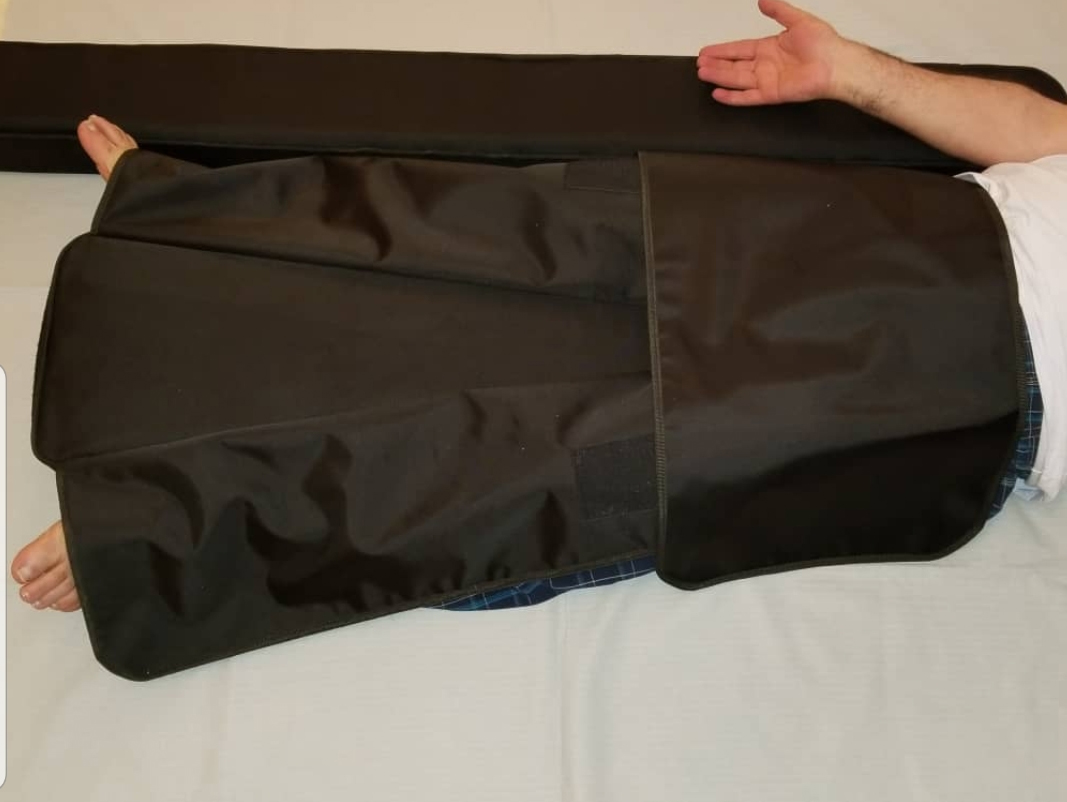 Fig. 1.
Using apron to cover entire lower extremities.
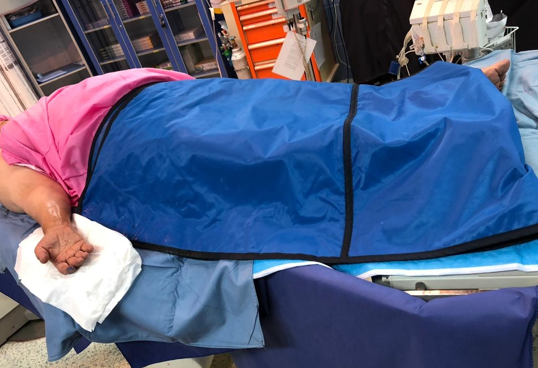 Fig. 2.
Another angel showing apron coverage.
Among 112 patients, 74 males and 38 females were studied. Mean
The risks posed by radiation exposure in the cardiac catheterization laboratory are underestimated by interventional cardiologists with growing concern [1].
Standard radiation protection devices utilized in the cardiac catheterization laboratory were designed for minimizing radiation exposure. Reduction in the scattered radiation is an important research for lowering radiation doses to patients and personnel. Chronic low-dose radiation exposure in the cardiac catheterization laboratory has been associated with a non-negligible increased risk of certain types of cancers [2]. In the last twenty years, radiation doses to primary operators in cardiac catheterization laboratories have not been changed [3].
Nowadays increasing complex cases are performed in cardiac catheterization laboratories. Despite improvements in reducing the scatter radiation emitted by fluoroscopy/cine-angiography in recent years, long complex cases have led to higher radiation exposure. Therefore, the need for alternative shielding techniques to reduce radiation exposure to both patients and operators are important. It has previously been shown that effort in reducing radiation scatter has led to significantly reduce radiation exposure to patients and operators during interventional fluoroscopic procedures [4].
Fetterly et al. [5] reported up to an 80% reduction in the scattered radiation with optimal use of radiation shielding. They used a movable upper body lead shield combined with lower body lead skirt with vertical extension as well as a scatter reduction drape placed in the conventional position for femoral access [5] with great success. In another study, a lead-free scatter reduction drape was shown to reduce radiation exposure to the operator by 23% when used in addition to conventional shielding [6]. In another study Ertel et al. [7] showed that a lead-free shielding drape specifically designed for a right radial cardiac catheterization can reduce radiation exposure to the operator by up to 72%.
Using dedicated shields for protecting this scattered radiation is costly. With our study, we proved that by using widely available operator apron for patients undergoing cardiac catheterization can reduce radiation doses with no additional cost. Our results showed more than 22 times lower radiation exposure using our simple method. This technique can also be used in patients with femoral approach. In this study, we only used large size apron skirt that was put on both lower extremities. It prevented the reflection of scatter radiation from the patients’ body surface. Unlike previous studies [8], our protection shield has less area but more thickness. It seems that less area is also protective needing future study. In this study, we only used one dosimeter in the room that detected the irradiation dose to our patients. In one study, Musallam A et al. [9] reported the use of a pelvic lead shield during radial angiography that led to reduction in the operator radiation exposure but there was an increased exposure to patients. However, in our study, we found less radiation to the patient assessed by dosimetry. Like our study, Sciahbasi A et al. [10] found the protective effect of drapes in radial artery procedures but they proved this protection for operators. Radial approach may increase radiation exposure to both patients and operators [11, 12, 13] therefore, by using our method, this risk may be significantly reduced warranting further investigation.
We only used a single detector for irradiation exposure that was placed somewhere far from the radiation field, so we were not able to access direct radiation doses to the operator. Total dose and cine time assessment were used for analysis. Different angiographic projections were not evaluated in our analysis. Finally, due to diversity in patients’ height and different position on the angiography table, the shield and detector had different distances to the x-ray tube limiting our results. Our sample size was small limiting our conclusion. However, despite small sample size we had great reduction in radiation. Furthermore, due to the small number of patients, further subgrouping based on procedures performed would reduce the number of patients dramatically making any statistical analysis meaningless. We did not randomize the patients based on height and weight further limiting our results. As the need for coronary intervention was only determined after the angiography, we could not randomized patients who were undergoing coronary angioplasty. In order to simplify our study, we utilized fluoroscopy time for our study as opposed to more accurate way of radiation measurement using DAP and Air Kerma which requires complex chambers mounted at the collimator with the need of specialized software.
AS, Designing data analysis and writing the manuscript. Ashkan Hashemi, Designing data analysis and writing the manuscript. BS, Designing data analysis and writing the manuscript. SN, Designing data analysis and writing the manuscript. Arash Hashemi, Designing data analysis and writing the manuscript. SE, Designing data analysis and writing the manuscript. MRM, Data analysis, editing and writing the manuscript.
All subjects gave their informed consent for inclusion before they participated in the study. The study was conducted in accordance with the Declaration of Helsinki, and the protocol was approved by the Ethics Committee of the hospital (IR.SBMU.NRITLD.REC.1400.023)
The authors would like to acknowledge the faculty of cath lab in Masih Daneshvari Hospital for their helpful support in this study with consent obtained from participants.
This research received no external funding.
The authors declare no conflict of interest.