IMR Press / CEOG / Volume 50 / Issue 2 / DOI: 10.31083/j.ceog5002023
Open Access Original Research
Relationship between Number of Mature Follicles and Pregnancy Rates in IUI Cycles in Women 38 to 43 Years Old
Show Less
1 Department of Obstetrics and Gynecology, Division of Reproductive Endocrinology and Infertility, McGill University, Montreal, QC H2L 4S8, Canada
2 MUHC Reproductive Center, McGill University, Montreal, QC H2L 4S8, Canada
*Correspondence: dahanhaim@hotmail.com (Michael H. Dahan)
Clin. Exp. Obstet. Gynecol. 2023, 50(2), 23; https://doi.org/10.31083/j.ceog5002023
Submitted: 7 August 2022 | Revised: 8 November 2022 | Accepted: 8 November 2022 | Published: 31 January 2023
Copyright: © 2023 The Author(s). Published by IMR Press.
This is an open access article under the CC BY 4.0 license.
Abstract

Background: Although the number of follicles at intrauterine insemination (IUI) is associated with the pregnancy rates and multiple pregnancy rates. Multiple pregnancy rates are low in older women. Therefore, this study was undertaken to determine the clinical pregnancy rate of IUI in women 38–43 years of age based on the number of stimulated mature follicles. Methods: A retrospective cohort study was performed including all the first to third stimulated IUI cycles conducted after the age of 38 years in a single academic fertility center between January 2011 and March 2018. Results: A total of 1574 IUI cycles were included in the study. The patients were divided according to the number of mature follicles (>14 mm in diameter) at the last ultrasound before the human chorionic gonadotropin (hCG) trigger. The total pregnancy rate was 9.1% and only 5 multiple pregnancies occurred. The parity (p = 0.049), the number of follicles 10–14 mm (p = 0.002), and the peak endometrial thickness (p = 0.003) were significantly different between the groups. No statistical difference was observed between the groups regarding pregnancy rates (p = 0.93) and clinical pregnancy rates (p = 0.21). Multivariate logistic regression controlling for confounding effects comparing clinical pregnancy rates with the standard as 1 follicle 14 mm or greater as benchmark did not alter the results. Conclusions: In women 38 to 43 years of age undergoing controlled ovarian hyperstimulation (COH)/IUI, one mature follicle yielded similar pregnancy and clinical pregnancy rates compared to multiple follicles, possibly due to the aneuploidy rate at this age.

Keywords
controlled ovarian hyperstimulation (COH)
intrauterine insemination (IUI)
older patients
infertility
1. Introduction

Controlled ovarian hyperstimulation (COH) with or without intrauterine insemination (IUI) is often used as a treatment for unexplained infertility, early-stage endometriosis, and borderline male-factor infertility [1]. COH combined with IUI is an important tool in infertility therapy, increasing the number of available oocytes thus enhancing the probability of conception.

Female age is an important predictor of both natural and treatment-related live birth rates (LBR), which decrease rapidly after 35 years of age [2]. Predictive factors for ongoing pregnancy with IUI other than woman age <40 years of age are cervical or anovulatory infertility, high total motile sperm count (TMS), and stimulation cycles [3]. Hence, if the pregnancy rate per IUI cycle is between 13% to 20% [4] and the birth rate per IUI treatment cycle is around 12% in young women, these rates decrease with increasing age [5]. In one study, cumulative pregnancy rates observed in up to 12 insemination cycles were 74% for women <31 years, 62% for women 31–35 years, and 54% for women 35–40 years [6]. Due to a significant decrease in efficacy and the poor prognosis for pregnancy in infertile elderly women, several studies have suggested that IUI should not be used or should be limited in women 40 years [7, 8, 9, 10]. Nevertheless, many patients insist on COH/IUI for cost or insurance coverage.

As women age, rates of aneuploidy increase with aneuploidy rates in human blastocysts rising from a 30% baseline in women younger than 35 years to >90% in women older than 44 years [11, 12]. Therefore, it makes inherent sense to maximize stimulation with in vitro fertilization (IVF) to obtain genetically normal embryos in older women. However, we do not stimulate to the same magnitude in women performing IUI. Current recommendations are to reach 3 mature follicles [13]. It is unknown if this mild stimulation improves outcomes in older women, due to the high chance of aneuploidy in such a small cohort of follicles.

Although COH/IUI is widely used, there is limited evidence to understand the role of the number of mature follicles and suggest the optimal protocol to maximize the likelihood of conception and LBR in older women while minimizing the risk of multiple-pregnancy. We aimed this study to determine the pregnancy-rate in IUI for women 38–43 years of age based on the number of mature follicles stimulated.

2. Material and Methods

We performed a retrospective cohort study of women 38–43 years of age at the time of IUI in a single academic fertility center between January 2011 and March 2018. Institutional review board approval was obtained (Approval number 2019-5254).

In our location, between 2011–2015, IVF was government funded. However, there were often waiting lists for IVF, so patients elected to perform COH/IUI which were more available, while in anticipation for IVF. In 2015 government coverage for IVF ceased but was continued for IUI (up to 9 cycles). Therefore, patients requested IUI despite the indication for care, due to the lack of cost of this treatment. It was felt that this would provide a rather unbiased population to study.

The primary outcome the was clinical pregnancy rate per IUI cycle. Secondary outcomes were pregnancy rates and multiple-pregnancy rates. Pregnancy was defined as a serum β-human chorionic gonadotropin (β-hCG) >10 mIU/mL. Clinical pregnancy was defined as an intrauterine-gestational sac with fetal pole and heartbeat seen on transvaginal ultrasound.

Inclusion and exclusion criteria: all first to third COH/IUI cycles for women ages 38–43 years were analyzed. Natural (unstimulated) cycles were excluded from the study.

All subjects had at least one patent fallopian tube. None of the women were known to have stage 3/4 endometriosis, submucosal fibroids, or polyps in situ.

Doses used were Clomiphene citrate 50 or 100 mg (Sanofi Canada, Laval, QC, Canada or EMD Serono, Montreal, QC, Canada), Letrozole 5 mg (Novartis, East Hanover, NJ, USA), and Gonadotrophins (GTs) as injectable recombinant follicle stimulating hormone (FSH) (Gonal-F; EMD Serono, Montreal, QC, Canada or Puregon, Organon/Merck Canada, Kirkland, QC, Canada), or human menopausal gonadotropin (hMG) (Menopur, Ferring Canada, Longueuil, QC, Canada) 50–300 IU daily, based on ovarian reserve. Patients using Clomiphene citrate or Letrozole ingested their medication on days 2–6 of the cycle and had an ultrasound at day 10 to monitor follicular growth. Patients in care would have a baseline ultrasound on cycle day 2–3. If ultrasound revealed endometrial thickness <5 mm and no ovarian cysts, the patient would begin COH and return for ultrasound monitoring per protocol.

When the leading follicle was >17 mm in diameter, the patient would receive hCG (Ovidrel 250MCG, SQ, Merck Serono, Canada, Montreal, QC, Canada) and IUI would be scheduled 36-hours later. Luteal support with vaginal progesterone (Prometrium®, Organon/Merck Canada, Kirkland, QC, Canada) 200 mg daily was started the day after insemination for all GTs-stimulated cycles.

For the sake of this study, mature follicles were defined 14 mm in mean diameter measured in 3 perpendicular planes, on transvaginal ultrasound.

Both partner sperm and commercially available donor-sperm insemination cycles were included in the study. When partner sperm was used, the partner was instructed to abstain from ejaculation for 48-hours before IUI. Samples were produced on-site and processed within 30 minutes of production. A basic wash and a density gradient centrifugation were performed on all semen samples. The concentrated pellet was reconstituted to a volume of 0.5 mL with tubal media (Ferticult, Beemm, Belgium). All samples were inseminated into the uterus using a Cook catheter (Cook Corporation, Bloomington, IN, USA).

A serum β-hCG pregnancy test was performed 16 days after IUI. If positive (β-hCG >10 mmol/L), a viability ultrasound was scheduled two weeks after the positive pregnancy test. If the initial day-16 serum β-hCG level was <100 IU/L, the serum test was repeated at 2-day intervals until evidence of the pregnancy outcome and location was determined.

Statistical analysis was performed with SPSS 23.0 (IBM Corp, Chicago, IL, USA). All data are presented as percentages or mean ± standard deviation (SD). Continuous data were compared with analysis of variance (ANOVA). Categorical data were compared using Chi-squared tests. Multivariate logistic regression controlling for confounding effects (Female age, TMS, antral follicle count (AFC) and endometrial thickness) of all baseline factors with p < 0.20 was done to compare the clinical pregnancy rate with the standard as one follicle of 14 mm or greater as a benchmark. Power analysis requires 785 cycles for an effect size of 0.1, alpha = 0.05, and power = 0.80. To put this effect size in perspective, this would be a change of 10% in outcomes or a clinical pregnancy rate of 11% vs. 10%.

3. Results

A total of 1574 IUI cycles were included in the study. All subjects were stimulated with either Clomiphene Citrate (n = 240), Letrozole (n = 176) or GTs (n = 1158). We divided the patients according to the number of mature follicles (14 mm) in diameter at the last ultrasound before IUI; 86 patients had 0 mature follicles, 983 had 1 mature follicle, 385 had 2 mature follicles, 94 had 3 mature follicles, 22 had 4 mature follicles and 4 women had 5 mature follicles. The 0 mature follicle group was triggered prior to 14 mm follicles developing due to the fact that they or their partners were unavailable to return for further care that cycle. Only 5 multiple-pregnancies in total occurred with no significant difference between the groups (p = 0.208).

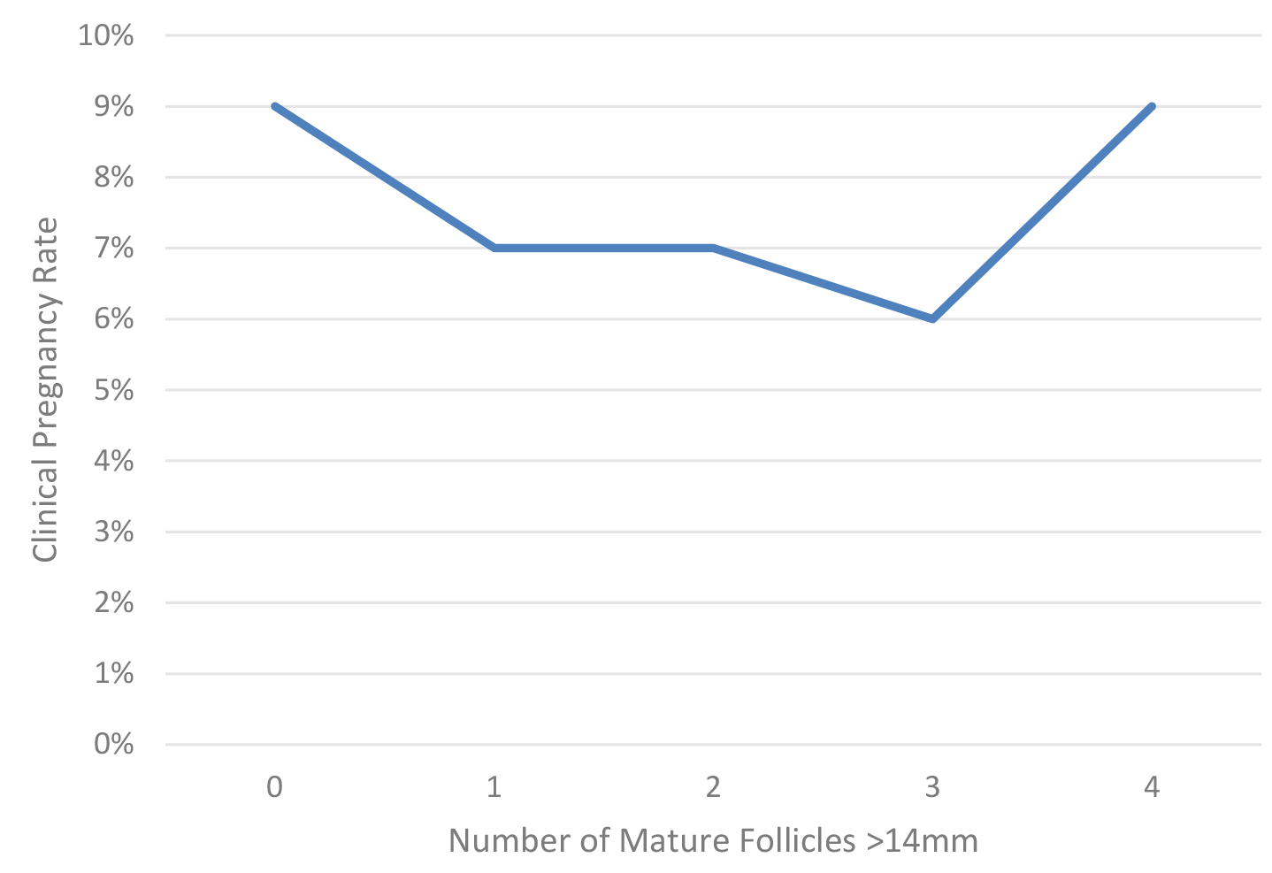
Table 1 presents the demographics and IUI outcomes by the number of mature follicles. The parity (P) (p = 0.049), the number of follicles 10–14 mm in diameter (p = 0.002) and the endometrial thickness (p = 0.003) were significantly different between the groups. There was no significant difference between the groups in terms of male age, TMS post processing, gravidity (G), AFC, basal estradiol, prolactin and thyroid stimulating hormon (TSH) levels. No statistical difference was observed between the groups regarding pregnancy rates and clinical pregnancy rates. Fig. 1 presents the clinical pregnancy rate as a function of the number of mature follicles. Multivariate stepwise-logistic regression controlling for confounders (Female age, TMS, AFC and endometrial thickness) comparing clinical pregnancy rates with the standard as 1 follicle 14 mm or greater as the benchmark is also presented in Table 1. After controlling for confounders effects, no statistical difference was observed in the clinical pregnancy rate for all the groups with one mature follicle stimulated being the gold standard selected: 0 follicles (adjusted odds ratio (OR) = 3.5, 95% confidence interval (CI) 0.33–37.6, p = 0.298); 2 follicles (adjusted OR = 1.8, 95% CI 0.53–6.5, p = 0.339); 3 follicles (adjusted OR = 2.5, 95% CI 0.43–13.9, p = 0.309); 4 follicles (adjusted OR = 0.72, 95% CI 0.16–3.2, p = 0.667); 5 follicles (adjusted OR = N/A, 95% CI N/A, p = 0.996).

Table 1.Demographic and IUI outcomes by ovarian stimulation protocol for women aged 38 years and older.
Number of mature follicles >14 0 1 2 3 4 5 Total p-value
N = 86 N = 983 N = 385 N = 94 N = 22 N = 4
Compared by ANOVA
Female age (years) 39.8 ± 1.7 39.8 ± 1.5 39.9 ± 1.6 40.1 ± 1.5 40.5 ± 1.7 41.0 ± 2.4 39.8 ± 1.5 0.033
Paternal age (years) 40.9 ± 5.2 41.5 ± 5.7 41.4 ± 5.7 42.2 ± 6.8 42.9 ± 7.2 41.8 ± 5.7 41.5 ± 5.7 0.649
TMS (post processing) 60.3 ± 71 49.9 ± 59 43.8 ± 51 47.2 ± 62 26.8 ± 29 24.1 ± 15 48.4 ± 58 0.224
G 1.1 ± 1.8 1.1 ± 1.4 1.1 ± 1.6 1.4 ± 1.9 1.0 ± 1.2 2.8 ± 1.9 1.1 ± 1.6 0.107
P 0.2 ± 0.6 0.3 ± 0.6 0.4 ± 0.8 0.3 ± 0.6 0.5 ± 0.1 0.3 ± 0.5 0.3 ± 0.6 0.049
Basal FSH (IU) 9.5 ± 5.0 8.4 ± 4.6 8.8 ± 5.9 9.1 ± 5.1 7.8 ± 3.5 9.2 ± 3.8 8.6 ± 5.0 0.265
Basal E2 (pmol/L) 178 ± 153 198 ± 156 189 ± 113 165 ± 76 237 ± 176 174 ± 138 193 ± 144 0.202
AFC 15.5 ± 11.4 14.0 ± 10.5 13.3 ± 9.1 12.3 ± 8.6 10.3 ± 7.3 14.8 ± 18.5 13.7 ± 10.1 0.169
Prolactin (ng/mL) 12.1 ± 12.6 10.4 ± 5.5 10.6 ± 10.0 10.8 ± 14.3 12.7 ± 8.1 8.3 ± 2.3 10.6 ± 8.0 0.393
TSH (mU/L) 1.9 ± 1.0 1.7 ± 1.2 1.8 ± 1.1 1.8 ± 1.1 1.6 ± 0.9 1.6 ± 0.7 1.8 ± 1.1 0.910
Number of follicles 10–14 1.6 ± 1.1 1.1 ± 1.3 1.1 ± 1.3 1.0 ± 1.4 1.7 ± 1.8 2.0 ± 1.8 1.1 ± 1.4 0.002
Endometrial thickness 7.6 ± 2.2 8.6 ± 3.0 8.9 ± 8.6 8.6 ± 2.5 8.6 ± 1.4 10.8 ± 2.0 8.6 ± 2.8 0.003
Pregnancy rate 8 (9.3%) 87 (8.9%) 35 (9.1%) 12 (12.8%) 2 (9.1%) 0 144/1574 0.933
Compared by multivariate logistic regression
Clinical pregnancy rate 8 (9.3%) 67 (6.8%) 28 (7.3%) 6 (6.4%) 2 (9.1%) 0 103/1574 0.208
Multiple-pregnancy 0 1 2 2 0 0 5/1574
95% CI 0.33–37.6 N/A 0.53–6.5 0.43–13.9 0.16–3.2 N/A
OR 3.5 N/A 1.8 2.5 0.72 N/A
p value 0.298 N/A 0.339 0.309 0.667 0.996

Data presented as mean ± SD or as percentage.

Clinical pregnancy rates; CI, OR and p value when controlling for the confounding effects (Female age, TMS, AFC and endometrial thickness) compared to 1 follicle as benchmark.

Multivariate logistic regression could not be performed for the group with 5 mature follicles since no pregnancies occulted in this group and numbers were small.

ANOVA, Analysis of Variance; TMS, total motile sperm count; G, gravidity; P, parity; E2, estradiol; FSH, follicle stimulating hormone; IUI, intra uterine insemination; IU, international units; AFC, antral follicle count; TSH, thyroid stimulating hormone; CI, confidence interval; OR, odds ratio; N/A, not available; N, number; SD, standard deviation.

Fig. 1.

Clinical pregnancy rate as a function of the number of mature follicles in women at least 38 years of age.

One might postulate that the group contains a wide range of ages, hence we also investigated the group of women ages 38–39 separately. Multivariate logistic regression controlling for the confounding effects comparing clinical pregnancy rate with the standard as one follicle 14 mm as the benchmark is presented in Table 2. After controlling for confounding effects, no statistical difference was observed in the clinical pregnancy rates for all the groups.

Table 2.Clinical pregnancy rate; CI, OR and p value when controlling for the confounding effects (Female age, partner age, TMS, baseline FSH, AFC, and endometrial thickness) compared to 1 follicle as a benchmark for women aged 38–39 years.
Number of mature follicles >14 0 1 2 3 4 5
N = 39 N = 476 N = 183 N = 35 N = 7 N = 1
Clinical pregnancy rate
95% CI N/A N/A 0.43–1.34 0.58–18.13 N/A N/A
OR 0.001 N/A 0.241 1.023 N/A N/A
p value 1.000 N/A 0.105 0.988 N/A N/A
Pregnancy rate
95% CI 0.53–31.9 N/A 0.517–1.79 0.279–3.43 0.048–4.9 N/A
OR 4.125 N/A 0.963 0.760 0.480 N/A
p value 0.175 N/A 0.906 0.970 0.536 N/A

TMS, total motile sperm count; FSH, follicle stimulating hormone; AFC, antral follicle count; CI, confidence interval; OR, odds ratio; N/A, not available; N, number.

All baseline demographics with p < 0.20 were controlled for in the stepwise multivariate logistic regression. All other demographics had p 0.20 with ANOVA in this age group. All demographics presented in Table 1 for the entire group, were compared in this group as well.

4. Discussion

The main finding of this study is that there were no association between the number of mature follicles and the pregnancy and clinical pregnancy rates in women aged 38–43 undergoing

COH/IUI. One mature follicle yielded similar pregnancy and clinical pregnancy rates compared to multiple follicles. These results remained unchanged when controlling for confounding effects. Interestingly, in subjects requiring triggering prior to development of any mature follicles, pregnancy rates remained robust (9%). Overall pregnancy rates in this older group were acceptable at 7% per-IUI cycle.

Multiple follicular growth is thought to increase the chances of pregnancy while increasing the risk of multiple-pregnancies, which in turn increases maternal risks, preterm delivery and perinatal morbidity and mortality [14]. However, this data primarily is derived from younger patients. Today, the risks of multiple-pregnancies are well known and therefore, clinics attempt to maintain multiple-pregnancies to a minimum. Therefore, a balance between the acceptable pregnancy rate and strict limitations of the number of mature follicles is practiced. In order to prevent high rates of multiple-gestations with IUI, Cohlen B. et al. [13] have suggested in a review and systematic assessment of the evidence that IUI should be withheld when more than two dominant follicles >15 mm or more than five follicles >10 mm at the time of hCG injection or luteinizing hormone (LH) surge are present. A meta-analysis and systematic-review of the literature including 14 studies (11,599 IUI cycles, mean female age 31–34 years), evaluated the relation between the number of follicles, pregnancy rates and multiple-pregnancy rates. They found that when multifollicular growth was achieved as compared with monofollicular growth, pregnancy rates per cycle increased from 8.4% to 15% and multiple-pregnancy rates per conceived cycle increased from 3.7% to 17% [4]. However, these two studies [4, 13] were performed in a young population with higher risks of multiple-pregnancy than older women. Obviously, the included studies demonstrated heterogeneity in various fields including inclusion criteria for couples involved, use of COH medication, follicle size cut-off values, etc. Their conclusion was that one stimulated follicle should be the goal if safety is the primary concern, whereas two follicles may be accepted after careful counseling of the patient [4]. A recent retrospective-cohort study examined the rates of clinical pregnancy and multiple-gestation in IUI cycles stratified by patient age and mature follicle number, and found that in women >40 years, up to four follicles tripled the odds of pregnancy (OR 3.1, 95% CI 2.1–4.5) while maintaining a less than 12% risk of multiple-gestation per-pregnancy and a 1.0% absolute risk of multiples [15]. These results differed from ours which may be due to the larger number of women in their study with 4 follicles as compared to ours. It is possible that this inclusion of a larger sample of women with high-order mature folliculogenesis, above the current American Society for Reproductive Medicine (ASRM) recommendations, may have altered the results of their study, as compared to ours. Contrary to these findings and similar to ours, a retrospective study included 180 patients undergoing COH/IUI, found that the clinical pregnancy rate does not seem to be affected by the number of follicles present at the time of IUI or on the day of hCG administration in a COH cycle. Nevertheless, the mean age in their study was 30.9 ± 4.2 and 31.4 ± 4.6 years for patients that conceived and those who didn’t, respectively [16] much younger than the patients evaluated in our study. The explanation of our finding may be related to the fact that advancing female age is one of the most important factors influencing fertility potential and multiple-pregnancy rates. From the age of 35 years onwards, oocyte quality decreases in parallel to the progressive loss of follicle numbers, becoming severely impaired after age 37–38 years [17]. Advancing female age not only affects natural conception but also the results of COH [2, 18], likely limiting the benefit of multiple follicular stimulations in the range obtained with IUI. It should also be noted that multiple-pregnancy rates were extremely low in this age group. Both the pregnancy outcomes being similar irrespective of the number of stimulated follicles and the multiple pregnancy rates being very low are likely related to the high aneuploidy rate seen in the age group studied.

Although we had 86 patients with no follicles 14 mm, it is not surprising that 9.3% of them conceived. Tur R. et al. [19] investigated the risk factors for high-order multiple implantations after COH. Multivariate and receiver-operating characteristic analysis of a large series of 1878 consecutive pregnancies obtained in stimulated cycles reveals that besides woman’s age and serum-estradiol level, the number of follicles >10 mm on the day of HCG injection was also associated with high-order multiple implantations. The number of follicles 10–14 mm was significantly different between the low-order pregnancies and high-order pregnancies (p < 0.01) [19]. This study confirms that although follicles may appear immature at the time of hCG trigger, they can ovulate and generate a pregnancy. Our data should be reassuring to older patients who need to trigger early (prior to 14 mm follicular diameter) since this seems not to affect their clinical pregnancy rates.

The strength of our study design is a relatively large number of IUI cycles, done in almost all patients seen at the center prior to undergoing IVF, due to the funding climate and waiting times at the time of funding. It gives us a unique possibility to analyze and compare different groups of women 38–43 years old with good statistical power. The limitation of this study is that it is a retrospective study, which could be masking possible undetected bias. The inability to track LBR is another limitation; IUI patients are not followed after clinical pregnancy for outcomes at our center. In lieu of this data, we used clinical pregnancy with a fetal heartbeat as our main outcome. Few subjects in our study had 4 or 5 mature follicles at the time of hCG and results of resultant comparisons should be taken with caution and confirmed in other studies. The role of vitamin D levels on the outcomes was not assessed and may have contributed to the results seen [20].

5. Conclusions

In women 38–43 years of age undergoing COH/IUI, the clinical pregnancy rate is not affected by the number of mature follicles in the range of 0 to 3 which were stimulated. One mature follicle yielded similar pregnancy and clinical pregnancy rates compared to three follicles and zero mature follicles when follicles were stimulated between 10–13.9 mm in average diameter.

Availability of Data and Materials

The data will be available on request for a period of up to 7 years from the time of IRB acceptance, per the McGill University Health Center protocols.

Author Contributions

MAS, RF and JRL collected the data. MHD conceived of the study. NS wrote the article. KRO and MHD edited the article. All authors read and approved the final manuscript.

Ethics Approval and Consent to Participate

Institutional review board approval was obtained (Approval number 2019-5254). Being a retrospective study patient consent was not required and approval was obtained for such from the Director of Services Professional of the hospital.

Acknowledgment

Not applicable.

Funding

This research received no external funding.

Conflict of Interest

The authors declare no conflict of interest. Michael H. Dahan is serving as Editor-in-Chief and Guest editors of this journal. We declare that Michael H. Dahan had no involvement in the peer review of this article and has no access to information regarding its peer review. Full responsibility for the editorial process for this article was delegated to Paolo Ivo Cavoretto.

References
[1]
Cantineau AEP, Cohlen BJ, Heineman MJ. Ovarian stimulation protocols (anti-oestrogens, gonadotrophins with and without GnRH agonists/antagonists) for intrauterine insemination (IUI) in women with subfertility. The Cochrane Database of Systematic Reviews. 2007; CD005356.
[2]
George K, Kamath M. Fertility and age. Journal of Human Reproductive Sciences. 2010; 3: 121.
[3]
Merviel P, Heraud MH, Grenier N, Lourdel E, Sanguinet P, Copin H. Predictive factors for pregnancy after intrauterine insemination (IUI): An analysis of 1038 cycles and a review of the literature. Fertility and Sterility. 2010; 93: 79–88.
[4]
van Rumste MM, Custers IM, van der Veen F, van Wely M, Evers JL, Mol BW. The influence of the number of follicles on pregnancy rates in intrauterine insemination with ovarian stimulation: a meta-analysis. Human Reproduction Update. 2008; 14: 563–570.
[5]
Kandavel V, Cheong Y. Does intra-uterine insemination have a place in modern ART practice? Best Practice & Research Clinical Obstetrics & Gynaecology. 2018; 53: 3–10.
[6]
Schwartz D, Mayaux MJ. Female Fecundity as a Function of Age. New England Journal of Medicine. 1982; 306: 404–406.
[7]
Plosker SM, Jacobson W, Amato P. Infertility: Predicting and optimizing success in an intra-uterine insemination programme. Human Reproduction. 1994; 9: 2014–2021.
[8]
Frederick JL, Denker MS, Rojas A, Horta I, Stone SC, Asch RH, et al. Infertility: is there a role for ovarian stimulation and intra-uterine insemination after age 40? Human Reproduction. 1994; 9: 2284–2286.
[9]
Harris ID, Missmer SA, Hornstein MD. Poor success of gonadotropin-induced controlled ovarian hyperstimulation and intrauterine insemination for older women. Fertility and Sterility. 2010; 94: 144–148.
[10]
Sahakyan M, Harlow BL, Hornstein MD. Influence of age, diagnosis, and cycle number on pregnancy rates with gonadotropin-induced controlled ovarian hyperstimulation and intrauterine insemination. Fertility and Sterility. 1999; 72: 500–504.
[11]
Franasiak JM, Forman EJ, Hong KH, Werner MD, Upham KM, Treff NR, et al. The nature of aneuploidy with increasing age of the female partner: a review of 15,169 consecutive trophectoderm biopsies evaluated with comprehensive chromosomal screening. Fertility and Sterility. 2014; 101: 656–663.e1.
[12]
Cimadomo D, Fabozzi G, Vaiarelli A, Ubaldi N, Ubaldi FM, Rienzi L. Impact of Maternal Age on Oocyte and Embryo Competence. Frontiers in Endocrinology. 2018; 9: 327.
[13]
Cohlen B, Bijkerk A, Van der Poel S, Ombelet W. IUI: review and systematic assessment of the evidence that supports global recommendations. Human Reproduction Update. 2018; 24: 300–319.
[14]
Doyle P. The outcome of multiple pregnancy. Human Reproduction. 1996; 11: 110–120.
[15]
Evans MB, Stentz NC, Richter KS, Schexnayder B, Connell M, Healy MW, et al. Mature Follicle Count and Multiple Gestation Risk Based on Patient Age in Intrauterine Insemination Cycles With Ovarian Stimulation. Obstetrics and Gynecology. 2020; 135: 1005–1014.
[16]
Özçakir HT, Tavmergen Göker EN, Terek MC, Adakan S, Ulukus M, Levi R, et al. Relationship of follicle number, serum estradiol level, and other factors to clinical pregnancy rate in gonadotropin-induced intrauterine insemination cycles. Archives of Gynecology and Obstetrics. 2002; 266: 18–20.
[17]
Faddy MJ, Gosden RG. A model conforming the decline in follicle numbers to the age of menopause in women. Human Reproduction 1996; 11: 1484–1486.
[18]
Faddy MJ, Gosden RG, Gougeon A, Richardson SJ, Nelson JF. Accelerated disappearance of ovarian follicles in mid-life: implications for forecasting menopause. Human Reproduction. 1992; 7: 1342–1346.
[19]
Tur R, Barri PN, Coroleu B, Buxaderas R, Martínez F, Balasch J. Risk factors for high-order multiple implantation after ovarian stimulation with gonadotrophins: evidence from a large series of 1878 consecutive pregnancies in a single centre. Human Reproduction. 2001; 16: 2124–2129.
[20]
Cavoretto PI, Viganò P. Time to implement vitamin D assessment and supplementation into routine obstetric practice? Fertility and sterility. 2022; 118: 123–124.

Publisher’s Note: IMR Press stays neutral with regard to jurisdictional claims in published maps and institutional affiliations.

Share
Back to top