1. Intrdouction
In addition to endocrine and metabolic problems, subfertility is a common
problem in patients with polycystic ovarian syndrome (PCOS). There is no single
cause of reduced fertility in PCOS patients and it may occur as a result of the
cumulative effect of the following factors: (i) phenotype of patients; (ii)
impaired peripheral and central peptide synthesis; (iii) failed receptivity gene
expression; (iv) pathological endometrial inflammation; (v) oocyte competence
varies depending on the patients phenotype and other comorbidities accompanying
the syndrome [1, 2, 3, 4, 5, 6]. In addition to these endometrium of PCOS patients differs from
both healthy non-PCOS controls and fertile subjects at the molecular level. In
addition to dsyregulated receptivity genes and sex steroid receptor expression
insulin resistance may adversely affect glucose utilization in endometrial cells
[6, 7]. Moreover, systemic chronic low-grade inflammation may cause implantation
defect in the endometrium of PCOS patients. These abnormal changes at the
metabolic and genomic level seen in the endometrial cells of PCOS patients may
cause failed trophoblast invasion and placentation resulting in both subfertility
and increased miscarriage rates [7]. It has been reported that one of the
possible causes of pregnancy complications in PCOS patients is increased by 2–3
times compared to healthy controls, and one of the possible causes is impaired
endometrial microenvironment [8].
Total embryo freezing is a widely used preventive method in patients who are
scheduled for Invitro fertilization (IVF)/Intracytoplasmic Sperm Injection (ICSI)
due to PCOS but who are also at risk of ovarian hyperstimulation syndrome (OHSS)
[9]. Women suffering PCOS constitute the main patient group that are at risk for
OHSS. The leading measure of life-threatening complications of OHSS is freezing
of all embryos and transfer back during a subsequent cycle. Although
physiological endometrial inflammation is required for a healthy implantation,
the state of endometrial inflammation on the day of egg collection is an unknown
entity [10, 11, 12]. A study by Koc et al. [5] has shown that both obese and
non-obese patients with PCOS have an increased amount of pathologic inflammation
in their endometrium during the mid-luteal phase. Nevertheless, the state of
inflammation in the endometrium on the day of egg collection in women with PCOS
undergoing total embryo freezing has not been investigated.
Nuclear Factor kappa B (NF-B) is the main cellular regulator of
endometrial inflammation [1, 5, 12]. It is also involved in cell proliferation,
apoptosis, invasion, and angiogenesis of the developing endometrium [11, 12, 13]. It
is a molecule consisting of five different subunits: p50/p105, p52/p100, p65
(RelA), c-Rel, and RelB. The subunits are bound to the inhibitory protein
IB and block the nuclear translocation of NF-B.
Following internal or external stimuli, IB is phosphorylated
and the release of NF-B occurs. NF-B dimers migrate to the
nucleus where they activate many genes related to inflammation [11, 12, 13]. Since
PCOS is a syndrome characterized by subclinical and chronic inflammation,
NF-B expression may change in the endometrium of patients with PCOS
undergoing controlled ovarian stimulation [1, 5]. Concordantly, pathologic
increase in NF-B expression was found in endometrial samples of
patients with PCOS [5]. There are no studies investigating NF-B levels
on the day of egg collection in the endometrium of patients with PCOS in whom
total embryo freezing is planned due to the potential risk of OHSS. This study
was designed to detect NF-B expression pattern in the endometrial
samples taken on the day of egg collection in women with PCOS scheduled for total
embryo freezing.
2. Materials and methods
Forty patients scheduled for IVF/ICSI due to PCOS were included in the study.
Participants were selected from the patients who applied to Istanbul IVF-Center
with complaints of infertility. Women in the PCOS group were selected from among
women with PCOS having a normal Body mass index (BMI: 18.5–24.9
kg/m). Patients with a BMI above 25 kg/m were not included in the
study. Some of these patients had previous unsuccessful IVF attempts and some had
a history of OHSS. In the preliminary interview with the patients, the decision
of total embryo freezing was made. The women in the control group consisted of 25
patients who were scheduled for total embryo freezing for reasons other than
PCOS. They were matched with the PCOS group in terms of BMI and age. Endometrial
samples taken from five fertile cases were selected as the second control group.
Age and BMI of the patients in the fertile group were similar to those in the
PCOS and control groups. The fertile women enrolled as the control group had at
least two children and had no history of primary or secondary infertility.
Patients were diagnosed as PCOS based on the revised Rotterdam criteria, which
require two of the following three manifestations: (1) oligo and/or anovulation,
(2) clinical and/or biochemical hyperandrogenism, and (3) polycystic ovaries
determined by ultrasonography. In order to be included in the control group, the
individual must not have any of the Rotterdam criteria. Women in the control
group who met at least one of these three criteria were not included in the
study. PCOS patients were not separated according to their phenotypes. However,
all participants in the PCOS group had clinical and laboratory findings of
phenotype A: hyperandrogenism (HA) + ovulatory dysfunction (OD) + polycystic
ovarian morphology (PCOM). Since phenotype B: HA + OD, phenotype C: HA + PCOM,
and phenotype D: OD + PCOM are very rare, we were not able to group patients
according to phenotype.
Excluded cases were the ones with: (1) previous endometrial pathology such as
Asherman syndrome, endometrial polyp, submucous fibroids, uterine septum and
other congenital uterine anomalies; (2) diagnosis of pelvic inflammatory disease,
deep endometriosis, or hydrosalpinx; (3) diagnosis of endometrioma or other
benign ovarian cysts; (4) hormonal medication and intrauterine contraception use
within the past 6 months before study enrollment; and (5) diagnosis of systemic
and/or rheumatologic disease that may lead to systemic inflammation and
receptivity defect; (6) previous ovarian surgery; (7) history of habitual
abortion; (8) subfertility etiology other than PCOS; (9) history of hypo/hyper
trodism and other endocrine disorders, such as diabetes mellitus.
Both groups of participants underwent routine laboratory and radiological
examination to diagnose the underlying factors for infertility. After 3–7 days
of abstinence, semen analysis was performed from all male partners. Those with
abnormal semen paramaters were excluded from the study. Hysterosalpingography was
performed in all participants and patients with bilateral tubal patency and
absence of intrauterine mass were included in the study. In addition to
demographics characteristics of women in PCOS and the control group, age, body
mass index (BMI) (kg/m), total testosterone, fasting glucose, insulin,
serum follicle stimulating hormone (FSH) and luteinizing hormone (LH) levels were
measured. Serum estradiol and progesterone levels on the day of human chorionic
gonadotropin (hCG) administration, the number of total oocytes, Metaphase II
(MII) oocytes and frozen embryos were recorded. Homeostatic model assessment
(HOMA-IR) Formula was used for calculating insulin resistance. The study was
performed according to the guidelines of the Helsinki Declaration on human
experimentation and was approved by the Local Ethics Committee.
Same protocol was used for ovarian stimulation in PCOS and control groups.
Recombinant follicle stimulating hormone (Gonal-F, Merck Pharmaceutical Group
Inc, Istanbul, Turkey) and/or human menopausal gonadotrophin (Merional, IBSA
Pharmaceutical Group Inc., Istanbul, Turkey) was started as the initial dose on
the second or third day of the menstrual cycle. Serial vaginal ultrasonography
was used for monitoring the ovarian response. In order to prevent premature
luteinization, 0.25 g GnRH antagonist (Cetrotide 250 g, Merck
Serono, Istanbul, Turkey) was added daily when the leading follicle reached a
diameter of 14 mm. When the mean diameter of two or three leading follicles
reached 17 mm or more, triptoreline acetate (Gonapeptyl 0.1 mg/mL, Ferring,
Istanbul, Turkey) was used to trigger ovulation. In the control group, a single
dose of recombinant hCG was used to induce ovulation. The oocyte pick-up was
carried out after trigger success, at a minimum of 35 and a maximum of 36 hours
after administration. After ICSI was performed on suitable oocytes, all embryos
obtained were subjected to total freezing. Following egg collection and while the
patient was under anesthesia, endometrial sampling was performed with a pipelle
cannula. The collected endometrial tissue was fixed in 10% formalin and embedded
in a paraffin block.
3. Immunohistochemical staining of oocyte retrieval day endometrial
samples for NF-B/p65
Four micrometer paraffin sections of endometrial samples obtained on the day of
oocyte collection were cut and placed on poly-l-lysine coated slides. The slides
were de-waxed in xylene, rehydrated in ethanol, and incubated for 10 minutes in
3% hydrogen peroxide. The sections were incubated for 8 to 10 minutes following
washing with PBS. The immuno-staining was performed by using NF-B/p65 Ab-1
antibody. Following washing with PBS, the poly-l-lysine coated slides were
incubated with horseradish peroxidase kit (NeoMarkers, Labvision Corp, Fremont,
CA, USA). To achieve a negative control, endometrial tissues were incubated with
rabbit serum with depleted immunogenic properties. All slides were exposed to
3-Amino-9-ethylcarbazole chromogen with hematoxylin and mounted with an aqueous
mount. Human placental samples were accepted as the positive control for
NF-B staining. To evaluate the intensity of endometrial NF-B/p65 expression, the H-score measurement method was used. This is an
immunohistochemical and semiquantitative method. It consists of the percentages
of positively stained endometrial cells multiplied by a weighted intensity of
staining: H-score = Pi (I + 1), where Pi is the percentage of stained
endometrial cells in each intensity step (0%–100%), and i is the intensity
showing weak (i = 1), moderate (i = 2), or strong (i = 3) staining.
4. Statistical analysis
All data analysis was performed using the Statistical Package for Social Sciences
software 21.0 for Windows package software (SPSS, Inc., Chicago, IL, USA). All
parameters studied in the PCOS and non-PCOS group showed normal distributions,
which were confirmed by the one sample Kolmogorov-Smirnov test. Comparisons
between the two groups were made using an independent samples t test or
Mann-Whitney U test. The relationship between the H-score values of NF-B and
other demographic, hormonal and reproductive parameters was evaluated by
Spearman’s correlations analysis. Data are presented as the means SD. A
p value of 0.05 was considered statistically significant.
5. Results
The demographic, hormonal and reproductive parameters of the patients in both
groups are shown in Table 1. There was no difference between the groups in terms
of age, BMI and duration of infertility. Age (28.7 0.11) and BMI (24.7
1.77) of the patients in the fertile group were similar to those in the
PCOS and control groups. In addition to serum LH, testosterone and insulin levels,
HOMA-IR of the patients in the PCOS group were found to be significantly higher
than the control group. Serum FSH evels were similar in both groups. The number
of MII oocytes collected and frozen embryos were significantly higher in the PCOS
group compared to the control group. Severe OHSS requiring hospitilization did
not develop in any of the patients in the PCOS group. Outpatient supportive care
was given to those patients with mild OHSS.
Table 1.Demographic, hormonal and reproductive characteristics of PCOS
and control groups undergoing total embryo freezing.
|
PCOS (n = 40) |
Non-PCOS (n = 25) |
p |
| Age (y) |
28.3 1.23 |
27.8 1.10 |
0.45 |
| BMI (kg/m) |
24.5 0.34 |
23.8 1.05 |
0.08 |
| Infertility duration (y) |
4.60 0.20 |
4.89 3.22 |
0.50 |
| The number of IVF-ET attempts |
2.1 0.1 |
2.4 1.9 |
0.42 |
| Endometrial thickness (mm) |
10.2 2.03 |
9.88 2.92 |
0.57 |
| Testosterone (ng/mL) |
0.66 1.90 |
0.42 2.10 |
0.02 |
| Glucose (mg/dL) |
85.3 2.12 |
79.4 1.01 |
0.58 |
| LH (mIU/mL) |
10.4 0.43 |
4.98 2.10 |
0.01 |
| FSH (mIU/mL) |
5.33 1.05 |
4.92 0.45 |
0.08 |
| Insulin (mU/L) |
11.4 1.22 |
6.90 0.23 |
0.01 |
| HOMA-IR |
3.76 1.02 |
1.23 1.09 |
0.01 |
| Total rFSH dose |
2102.4 345.3 |
2450.3 566.4 |
0.03 |
| E2 on hCG day (pg/mL) |
2702.4 980.1 |
2105.3 665.3 |
0.001 |
| Progesterone on hCG day |
1.43 0.34 |
0.88 0.11 |
0.03 |
| Total oocyte |
20.2 1.23 |
13.2 0.33 |
0.02 |
| MII oocyte |
13.5 3.22 |
9.30 2.01 |
0.01 |
| The number of frozen embryo |
10.2 1.08 |
6.3 2.81 |
0.02 |
| Data presented as means SD. BMI, body mass index; FSH,
follicle-stimulating hormone; LH, luteinizing hormone; PCOS, polycystic ovary
syndrome; MII, mature oocyte. |
Endometrial samples from PCOS, non-PCOS and fertile cases demonstrated adequate
staining with NF-B/p65 antibody. NF-B/p65 immunoreactivity
was detected in both luminal and glandular endometrial cells of all samples.
Staining was mostly concentrated in the cytoplasm of endometrial cells. The mean
H-score of endometrial NF-B/p65 expression in the PCOS group was
significantly increased compared to the non-PCOS group and fertile controls.
NF-B/p65 immunoreactivity of the non-PCOS and fertile groups were found
to be similar (Table 2). There was no statistically significant difference in the
mean H-score of endometrial NF-B/p65 expression between the non-PCOS
group and fertile controls. The increase in NF-B/p65 immunoreactivity
in endometrial samples of PCOS cases was evaluated as the level of pathologic
endometrial inflammation. Fig. 1 shows in detail the distribution and intensity
of NF-B/p65 immunoreactivity in endometrial samples of PCOS, non-PCOS
and the fertile group. A positive and significant correlation was found between
H-score values of NF-B and E2, endometrial thickness, total oocyte
count and total rFSH dose on hCG day. Similarly, a strong positive correlation
was found between serum testosterone, insulin levels, HOMA-IR values and
NF-B values. In addition to progesterone values on hCG day, no
significant correlation was found between other parameters and NF-B
(Table 3).
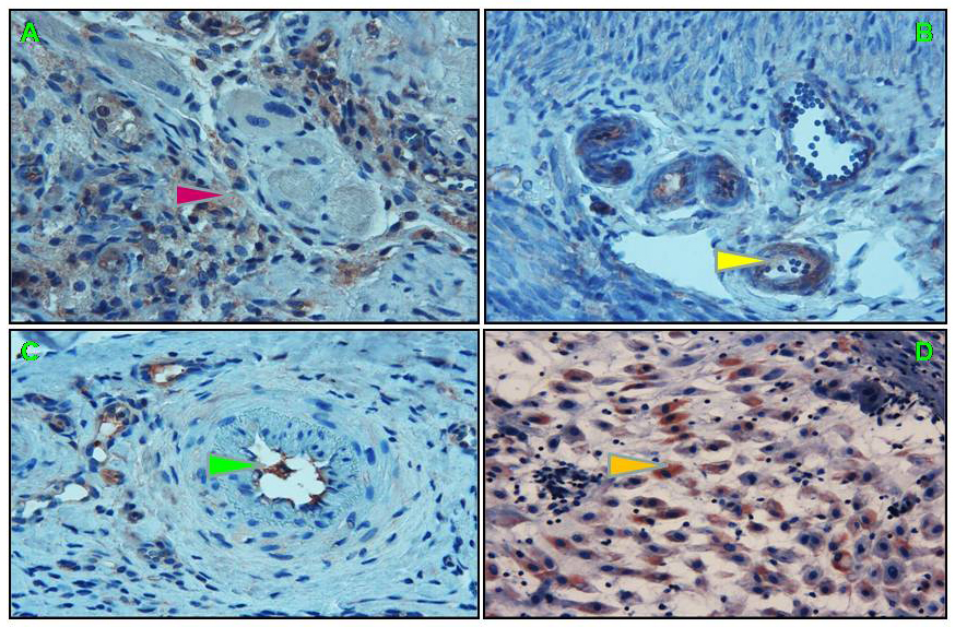 Fig. 1.
Immunohistochemical staining of endometrial samples for NF-B/p65.
(A) Increased NF-B/p65 immunoreactivity in the
endometrium collected from women with PCOS (red arrowhead, 20). (B) Weak
NF-B/p65 staining in the endometrium of non-PCOS controls (yellow
arrowhead, 20). (C) Weak NF-B/p65 immunoreactivity
predominantly localized into the cytoplasm of glandular epithelial cells in
fertile control (green arrowhead). (D) 3th trimester human placenta accepted as
positive control for NF-B/p65 (pink arrowhead, 20).
Table 2.Endometrial H-Score values of NF-B/p65
in PCOS and control groups undergoing total embryo freezing.
| Groups |
Endometrial H-Score of NF-B/p65 |
| I- PCOS with total embryo freezing (n = 40) |
H-score = 3.80 + 1.91 |
| II- Age and BMI matched control with total embryo freezing (n = 25) |
H-score = 2.03 + 1.14 |
| III- Fertile control (n = 5) |
H-score = 1.90 + 1.02 |
| I vs II |
0.001* |
| I vs III |
0.002* |
| II vs III |
0.040 |
| Data are presented as mean and SD. *p 0.05. H-score = Pi (I +
1), where Pi is the percentage of stained endometrial cells in each intensity
step (0%–100%), and i is the intensity showing weak (i = 1), moderate (i = 2),
or strong (i = 3) staining. |
Table 3.Correlation analysis between endometrial H-Score
of NF-B/p65 and measured parameters.
|
H-Score of NF-B/p65 expression in subjects undergoing total embryo freezing |
| r |
p |
| E2 on the day of hCG |
0.65 |
0.01 |
| Progesteron on the day of hCG |
–0.33 |
0.55 |
| Testosterone |
0.74 |
0.02 |
| Insulin |
0.66 |
0.01 |
| HOMA-IR |
0.58 |
0.04 |
| Total oocyte |
0.40 |
0.03 |
| Endometrial thickness |
0.32 |
0.04 |
| Total rFSH dose |
0.43 |
0.01 |
6. Discussion
Although it is not included in the diagnostic criteria of PCOS, systemic
inflammation is an important feature of PCOS [2, 3]. On the other hand, the number
of studies showing the presence of inflammation at the tissue level are few [1, 5].
It has been reported that NF-B phosphorylation is increased in
endothelial cell cultures of women with PCOS, while high androgen levels in
Ishikawa cell cultures block estrogen-induced receptivity gene expression [4, 14].
In the mid-luteal endometrial samples of PCOS cases, a significant increase in
NF-B/p65 expression has been reported [5].
In addition to adequate decidualization and activation of receptivity genes in
the endometrium, a physiologic amount of inflammation is also required for
successful implantation [12, 15]. The physiologic inflammatory response is
characterized by coordinate activation of different signaling pathways that
regulate expression of both pro- and anti-inflammatory mediators and receptivity
genes in endometrium [12]. Inflammation also allows other leukocytes to
accumulate at the implantation site, especially the uterine natural killer cell.
NF-B activation is critical for the flawless functioning of this
inflammatory process throughout implantation [12, 15]. In many tissues,
NF-B is activated through canonical and alternative pathways [13, 16].
Endometrial cells may be using canonical or alternative pathways for NF-B/p65 activation [15]. The canonical pathway is triggered by
proinflammatory cytokines such as tumor necrosis factor- and
IL-1 and leading to activation of proinflammatory molecules and receptivity genes
[11, 15]. Although the exact cause of the process initiating NF-B activation in
the endometrium is unknown, it has been accepted that the morphologic and
molecular changes that occur during the decidualization process are the main
inductors. Since all the pre-implantation preparation steps of endometrial tissue
are hormone-dependent, regulation of the synthesis and release of NF-B
may be regulated by sex hormones, differing from other tissues [11, 12, 15].
Despite the increase in the number of studies on implantation and
NF-B, the role of NF-B in embryo implantation has not been
extensively studied [5, 12]. Our study demonstrated increased pathologic
endometrial inflammation in endometrial samples taken on the day of egg
collection in PCOS cases with total embryo freezing due to the risk of OHSS. The
increased NF-B expression we detected in the PCOS patient group
is reliable evidence of pathologic endometrial inflammation, and it is a known
fact that this inflammatory process blocks the release of homeobox genes
responsible for basic receptivity [12, 17]. Possible causes of increased
endometrial inflammation in PCOS cases may be increased estradiol and
progesterone levels due to controlled ovarian stimulation. The positive
correlation between endometrial NF-B levels and estrogen levels
measured on the day of hCG in our study supports this idea. However, a recent
study conducted on patients with PCOS who did not undergo ovarian stimulation
reported that there is an intense pathologic endometrial inflammation in
mid-luteal samples which weakens our hypothesis [5]. Similarly, the normal
endometrial NF-B levels in our control group patients who underwent
ovarian stimulation despite high estrogen levels suggest that estrogens have no
effect on pathologic endometrial inflammation. Indeed, we found a positive
correlation between endometrial thickness, total oocyte count, rFSH dose and
endometrial NF-B levels which support that estrogens play a role in
endometrial NF-B expression. On the other hand, we found a negative and
insignificant correlation between pogesterone and NF-B levels. Since
physiologic levels of estrogen and progesterone regulate the expression of
endometrial receptivity genes, a supraphysiological increase in estrogen levels
due to ovarian stimulation may contribute to increased endometrial NF-B
expression in patients with PCOS [12, 15, 18].
Another possible cause of pathologic endometrial inflammation may be the high
androgen and insulin levels we detected in patients with PCOS. Increased levels
of inflammatory markers have been reported in most studies from the peripheral
blood of PCOS patients [19]. However, there is only one study investigating
inflammatory markers in the endometrial tissue of PCOS patients. In that study,
Koc et al. [5] reported that NF-B/p65 expression
levels in the endometrium of both normal and overweight PCOS patients increased
significantly compared to non-PCOS control subjects. The authors emphasized that
increasing NF-B levels independent of BMI is due to increased androgen
and insulin levels. Many studies have confirmed that chronic inflammation due to
PCOS is associated with increased androgen levels and insulin resistance
[1, 2, 3, 14]. While chronic inflammation in PCOS cases induces androgen increase,
increasing androgens also increase both androgen synthesis and insulin resistance
with a positive feedback effect [1, 2, 3, 14]. Although our cases were selected from
PCOS patients with normal BMI, HOMA-IR levels were found to be high. Previous
studies conducted on lean women with PCOS have showed that they are as equally
insulin-resistant as overweight women with PCOS, suggesting that insulin
resistance is independent of BMI [20, 21]. Therefore, it is not surprising that
our PCOS cases have high insulin levels despite a normal BMI and is consistent
with the literature. In our study, we found a positive and strong correlation
between serum testosterone and insulin levels and NF-B levels within the
endometrium. Consistent with our findings, Koc et al. [5] also found a
significant correlation between increased endometrial NF-B levels and
serum androgen and insulin levels in PCOS patients. Similarly, Cermik et
al. [4]. reported that increased androgen levels in PCOS patients caused
subfertility by decreasing Homeobox10 (HOXA10) gene expression. Gonzales
et al. [19] reported that intranuclear NF-B levels increased
significantly in PCOS patients with hyperglycemia and that this increase was
associated with insulin resistance and hyperandrogenism. Since serum glucose
levels of the PCOS group and non-PKOS cases were found to be similar in our
patients, we cannot make a clear comment on this issue. However, it has been
reported that increased circulating androgen levels of PCOS patients in
reproductive age increase the interest of cells in glucose use, leading to an
increase in NF-B expression [15].
There are many experimental and clinical studies showing the relationships
between androgens and insulin levels and chronic inflammation in PCOS patients.
It has been reported that by reducing phosphorylation of NF-B/p65, some
herbals could reduce hyperandrogenism in an animal model of PCOS [22, 23].
Inagreement with this, iridoids can protect patients with PCOS from inflammatory
damage by regulating the NF-B expression [23]. Exogenous application of
sera taken from PCOS patients to endothelial cell cultures led to a significant
increase in NF-B activation [24]. Similarly, in mononuclear cell lines
obtained from PCOS patients, the percent change in NFB activation was
positively correlated with androgens [15]. Another study demonstrated that the use
of exogenous androgens significantly decreased the expression of endometrial
receptivity genes in women with PCOS [4]. A recent study reported that serum
NF-B levels were significantly decreased in patients given metformin or
cyproterone acetate therapy for PCOS compared with those who were not treated.
The reduction of NF-B levels by antiandrogen or antiprogestin therapy
strongly supports the role of androgens in the pathologic inflammation [25]. The
positive correlation between serum testosterone and insulin levels and
endometrial NF-B/p65 found in our study is strong evidence of the clear
relationship between hyperandrogenemia, HOMA-IR and increased pathologic
inflammation.
As in all other tissues, NF-B has long been accepted as a prototypical
proinflammatory signaling pathway in the endometrium. Endometrial NF-B
expression levels measured in the follicular phase in healthy and fertile
individuals were reported to be higher than the NF-B levels detected in
both the secretory and menstrual phases [15, 26, 27, 28]. Unlike the endometrial
NF-B expression patterns of healthy individuals, NF-B
expression patterns in the eutopic endometrium of women with an endometrioma,
endometriosis, hydrosalpinx and PCOS is disrupted [26, 27, 28, 29]. Implantation rates
decrease significantly in women with endometriosis or hydrosalpinx that cause an
increase in inflammation in the endometrium [4, 12]. Surgical removal of the
endometrioma or hydrososalpinx normalized the pathologic increase in endometrial
NF-B levels [1, 12]. All of these findings are important in terms of
showing that some benign gynecologic diseases located outside the endometrium may
trigger pathologic inflammation in the eutopic endometrium. Similarly, although
PCOS is located outside the endometrium, it can trigger pathologic inflammation
in the endometrium due to hormonal changes and chronic inflammation [1, 2, 3].
Consistent with our findings, it has been reported that NF-B expression levels
in the endometrium of PCOS patients with normal and high BMI increased
significantly compared to healthy controls [5]. Likewise, in accordance with the
other studies, expression levels of HOXA10 and HOXA11, which are the main
regulator genes of endometrial receptivity, have been reported to be low in women
with PCOS [15]. Senturk et al. [30] showed that endometrial HOXA10 and 11
levels of PCOS patients were significantly decreased compared to fertile controls
and that their expression progressed to normal levels after laparoscopic ovarian
drilling. When the results of these studies and our findings are evaluated
together, they indicate that the functions of receptivity molecules and
NF-B pathway, which are responsible for implantation and physiologic
inflammation, are impaired in women with PCOS.
Our study showed that total embryo freezing not only prevented the development
of OHSS, but also delayed embryo transfer, thus preventing an unsuccessful
implantation. Delaying transfer by freezing embryos in PCOS cases with a high
risk of OHSS may save the clinician time required for the recovery of the
endometrium. In the following cycles of these patients, re-preparing the
endometrium and performing frozen embryo transfer may allow improvement for
successful implantation. However, it is not known how many cycles the pathologic
inflammation in the endometrium is decreased in PCOS cases with embryo freezing.
Endometrioma and hydrosalpinx studies potentially could reveal clarifying data on
this issue. Expression levels of endometrial receptivity genes have been
demonstrated to reach fertile levels within 3 to 4 months after endometrioma
resection or salpingectomy [11, 16]. Similarly, in PCOS cases with laparoscopic
ovarian drilling, endometrial receptivity was normalized in control endometrial
sampling 3 months following surgery [30].
Our study has three main limitations. First, since the effect of OHSS in the
patients in the control group was not excluded, OHSS-related changes in the
endometrium of these patients continue. For this reason, there is a need for
future studies in which a group of only non-PCOS patients who had OHSS, whose age
and BMI are matched, are added to the study. The second limitation is that it was
not questioned whether the cases in the fertile group had PCOS findings. The last
limitation is that comparisons could not be made in the patient and control
groups with similar BMI in our study.
7. Conclusions
In conclusion, endometrium of women with PCOS who underwent total embryo
freezing due to the risk of OHSS lack the physiologic inflammation conditions
which are favorable for implantation. Freezing the embryo in these patients will
both prevent the deterioration of OHSS and save time for the clinician in order
to have the endometrium suitable for implantation.
Author contributions
CK, NT and RO conceived, designed and performed the study procedures; CK, NT and
RO analyzed the data and contributed reagents and materials; CK wrote the paper.
All authors contributed to editorial changes in the manuscript. All authors read
and approved the final manuscript.
Ethics approval and consent to participate
This study was conducted in accordance with the Declaration of Helsinki. Ethical
approval was obtained from the local Ethics Committee of the Beykent University
(Approval number: 2020/11). Informed consent was obtained from all participants
at the time of enrollment.
Acknowledgment
We thank participants, anonymous reviewers for excellent criticism of the
article.
Funding
This research received no external funding.
Conflict of interest
The authors declare no conflict of interest.


 , Nurettin Turktekin 2, Ramazan Ozyurt 3