
Academic Editor: Shigeki Matsubara
Background: To investigate the
clinical effect of sphincter repair combined with perineal body reconstruction in
the treatment of old fourth-degree perineal tears. Method: A prospective
database of five patients with old fourth-degree perineal tears treated between
January 2015 and January 2021 was established and studied retrospectively.
Results: All five patients were followed-up with for 1 year, and anal
incontinence was cured. The anal function recovered, and the anal shape was
satisfactory. Three months after the operation, transperineal ultrasonography
showed continuity of the anal sphincter, anal mucosa, and perianal skin.
Anorectal manometry was performed 3 months after the operation. The anal resting
pressure (56.20
With the improved awareness of perineal laceration and the strengthening of professional training, the reporting rate of third- and fourth-degree perineal tears has increased annually [1]. Among women who give birth vaginally, 85% suffer from perineal trauma, 69% of whom require suturing [2]. Fourth-degree perineal tear is the most serious perineal trauma after delivery, although the effect is satisfactory after timely repair. Therefore, old fourth-degree perineal tears are relatively rare in the clinic. However, in the past, in China’s rural areas, the medical level was relatively low, the number of parturients was higher, and many delivered at home. Thus, if perineal tear was not treated in time, it resulted in perineal laceration. With increasing age, pelvic floor muscle relaxation and anal incontinence became more serious, which greatly affected the physical and mental health and quality of life of patients.
At present, there are many repair methods for perineal laceration, including transvaginal repair, rectal moving valve technology, and biological patch repair technology [3]; however, these are difficult to perform during operation, and the postoperative recurrence rate is high. Here, we report the treatment of five cases of old fourth-degree perineal tears with transperineal sphincter repair combined with perineal body reconstruction.
This retrospective study was approved by the Ethics Committee of the Affiliated Hospital of Southwest Medical University. Signed informed consent was obtained from each patient preoperatively. The clinical data of five patients with old fourth-degree perineal tears who were treated between January 2015 and January 2021 were analyzed retrospectively. On admission, physical examination showed that the perineum was looking abnormal; the anus, rectum, and vagina were completely connected, and the anal sphincter was loose and weak (Fig. 1). All patients underwent routine laboratory examinations, colonoscopy, and anorectal manometry (ARM), excluding serious systemic diseases and psychosis.
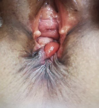 Fig. 1.
Disappearance of the normal perineal structure, with complete connection of the anus, rectum, and vagina.
All patients took oral intestinal anti-microbial agent drugs (metronidazole tablets, 0.2–0.4 g/tablet, three times/day) 3 days before the operation and consumed a residue-free diet. Intestinal preparation was performed by oral administration of compound polyglycol electrolyte powder 1 day before the operation, and enema was performed once in the evening of the day before and in the morning of the day of the operation. The vagina was rinsed with dilute iodophor 1 day before and on the day of the operation. When the intestine and vagina were fully prepared, antibiotics (cefuroxime) were injected intravenously 0.5–2 h before the operation to prevent infection.
All patients were treated with spinal anesthesia. After satisfactory anesthesia, the bladder lithotomy position was taken, an indwelling catheter was inserted, a routine disinfection towel was placed, an iodophor cotton ball was used to disinfect the lower segment of the vagina, anal canal, and rectum, and a gauze roll was used to fill the anus. The specific instructions were as follows. (1) The operating field was exposed, before separating the labia minora to both sides and sewing them onto the labia majora and medial thigh (Fig. 2); (2) an “X” incision was made, before cutting open the skin and subcutaneous tissue, and freeing it fully as a flap; (3) after local submucosal injection of 1:200,000 U epinephrine, arcuate incision of the rectovaginal septum, separation of the rectovaginal septum, and exposure of the levator ani muscle, the broken ends of the sphincter were freed on both sides; (4) the broken ends of the levator ani and sphincter were sutured, layer-by-layer from top to bottom, and each cavity was closed (the rectal wall and posterior vaginal wall were sutured intermittently); (5) the flap was cross-transferred and then suture to reconstruct the perineal body; and (6) a drainage strip was placed under the skin and an anal tube was inserted in the anus.
 Fig. 2.
Operation steps. (A) An “X” incision was made, before cutting the rectovaginal septum, separating the rectovaginal septum, exposing the levator ani muscle, and freeing the broken end of the sphincter to both sides. (B) The broken ends of the levator ani and sphincter were sutured layer-by-layer from top to bottom, and each cavity was closed. (C) Drainage strips were placed subcutaneously and anal tubes were inserted in the anus.
The patients were asked to stay in bed until the sutures were removed. Generally, the suture was removed 8–10 days after the operation. Postoperative defecation was controlled for 5–7 days. Patients fasted for 1–2 days and consumed a residue-free diet for 3–5 days. Patients took a laxative on the 4th day after the operation and it was ensured that loose and soft stool was discharged within 2 weeks of the operation. Patients consumed soft, easily digested, high-calorie food to prevent constipation and excessive force. The catheter was retained for 5–7 days, the anal canal was maintained for as long as possible until defecation, and infection was prevented with antibiotics for 72 h.
One month after the operation, anal function was evaluated based on anal incontinence. Firm and loose stools were cured by self-control; automatic control of dry stool and poor control of loose stool were considered as positive treatment outcomes; and no improvement in defecation was considered as a negative treatment outcome. The shapes of the perineum and anus were visually observed to check if the skin was continuous. ARM and transperineal ultrasonography were performed 3 months after the operation to observe the continuity of the anal sphincter, anal mucosa, and perianal skin. The anal continence was evaluated using the Wexner continence grading system [4] (Table 1), and the score was calculated before the operation and 3, 6, and 12 months after the operation. A Wexner score of 0 indicated normal function, while a score of 20 indicated complete incontinence.
| Variables | Never | Occasionally | Sometimes | Usually | Always |
| Flatus | 0 | 1 | 2 | 3 | 4 |
| Liquid stools | 0 | 1 | 2 | 3 | 4 |
| Solid stools | 0 | 1 | 2 | 3 | 4 |
| Wears pad | 0 | 1 | 2 | 3 | 4 |
| Alteration in lifestyle | 0 | 1 | 2 | 3 | 4 |
SPSS 25.0 statistical software (IBM Corp., Chicago, IL, USA) was used to conduct statistical analysis. The
measurement data are expressed as the mean measurement data
(
Five patients with old fourth-degree perineal tears with an average age of 56
The mean operative time was 110
All five patients were followed-up with for 12 months, at which point, anal
incontinence was significantly improved, defecation and exhaust could be
controlled, and all patients were cured. One month after the operation, the shape
of the perineum and anus of the patients was complete, and the skin was well
aligned and continuous (Fig. 3). The mean perineal body length was 3.58
 Fig. 3.
Conditions after the operation. (A) There was no obvious blood and fluid leakage at the incision on the first day after the operation. (B) There was no incision dehiscence after suture removal on the 10th day after the operation. (C) One month after the operation, the shape of the perineum and anus was complete, and the skin was well aligned and continuous.
Perineal laceration is a common complication among pregnant women who give birth vaginally. The classification of perineal laceration was first proposed by Sultan et al. [5], before being adjusted by the International Consultation on Incontinence and the Royal College of Obstetricians and Gynaecologists [6]. Fourth-degree perineal tears occur due to damage to the inner and outer anal sphincter and anorectal mucosa, which will not only cause physiological diseases but also cause serious psychological distress. The risk factors of third- and fourth-degree perineal tears include Asian primipara, macrosomia, shoulder dystocia, occipital posterior position, extension of the second stage of labor, and instrumental midwifery [7, 8]. It is crucial that clinicians have a full understanding of the risk factors for of third- and fourth-degree perineal tears.
The main clinical manifestation after fourth-degree perineal tears is anal incontinence. If fourth-degree perineal tears are not treated in a timely manner, or after treatment failure, they will lead to old perineal laceration. With increasing age, pelvic floor muscle relaxation, muscle weakness, muscle retraction at the torn ends on both sides of the anus, and the symptoms of anal incontinence become more serious; at which point, patients often seek medical treatment. The effective treatment of fourth-degree perineal tears is still a challenging topic, although for most patients, surgical treatment should be the first choice. The key lies in the repair of the anal sphincter and the reconstruction of the perineal body. However, the main reason for the failure of surgery is incision infection and incision tension. Some surgeons have improved the complex injury of anal sphincter repair by including inner anal sphincter repair and outer anal sphincter repair [9]. In cases with external anal sphincter tears, the risk of fecal incontinence caused by overlapping suture is lower [10]. The overlap technique requires more dissection and mobilization of the sphincter ends, along the ischioannal fossa [11]. However, in the actual repair operation of anal sphincter laceration, because the boundary between the internal and external sphincter of the anus is unclear, we did not distinguish them for suturing in our patient group. The advantages are rich blood supply at the torn ends of the free sphincter and better tension resistance at the broken end of the muscle, which can effectively avoid the risk of postoperative incision laceration and recurrence.
In the present study, all patients had good perineal and anal healing after the operation. Perineal ultrasonography showed continuity of the anal sphincter, anal mucosa, and perianal skin. The postoperative anal resting pressure, maximum anal systolic pressure, and maximum anal systolic time were better than those before the operation, and the postoperative Wexner score was significantly improved.
We are of the opinion that surgeons should still be cautious. For example, with respect to perioperative management, with respect to perioperative management, the bowel and vagina should be fully prepared before the operation, the incision must be kept clean to prevent incision infection, stool must be kept soft after the operation, and excessive force must be avoided. With respect to intraoperative problems, strict aseptic operation is required during the operation, a full layer suture must be performed during the reconstruction and suture of the perineum without dead space, and the suturing of the skin and muscle must be tension-free.
The main purpose of the treatment of fourth-degree perineal tears is to improve anal function. We used transperineal sphincter repair combined with perineal body reconstruction to provide a new idea for the clinical treatment of anal incontinence after grade IV perineal laceration. However, we acknowledge that the small number of cases as well as the short duration of follow-up are limitations of this preliminary study. This is only a short term management evidence, the patients will be followed up for a longer time, and long term follow-up data will be published later.
Transperineal sphincter repair combined with perineal body reconstruction is safe and effective in the treatment of old fourth-degree perineal tears. The initial results are encouraging, indicating the need for a more formal evaluation of the technology.
Study concept and design—QL; Acquisition of data—JL; Analysis and interpretation—JL; Study supervision—QL. All authors read and approved the final manuscript.
This study was reviewed and approved by the Ethics Committee of the Affiliated Hospital of Southwest Medical University (KY2020142). All patients signed informed consent.
We wish to acknowledge the hard and dedicated work of all the staff that implemented the intervention and evaluation components of the study.
This research received no external funding.
The authors declare no conflict of interest.
Publisher’s Note: IMR Press stays neutral with regard to jurisdictional claims in published maps and institutional affiliations.