IMR Press / FBL / Volume 28 / Issue 2 / DOI: 10.31083/j.fbl2802035
Open Access Review
Aquaporins and Neuropathic Pain
Show Less
1 Department of Anesthesiology, The Affiliated Hospital of North Sichuan Medical College, 637000 Nanchong, Sichuan, China
*Correspondence: linjingyan@nsmc.edu.cn (Jing-yan Lin)
Front. Biosci. (Landmark Ed) 2023, 28(2), 35; https://doi.org/10.31083/j.fbl2802035
Submitted: 13 October 2022 | Revised: 28 November 2022 | Accepted: 8 December 2022 | Published: 24 February 2023
Copyright: © 2023 The Author(s). Published by IMR Press.
This is an open access article under the CC BY 4.0 license.
Abstract

Neuropathic pain is a chronic secondary pain condition resulting from lesions or diseases of the peripheral or central nervous system (CNS). Neuropathic pain is closely related to edema, inflammation, increased neuronal excitability, and central sensitization caused by glutamate accumulation. Aquaporins (AQPs), mainly responsible for the transport and clearance of water and solute, play important roles in developing CNS diseases, especially neuropathic pain. This review focuses on the interaction of AQPs with neuropathic pain, and the potential of AQPs, especially aquaporins 4, as therapeutic targets.

Keywords
aquaporins
neuropathic pain
glymphatic system
review
1. Introduction

Aquaporins (AQPs) are water channel proteins essential to life and expressed in all kingdoms [1]. To date, thirteen AQPs were identified in human body and found widely distributed in specific cell types in various organs and tissues [2]. Four AQP monomers, each consisting of six transmembrane
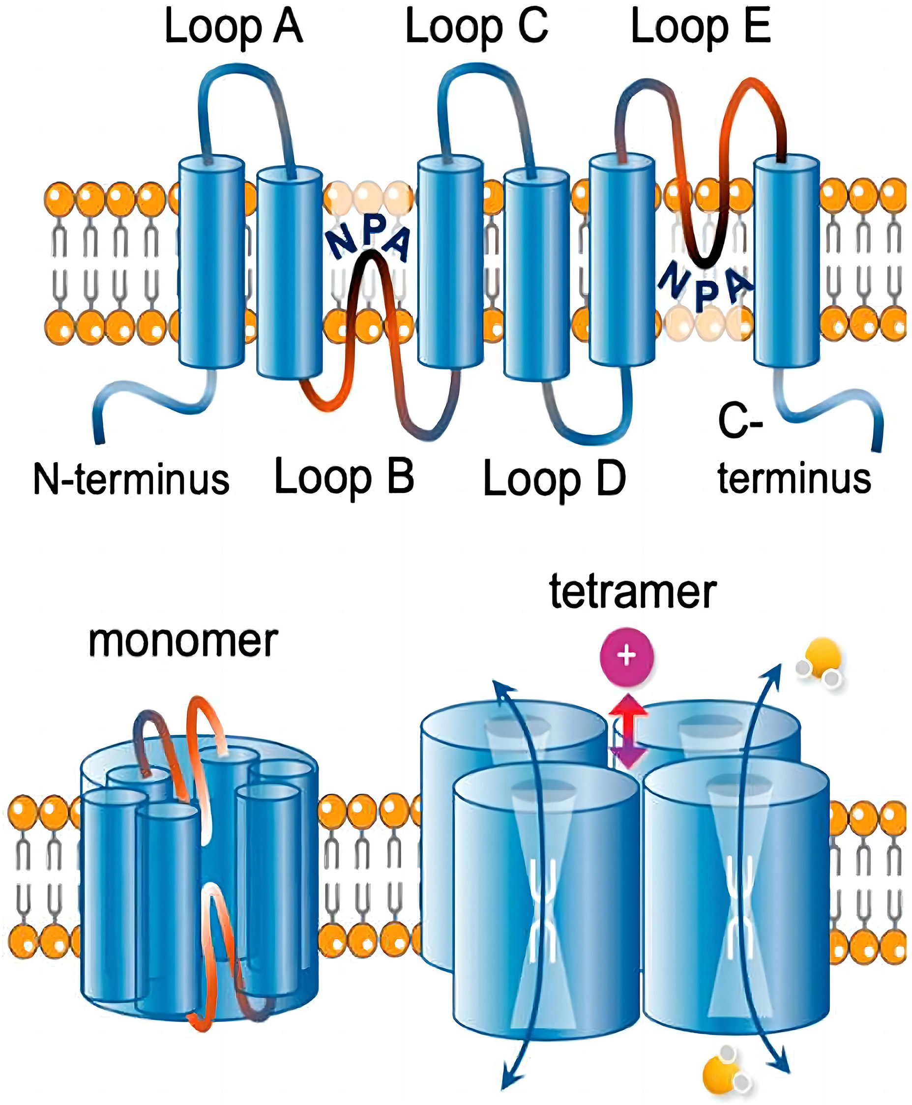
 α-helices with a central water transport pore, aggregate to form tetramers that are functional units in the membrane, thereby facilitating fluid movement across the cell [2]. These thirteen AQPs contribute to the secretion of body fluids such as cerebrospinal fluid, tears, saliva, sweat and bile, and the concentration of urine [3]. In addition to mediating water transportation, AQPs also facilitate gas (CO2) and cation transport, as well as participate in cell signaling [4]. Moreover, AQPs have been implicated in the development of a range of diseases, such as vision loss, skin barrier loss, kidney disease, brain edema after stroke or head trauma, breast and reproductive cancers, and Parkinson’s disease [5, 6, 7] (Fig. 1, Ref. [6]).

Fig. 1.

Transmembrane topology of an aquaporin. Reproduced from [6] under the terms and conditions of the Creative Commons Attribution (CC BY) license (https://creativecommons.org/licenses/by/4.0/). Copyright 2020 Khan, Ricciardelli and Yool. Each monomer consists of six transmembrane helices connected by loops A to E. Loops B and E typically carry signature NPA (Asn-Pro-Ala) motifs that fold together within each subunit to form a water pore. Four monomers form a tetramer (the functional channel). A subset of AQPs uses the central pore as a gated ion channel. AQPs, aquaporins.

Neuropathic pain is a chronic secondary pain condition resulting from lesions or diseases of the peripheral or central nervous system (CNS) [8, 9, 10]. It is estimated that around 5% of the population suffers from neuropathic pain [11]. Most patients complain of an ongoing or intermittent spontaneous pain of, for example, burning, pricking, or squeezing quality, which may be accompanied by evoked pain, particularly to light touch and cold [12]. Studies have indicated that the ectopic activity in damaged or adjacent nerves, dorsal root ganglia or central pathways, peripheral and central sensitization and a range of molecular mechanisms are involved in the development of neuropathic pain [11, 12] (Fig. 2).
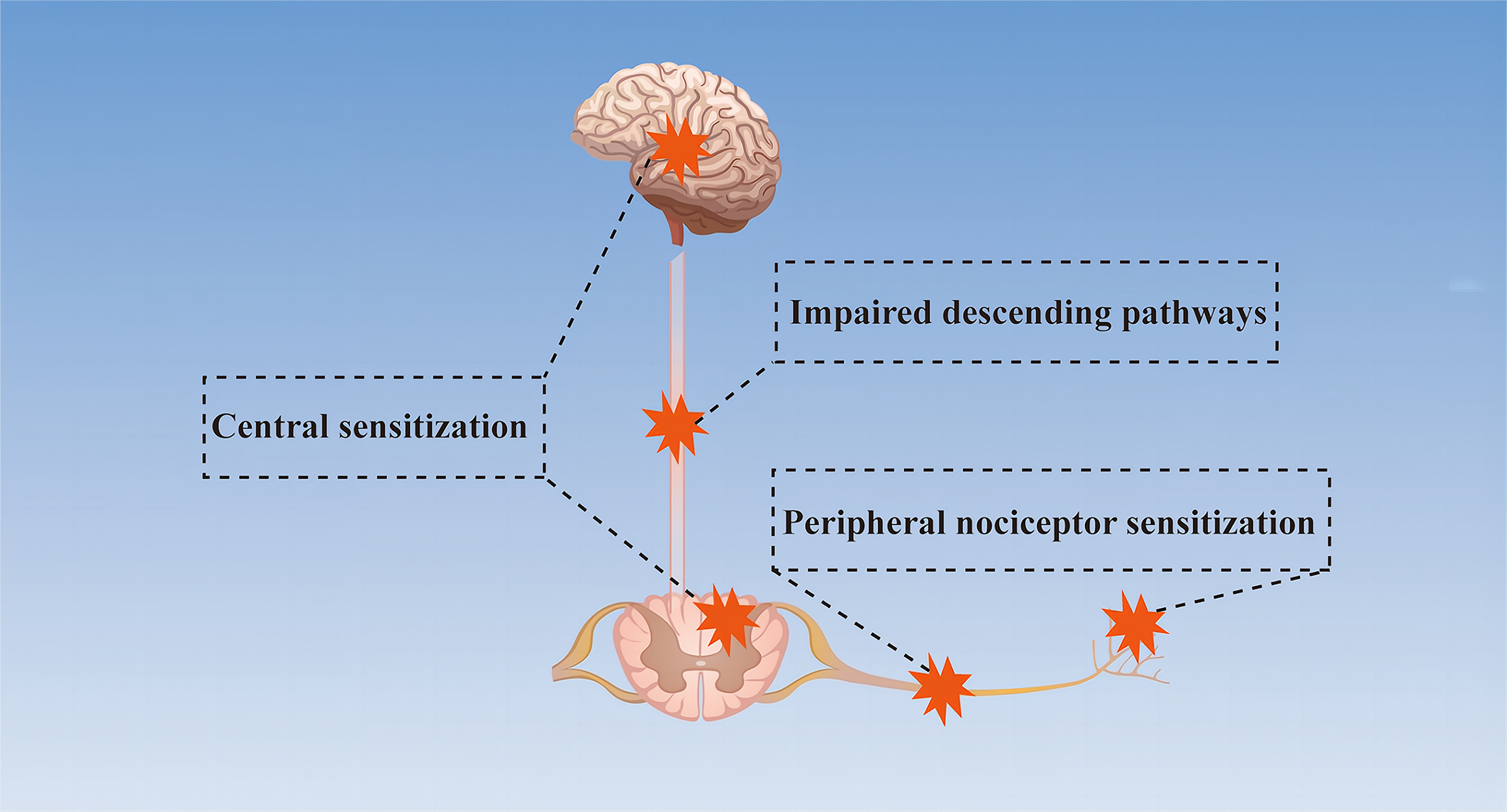
Fig. 2.

Simplified mechanism diagram of neuropathic pain. Peripheral nociceptor sensitization due to abnormal nociceptors and primary afferents, impaired descending pathways between the brain and spinal cord, and central sensitization caused by the imbalance between excitatory and inhibitory neurotransmitters are the mechanisms of neuropathic pain.

Studies have shown that several aquaporins are closely related to neuropathic pain, including aquaporin 1 (AQP1), aquaporin 2 (AQP2), aquaporin 4 (AQP4), aquaporin 5 (AQP5) and aquaporin 9 (AQP9) [13, 14, 15, 16, 17]. Since AQPs promote water and solute transport and are involved in the occurrence and development of many diseases, it is logical that AQPs are involved in the occurrence and development of neuropathic pain. This review focuses on our current understanding of AQPs and neuropathic pain and the potential of these AQPs as therapeutic targets.

2. AQP1

AQP1, the first water channel being identified, functions as a gas channel and can increase CO2 permeation of lipid bilayers, likely through the water pore of the protein [2, 18]. In addition, AQP1 is widely distributed in the body. It controls the transport and flow of water bi-directionally around the cell membranes and is distributed in kidney, various membranes (epithelial cells), red blood cells, sweat glands, lungs, and gastrointestinal trajectories [2, 19]. At present, AQP1 has been manifested in many diseases. For example, Mir-3194-3p inhibits the progression of breast cancer by targeting AQP1, silencing AQP1 can improve the cognitive function of Alzheimer’s disease mouse model through the Wnt signaling pathway, glioma-associated oncogene homolog 1 can promote glioma cell metastasis by regulating AQP1, and naringin can alleviate airway inflammation by upregulating AQP1 expression [20, 21, 22, 23].

Interestingly, in addition to regulating water and gas function, AQP1 has also been implicated in pain. Studies have indicated that AQP1-/- mice were shown to be less reactive to thermal and capsaicin chemical stimulation, but not to mechanical and formalin stimulation, suggesting a possible role for AQP1 in pain signaling [24]. In addition, AQP1 in dorsal root ganglion neurons is involved in the perception of inflammatory heat and cold pain, and its molecular basis is partly caused by reduced Na(V) 1.8-dependent membrane Na (+) current [25]. These results suggest that AQP1 may participate in pain perception through ion channels. AQP1 is involved in pain perception, but the effect of AQP1 knockout on pain is still controversial [26]. In addition, studies have shown that the demise of nociceptive Schwann cells with AQP1 as one of its markers can cause neuropathic-like pain in mice [27]. These results suggest that AQP1 is closely related to neuropathic pain.

In a clinical study on leprosy, Salgado et al. [28] found that AQP1 may be involved in the loss of sensation or leprosy neuropathic pain. Moreover, animal experiments have shown that inhibition of AQP1 expression alleviated neuropathic pain caused by chronic dorsal root ganglion compression and melatonin decreased mechanical allodynia by regulating AQP1 [14, 29]. In the treatment of a series of diseases targeting AQP1, many traditional Chinese medicines were found to play therapeutic effects by regulating AQP1, such as alpinetin, dachengqi, and shenmai injection [30, 31, 32]. Interestingly, AQP1 was shown to increase the sensitivity of acetazolamide and cisplatin in the treatment of cancer, suggesting a positive potential antitumor role of AQP1 [33, 34]. Overall, AQP1 may be involved in pain perception through ion channels and, in this way, in the development of neuropathic pain.

3. AQP2

Water balance plays an important role in maintaining the normal function of the body, and water reabsorption by the collecting duct of the kidney is one of its key factors, which is regulated by AQP2 [35]. AQP2, a channel that is selective only for water molecules and impermeable to ions or other small molecules, is an aquaporin regulated by arginine vasopressin [2, 35]. Normal functioning of AQP2 depends on multiple posttranslational modifications, and aberrant arginine vasopressin signaling and altered AQP2 expression or transport can lead to diseases characterized by dysregulation of mechanisms controlling water homeostasis [36]. Impairment of AQP2 results in various water balance disorders, including disorders associated with polyuria (urinary tract obstruction, hypokalemia, inflammation, and lithium intoxication), dilutive hyponatremia (improper antidiuretic syndrome, congestive heart failure, cirrhosis syndrome), and Ménière’s Disease [37, 38].

In addition to its role in transporting water, mainly to the kidneys, AQP2 also appears to be involved in pain. In an acute inflammatory pain animal model, expression of AQP2 in the trigeminal ganglia was increased, and the redistribution of AQP2 was mainly identified in small-sized neurons and Schwann cells. The above results indicated the involvement of AQP2 in pain transmission in the peripheral nervous system [39]. Sciatic nerve injury, a classic pain model, is widely used in the study of neuropathic pain. The expression of AQP2 was increased in the small dorsal root ganglion neurons of rats with chronic contraction injury and in sciatic nerve crush injury models [15, 40]. Moreover, it is worth mentioning that erythropoietin protects against neuropathic pain by regulating AQP2 expression in a rat model of chronic contractile injury in vivo [41]. These results suggest that AQP2 may be involved in inflammatory nerve injury and may play a role in promoting regeneration after nerve injury as well as in the treatment of neuropathic pain.

Since AQP2 is highly involved in the water transportation of the kidneys, the research on renal diseases targeting AQP2 has become one of the current hot spots. For example, statins ameliorate cholesterol-induced inflammation and improve AQP2 expression by inhibiting NOD-like receptor thermal protein domain associated protein 3 (NLRP3) activation in the kidney, thus contributing to the treatment of chronic kidney disease [41]. Protein kinase A may treat congenital nephrogenic diabetes insipidus by activating AQP2 [42]. Zhen-wu-tang reduces renal edema induced by doxorubicin by regulating AQP2 and Mir-92b [43]. Steviol slows renal cyst growth by reducing AQP2 expression and promoting AQP2 degradation [44]. In addition, electroacupuncture alleviates arginine vasopressin-induced endolymphatic hydrops by regulating AQP2 and vincamine is capable of suppressing endolymphatic hydrops formation by down-regulating the VAP/AQP2 signaling pathway [45, 46]. These results suggest that AQP2 can be used as a target for the treatment of renal diseases and Ménière’s Disease. However, the role of AQP2 in neuropathic pain and its potential as a therapeutic target of neuropathic pain needs to be further studied.

4. AQP4

Among the 13 identified AQPs, AQP4 has become an interesting therapeutic target in various neurological disorders, due to its variety of functions and widespread expression in the CNS [47]. AQP4 is the most abundant water channel in the brain, spinal cord, and optic nerve. It controls brain water homeostasis [48]. AQP4 monomers have a molecular size of ~30 kDa and contain six membrane-spanning helical segments and two shorter helical segments that only partly span the membrane [49]. According to the translation start sites, AQP4 can be divided into 2 major isoforms, M1 and M23 [48, 49]. AQP4 can form crystal-like supramolecular assemblies in the plasma membrane, which are called orthogonal arrays of particles (OAPs) [50, 51]. Moreover, OAPs have been identified as the target of anti-AQP4 antibodies in neuromyelitis optica spectrum disorder (NMOSD) [50, 51, 52].

In addition to its primary water transport function, AQP4 also regulates astrocyte migration, participates in neural signal transduction, and regulates neuroinflammation [49]. It is worth mentioning that the increased AQP4 expression and the redistribution/surface localization are two different concepts. Previous studies have shown an increase in AQP4 membrane localization in primary human astrocytes which wasn’t accompanied by a change in AQP4 protein expression. This mislocalization was supposed to be a potential therapeutic target [53, 54, 55]. Many diseases such as hypothermia, painful diabetic neuropathy, Alzheimer’s disease and epilepsy have been manifested to be associated with impaired expression or mislocalization of AQP4 [12, 17, 56, 57, 58]. Interestingly, recent studies have demonstrated the existence of the glymphatic system, which consists of AQP4 and astrocytes and is responsible for removing metabolic waste products from the CNS [17, 59] (Fig. 3, Ref. [6]). Currently, there are many studies on the glymphatic system, and MRI is the most commonly used device. In terms of contrast agent selection, the newly discovered contrast agent that can be used to observe the paracellular flow and diffusive transcellular exchange of water is H
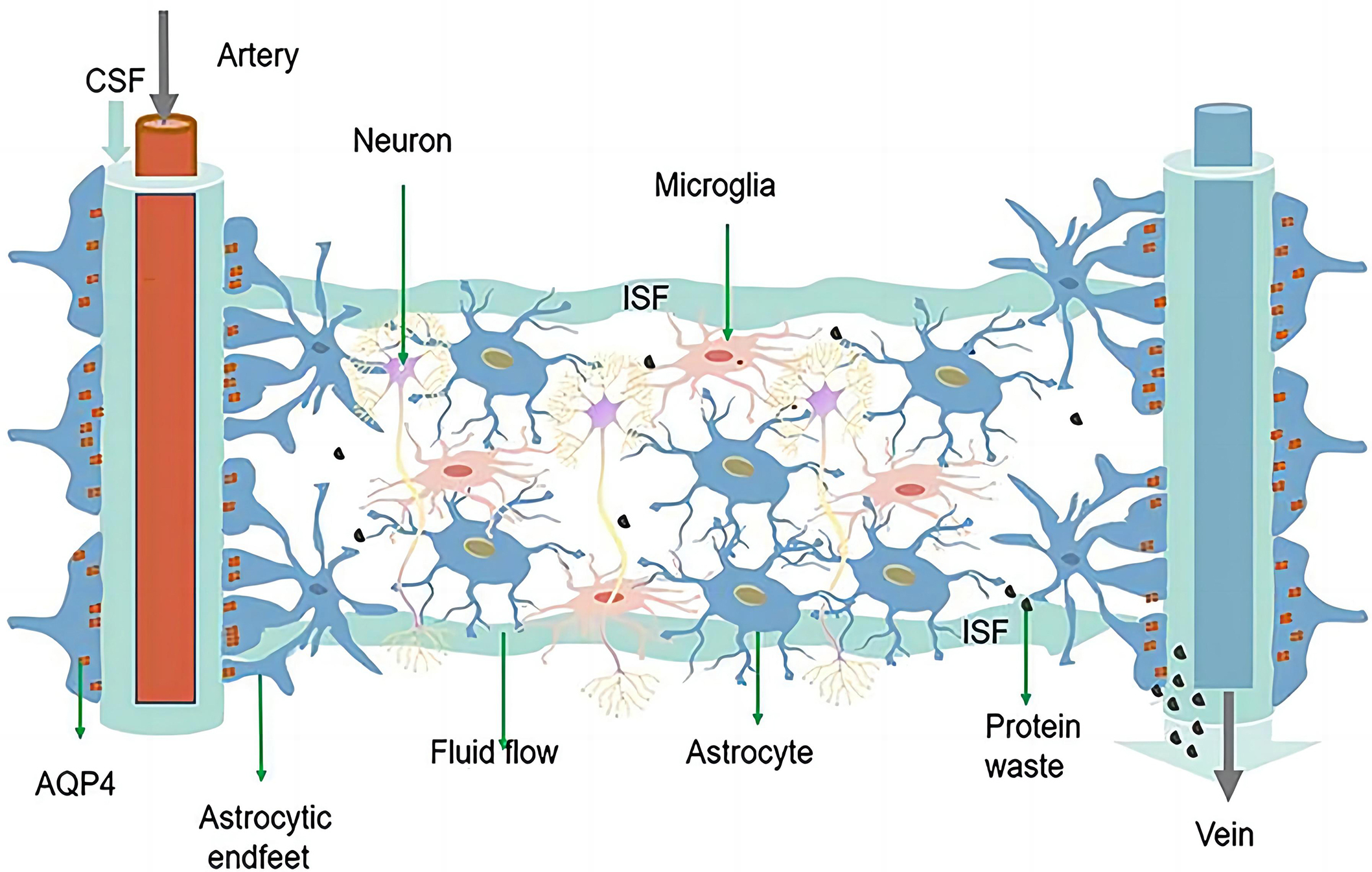
217O, which contributes a lot to our better understanding of the glymphatic system [60, 61]. Studies have demonstrated that both AQP4 and the glymphatic system are involved in the occurrence and development of many diseases, like Alzheimer’s disease, chronic pain, painful diabetic neuropathy, and perioperative neurocognitive disorders [11, 12, 17, 59, 62, 63]. Since waste removal is essential for the maintenance of normal function in the CNS, which lacks traditional lymphatic vessels, the emergence of the glymphatic system provides new insights into further understanding of neurological diseases [64, 65]. The glymphatic system with AQP4 as the core is responsible for the excretion of metabolic waste such as glutamate (excitatory neurotransmitter) and water balance in the CNS [17, 59]. The inflammation, edema, and neuronal hyperexcitability are also one of the pathogeneses of neuropathic pain [66, 67]. Therefore, there is a strong link between AQP4 and neuropathic pain, which has been demonstrated by many studies [68, 69, 70]. Since AQP4 is the aquaporin that is most closely associated with neuropathic pain discovered so far, we will discuss several diseases that are accompanied by neuropathic pain in detail (Fig. 3).

Fig. 3.

The schematic diagram of the glymphatic system. Reproduced from [6] under the terms and conditions of the Creative Commons Attribution (CC BY) license (https://creativecommons.org/licenses/by/4.0/). Copyright 2021 Ren, Liu, Lian, Li, Li, Li and Zhao. The glymphatic system is composed of astrocytes and AQP4 located at the endfeet of astrocytes. CSF enters the brain parenchyma through the periarterial space, exchanges with ISF, and finally exits through the perivenous space. AQP4 can facilitate this fluid exchange, through which solutes and metabolic waste products (e.g., glutamate) from the interstitium are exchanged, and finally expelled from the brain by the meninges and cervical lymphatics. AQP4, aquaporin 4; CSF, Cerebrospinal fluid; ISF, interstitial fluid.

4.1 Neuromyelitis Optica

Neuromyelitis Optica (NMO) is a clinical syndrome characterized by attacks of acute optic neuritis and transverse myelitis [51, 52, 68]. In most patients, NMO is caused by pathogenetic serum IgG autoantibodies to AQP4 (AQP4-IgG) and IgG autoantibodies to myelin oligodendrocyte glycoprotein (MOG-IgG), and the term NMOSD is used to refer to NMO and its formes frustes [68]. It has been reported that NMO occurs in all ethnicities around the world, with significant regional differences, and non-white individuals have higher incidence and prevalence rates [71]. AQP4-IgG associated NMO is characterized by IgG and complement deposition occurring at the endfeet of astrocytes, often accompanied by loss of astrocytes, oligodendrocytes, and neurons, whereas MOG-IgG associated NMO is characterized by demyelination with dominant loss of MOG and relative preservation of axons and oligodendrocytes [68].

Previous studies have shown a high negative correlation between pain severity and quality of life in NMO patients [72]. At present, the mechanism of neuropathic pain associated with NMO is still unclear. The loss of excitatory amino acid transporter 2 mediated by AQP4-IgG leads to the imbalance between excitation and inhibition in the nociceptive pathway caused by extracellular glutamate accumulation, the increase of local nerve growth factor concentration mediated by MOG-IgG, abnormal sprouting of injurious spinal cord fibers and inflammatory lesions have been reported to be involved in the occurrence and development of NMO [68, 73, 74]. The AQP4-based glymphatic system is responsible for the transport of glutamate and other metabolic wastes and has been reported to be a therapeutic target for Alzheimer’s Disease [75]. Since NMO mostly manifests as AQP4-IgG, its pathogenesis is mainly central sensitization caused by glutamate transport disorder, and the AQP4-based glymphatic system can promote glutamate transport [68, 75]. Therefore, there must be a close relationship between NMO and the glymphatic system based on AQP4.

There are several treatments available for NMOSD, such as conventional immunosuppressants, B cell-depleting agents, interleukin-6 signaling blocking agents, complement blocking agents, and intravenous immunoglobulins [76]. It is worth noting that the research on the interleukin-6 (IL-6) signaling pathway is hot at present. The plasmablasts in the peripheral blood of NMOSD patients with positive AQP4-IgG are increased, IL-6 drives these plasma cells to produce and secrete AQP4-IgG, whereas an IL-6 blocking agent reduces the production of AQP4-IgG [77]. For example, two IL-6 blocking agents, tocilizumab and satralizumab, have been shown to have good therapeutic effects on NMOSD and are potentially effective and safe therapeutic methods for relapse prevention in NMOSD [76].

4.2 Spinal Cord Injury

Spinal cord injury (SCI) refers to the direct or indirect spinal cord injury caused by external factors and accompanied by anatomical structure or functional changes [78, 79, 80]. It has been reported that SCI affects about 250,000–500,000 people every year, with road traffic accidents as the main cause and young males as the main population, which can seriously affect the quality of life of patients and even cause death [80]. Clinical and animal studies have shown that many factors are involved in the pathogenesis of SCI, including changes in the expression of glutamate, Gamma-aminobutyric acid (GABA), serotonin, reactive oxygen species (ROS), and proinflammatory cytokines, as well as the neuroanatomical shifts of receptors/ion channels, budding/denervation of primary afferent fibers, and activation of glial cells [80]. Due to the difficulty in conducting clinical studies and the fact that animal studies mainly focus on the use of drugs or injuries to build rodent models, the mechanism of SCI is still not clear [78, 79, 80, 81, 82].

Pain is one of the main symptoms of SCI patients, which can be roughly divided into nociceptive pain (pain caused by nociceptive receptors) or neuropathic pain (pain caused by damage to the somatosensory nervous system), and more than half of the patients will be accompanied by neuropathic pain [79, 83, 84]. Neuropathic SCI pain can arise as a consequence of altered sensory processing at any point along the path from peripheral sensation to conscious perception either at or below the level of injury [79]. Abnormal activation of nociceptors and its increased excitability, changes in the expression of ion channels, abnormal activation of dorsal root ganglion neurons and their associated cells, and changes in the expression of connexin-43 are all components of the peripheral mechanism of SCI, whereas reactive astrocytosis, decreased glutamate reuptake caused by decreased expression of glutamate transporter glutamate transporter 1 (GLT-1), and increased AQP4 expression are all components of the central mechanism of SCI [80, 85, 86, 87, 88, 89]. In addition, the reorganization of the neural axis as well as the de-afferentation of different regions have also been confirmed as one of the pathogenesis of neuropathic pain [90].

Electrical stimulation, cell transplantation, drugs, and stem cell therapy have certain therapeutic effects on spinal cord injury, and the most important thing to promote the recovery of neurological function is to restore good blood perfusion [79]. Moreover, studies have reported that AQP4 increases in the chronic post-injury phase are associated with the development of pain-like behavior in SCI rats, while possible mechanisms underlying pain development may involve astrocytic swelling-induced glutamate release [91]. Studies have indicated that inhibition of AQP4 expression can reduce edema and improve neuropathic pain after SCI [92, 93]. In combination with the aforementioned findings and the roles of AQP4 such as water transport, astrocyte migration, and inflammation regulation, AQP4 must be closely related to SCI and the so associated neuropathic pain, and thus plays an important role in the secondary pathological process (spinal cord edema, glial scar formation, and inflammation) after SCI [49, 94].

4.3 Painful Diabetic Neuropathy

Peripheral neuropathy is one of the most common complications of type 1 and type 2 diabetes, with up to half of the patients with diabetes developing neuropathy during the course of their disease, of which 30%–40% are accompanied by neuropathic pain [95]. Painful diabetic neuropathy (PDN) is a frequent subtype of peripheral neuropathic pain and is defined as pain as a direct consequence of abnormalities in the peripheral somatosensory system in people with diabetes [10]. PDN usually manifests as spontaneous (that is, stimulus-independent) burning pain of the feet or other positive sensory symptoms, such as brush-evoked allodynia (when a normally non-noxious stimulus evokes pain) and paresthesias, which can result in sleep disturbances, fatigue, decreased activity, reduced quality of life, and high medical costs [96, 97, 98]. So far, the pathogenesis of PDN is not clear, and the treatment is mainly symptomatic, which is not ideal [98, 99, 100].

Studies have shown that the hyperexcitability of primary afferents, the local factors around the dorsal root ganglia, such as tumor necrosis factor and IL-1β, the peripheral and central sensitization, the abnormal activation of microglia and astrocytes, and the spinal cord disinhibition, are all involved in the development of PDN [101, 102, 103, 104, 105, 106, 107, 108]. Moreover, the production and accumulation of abnormal metabolites such as reactive oxygen species (ROS), proinflammatory transcription factors, and impaired clearance of glutamate (excitatory neurotransmitter) may also be involved in the occurrence and development of PDN [98, 109, 110]. Since inflammation, abnormal glutamate metabolism, and abnormal activation of astrocytes are involved in the pathogenesis of PDN, and combined with the regulatory effect of AQP4 on the above metabolic abnormalities, AQP4 may be used as a therapeutic target for PDN. It is worth mentioning that Wang et al. [17, 59] found that compared with normal rats, the clearance function of the spinal glymphatic system in PDN rats was significantly deceased, and the expression and polarization of AQP4 in lumbar enlargement of the spinal cord were significantly altered.

Currently, there are three main treatment modalities for diabetic neuropathy (glycemic control, foot care, and symptomatic treatment). In addition, for PDN, Calcium channel a2δ ligands, serotonin and noradrenaline reuptake inhibitors (SNRIs), and tricyclic antidepressants (TCA) have been shown to have good therapeutic effects [111, 112, 113, 114, 115, 116, 117, 118, 119]. Interestingly, some new treatments for PDN are beginning to emerge, such as high-frequency (10-kHz) spinal cord stimulation, invasive electrical neuromodulation, human hepatocyte growth factor gene therapy, intravenous immunoglobulin injection, etc. [120, 121, 122, 123]. Moreover, Wang et al. [17, 59] found that β-hydroxybutyrate could alleviate PDN by restoring the distribution of AQP4 around blood vessels in the spinal glymphatic system based on the abnormal changes of AQP4 in PDN rats. This study further revealed the connection between the AQP4-based glymphatic system and PDN and provided a new therapeutic target for PDN.

5. AQP5

AQP5 was first cloned from the rat submandibular gland [124]. It is mainly permeable to water and CO2 and is expressed in digestive, renal, respiratory, integumentary, and reproductive systems as well as in sense organs [2, 124]. Due to its broad distribution and permeability to water and CO2, AQP5 is involved in the progression of many diseases, such as breast cancer, airway inflammation, asthma, pulmonary edema, and chronic myelogenous leukemia [124, 125, 126, 127, 128]. At present, the relationship between AQP5 and cancer is one of the hot spots. Studies have shown that AQP5 promotes proliferation, migration and activation of cancer cells through a variety of pathways that are not fully understood, such as phosphorylation of serine 156, activation of Ras and Wnt pathways, mitogen-activated protein kinase and epidermal growth factor receptor [124, 125].

Given the close relationship between AQP5 and tumors, AQP5 has been used as one of the biomarkers of some tumors and has been involved in some experiments on drug resistance and drug screening against cancer cells [129, 130, 131]. In addition, since its wide distribution in various systems of the body, AQP5 can be found to be involved in many glandular secretion-related diseases, such as poor salivary gland secretion in patients with Sjogren’s syndrome, salivary gland dysfunction induced by ovariectomy in rats, and pulmonary edema in mice with allergic asthma induced by ovalbumin [128, 132, 133].

Studies have found that AQP5 is also expressed in the CNS, suggesting that AQP5 may be involved in the development of some neurological diseases caused by water imbalance [40, 134]. In a study of rats and mice, the expression of AQP5 was up-regulated after crushed sciatic nerve injury, and neuropathic pain is one of the main symptoms of sciatic nerve injury, suggesting that AQP5 may be related to neuropathic pain [40]. In addition, topical administration of gabapentin was found to alleviate neuropathic ocular pain by inducing AQP5 expression in the lacrimal gland, and resveratrol was found to mitigate neuropathic pain by inhibiting AQP5 activation caused by chronic contractile injury [16, 135]. Moreover, studies have indicated that AQP5 colocalizes with AQP2 in the kidney, and AQP5 can also regulate AQP2 expression, which is associated with neuropathic pain [15, 41, 136]. These pieces of evidence suggest a direct or indirect link between AQP5 and neuropathic pain, and further studies are needed to confirm the specific signaling pathways.

6. AQP9

AQP9 is expressed in multiple organs and systems, such as the liver, epididymis, testis, spleen, and brain [137]. AQP9 is permeable to water, glycerol, urea, carbides, CO2, and NH3 [138]. In addition, AQP9 can also transport larger substrates such as lactate, purines, and pyrimidines [2]. Since AQP9 can promote the metabolism and transport of glycerol and urea, AQP9 has become one of the research hotspots in the study of the liver, especially liver cancer [139]. Many studies have reported that AQP9 inhibited the growth and metastasis of hepatocellular carcinoma and promote liver regeneration after hepatectomy [140, 141, 142]. AQP9 can also be used as an intervention target to antagonize the development of early chronic liver injury [143]. More, AQP9 also plays a role in several other diseases, such as androgen-independent prostate cancer, microgravity-induced bone loss, Parkinson’s disease, colorectal cancer, Crohn’s disease, and so on [7, 144, 145, 146, 147].

Due to its wide distribution, studies have shown that AQP9 can be used as a therapeutic target for some diseases. For example, RG100204, a novel AQP9 inhibitor, mitigated septic cardiomyopathy and multiple organ failure in septic mice [148]. Curcumin attenuated cerebral edema in mice with cerebral hemorrhage by inhibiting the expression of AQP4 and AQP9 [149]. The herbal medicines Inchinkoto and Saireito improved hepatic fibrosis by regulating AQP9 in the liver of a rat bile duct ligation model [150]. Leptin administration ameliorated nonalcoholic fatty liver in ob/ob mice by coordination regulation of liver-specific AQP9 [151]. Of course, the effect of drugs targeting AQP9 to play a role in other diseases needs to be further investigated.

In addition to its involvement in the previously described diseases, AQP9 also appears to be associated with pain. Studies have shown that the expression of AQP9 increased after crushed sciatic nerve injury in rats, and AQP9 was proved to be one of the biomarkers selected for the diagnosis of lumbar disc herniation [40, 152]. AQP9 also promotes astrocytoma invasion and motility while astrocyte activation has been proven to be a high-risk factor for neuropathic pain [153, 154, 155]. On the contrary, other studies have shown that AQP9 knockdown promoted the occurrence of neuropathic pain in rat chronic constriction injury model [13]. These results indicate that the effect of AQP9 on neuropathic pain is still inconclusive at present, and the link and signaling pathways between AQP9 and neuropathic pain are not clear, but it can be confirmed that AQP9 and neuropathic pain are indeed correlated, and more experiments are needed to explore it.

7. Conclusions

The incidence of neurological diseases is high, and most neurological diseases are usually accompanied by neuropathic pain, making the study of neuropathic pain the current research hotspot and has profound significance. Neuropathic pain is closely related to edema, inflammation, increased neuronal excitability, and central sensitization caused by glutamate accumulation [66, 67, 156]. Aquaporins, mainly responsible for water transport and clearance, play an important role in the development of CNS diseases. Studies have shown that AQP1, AQP2, AQP4, AQP5, and AQP9 are directly or indirectly related to neuropathic pain, and among them, AQP4 is closely related to neuropathic pain [13, 14, 15, 16, 17]. The CNS is sensitive to changes in the surrounding environment and lacks traditional lymphatic pathways. The newly discovered glymphatic system promotes the elimination of metabolic wastes and maintains water balance in the CNS, which provides a new direction for further understanding the pathogenesis of nervous system diseases [64, 65]. It is reported that many diseases are associated with the impaired glymphatic system, such as Alzheimer’s disease, traumatic brain injury, and cognitive deficiency associated with diabetes [157, 158, 159].

As components of the glymphatic system, astrocytes and AQP4 have been proven to be closely related to neuropathic pain. Reducing the expression of AQP4, restoring the polarization of AQP4, and inhibiting reactive astrogliosis can alleviate neuropathic pain [17, 59, 69, 154, 155]. AQP4 not only serves as the structural basis of the glymphatic system, but also regulates astrocyte migration, participates in nerve signal transduction, and regulates neuroinflammation. Hereby it has undoubtedly become a new therapeutic target for neuropathic pain [44, 66, 67]. In recent years, on the basis that the glymphatic system is involved in improving the effectiveness of intrathecal drug delivery, the research and development of drugs targeting AQPs for the treatment of a variety of diseases have become a hot topic [1, 160, 161, 162]. TGN020 is the most effective AQP4 inhibitor found in current studies and has been proven to have therapeutic effects on diabetic retinopathy, cerebral ischemia edema, Alzheimer’s disease, etc. [163, 164, 165, 166]. In addition, trifluoperazine has been confirmed to play a therapeutic role in CNS edema by regulating the expression and localization of AQP4 [167, 168]. The interaction between AQPs, NLRP3 inflammasome and Sigma1 receptor points out the way to develop new drugs targeting oxidative stress [169, 170]. These pieces of evidence suggest that the direct or indirect interactions between AQPs and intermediate proteins and ion channels will be the focus of future drug development.

Studies have demonstrated that AQP plays a key role in fluid homeostasis, glandular secretions, signal transduction and sensation, barrier function, immunity and inflammation, cell migration, and angiogenesis [171]. Recent hot studies have shown that targeting the trafficking of AQP proteins to the plasma membrane is a viable alternative drug target to direct inhibition of the water-conducting pore [161]. Hence, AQPs have been validated as an important drug target but there is no single drug that has yet been approved to successfully target it, given the close link between AQPs and many diseases, drug development targeting aquaporins will meet the urgent, unmet clinical need of millions of patients for whom no pharmacological interventions are available [61, 172]. Interestingly, the advent of human-scale self-organizing models, organoids, 3D cultures, human microvascular platforms on a chip and calcein fluorescent dye, as well as more advanced systems capable of real-time imaging, have provided great help to our further understanding of AQPs [173, 174, 175, 176]. Moreover, the emergence of high-throughput screening platforms and computer-aided drug design provides new insights into drug development and supports AQP4 target validation in future studies [177, 178].

In general, water balance is essential for maintaining the normal function of the CNS, which is further confirmed by neuropathic pain caused by the imbalance of water and solute. It also further demonstrates the importance of AQPs for CNS disorders such as neuropathic pain. Since the glymphatic system has been manifested to improve the effectiveness of intrathecal drug delivery and several AQPs are involved in neuropathic pain, we expect more and more new treatments or drugs based on AQPs especially AQP4 to appear with the deepening of studies, aiming to improve treatments and prognosis of the millions suffering from neuropathic pain.

Abbreviations

AQPs, aquaporins; CNS, central nervous system; AQP1, aquaporin 1; AQP2, aquaporin 2; AQP4, aquaporin 4; AQP5, aquaporin 5; AQP9, aquaporin 9; NLRP3, NOD-like receptor thermal protein domain associated protein 3; OAPs, orthogonal arrays of particles; NMOSD, neuromyelitis optica spectrum disorder; NMO, Neuromyelitis Optica; AQP4-IgG, IgG autoantibodies to AQP4; MOG-IgG, IgG autoantibodies to myelin oligodendrocyte glycoprotein; SCI, spinal cord injury; GABA, Gamma-aminobutyric acid; ROS, reactive oxygen species; GLT-1, glutamate transporter 1; PDN, painful diabetic neuropathy; SNRIs, serotonin and noradrenaline reuptake inhibitors; TCA, tricyclic antidepressants.

Author Contributions

FW and JYL designed the research study. FW and WX performed the research. CX and JL provided help and advice. FW and WX analyzed the data. FW wrote the manuscript. All authors contributed to editorial changes in the manuscript. All authors read and approved the final manuscript.

Ethics Approval and Consent to Participate

Not applicable.

Acknowledgment

We acknowledge the support of the Nanchong Science and Technology Bureau.

Funding

This research was funded by the Grant of the Nanchong Science and Technology Bureau, grant number 22SXQT0125.

Conflict of Interest

The authors declare no conflict of interest.

References
[1]
Abir-Awan M, Kitchen P, Salman MM, Conner MT, Conner AC, Bill RM. Inhibitors of Mammalian Aquaporin Water Channels. International Journal of Molecular Sciences. 2019; 20: 1589.
[2]
Li C, Wang W. Molecular Biology of Aquaporins. Advances in Experimental Medicine and Biology. 2017; 969: 1–34.
[3]
Ishibashi K, Hara S, Kondo S. Aquaporin water channels in mammals. Clinical and Experimental Nephrology. 2009; 13: 107–117.
[4]
Kitchen P, Day RE, Salman MM, Conner MT, Bill RM, Conner AC. Beyond water homeostasis: Diverse functional roles of mammalian aquaporins. Biochimica et Biophysica Acta. 2015; 1850: 2410–2421.
[5]
Tradtrantip L, Jin B, Yao X, Anderson MO, Verkman AS. Aquaporin-Targeted Therapeutics: State-of-the-Field. Advances in Experimental Medicine and Biology. 2017; 969: 239–250.
[6]
Khan S, Ricciardelli C, Yool AJ. Targeting Aquaporins in Novel Therapies for Male and Female Breast and Reproductive Cancers. Cells. 2021; 10: 215.
[7]
Stahl K, Rahmani S, Prydz A, Skauli N, MacAulay N, Mylonakou MN, et al. Targeted deletion of the aquaglyceroporin AQP9 is protective in a mouse model of Parkinson’s disease. PLoS ONE. 2018; 13: e0194896.
[8]
Szok D, Tajti J, Nyári A, Vécsei L. Therapeutic Approaches for Peripheral and Central Neuropathic Pain. Behavioural Neurology. 2019; 2019: 8685954.
[9]
Bouhassira D. Neuropathic pain: Definition, assessment and epidemiology. Revue Neurologique. 2019; 175: 16–25.
[10]
Rosenberger DC, Blechschmidt V, Timmerman H, Wolff A, Treede R. Challenges of neuropathic pain: focus on diabetic neuropathy. Journal of Neural Transmission. 2020; 127: 589–624.
[11]
Li G, He X, Li H, Wu Y, Guan Y, Liu S, et al. Overexpression of Slit2 improves function of the paravascular pathway in the aging mouse brain. International Journal of Molecular Medicine. 2018; 42: 1935–1944.
[12]
Reeves BC, Karimy JK, Kundishora AJ, Mestre H, Cerci HM, Matouk C, et al. Glymphatic System Impairment in Alzheimer’s Disease and Idiopathic Normal Pressure Hydrocephalus. Trends in Molecular Medicine. 2020; 26: 285–295.
[13]
Wu J, Wang C, Ding H. LncRNA MALAT1 promotes neuropathic pain progression through the miR 154 5p/AQP9 axis in CCI rat models. Molecular Medicine Reports. 2020; 21: 291–303.
[14]
Wei H, Gao W, Qi L, Jia L, Qu Y, Yue S, et al. Effect of cGMP-activated aquaporin 1 on TRPV4 in rats with allodynia induced by chronic compression of the dorsal root ganglion. Neuroscience Letters. 2020; 716: 134630.
[15]
Buffoli B, Borsani E, Rezzani R, Rodella LF. Chronic constriction injury induces aquaporin-2 expression in the dorsal root ganglia of rats. Journal of Anatomy. 2009; 215: 498–505.
[16]
Yu Y, Wang M, Yu X, Yan Y, Yu B, Zhang D. Targeting Forkhead box O1-aquaporin 5 axis mitigates neuropathic pain in a CCI rat model through inhibiting astrocytic and microglial activation. Bioengineered. 2022; 13: 8567–8580.
[17]
Wang F, Xu C, Su C, Li J, Lin J. β-Hydroxybutyrate Attenuates Painful Diabetic Neuropathy via Restoration of the Aquaporin-4 Polarity in the Spinal Glymphatic System. Frontiers in Neuroscience. 2022; 16: 926128.
[18]
Prasad GV, Coury LA, Finn F, Zeidel ML. Reconstituted aquaporin 1 water channels transport CO2 across membranes. The Journal of Biological Chemistry. 1998; 273: 33123–33126.
[19]
Rangubpit W, Sompornpisut P, Pandey R. Structure and dynamics of aquaporin-1. Vitamins and Hormones. 2020; 112: 29–46.
[20]
Wei M, Yu H, Cai C, Gao R, Liu X, Zhu H. MiR-3194-3p Inhibits Breast Cancer Progression by Targeting Aquaporin1. Frontiers in Oncology. 2020; 10: 1513.
[21]
Yu B, Zhang J, Li H, Sun X. Silencing of aquaporin1 activates the Wnt signaling pathway to improve cognitive function in a mouse model of Alzheimer’s disease. Gene. 2020; 755: 144904.
[22]
Liao Z, Ye M, Yu P, Xiao C, Lin F. Glioma-Associated Oncogene Homolog1 (Gli1)-Aquaporin1 pathway promotes glioma cell metastasis. BMB Reports. 2016; 49: 394–399.
[23]
Zhang H, Zhou X, Zhong Y, Ji L, Yu W, Fang J, et al. Naringin suppressed airway inflammation and ameliorated pulmonary endothelial hyperpermeability by upregulating Aquaporin1 in lipopolysaccharide/cigarette smoke-induced mice. Biomedicine & Pharmacotherapy. 2022; 150: 113035.
[24]
Oshio K, Watanabe H, Yan D, Verkman AS, Manley GT. Impaired pain sensation in mice lacking Aquaporin-1 water channels. Biochemical and Biophysical Research Communications. 2006; 341: 1022–1028.
[25]
Zhang H, Verkman AS. Aquaporin-1 tunes pain perception by interaction with Na(v)1.8 Na+ channels in dorsal root ganglion neurons. The Journal of Biological Chemistry. 2010; 285: 5896–5906.
[26]
Hua Y, Ying X, Qian Y, Liu H, Lan Y, Xie A, et al. Physiological and pathological impact of AQP1 knockout in mice. Bioscience Reports. 2019; 39: BSR20182303.
[27]
Rinwa P, Calvo-Enrique L, Zhang M, Nyengaard JR, Karlsson P, Ernfors P. Demise of nociceptive Schwann cells causes nerve retraction and pain hyperalgesia. Pain. 2021; 162: 1816–1827.
[28]
Salgado CG, Pinto P, Bouth RC, Gobbo AR, Messias ACC, Sandoval TV, et al. miRNome Expression Analysis Reveals New Players on Leprosy Immune Physiopathology. Frontiers in Immunology. 2018; 9: 463.
[29]
Nesic O, Lee J, Unabia GC, Johnson K, Ye Z, Vergara L, et al. Aquaporin 1 - a novel player in spinal cord injury. Journal of Neurochemistry. 2008; 105: 628–640.
[30]
Liang X, Zhang B, Chen Q, Zhang J, Lei B, Li B, et al. The mechanism underlying alpinetin-mediated alleviation of pancreatitis-associated lung injury through upregulating aquaporin-1. Drug Design, Development and Therapy. 2016; 10: 841–850.
[31]
Hu X, Liu S, Zhu J, Ni H. Dachengqi decoction alleviates acute lung injury and inhibits inflammatory cytokines production through TLR4/NF-κB signaling pathway in vivo and in vitro. Journal of Cellular Biochemistry. 2019; 120: 8956–8964.
[32]
Bai Y, Yao H, Hu M, Wang L, Jin L, Wang W, et al. Effects of shenmai injection on pulmonary aquaporin 1 in rats following traumatic brain injury. Chinese Medical Journal. 2011; 124: 457–460.
[33]
Gao H, Dong H, Li G, Jin H. Combined treatment with acetazolamide and cisplatin enhances chemosensitivity in laryngeal carcinoma Hep-2 cells. Oncology Letters. 2018; 15: 9299–9306.
[34]
Jia T, Ming S, Cao Q, Xu F. Combined treatment with acetazolamide and cisplatin enhances the chemosensitivity of human head and neck squamous cell carcinoma TU868 cells. Archives of Oral Biology. 2020; 119: 104905.
[35]
Noda Y, Sasaki S. Updates and Perspectives on Aquaporin-2 and Water Balance Disorders. International Journal of Molecular Sciences. 2021; 22: 12950.
[36]
Centrone M, Ranieri M, Di Mise A, D’Agostino M, Venneri M, Ferrulli A, et al. AQP2 trafficking in health and diseases: an updated overview. The International Journal of Biochemistry & Cell Biology. 2022; 149: 106261.
[37]
Mom R, Robert-Paganin J, Mom T, Chabbert C, Réty S, Auguin D. A Perspective for Ménière’s Disease: In Silico Investigations of Dexamethasone as a Direct Modulator of AQP2. Biomolecules. 2022; 12: 511.
[38]
Radin MJ, Yu M, Stoedkilde L, Miller RL, Hoffert JD, Frokiaer J, et al. Aquaporin-2 regulation in health and disease. Veterinary Clinical Pathology. 2012; 41: 455–470.
[39]
Borsani E, Bernardi S, Albertini R, Rezzani R, Rodella LF. Alterations of AQP2 expression in trigeminal ganglia in a murine inflammation model. Neuroscience Letters. 2009; 449: 183–188.
[40]
Segura-Anaya E, Martínez-Gómez A, Dent MAR. Differences in the localization of AQP1 and expression patterns of AQP isoforms in rat and mouse sciatic nerve and changes in rat AQPs expression after nerve crush injury. IBRO Neuroscience Reports. 2021; 12: 82–89.
[41]
Kong Y, Feng W, Zhao X, Zhang P, Li S, Li Z, et al. Statins ameliorate cholesterol-induced inflammation and improve AQP2 expression by inhibiting NLRP3 activation in the kidney. Theranostics. 2020; 10: 10415–10433.
[42]
Ando F. Activation of AQP2 water channels by protein kinase A: therapeutic strategies for congenital nephrogenic diabetes insipidus. Clinical and Experimental Nephrology. 2021; 25: 1051–1056.
[43]
Liang C, Zhang P, Wu J, Liu B, Yu-He, Lu R, et al. Zhen-wu-tang attenuates Adriamycin-induced nephropathy via regulating AQP2 and miR-92b. Biomedicine & Pharmacotherapy. 2019; 109: 1296–1305.
[44]
Noitem R, Yuajit C, Soodvilai S, Muanprasat C, Chatsudthipong V. Steviol slows renal cyst growth by reducing AQP2 expression and promoting AQP2 degradation. Biomedicine & Pharmacotherapy. 2018; 101: 754–762.
[45]
Jiang L, He J, Chen X, Chen H. Effect of electroacupuncture on arginine vasopressin-induced endolymphatic hydrops. Journal of Traditional Chinese Medicine. 2019; 39: 221–228.
[46]
Li Y, Zou Q, Zhang J. Vincamine exerts protective effect on spiral ganglion neurons in endolymphatic hydrops guinea pig models. American Journal of Translational Research. 2018; 10: 3650–3663.
[47]
Vandebroek A, Yasui M. Regulation of AQP4 in the Central Nervous System. International Journal of Molecular Sciences. 2020; 21: 1603.
[48]
Mader S, Brimberg L. Aquaporin-4 Water Channel in the Brain and Its Implication for Health and Disease. Cells. 2019; 8: 90.
[49]
Verkman AS, Smith AJ, Phuan P, Tradtrantip L, Anderson MO. The aquaporin-4 water channel as a potential drug target in neurological disorders. Expert Opinion on Therapeutic Targets. 2017; 21: 1161–1170.
[50]
Ho JD, Yeh R, Sandstrom A, Chorny I, Harries WEC, Robbins RA, et al. Crystal structure of human aquaporin 4 at 1.8 A and its mechanism of conductance. Proceedings of the National Academy of Sciences of the United States of America. 2009; 106: 7437–7442.
[51]
Papadopoulos MC, Verkman AS. Aquaporin water channels in the nervous system. Nature Reviews: Neuroscience. 2013; 14: 265–277.
[52]
Miyazaki K, Abe Y, Iwanari H, Suzuki Y, Kikuchi T, Ito T, et al. Establishment of monoclonal antibodies against the extracellular domain that block binding of NMO-IgG to AQP4. Journal of Neuroimmunology. 2013; 260: 107–116.
[53]
Kitchen P, Day RE, Taylor LHJ, Salman MM, Bill RM, Conner MT, et al. Identification and Molecular Mechanisms of the Rapid Tonicity-induced Relocalization of the Aquaporin 4 Channel. The Journal of Biological Chemistry. 2015; 290: 16873–16881.
[54]
Ciappelloni S, Bouchet D, Dubourdieu N, Boué-Grabot E, Kellermayer B, Manso C, et al. Aquaporin-4 Surface Trafficking Regulates Astrocytic Process Motility and Synaptic Activity in Health and Autoimmune Disease. Cell Reports. 2019; 27: 3860–3872.e4.
[55]
Salman MM, Kitchen P, Woodroofe MN, Brown JE, Bill RM, Conner AC, et al. Hypothermia increases aquaporin 4 (AQP4) plasma membrane abundance in human primary cortical astrocytes via a calcium/transient receptor potential vanilloid 4 (TRPV4)- and calmodulin-mediated mechanism. The European Journal of Neuroscience. 2017; 46: 2542–2547.
[56]
Salman MM, Kitchen P, Woodroofe MN, Bill RM, Conner AC, Heath PR, et al. Transcriptome Analysis of Gene Expression Provides New Insights into the Effect of Mild Therapeutic Hypothermia on Primary Human Cortical Astrocytes Cultured under Hypoxia. Frontiers in Cellular Neuroscience. 2017; 11: 386.
[57]
Binder DK, Nagelhus EA, Ottersen OP. Aquaporin-4 and epilepsy. Glia. 2012; 60: 1203–1214.
[58]
Salman MM, Sheilabi MA, Bhattacharyya D, Kitchen P, Conner AC, Bill RM, et al. Transcriptome analysis suggests a role for the differential expression of cerebral aquaporins and the MAPK signalling pathway in human temporal lobe epilepsy. The European Journal of Neuroscience. 2017; 46: 2121–2132.
[59]
Wang G, Wang F, He Y, Lin J. Plasticity of the spinal glymphatic system in male SD rats with painful diabetic neuropathy induced by type 2 diabetes mellitus. Journal of Neuroscience Research. 2022; 100: 1908–1920.
[60]
Salman MM, Kitchen P, Iliff JJ, Bill RM. Aquaporin 4 and glymphatic flow have central roles in brain fluid homeostasis. Nature Reviews: Neuroscience. 2021; 22: 650–651.
[61]
Salman MM, Kitchen P, Halsey A, Wang MX, Törnroth-Horsefield S, Conner AC, et al. Emerging roles for dynamic aquaporin-4 subcellular relocalization in CNS water homeostasis. Brain. 2022; 145: 64–75.
[62]
Ren X, Liu S, Lian C, Li H, Li K, Li L, et al. Dysfunction of the Glymphatic System as a Potential Mechanism of Perioperative Neurocognitive Disorders. Frontiers in Aging Neuroscience. 2021; 13: 659457.
[63]
Lu G, Pang C, Chen Y, Wu N, Li J. Aquaporin 4 is involved in chronic pain but not acute pain. Behavioural Brain Research. 2020; 393: 112810.
[64]
Carare RO, Hawkes CA, Weller RO. Afferent and efferent immunological pathways of the brain. Anatomy, function and failure. Brain, Behavior, and Immunity. 2014; 36: 9–14.
[65]
Jessen NA, Munk ASF, Lundgaard I, Nedergaard M. The Glymphatic System: A Beginner’s Guide. Neurochemical Research. 2015; 40: 2583–2599.
[66]
Bannister K, Sachau J, Baron R, Dickenson AH. Neuropathic Pain: Mechanism-Based Therapeutics. Annual Review of Pharmacology and Toxicology. 2020; 60: 257–274.
[67]
Finnerup NB, Kuner R, Jensen TS. Neuropathic Pain: From Mechanisms to Treatment. Physiological Reviews. 2021; 101: 259–301.
[68]
Jarius S, Paul F, Weinshenker BG, Levy M, Kim HJ, Wildemann B. Neuromyelitis optica. Nature Reviews: Disease Primers. 2020; 6: 85.
[69]
Aktas O, Kümpfel T. Von der Neuromyelitis optica zur Neuromyelitis-optica-Spektrum-Erkrankung: vom klinischen Syndrom zur Klassifikation [From neuromyelitis optica to neuromyelitis optica spectrum disorder: from clinical syndrome to diagnistic classification]. Nervenarzt. 2021; 92: 307–316.
[70]
Guo S, Song Z, He J, Yin G, Zhu J, Liu H, et al. Akt/Aquaporin-4 Signaling Aggravates Neuropathic Pain by Activating Astrocytes after Spinal Nerve Ligation in Rats. Neuroscience. 2022; 482: 116–131.
[71]
Mori M, Kuwabara S, Paul F. Worldwide prevalence of neuromyelitis optica spectrum disorders. Journal of Neurology, Neurosurgery, and Psychiatry. 2018; 89: 555–556.
[72]
Fujihara K, Hattori S, Kleiter I, Levy M, Matsuda Y, Mitsutake A, et al. Patient-reported burden of symptoms in neuromyelitis optica: A secondary analysis on pain and quality of life. Journal of the Neurological Sciences. 2021; 428: 117546.
[73]
Bradl M, Kanamori Y, Nakashima I, Misu T, Fujihara K, Lassmann H, et al. Pain in neuromyelitis optica–prevalence, pathogenesis and therapy. Nature Reviews: Neurology. 2014; 10: 529–536.
[74]
von Büdingen H, Mei F, Greenfield A, Jahn S, Shen YA, Reid HH, et al. The myelin oligodendrocyte glycoprotein directly binds nerve growth factor to modulate central axon circuitry. The Journal of Cell Biology. 2015; 210: 891–898.
[75]
Lan Y, Chen J, Hu G, Xu J, Xiao M, Li S. Aquaporin 4 in Astrocytes is a Target for Therapy in Alzheimer’s Disease. Current Pharmaceutical Design. 2017; 23: 4948–4957.
[76]
Chan K, Lee C. Treatment of Neuromyelitis Optica Spectrum Disorders. International Journal of Molecular Sciences. 2021; 22: 8638.
[77]
Chihara N, Aranami T, Sato W, Miyazaki Y, Miyake S, Okamoto T, et al. Interleukin 6 signaling promotes anti-aquaporin 4 autoantibody production from plasmablasts in neuromyelitis optica. Proceedings of the National Academy of Sciences of the United States of America. 2011; 108: 3701–3706.
[78]
Quadri SA, Farooqui M, Ikram A, Zafar A, Khan MA, Suriya SS, et al. Recent update on basic mechanisms of spinal cord injury. Neurosurgical Review. 2020; 43: 425–441.
[79]
Shiao R, Lee-Kubli CA. Neuropathic Pain After Spinal Cord Injury: Challenges and Research Perspectives. Neurotherapeutics. 2018; 15: 635–653.
[80]
Kang J, Cho SS, Kim HY, Lee BH, Cho HJ, Gwak YS. Regional Hyperexcitability and Chronic Neuropathic Pain Following Spinal Cord Injury. Cellular and Molecular Neurobiology. 2020; 40: 861–878.
[81]
Li Y, Ritzel RM, Khan N, Cao T, He J, Lei Z, et al. Delayed microglial depletion after spinal cord injury reduces chronic inflammation and neurodegeneration in the brain and improves neurological recovery in male mice. Theranostics. 2020; 10: 11376–11403.
[82]
Harikrishnan VS, Palekkodan H, Fasaludeen A, Krishnan LK, Abelson KSP. Refinement of the spinal cord injury rat model and validation of its applicability as a model for memory loss and chronic pain. Heliyon. 2021; 7: e07500.
[83]
Bryce TN, Biering-Sørensen F, Finnerup NB, Cardenas DD, Defrin R, Lundeberg T, et al. International spinal cord injury pain classification: part I. Background and description. March 6-7, 2009. Spinal Cord. 2012; 50: 413–417.
[84]
Burke D, Fullen BM, Stokes D, Lennon O. Neuropathic pain prevalence following spinal cord injury: A systematic review and meta-analysis. European Journal of Pain. 2017; 21: 29–44.
[85]
Ritter DM, Zemel BM, Hala TJ, O’Leary ME, Lepore AC, Covarrubias M. Dysregulation of Kv3.4 channels in dorsal root ganglia following spinal cord injury. The Journal of Neuroscience. 2015; 35: 1260–1273.
[86]
Zhang X, Chen Y, Wang C, Huang LM. Neuronal somatic ATP release triggers neuron-satellite glial cell communication in dorsal root ganglia. Proceedings of the National Academy of Sciences of the United States of America. 2007; 104: 9864–9869.
[87]
Orr MB, Gensel JC. Spinal Cord Injury Scarring and Inflammation: Therapies Targeting Glial and Inflammatory Responses. Neurotherapeutics. 2018; 15: 541–553.
[88]
Nesic O, Lee J, Johnson KM, Ye Z, Xu G, Unabia GC, et al. Transcriptional profiling of spinal cord injury-induced central neuropathic pain. Journal of Neurochemistry. 2005; 95: 998–1014.
[89]
Falnikar A, Hala TJ, Poulsen DJ, Lepore AC. GLT1 overexpression reverses established neuropathic pain-related behavior and attenuates chronic dorsal horn neuron activation following cervical spinal cord injury. Glia. 2016; 64: 396–406.
[90]
Nardone R, Höller Y, Brigo F, Seidl M, Christova M, Bergmann J, et al. Functional brain reorganization after spinal cord injury: systematic review of animal and human studies. Brain Research. 2013; 1504: 58–73.
[91]
Nesic O, Guest JD, Zivadinovic D, Narayana PA, Herrera JJ, Grill RJ, et al. Aquaporins in spinal cord injury: the janus face of aquaporin 4. Neuroscience. 2010; 168: 1019–1035.
[92]
Yan X, Liu J, Wang X, Li W, Chen J, Sun H. Pretreatment with AQP4 and NKCC1 Inhibitors Concurrently Attenuated Spinal Cord Edema and Tissue Damage after Spinal Cord Injury in Rats. Frontiers in Physiology. 2018; 9: 6.
[93]
Xian S, Ding R, Li M, Chen F. LncRNA NEAT1/miR-128-3p/AQP4 axis regulating spinal cord injury-induced neuropathic pain progression. Journal of Neuroimmunology. 2021; 351: 577457.
[94]
Pan Q, Lin F, Liu N, Chen R. The role of aquaporin 4 (AQP4) in spinal cord injury. Biomedicine & Pharmacotherapy. 2022; 145: 112384.
[95]
Jensen TS, Karlsson P, Gylfadottir SS, Andersen ST, Bennett DL, Tankisi H, et al. Painful and non-painful diabetic neuropathy, diagnostic challenges and implications for future management. Brain. 2021; 144: 1632–1645.
[96]
Galer BS, Gianas A, Jensen MP. Painful diabetic polyneuropathy: epidemiology, pain description, and quality of life. Diabetes Research and Clinical Practice. 2000; 47: 123–128.
[97]
Schmader KE. Epidemiology and impact on quality of life of postherpetic neuralgia and painful diabetic neuropathy. The Clinical Journal of Pain. 2002; 18: 350–354.
[98]
Feldman EL, Callaghan BC, Pop-Busui R, Zochodne DW, Wright DE, Bennett DL, et al. Diabetic neuropathy. Nature Reviews: Disease Primers. 2019; 5: 42.
[99]
Javed S, Petropoulos IN, Alam U, Malik RA. Treatment of painful diabetic neuropathy. Therapeutic Advances in Chronic Disease. 2015; 6: 15–28.
[100]
Iqbal Z, Azmi S, Yadav R, Ferdousi M, Kumar M, Cuthbertson DJ, et al. Diabetic Peripheral Neuropathy: Epidemiology, Diagnosis, and Pharmacotherapy. Clinical Therapeutics. 2018; 40: 828–849.
[101]
Ørstavik K, Namer B, Schmidt R, Schmelz M, Hilliges M, Weidner C, et al. Abnormal function of C-fibers in patients with diabetic neuropathy. The Journal of Neuroscience. 2006; 26: 11287–11294.
[102]
Bennett DLH, Woods CG. Painful and painless channelopathies. The Lancet Neurology. 2014; 13: 587–599.
[103]
Cooper MA, Ryals JM, Wu P, Wright KD, Walter KR, Wright DE. Modulation of diet-induced mechanical allodynia by metabolic parameters and inflammation. Journal of the Peripheral Nervous System. 2017; 22: 39–46.
[104]
Woolf CJ. Central sensitization: implications for the diagnosis and treatment of pain. Pain. 2011; 152: S2–S15.
[105]
Salter MW, Beggs S. Sublime microglia: expanding roles for the guardians of the CNS. Cell. 2014; 158: 15–24.
[106]
Tsuda M, Ueno H, Kataoka A, Tozaki-Saitoh H, Inoue K. Activation of dorsal horn microglia contributes to diabetes-induced tactile allodynia via extracellular signal-regulated protein kinase signaling. Glia. 2008; 56: 378–386.
[107]
Liao Y, Zhang G, Jia D, Wang P, Qian N, He F, et al. Spinal astrocytic activation contributes to mechanical allodynia in a mouse model of type 2 diabetes. Brain Research. 2011; 1368: 324–335.
[108]
Marshall AG, Lee-Kubli C, Azmi S, Zhang M, Ferdousi M, Mixcoatl-Zecuatl T, et al. Spinal Disinhibition in Experimental and Clinical Painful Diabetic Neuropathy. Diabetes. 2017; 66: 1380–1390.
[109]
Rivera-Aponte DE, Méndez-González MP, Rivera-Pagán AF, Kucheryavykh YV, Kucheryavykh LY, Skatchkov SN, et al. Hyperglycemia reduces functional expression of astrocytic Kir4.1 channels and glial glutamate uptake. Neuroscience. 2015; 310: 216–223.
[110]
Rendra E, Riabov V, Mossel DM, Sevastyanova T, Harmsen MC, Kzhyshkowska J. Reactive oxygen species (ROS) in macrophage activation and function in diabetes. Immunobiology. 2019; 224: 242–253.
[111]
Qaseem A, Wilt TJ, Kansagara D, Horwitch C, Barry MJ, Forciea MA, et al. Hemoglobin A1c Targets for Glycemic Control With Pharmacologic Therapy for Nonpregnant Adults With Type 2 Diabetes Mellitus: A Guidance Statement Update From the American College of Physicians. Annals of Internal Medicine. 2018; 168: 569–576.
[112]
Laiteerapong N, Ham SA, Gao Y, Moffet HH, Liu JY, Huang ES, et al. The Legacy Effect in Type 2 Diabetes: Impact of Early Glycemic Control on Future Complications (The Diabetes & Aging Study). Diabetes Care. 2019; 42: 416–426.
[113]
Kavitha KV, Tiwari S, Purandare VB, Khedkar S, Bhosale SS, Unnikrishnan AG. Choice of wound care in diabetic foot ulcer: A practical approach. World Journal of Diabetes. 2014; 5: 546–556.
[114]
Cruccu G, Truini A. A review of Neuropathic Pain: From Guidelines to Clinical Practice. Pain and Therapy. 2017; 6: 35–42.
[115]
Attal N, Cruccu G, Baron R, Haanpää M, Hansson P, Jensen TS, et al. EFNS guidelines on the pharmacological treatment of neuropathic pain: 2010 revision. European Journal of Neurology. 2010; 17: 1113–e88.
[116]
Bril V, England J, Franklin GM, Backonja M, Cohen J, Del Toro D, et al. Evidence-based guideline: Treatment of painful diabetic neuropathy: report of the American Academy of Neurology, the American Association of Neuromuscular and Electrodiagnostic Medicine, and the American Academy of Physical Medicine and Rehabilitation. Neurology. 2011; 76: 1758–1765.
[117]
Finnerup NB, Attal N, Haroutounian S, McNicol E, Baron R, Dworkin RH, et al. Pharmacotherapy for neuropathic pain in adults: a systematic review and meta-analysis. The Lancet Neurology. 2015; 14: 162–173.
[118]
Griebeler ML, Morey-Vargas OL, Brito JP, Tsapas A, Wang Z, Carranza Leon BG, et al. Pharmacologic interventions for painful diabetic neuropathy: An umbrella systematic review and comparative effectiveness network meta-analysis. Annals of Internal Medicine. 2014; 161: 639–649.
[119]
Waldfogel JM, Nesbit SA, Dy SM, Sharma R, Zhang A, Wilson LM, et al. Pharmacotherapy for diabetic peripheral neuropathy pain and quality of life: A systematic review. Neurology. 2017; 88: 1958–1967.
[120]
Petersen EA, Stauss TG, Scowcroft JA, Brooks ES, White JL, Sills SM, et al. Effect of High-frequency (10-kHz) Spinal Cord Stimulation in Patients With Painful Diabetic Neuropathy: A Randomized Clinical Trial. JAMA Neurology. 2021; 78: 687–698.
[121]
Raghu ALB, Parker T, Aziz TZ, Green AL, Hadjipavlou G, Rea R, et al. Invasive Electrical Neuromodulation for the Treatment of Painful Diabetic Neuropathy: Systematic Review and Meta-Analysis. Neuromodulation. 2021; 24: 13–21.
[122]
Bloomgarden Z. Acupuncture for painful diabetic neuropathy. Journal of Diabetes. 2019; 11: 924.
[123]
Gemignani F. Intravenous Immunoglobulin Therapy in Painful Diabetic Neuropathy. Pain Medicine. 2021; 22: 2775–2776.
[124]
Direito I, Madeira A, Brito MA, Soveral G. Aquaporin-5: from structure to function and dysfunction in cancer. Cellular and Molecular Life Sciences. 2016; 73: 1623–1640.
[125]
Bystrup M, Login FH, Edamana S, Borgquist S, Tramm T, Kwon T, et al. Aquaporin-5 in breast cancer. Acta Pathologica, Microbiologica, et Immunologica Scandinavica. 2022; 130: 253–260.
[126]
Xue X, Liu X, Wei S, Wang X, Yang Y. Wuling San and Xiao Chaihu Decoction affect airway inflammatory response and airway smooth muscle cell proliferation in mice with allergic asthma via miR-486-5p/AQP5 axis. American Journal of Translational Research. 2021; 13: 11341–11352.
[127]
Chae YK, Kang SK, Kim MS, Woo J, Lee J, Chang S, et al. Human AQP5 plays a role in the progression of chronic myelogenous leukemia (CML). PLoS ONE. 2008; 3: e2594.
[128]
Ijaz B, Shabbir A, Shahzad M, Mobashar A, Sharif M, Basheer MI, et al. Amelioration of airway inflammation and pulmonary edema by Teucrium stocksianum via attenuation of pro-inflammatory cytokines and up-regulation of AQP1 and AQP5. Respiratory Physiology & Neurobiology. 2021; 284: 103569.
[129]
Wang L, Huo D, Zhu H, Xu Q, Gao C, Chen W, et al. Deciphering the structure, function, expression and regulation of aquaporin-5 in cancer evolution. Oncology Letters. 2021; 21: 309.
[130]
Shi X, Wu S, Yang Y, Tang L, Wang Y, Dong J, et al. AQP5 silencing suppresses p38 MAPK signaling and improves drug resistance in colon cancer cells. Tumour Biology. 2014; 35: 7035–7045.
[131]
Villandre J, White V, Lear TB, Chen Y, Tuncer F, Vaiz E, et al. A Repurposed Drug Screen for Compounds Regulating Aquaporin 5 Stability in Lung Epithelial Cells. Frontiers in Pharmacology. 2022; 13: 828643.
[132]
Zhao L, Xu J, Li S, Li B, Jia M, Pang B, et al. Resveratrol alleviates salivary gland dysfunction induced by ovariectomy in rats. Biochemical and Biophysical Research Communications. 2022; 630: 112–117.
[133]
Yoshimura S, Nakamura H, Horai Y, Nakajima H, Shiraishi H, Hayashi T, et al. Abnormal distribution of AQP5 in labial salivary glands is associated with poor saliva secretion in patients with Sjögren’s syndrome including neuromyelitis optica complicated patients. Modern Rheumatology. 2016; 26: 384–390.
[134]
Benga O, Huber VJ. Brain water channel proteins in health and disease. Molecular Aspects of Medicine. 2012; 33: 562–578.
[135]
Cammalleri M, Amato R, Olivieri M, Pezzino S, Bagnoli P, Dal Monte M, et al. Effects of Topical Gabapentin on Ocular Pain and Tear Secretion. Frontiers in Pharmacology. 2021; 12: 671238.
[136]
Wu H, Chen L, Zhang X, Zhou Q, Li J, Berger S, et al. Aqp5 is a new transcriptional target of Dot1a and a regulator of Aqp2. PLoS ONE. 2013; 8: e53342.
[137]
Elkjaer M, Vajda Z, Nejsum LN, Kwon T, Jensen UB, Amiry-Moghaddam M, et al. Immunolocalization of AQP9 in liver, epididymis, testis, spleen, and brain. Biochemical and Biophysical Research Communications. 2000; 276: 1118–1128.
[138]
Finn RN, Cerdà J. Evolution and functional diversity of aquaporins. The Biological Bulletin. 2015; 229: 6–23.
[139]
Zhu S, Ran J, Yang B, Mei Z. Aquaporins in Digestive System. Advances in Experimental Medicine and Biology. 2017; 969: 123–130.
[140]
Zhang B, Lv D, Chen Y, Nie W, Jiao Y, Zhang J, et al. Aquaporin-9 facilitates liver regeneration following hepatectomy. Redox Biology. 2022; 50: 102246.
[141]
Qian Y, Liu F, Zhang W, Zheng X, Liao S, Lv L, et al. AQP9 suppresses hepatocellular carcinoma cell invasion through inhibition of hypoxia-inducible factor 1α expression under hypoxia. Journal of Gastroenterology and Hepatology. 2020; 35: 1990–1997.
[142]
Liao S, Chen H, Liu M, Gan L, Li C, Zhang W, et al. Aquaporin 9 inhibits growth and metastasis of hepatocellular carcinoma cells via Wnt/β-catenin pathway. Aging. 2020; 12: 1527–1544.
[143]
Cheng Q, Ding H, Fang J, Fang X, Liu H, Wang J, et al. Aquaporin 9 Represents a Novel Target of Chronic Liver Injury That May Antagonize Its Progression by Reducing Lipotoxicity. Oxidative Medicine and Cellular Longevity. 2021; 2021: 5653700.
[144]
Chen Q, Zhu L, Zheng B, Wang J, Song X, Zheng W, et al. Effect of AQP9 Expression in Androgen-Independent Prostate Cancer Cell PC3. International Journal of Molecular Sciences. 2016; 17: 738.
[145]
Bu G, Shuang F, Wu Y, Ren D, Hou S. AQP9: a novel target for bone loss induced by microgravity. Biochemical and Biophysical Research Communications. 2012; 419: 774–778.
[146]
Huang D, Feng X, Liu Y, Deng Y, Chen H, Chen D, et al. AQP9-induced cell cycle arrest is associated with RAS activation and improves chemotherapy treatment efficacy in colorectal cancer. Cell Death & Disease. 2017; 8: e2894.
[147]
Yu B, Yin Y, Tang Y, Wei K, Pan Z, Li K, et al. Diagnostic and Predictive Value of Immune-Related Genes in Crohn’s Disease. Frontiers in Immunology. 2021; 12: 643036.
[148]
Mohammad S, O’Riordan CE, Verra C, Aimaretti E, Alves GF, Dreisch K, et al. RG100204, A Novel Aquaporin-9 Inhibitor, Reduces Septic Cardiomyopathy and Multiple Organ Failure in Murine Sepsis. Frontiers in Immunology. 2022; 13: 900906.
[149]
Wang B, Cui Z, Zhong Z, Sun Y, Sun Q, Yang G, et al. Curcumin attenuates brain edema in mice with intracerebral hemorrhage through inhibition of AQP4 and AQP9 expression. Acta Pharmacologica Sinica. 2015; 36: 939–948.
[150]
Hashizume N, Shin R, Akiba J, Sotogaku N, Asagiri K, Hikida S, et al. The herbal medicines Inchinkoto and Saireito improved hepatic fibrosis via aquaporin 9 in the liver of a rat bile duct ligation model. Pediatric Surgery International. 2021; 37: 1079–1088.
[151]
Rodríguez A, Moreno NR, Balaguer I, Méndez-Giménez L, Becerril S, Catalán V, et al. Leptin administration restores the altered adipose and hepatic expression of aquaglyceroporins improving the non-alcoholic fatty liver of ob/ob mice. Scientific Reports. 2015; 5: 12067.
[152]
Li K, Li S, Zhang H, Lei D, Lo WLA, Ding M. Computational Analysis of the Immune Infiltration Pattern and Candidate Diagnostic Biomarkers in Lumbar Disc Herniation. Frontiers in Molecular Neuroscience. 2022; 15: 846554.
[153]
Lv Y, Huang Q, Dai W, Jie Y, Yu G, Fan X, et al. AQP9 promotes astrocytoma cell invasion and motility via the AKT pathway. Oncology Letters. 2018; 16: 6059–6064.
[154]
Lee JY, Choi HY, Ju B, Yune TY. Estrogen alleviates neuropathic pain induced after spinal cord injury by inhibiting microglia and astrocyte activation. Biochimica et Biophysica Acta. Molecular Basis of Disease. 2018; 1864: 2472–2480.
[155]
Jiang B, Cao D, Zhang X, Zhang Z, He L, Li C, et al. CXCL13 drives spinal astrocyte activation and neuropathic pain via CXCR5. The Journal of Clinical Investigation. 2016; 126: 745–761.
[156]
Dumurgier J, Tzourio C. Epidemiology of neurological diseases in older adults. Revue Neurologique. 2020; 176: 642–648.
[157]
Kress BT, Iliff JJ, Xia M, Wang M, Wei HS, Zeppenfeld D, et al. Impairment of paravascular clearance pathways in the aging brain. Annals of Neurology. 2014; 76: 845–861.
[158]
Peng W, Achariyar TM, Li B, Liao Y, Mestre H, Hitomi E, et al. Suppression of glymphatic fluid transport in a mouse model of Alzheimer’s disease. Neurobiology of Disease. 2016; 93: 215–225.
[159]
Jiang Q, Zhang L, Ding G, Davoodi-Bojd E, Li Q, Li L, et al. Impairment of the glymphatic system after diabetes. Journal of Cerebral Blood Flow and Metabolism. 2017; 37: 1326–1337.
[160]
Verkman AS, Anderson MO, Papadopoulos MC. Aquaporins: important but elusive drug targets. Nature Reviews: Drug Discovery. 2014; 13: 259–277.
[161]
Markou A, Unger L, Abir-Awan M, Saadallah A, Halsey A, Balklava Z, et al. Molecular mechanisms governing aquaporin relocalisation. Biochimica et Biophysica Acta. Biomembranes. 2022; 1864: 183853.
[162]
Lohela TJ, Lilius TO, Nedergaard M. The glymphatic system: implications for drugs for central nervous system diseases. Nature Reviews: Drug Discovery. 2022; 21: 763–779.
[163]
Zhao L, Li D, Liu N, Liu L, Zhang Z, Gao C, et al. Correlation of TGN-020 with the analgesic effects via ERK pathway activation after chronic constriction injury. Molecular Pain. 2018; 14: 1744806918796057.
[164]
Oosuka S, Kida T, Oku H, Horie T, Morishita S, Fukumoto M, et al. Effects of an Aquaporin 4 Inhibitor, TGN-020, on Murine Diabetic Retina. International Journal of Molecular Sciences. 2020; 21: 2324.
[165]
Sun C, Lin L, Yin L, Hao X, Tian J, Zhang X, et al. Acutely Inhibiting AQP4 With TGN-020 Improves Functional Outcome by Attenuating Edema and Peri-Infarct Astrogliosis After Cerebral Ischemia. Frontiers in Immunology. 2022; 13: 870029.
[166]
Harrison IF, Ismail O, Machhada A, Colgan N, Ohene Y, Nahavandi P, et al. Impaired glymphatic function and clearance of tau in an Alzheimer’s disease model. Brain. 2020; 143: 2576–2593.
[167]
Kitchen P, Salman MM, Halsey AM, Clarke-Bland C, MacDonald JA, Ishida H, et al. Targeting Aquaporin-4 Subcellular Localization to Treat Central Nervous System Edema. Cell. 2020; 181: 784–799.e19.
[168]
Sylvain NJ, Salman MM, Pushie MJ, Hou H, Meher V, Herlo R, et al. The effects of trifluoperazine on brain edema, aquaporin-4 expression and metabolic markers during the acute phase of stroke using photothrombotic mouse model. Biochimica et Biophysica Acta. Biomembranes. 2021; 1863: 183573.
[169]
Pellavio G, Rossino G, Gastaldi G, Rossi D, Linciano P, Collina S, et al. Sigma-1 Receptor Agonists Acting on Aquaporin-Mediated H_2O_2 Permeability: New Tools for Counteracting Oxidative Stress. International Journal of Molecular Sciences. 2021; 22: 9790.
[170]
da Silva IV, Cardoso C, Martínez-Banaclocha H, Casini A, Pelegrín P, Soveral G. Aquaporin-3 is involved in NLRP3-inflammasome activation contributing to the setting of inflammatory response. Cellular and Molecular Life Sciences. 2021; 78: 3073–3085.
[171]
Wagner K, Unger L, Salman MM, Kitchen P, Bill RM, Yool AJ. Signaling Mechanisms and Pharmacological Modulators Governing Diverse Aquaporin Functions in Human Health and Disease. International Journal of Molecular Sciences. 2022; 23: 1388.
[172]
Salman MM, Kitchen P, Yool AJ, Bill RM. Recent breakthroughs and future directions in drugging aquaporins. Trends in Pharmacological Sciences. 2022; 43: 30–42.
[173]
Salman MM, Marsh G, Kusters I, Delincé M, Di Caprio G, Upadhyayula S, et al. Design and Validation of a Human Brain Endothelial Microvessel-on-a-Chip Open Microfluidic Model Enabling Advanced Optical Imaging. Frontiers in Bioengineering and Biotechnology. 2020; 8: 573775.
[174]
Wevers NR, Kasi DG, Gray T, Wilschut KJ, Smith B, van Vught R, et al. A perfused human blood-brain barrier on-a-chip for high-throughput assessment of barrier function and antibody transport. Fluids and Barriers of the CNS. 2018; 15: 23.
[175]
Akide Ndunge OB, Kilian N, Salman MM. Cerebral Malaria and Neuronal Implications of Plasmodium Falciparum Infection: From Mechanisms to Advanced Models. Advanced Science. 2022; e2202944.
[176]
Kitchen P, Salman MM, Abir-Awan M, Al-Jubair T, Törnroth-Horsefield S, Conner AC, et al. Calcein Fluorescence Quenching to Measure Plasma Membrane Water Flux in Live Mammalian Cells. STAR Protocols. 2020; 1: 100157.
[177]
Salman MM, Al-Obaidi Z, Kitchen P, Loreto A, Bill RM, Wade-Martins R. Advances in Applying Computer-Aided Drug Design for Neurodegenerative Diseases. International Journal of Molecular Sciences. 2021; 22: 4688.
[178]
Aldewachi H, Al-Zidan RN, Conner MT, Salman MM. High-Throughput Screening Platforms in the Discovery of Novel Drugs for Neurodegenerative Diseases. Bioengineering. 2021; 8: 30.

Publisher’s Note: IMR Press stays neutral with regard to jurisdictional claims in published maps and institutional affiliations.

Share
Back to top