IMR Press / FBL / Volume 27 / Issue 9 / DOI: 10.31083/j.fbl2709253
Open Access Review
The Role of Epigenetic Regulator SIRT1 in Balancing the Homeostasis and Preventing the Formation of Specific “Soil” of Metabolic Disorders and Related Cancers
Show Less
1 1st Department of Internal Medicine and Oncology, Semmelweis University, 1083 Budapest, Hungary
2 1st Department of Internal Medicine and Oncology, Oncology Profile, Semmelweis University, 1083 Budapest, Hungary
*Correspondence: nemeth.zsuzsanna@med.semmelweis-univ.hu; nsusanna@gmail.com (Zsuzsanna Nemeth)
Academic Editors: Josef Jampílek and Graham Pawelec
Front. Biosci. (Landmark Ed) 2022, 27(9), 253; https://doi.org/10.31083/j.fbl2709253
Submitted: 17 April 2022 | Revised: 29 April 2022 | Accepted: 28 June 2022 | Published: 31 August 2022
Copyright: © 2022 The Author(s). Published by IMR Press.
This is an open access article under the CC BY 4.0 license.
Abstract

SIRT1 was discovered in 1979 but growing interest in this protein occurred only 20 years later when its overexpression was reported to prolong the lifespan of yeast. Since then, several studies have shown the benefits of its increased expression in preventing or delaying of many diseases. SIRT1, as a histone deacetylase, is an epigenetic regulator but it has wide range of non-histone targets which are involved in metabolism, energy sensing pathways, circadian machinery and in inflammatory regulation. Disturbances in these interconnected processes cause different diseases, however it seems they have common roots in unbalanced inflammatory processes and lower level or inactivation of SIRT1. SIRT1 inactivation was implicated in coronavirus disease (COVID-19) severity as well and its low level counted as a predictor of uncontrolled COVID-19. Several other diseases such as metabolic disease, obesity, diabetes, Alzheimer’s disease, cardiovascular disease or depression are related to chronic inflammation and similarly show decreased SIRT1 level. It has recently been known that SIRT1 is inducible by calorie restriction/proper diet, physical activity and appropriate emotional state. Indeed, a healthier metabolic state belongs to higher level of SIRT1 expression. These suggest that appropriate lifestyle as non-pharmacological treatment may be a beneficial tool in the prevention of inflammation or metabolic disturbance-related diseases as well as could be a part of the complementary therapy in medical practice to reach better therapeutic response and quality of life. We aimed in this review to link the beneficial effect of SIRT1 with those diseases, where its level decreased. Moreover, we aimed to collect evidences of interventions or treatments, which increase SIRT1 expression and thus, open the possibility to use them as preventive or complementary therapies in medical practice.

Keywords
health
lifestyle
SIRT1
methylation
metabolism
cancer prevention
1. Introduction

Discovery of SIRT1 goes back to 1979 when it was reported as mating-type regulator protein in Saccharomyces cerevisiae [1]. Emerging interest in this protein occurred 20 years later when its overexpression was reported to extend yeast lifespan [2]. SIRT1 is a conserved protein but its increased complexity of function was described in mammals where seven enzymes are belonging to the sirtuin family [3]. In 2010 it was demonstrated first time that resveratrol induces the expression of SIRT1 protein in human cancer cells [4]. Resveratrol is a polyphenol found in grapes, wines, peanuts and various herbs and functions as phytoestrogen [5]. Since then, numerous studies have shown its beneficial effects through preventing or slowing down the progression of wide range of diseases like diabetes, ischemic injuries, cardiovascular disease, cancer and other age-related diseases [6, 7, 8, 9]. SIRT1 inactivation is implicated in COVID-19 severity and its lower level is a predictor of uncontrolled COVID-19 [10]. Its agonistic effect was described in relation to drugs like fenofibrate or statins [11, 12].

Metabolic syndrome is described as a disease where at least three of the next symptoms are represented: abdominal obesity, high triglyceride and low high density lipoprotein (HDL) level, high blood pressure, high fasting glucose level, increased risk of blood clotting and tendency to develop inflammation. Severe obesity is a metabolic disease associated with chronic inflammation. Furthermore, chronic inflammation when expression of pro-inflammatory cytokines IL-1β, IL-6, IL-8 and NFκB activation is presented for months or longer, is considered as the primary cause of inflammatory diseases, which represents appropriate physiological soil to cancer development as well [13]. Metabolic syndrome and subsequent cancer development are important parts in recent cancer research, although this link was described more than a century ago [14, 15, 16, 17, 18, 19]. It has long been proven that bacterial or viral infections can cause inflammation, but only has recently been identified that saturated fatty acids also can induce pro-inflammatory TNFα release with subsequent oxidative stress and insulin resistance [20], and lead to JNK/NFκB activation [21, 22, 23]. Obesity and depression which are associated with inflammatory dysregulation has recently been implicated as risk factors in COVID-19 mortality [24]. It has been known as well that SIRT1 level is elevated as a result of proper diet, appropriate amount of physical activity or positive emotions which lead to improved metabolic-, energy- and lipid profile compared to inadequate diet, sedentary lifestyle and imbalanced emotional states. Indeed, a healthier metabolic state is characterized by higher expression of SIRT1 (Table 1, Ref. [25, 26, 27, 28, 29, 30, 31, 32, 33, 34, 35, 36, 37, 38, 39, 40, 41]) [25]. These data suggest that those evolutionary mechanisms which activate the SIRT1/AMPK pathways may be influenced by appropriate lifestyle in order to restore a healthy balance of homeostasis. Therefore, this may serve as useful tool in disease prevention or as complementary therapy along with conventional treatments to reach better therapeutic response and quality of life. It is known that appropriate lifestyle can reduce the number of cancer cases by 40% [42, 43] as well as COVID-19 infection and severity [10].

Table 1.Studies investigated SIRT1 expression in relation to specific physiological states, lifestyles and mindsets.
Numbered Life style or treatment Disease Tissue type SIRT1 level Metabolic changes, clinical parameters after intervention OR disease & states Signaling/ cellular mechanism Study type Cases no. Reference
mRNA protein activity
1 CPAP healthy OSAS vs. healthy blood NA increased* increased* increased*NOx, decreased*AHI&ODI&AI, improved* sleeping architecture&efficiency NFkB, AP-1/decreased oxidative stress, Controlled clinical trial 35 [35]
2 aging healhty blood NA increased* NA decreased*LDL, increased*HDL, decreased*eNOS PON-1/oxidative stress, SNP effect Randomized controlled trial 120+115+103 [39]
3 CR (short) healhty blood, skeletal muscle NA decreased NA decreased*weight, BMI, insulin, leptin, triglycerides, T3 and increased* growth hormon, FFA, ketone bodies, FT4, SHGB, cortisol, ALP, creatinine Akt ,AS160, S6kinase, 4E-BP1, FOXO1; insulin/mTOR; ERK; CRE/energy-nutrient sensing pathways Original article 12 [27]
4 CR (long) obease and lean women subcutaneous adipose tissue increased* NA NA decreased*weight, serum insulin, leptin and increased* FFA NA Original article 9/12 [26]
5 CR (long) healthy overweight PBMC NA increased* increased* decreased*weight, BMI, %fat, visceral fat area, mean blood pressure, HOMA-R, IRI, FFA, IL-6 and increased* VO2 max p-SIRT1, p-AMPK Original article 4 [25]
6 CR /E (long) healthy overweight skeletal muscle increased* NA NA decreased* EE, T3, DNA-damage, insulin and increased* insulin-sensitivity PPARGC1A, TFAM, eNOS, PARL, mtDNA/mitochondrial function Randomized controlled trial 36 [31]
7 CR/E (long) obese white adipose tissue increased* NA NA decreased* PARP activity and increased* NAD+ biosynthesis, NAMPT PARP activity/NAD+ biosynthesis, NAMPT Controlled clinical trial 19 [32]
8 CBT overweight/obese plasma NA NA NA dietary intake preferences, ghrelin concentration SIRT1 and CLOCK SNP effects Original article 1465 [41]
9 diet healthy plasma, PBMC increased* NA NA decreased* AGEs, 8-iso-prostanes, VCAM-1, TNFa, RAGE and increased* SIRT1, PPARg PPARg, SIRT1/oxidative stress, inflammation Randomized controlled trial 67+18 [37]
10 diet healthy serum, PBMC NA NA NA decreased* LDL-C and increased* HDL-C SIRT1 haplotype2 Original article 707+723 [29]
11 diet healthy blood NA NA NA decreased* airway abstruction and increased* FEV1 SIRT1 SNPs Original article 3224/ 1152+1390 [36]
12 E (aerob) healthy skeletal muscle increased* NA NA increased* PGC1α, SIRT1, citrate synthase and cytochrome c mitochondrial biogenesis Clinical trial 40 [30]
13 E (aerob) healthy blood increased* NA NA decreased* IFNγ, IP-10, IL-1β, IL-6, IL-8, TNFα , p53, H2O2 and increased* PPARγ, PGC-1α, FOXO1/3, catalase, SOD TLR4 pro-inflammatory; IFNγ, PPARγ, PGC-1α, FOXO1,3/oxidative stress Clinical trial 30 [38]
14 E (anaerob) healthy skeletal muscle NA increased* NA increased* p-AMPK, SIRT1 p-AMPK Randomized controlled trial 15 [33]
15 E (aerob) healthy skeletal muscle no change NA NA decreased* total body and intraabdominla fat-mass, glycated haemoglobin and increased* oxygen consumption, insulin sensitivity, total body fat-free mass no activation of mitochondrial biogenesis Randomized controlled trial 11+11+11 [34]
16 depressed emotional state emotional disorders PBMC decreased* NA NA no change in cortisol NA Original article 28/59+44 [40]
17 LAGB sever obesity adipose tissue, liver increased* increased* NA decreased* BMI, fasting glucose, insulin, HOMA, ALT, GGT, CRP, leukocyte count and increased* SIRT1,3,6 liver inflammation, fibrosis, steatosis, adipocytokine expression Original article 29 [28]
AGEs, advanced glycation end-products; AHI, apnea-hypopnea index; AI, arousal index; ALP, alkaline phosphatase; BMI, body mass index; CBT, cognitive behaviour therapy; CPAP,continous positive arway pressure; CR, calorie restriction; E,exercise; F3, triiodothyronine; FFA, free fatty acid; FT4, free thyroxine; LAGB, laparoscopic adjustable gastric banding; NOx, nitic oxide derivatives; ODI, oxygen desaturation index; OSAS, obstructive sleep apnea syndrome; PBMC, peripherial blood mononuclear cells; SNP, single nucleotide polymorphysm; WL, weight loss; *, significant change.

With this review, we aimed to demonstrate that well designed interventions can increase SIRT1 expression and, consequently, improve the metabolic and energy profile in diseases where SIRT1 levels are attenuated including COVID-19. Moreover, we aimed to discuss whether these interventions could be part of preventive and complementary therapies in the medical practice.

2. SIRT1 Protein and Its Regulation

SIRT1 is a class III human histone deacetylase enzyme (HDAC) and as the member of sirtuin family, plays crucial role in orchestrating homeostasis [44, 45]. As an epigenetic modifier, this protein regulates chromatin structure and transcription processes as well. The sirtuin group is structurally and functionally different from the other three HDAC groups, so the members of this group cannot be modulated by classic HDAC inhibitors [45, 46, 47]. SIRT1 is a monomer protein consisting of a highly conserved catalytic domain (blue region in Fig. 1A), two nuclear localization sites (NLS) and two nuclear export sites (NES) (Fig. 1A) [48, 49].
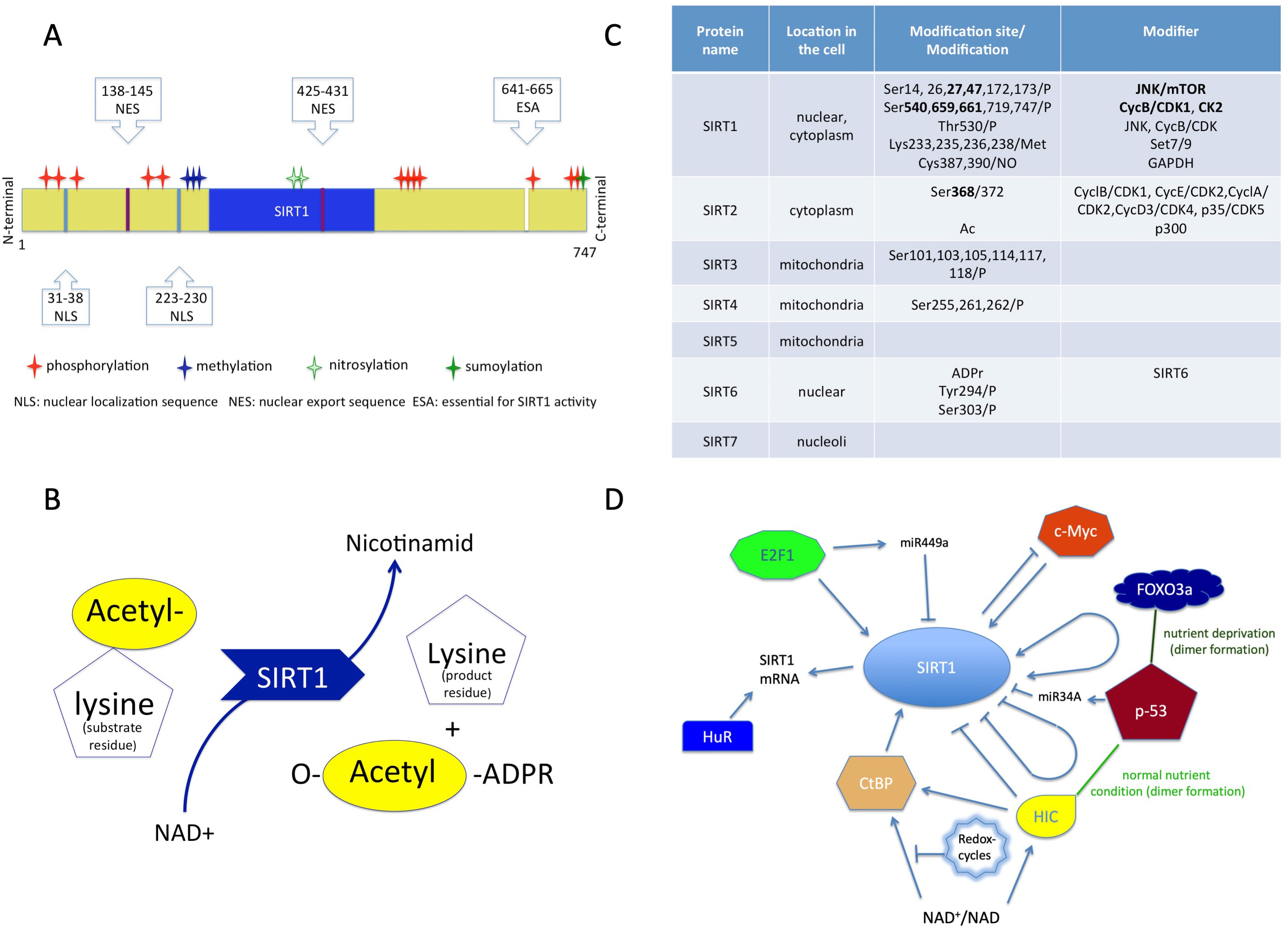
Fig. 1.

Structure and function of sirtuins. (A) Structure of SIRT1 protein consisting of a conserved (blue) region, two nuclear localization sites (NLS), two nuclear export sites (NES) with the modifications. (B) The transacetylation process. (C) The localization of the sirtuins in the cell, and their modifiers with modifications. (D) Molecules which affect SIRT1 expression. NAD+, Nicotinamid-adenin-dinucleotid; ADPR, Adenozin-diphosphate-ribose.

The catalytic pocket engages both the NAD+ co-factor and the acetyl-lysine substrates [46, 48]. The enzymatic reaction is transacetylation (Fig. 1B) [46, 50].

The activity of SIRT1 protein can be modulated by the availability of NAD+ and also by posttranslational modifications (Fig. 1C). Phosphorylation at the terminal sites orchestrates substrate binding, whereas desumoylation reduces and nytrosilation impairs its catalytic activity [47].

The modulation of SIRT1 activity can occur through protein-protein interactions as well [50]. The active regulator of SIRT1 (AROS) enhances SIRT1 activity by binding to its NH2-terminal [47]. The protein called deleted in breast cancer 1 (DBC1) abrogates SIRT1 activity through binding to the catalytic domain. Additionally, SUMO1/Sentrin specific peptidase 1 (SENP1) has also been identified in the reduction of SIRT1 activity [47, 51].

The gene expression of SIRT1 is regulated by cellular redox-changes and environmental factors (e.g., nutrients).

The protein expression level of SIRT1 can be modulated by both transcriptional and translational pathways by E2F1 and p53 (Fig. 1D) [52]. The RNA-binding protein HuR increases the stability of SIRT1 mRNA [52, 53]. Additionally, the tumor suppressor miR-34a downregulates the expression of SIRT1 and attenuates the resistance to chemotherapy agent 5-fluoro-uracil [54]. The miR499a inhibits SIRT1 expression and miR199a attenuates SIRT1 activity [52].

3. Metabolic Effects of SIRT1

Non-histone proteins deacetylated by SIRT1 are closely related to pathways involved in metabolic adaptation to fasting [55]. During this adaptation process specific gene transcriptions are induced through activation of transcription factor FOXO3 by SIRT1 [56]. SIRT1 has tissue-specific functions in metabolic and energy homeostasis, and plays a significant role in maintaining serum glucose levels during starvation [56]. To achieve the latter, SIRT1 deacetylates and activates PPARα to stimulate fatty acid oxidation and activates the necessary coactivator mammalian target-of-rapamycin comlpex-1 (mTORC2) in the liver [55]. Furthermore, it represses the differentiation program of adipocytes as well as triggers lipolysis and fat loss in differentiated fat cells by suppressing peroxisome proliferator-activated receptor-γ (PPARγ), which is responsible for fat storage in white adipocytes [57]. Like in white adipose tissues and liver, SIRT1 induces fatty acid oxidation in skeletal muscle as well, mediating essential anti-diabetic function [55]. It also induces secretion of insulin from pancreatic islet cells to promote optimal glucose uptake into the cells of the human body [56]. SIRT1 upregulates the protein levels of oxysterol receptor (LXRα) and the ATP-binding cassette transporter A1 (ABCA1) to reverse cholesterol transport from peripheral tissues. Similarly, it increases the level of bile acid receptor farnesoid X receptor (FXR) which is required for bile acid biosynthesis and cholesterol catabolism [55]. Additionally, SIRT1 deacetylates two central components of the circadian clock (BMAL1 and PER2) in the liver and thus controls liver specific circadian regulation of metabolic processes [55]. As an important part of its effect, SIRT1 also activates PPARγ coactivator-1α (PGC-1α) which connects metabolic and energy dysfunction to the transcriptional output [58]. PGC-1α is expressed mainly in those tissues which have high oxidative capacity included heart, skeletal muscle, liver, brown adipose tissue and brain [59]. PGC-1α is a key factor in glucose production in the liver where it activates the entire gluconeogenetic pathway while interacts with FOXO1 in insulin-regulated gluconeogenesis [60]. Additionally, PGC-1α facilitates mitochondrial biogenesis for adaptation to reduced energy status and it is a key player in energy expenditure (EE) as well [55, 61]. Therefore, increased expression of PGC-1α resulted in increased number and function of mitochondria and a relatively higher number of type I oxidative fibers in skeletal muscle [62]. Defects in mitochondrial oxidative function with decreased EE may contribute to the metabolic conditions observed in insulin resistance (IR), type 2 diabetes mellitus (T2DM) and aging, and appear to be parallel to intramuscular triglyceride accumulation, which is the most recognized and consistent marker of whole-body IR [58, 63]. Insulin resistance is associated with a decrease in the use of fatty acids as an energy source in the skeletal muscle, resulting in storage and increased ectopic lipid deposition [58]. Additionally, obese and diabetic patients have a lower proportion of type I muscle fibers. These fibers have high mitochondrial content and high oxidative capacity compared to type IIb fibers which are glycolytic in nature. This observation is important since insulin sensitivity is positively correlated with the oxidative capacity of the muscle [64]. Indeed, decreased PGC-1α levels have been reported in skeletal muscle of insulin resistant and T2DM patients [65, 66], whereas expression has been restored by treatments known to normalize body weight and/or glucose homeostasis [67]. Similarly, high fat diet inhibits PGC-1α by hyperacetylation, and not surprisingly, SIRT1 expression diminished in such circumstances [58].

SIRT1 can activate AMPK through deacetylating and activating its upstream activator, liver-kinase-B1 (LKB1). Similarly, AMPK can induce SIRT1 expression by increasing NAD+ level [68]. Moreover, AMPK can activate PGC-1α by both direct phosphorylation and NAD+-dependent activation via SIRT1 [69]. This means that there is a positive regulatory cycle between SIRT1 and AMPK, and both can activate PGC-1α [68]. AMPK maintains the energy homeostasis, senses the energy stress at cellular level through the increased AMP/ADP to ATP ratio, resulting in its activation [70]. AMPK is a kinase which can activate or inactivate the targeted metabolic enzymes by phosphorylation, and interestingly, many of these targets are regulated by SIRT1 as well [71]. Generally, AMPK activates catabolic processes to produce ATP, while inhibits almost all anabolic processes consuming ATP. In this regard, it inhibits protein synthesis by inhibiting mammalian target-of-rapamycin comlpex-1 (mTORC1) which promotes cell growth and proliferation in the presence of growth factors, nutrient availability and energy status [69]. AMPK also promotes the mRNA expression of bile salt export pump protein in hepatocytes which allows the efficient absorption of lipids from food [70]. During evolution, this system adopted to the multicellular organism and interacted with hormones to balance energy intake and expenditure at the whole-body level [70]. Indeed, ghrelin released from gut activates AMPK in hypothalamus, increasing food intake. Adiponectin and thyroid hormones induce fatty acid oxidation in liver, skeletal muscle and in brown adipose tissue. In contrast, leptin released from adipocytes inhibits AMPK, reduces appetite, and increases biosynthesis and energy storage. Leptin/adiponectin ratio is also a known marker of insulin resistance [69, 70]. Leptin activates the PI3-kinase-Akt-mTORC1-S6K1 pathway which is implicated in obesity-related pathophysiological conditions such as diabetes, cardiovascular disease and cancer [72]. Additionally, AMPK is the site of action of metformin [61]. These data show that SIRT1 and AMPK can acutely restore energy balance but also reprogram the cell metabolism through transcription [69].

3.1 Effect of Calorie Restriction on SIRT1 Levels and Associated Metabolic Changes

Clinical trials and human studies investigating calorie restriction (CR) (Table 1) described that adipose tissues in lean people showed 120% higher SIRT1 level compared to obese patients at the baseline [26]. Long-term CR significantly increased the SIRT1 mRNA expression levels (130% higher) compared to baseline levels in obese women [26], and significantly increased the activation levels of SIRT1 and AMPK proteins in peripheral blood monocytes (PBMCs) as well [25]. However, in short-term CR, neither SIRT1 level nor activation of AMPK pathway was changed, although activation of insulin/mTOR pathway was significantly reduced [27]. On the contrary, both short and long-term CR significantly decreased body weight, BMI, insulin level (with unchanged level of insulin receptor β/IRβ), leptin, triglycerides, T3 levels, while significantly increased growth hormones, free fatty acids, ketone bodies, FT4, SHBG, cortisol, alkaline phosphatase and creatinine levels [26, 27]. One of the studies investigating long-term CR showed a significant decrease in basal metabolic rate, mean blood pressure, homeostasis model assessment for insulin resistance (HOMA-R) and insulin resistance index (IRI). Moreover, there was a significant increase in VO2 max and a significant decrease in inflammation-related IL-6 and visfatin, but not the CRP and ICAM-1 [25].

CR also has a beneficial effect on the circulatory system based on studies showing that SIRT1 and endothelial nitric oxide synthase (eNOS) positively regulate each other upon CR when eNOS induces SIRT1 expression, while SIRT1 deacetylates eNOS and promotes its activity. Vascular endothelium produces nitric oxide (NO) through eNOS and supports general endothelial health; as NO is important in relaxing muscle and lowering blood pressure. Additionally, CR improves myocardial ischaemic tolerance by facilitating SIRT1 entry into the nucleus by eNOS, and upregulation of SIRT1 protects against cardiac hypertrophy resulting from PPARα activation and increased fat oxidation [55, 56]. Additional cardiovascular benefit of SIRT1 in addition to promote lipolysis and improve insulin sensitivity, is the ability to limit proinflammatory macrophage activity [73]. The effect of COVID-19 in patients with low SIRT1 levels may lead to mitophagy and programmed cell death with relevance to myocardial infarction and ischemic heart disease [10]. SIRT1 upregulation through low calorie diets, lifestyle changes and physical activity may also protect COVID-19 patients against chronic and cardiovascular disease and complications [74, 75].

Hypothalamus is the main systemic coordinator of mammalian physiology including regulation of diurnal activities like feeding, body temperature, energy expenditure and other metabolic functions. CR increases SIRT1 levels in the dorsomedial and lateral hypothalamus triggering behavioral changes to higher physical activity and increased body temperature [55]. SIRT1 upregulates the core transcription factors, BMAL1 and CLOCK, and directly regulates their cyclic repressors as well by leading to degradation. Because AMPK is also able to induce degradation in cyclic repressors which suppress many nuclear receptors thus it is also able to modify nuclear receptor-dependent gene expression programs [69].

Metabolic parameters negatively correlated by SIRT in a clinical trial using laparoscopic adjustable gastric banding (LAGB) to ensure CR. Significantly decreased BMI, fasting glucose, insulin, HOMA, liver enzymes such as ALT, GGT, inflammatory CRP and leukocyte counts after 6 months of weight loss compared to the baseline. The mRNA levels of SIRT1, 3 and 6 significantly increased in the adipose tissue. In the liver, SIRT1 and 3 protein levels increased as well. Expression of all three mRNA significantly and negatively correlated with BMI and serum glucose, and SIRT1 mRNA expression showed significantly negative correlation with liver portal fibrosis, steatosis, GGT levels, adipo-cytokines and receptor expressions in both tissue types (Table 1) [28].

SIRT1 regulates lipid and glucose metabolism, thus consequently lipid profile, where dyslipidaemia is a cardiovascular risk with high LDL-C (low-density lipoprotein-C) and low HDL-C (high-density lipoprotein-C) levels. The genetic variant haplotype 2 of SIRT1 significantly decreased LDL-C and increased HDL-C levels in both sexes but only with low n-6/n-3 polyunsaturated fatty acid (PUFA) intake [29].

3.2 Effect of Physical Activity on SIRT1 Levels and Associated Metabolic Changes

Clinical trials investigating the effect of exercise alone or with CR have found similar result to CR interventions (Table 1). In more details, acute and chronic stimuli due to one- and three-days cycling, respectively, increased mitochondrial biogenesis through significantly increased PGC-1α, SIRT1, citrate synthase and cytochrome-c levels in skeletal muscle compared to pre-exercise samples [30]. Furthermore, a high positive correlation between PGC-1α and SIRT1 mRNA levels and significantly increased mitochondrial function were detected based on PGC-1α, eNOS, SIRT1, TFAM, PARL levels, and mitochondrial DNA content. Moreover, significantly decreased DNA-damage were found. Additionally, whole-body EE, triiodothyronine and insulin levels decreased significantly with improved insulin-sensitivity, although TCA cycle and β-oxidation did not change significantly [31]. Another study found a significant correlation between higher baseline level of SIRT1 mRNA and weight lost. In the SIRT1 high group weight loss improved continuously over the entire 12 months intervention, while the SIRT1 low group for only 5-month period. Weight loss highly correlated with significantly increased SIRT1 mRNA expression, NAD+ biosynthesis, and significant decrease in PPARγ in white adipose tissue [32]. Interesting results were seen after a 30 second sprint anaerobic exercise, where glucose ingested and fasting control groups were compared. In fasting control group SIRT1 protein expression levels significantly increased after a single, 30 seconds sprint exercise with elevated Thr172 -AMPKα phosphorylation. Both effects were blunted in the glucose group. PGC-1α level, however, remained unchanged in both groups. It suggests that AMPK phosphorylation induced by sprint exercise, as a short-term and anaerobe exercise, is not sufficient alone to induce PGC-1α level, regardless of glucose intake [33]. In a clinical trial comparing small-side rugby as an alternative to stationary cycling, glycated haemoglobin, total body fat-mass and intra-abdominal fat-mass were significantly reduced in both groups, but oxygen consumption, insulin sensitivity and total body fat-free mass were significantly increased only in the rugby group. However, they found no significant differences in the protein levels related to glucose regulation and mitochondrial biogenesis (PGC-1α, SIRT1, p53, GLUT4, AKT, complex I-IV subunits, ATP synthase subunits) between the two groups after the interventions [34].

4. Effect of SIRT1 on Inflammatory Processes

The sequential course of inflammation is directed by reprogrammed metabolism and bioenergy [73]. SIRT1 is involved in this process, and switching glycolysis to fatty acid oxidation in monocytes during adaptation to acute inflammation [76]. Reactive oxygen species and ATP generated by anabolic glycolysis are required for rapid defense against invading microorganisms, while lipolysis/fatty acid oxidation is necessary to regeneration and repair, and it leads to immune repression. Failure in this homeostasis results in many chronic and acute inflammatory diseases such as obesity, diabetes, metabolic syndrome, atherosclerosis, Alzheimer’s disease or sepsis [73]. The repression of anti-aging gene SIRT1, which is involved in epigenetic processes that alter the immune system, was implicated to diabetes and multiple organ dysfunction syndrome [77]. During acute systemic inflammation response, which could be induced by COVID-19 as well, cardiovascular and microvascular functions are impaired leading to multiple organ failure [73]. On the contrary, at the molecular level, the shift in NAD+ availability increases nuclear NAD+ level and activates SIRT1, which then induces facultative heterochromatin formation for gene silencing in promoter regions of proinflammatory genes such as TNF-α and IL-1β [78]. In this process activated SIRT1 binds, deactivates and leads NFκB RelA/p65 to proteasome degradation. Additionally, in association with RelB which is also induced, SIRT1 deacetylates histone H1 and recruits a multiunit repressor complex to form heterochromatin at proinflammatory genes [79, 80].

Obstructive sleep apnoea syndrome (OSAS) is described as poor sleeping quality with decreased blood oxygen levels due to repeated airway obstruction. This increases blood pressure, hypoxia/re-oxygenation events, activates the pro-inflammatory NFκB and AP-1, and results in oxidative stress, systemic inflammation, nervous system dysfunction, reduced NO levels, atherosclerosis and increased aging. SIRT1 protein levels and its activity are significantly reduced in blood of OSAS patients, but may be reverted in parallel with nitrogen oxide derivatives by continuous positive airway pressure treatment (Table 1). This change can be explained as a consequence of decreased oxidative stress [35]. In addition, consumption of white wine as a source of resveratrol significantly increased lung function based on forced expiratory volume in 1 second (FEV1) and also reduced the risk of airway obstruction [36].

It has been known for a long time that PPARγ regulates adipocyte differentiation, fat storage, glucose metabolism, and it is a target of antidiabetics. But it has recently become known that it can also direct inflammatory processes toward the anti-inflammatory phenotype [81]. The dietary composition called advanced glycation end products (AGEs) caused high oxidative stress and inflammation, and suppressed SIRT1 and PPARγ, the two host defense molecules which regulate inflammation and metabolic functions [81]. Similarly, the AGE restriction diet restored mRNA levels of SIRT1 and PPARγ and significantly decreased inflammatory and stress markers (i.e., AGEs, 8-iso-prostanes, VCAM-1, mononuclear TNFα and RAGE) in healthy people (Table 1) [37]. Furthermore, it was effective in normalization of expression levels of SIRT1 mRNA, AGE receptor 1 (AGER1), and marked increases in adiponectin levels, as well as decreases in insulin, HOMA, leptin, TNFα, NFκB, serum AGEs, 8-isoprostane levels in T2DM patients (Table 1) [82].

The association between low back pain and oxidative stress has already been reported [83, 84]. However, a recent clinical study also found that exercise intervention was effective in reducing nonspecific low back pain due to significantly decreased TLR-4 related pro-inflammatory signals (TLR-4 mRNA, IFNγ and IP-10, IL-1β, IL-6, IL-8, TNFα). SIRT1 and PGC-1α mRNA levels were significantly increased, while p53 mRNA levels significantly decreased. FOXO1/3 mRNA levels as well as activity of targeted antioxidant enzymes, catalase and SOD were significantly up-regulated in parallel with decreased oxidative stress as measured by blood H2O2 levels [38].

Age-related diseases, such as cardiovascular disease, Alzheimer’s and Parkinson’s diseases or cancers, are supposed to be developed by a low degree of systemic inflammation with advanced age [73, 85, 86]. Oxidative stress is significantly increased by aging and is closely related to mitochondrial dysfunction, which promotes the production of reactive oxygen species [87]. SIRT1 and AMPK, however, regulate mitochondrial biogenesis and clearance of defected mitochondria (mitochondria fission) through PGC-1α [69]. This might be the reason why aging significantly increases SIRT1 protein levels. SIRT1 shows significant positive correlation with HDL in older people. Moreover, a significant negative correlation was detected between SIRT1 level and LDL in adults [39]. SIRT1 has anti-aging activity by activating AMPK, PGC-1α, FOXO and inactivating p53 and NFκB [88]. SIRT1 and AMPK promote each other and their cyclic regulation mechanism highlights their common roles both in energy balance metabolism and aging processes [68, 89]. SIRT1 prevents endothelial senescence by activating PGC-1α and PPARα through deacetylation. Consequently, this results in the downregulation of production of NADPH oxidase-mediated reactive oxygen species and reactivation of endothelial smooth muscle relaxation due to reverted nitrogen-monoxide level [90]. Swimming inhibits apoptosis in aging hippocampal cells, improves muscle functions and reduces inflammation in aging rats through AMPK/SIRT1/PGC-1α pathway [89, 91, 92].

5. SIRT1 Level in Emotional and Cognitive States

Stress level and emotional state are also important risk factors in development of metabolic disorders and cancer [93, 94]. Patients with mood disorders such as major depressive disorder and bipolar disorder have been studied in both depressed and remission states. The mRNA levels of SIRT1, 2 and 6 were significantly decreased in peripheral blood monocytes in mood disorders and depressed states compared to healthy controls. In contrast, in remission state these were comparable to those seen in healthy subjects. This result suggests that SIRT1, 2 and 6 are state-dependent and might be associated with pathogenesis and pathophysiology of mood-disorders. However, serum cortisol level did not change dramatically during remission and there was no significant correlation with SIRT levels. It suggests that chronic stress factor was still present (Table 1) [40].

Cognitive behavioral therapy has been applied to support weight loss in another study. Carriers of double minor allele of SIRT1 (rs1467568; AG or AA) and CLOCK (rs1801260, TC or CC) showed significantly higher resistance to weight loss, lower weekly weight loss at higher plasma ghrelin concentration, evening preference and lower adherence to Mediterranean diet compared to homozygotes for both major alleles (GG and TT, respectively) (Table 1) [41].

Calorie restriction, adequate physical activity and emotional state increase the level of SIRT1 in the body, resulting in beneficial changes in levels of inflammatory and stress markers, thus promoting balanced epigenetic regulation and increasing healthy life expectancy (Table 1) [88, 95].

6. SIRT1 in Cellular Processes of Autophagy and Apoptosis

SIRT1 regulates the availability of several target genes and transcription factor genes involved in epigenetic regulation [96], and many of which are also involved in aging and age-related diseases [52].

Autophagy is a process in which organelles and macromolecules are directed into lysosomes for degradation. This process is used by cells for normal turnover or production of nutrients in response to energy shortage [69]. AMPK is also involved in this process by activating pro-autophagic complexes, directly triggering the autophagy cascade, initiating autophagosome formation or inhibiting mTOR, an autophagy suppressor molecule [97, 98]. P53 can also inhibit mTOR and activate AMPK thus inducing autophagy. Although in most cases p53 is positively regulated by AMPK [68], it can also inhibit autophagy [99].

P53 is a tumor suppressor gene and is also able to arrest the cell cycle by DNA repair and apoptosis [68]. It controls the metabolic switch between glycolysis and oxidative phosphorylation (Wartburg effect) and favors TCA cycle while limiting glycolytic flux [100]. P53 can regulate SIRT1 expression depending on the nutrient status of the cells [52, 56]. In starving cells, p53 complexed with FOXO3 induces SIRT1 expression, while under normal conditions HIC1, an epigenetically regulated repressor, mediates the suppression of SIRT1 gene expression [52].

However, SIRT1 can also regulate p53 through deacetylation thus inhibiting DNA damage and stress induced cellular senescence [88]. SIRT1 prevents nuclear translocation of p53 and blocks transcription-dependent induction of apoptosis, while promotes its cytosolic accumulation and transport to mitochondria, suggesting transcription-independent induction of apoptosis. Although firstly SIRT1 was considered as a tumor promoter, recent studies have suggested it to be a tumor suppressor by facilitating mitochondria-dependent apoptosis [101]. Moreover, in chronic stress condition SIRT1 can inhibit corticosterone-induced autophagy and enhances apoptosis [102].

The main role of SIRT1 in biological processes and its regulatory targets for normal healthy homeostasis are summarized in Fig. 2 [27, 31, 48, 52, 56, 101, 103, 104, 105].
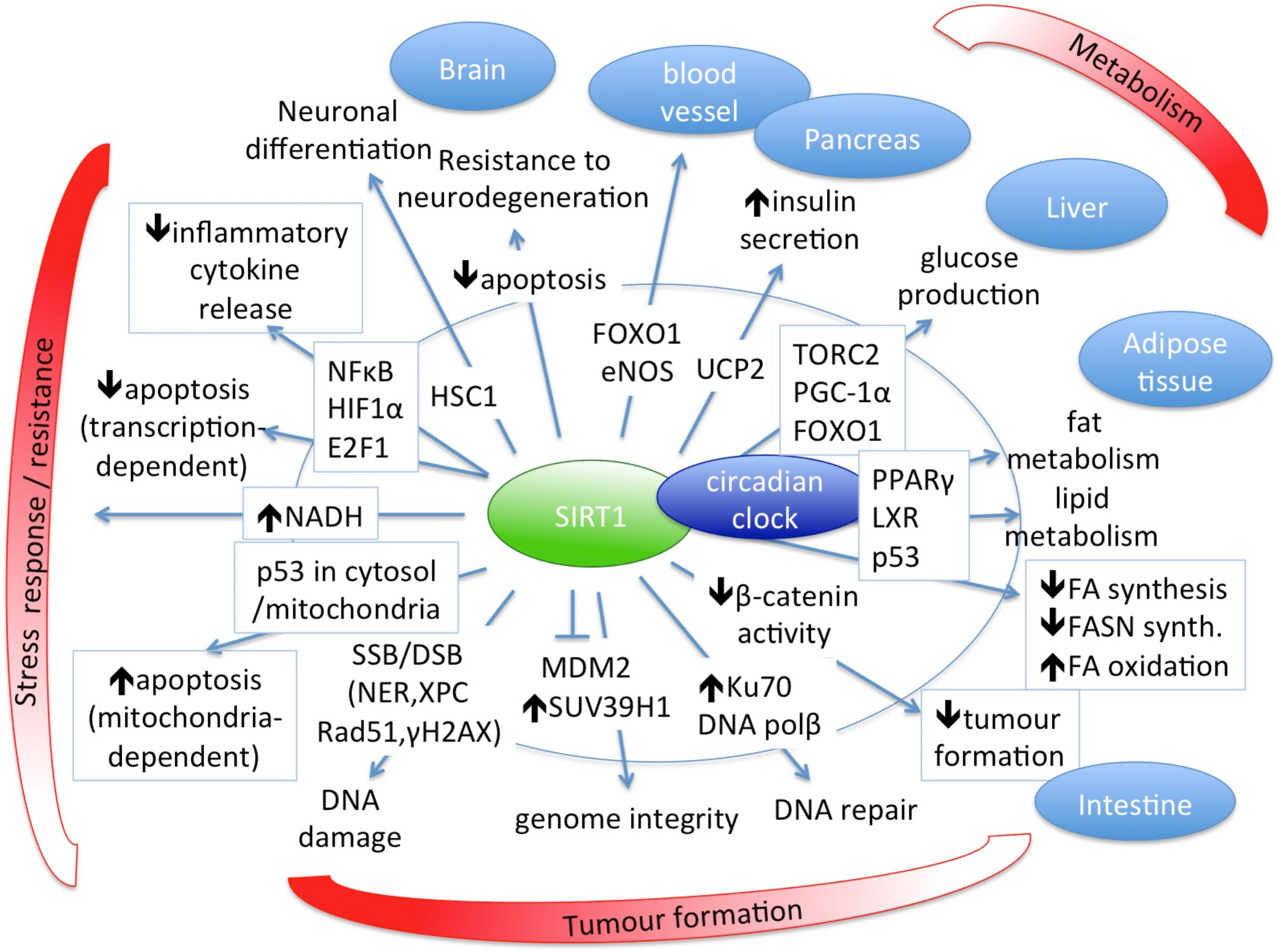
Fig. 2.

SIRT1 has central role in several biological processes and is an important regulator of normal healthy homeostasis.

7. SIRT1 in the Four Main Cancer Types

Research on metabolic disorders and cancers has increased interest in lifestyle factors such as diet, physical activity, stress level, habits and behaviors as these significantly influence human physiology during the actual duration of their effects. These factors, whether they exist continuously or for a short time but regularly, take their effects into account over a long period of time. Thus, because they act through specific epigenetic modifications [44, 103], they can modify homeostasis, resulting in further genetic alterations [106, 107]. It has been established that lifestyle factors may have a greater impact on colorectal cancer risk than genetics, based on a Spanish cohort study [108].

Although the clear positive effect of SIRT1 and AMPK is still controversial in the development of cancer, several studies have found a positive correlation between SIRT1 expression and longer overall survival (OS) in cancer patients [109, 110].

Colorectal cancer. Several studies have found significant correlation between higher expression or overexpression of SIRT1 and TNM stages (classification of malignancies, T - tumor, N - Node, M - metastases), depth of invasion, lymph node metastasis, increased stem cell markers and decreased suppressors of metastasis [111, 112, 113, 114, 115]. Similarly, in serrated lesions, which are alternative routes for colorectal cancer formation, high expression of SIRT1 and c-Myc was strongly associated with higher grades of malignancy along with the presence of KRAS or BRAF mutations [116]. In two prospective studies, overexpression of SIRT1 was more common in microsatellite instable-high (MSI-high), CpG island methylator phenotype (CIMP-high) and CIMP-high/MSI-high tumors, but only in BRAF-mutated tumors that were positively and significantly associated with CIMP-high/MSI-high tumors. SIRT1 was also associated with overexpression of FASN and hypermethylation of several promoters and CpG islands [117]. In contrast, another study reported that lymph node metastasis was negatively correlated with SIRT1 [118]. Beneficial effect of resveratrol through apoptosis induced in colon cancer cells and sensitization of chemoresistant colon cancer cells to various drugs has been demonstrated in another study as well [119]. Moreover, it has been suggested that exercise-induced alterations in the systemic milieu can influence important regulatory mechanisms in the tumor microenvironment such as angiogenesis, immune regulation and metabolism, and lead to cumulative antitumorigenic effect (Table 2, Ref. [17, 111, 112, 113, 114, 115, 116, 117, 118, 120, 121, 122, 123, 124, 125, 126, 127, 128, 129, 130, 131, 132, 133, 134, 135]) [136].

Table 2.Studies investigating SIRT1 expression in relation to colorectal, lung, breast and prostate cancers.
Numbered Cancer type/SIRT1 induccer Disease Tissue type SIRT1 level Metabolic changes, clinical parameters after intervention OR disease & states Signaling/ cellular mechanism Study type Cases no. Reference
mRNA protein activity
18 CRC cancer colorectal cancer tissue (FFPE- IHC) NA increased* NA depth of invasion, lymph node metastase, TNM stage/poor prognosis NA Systematic review and meta analysis 50-497/7 studies [111]
19 CRC cancer colorectal adenocarcinoma tissue (FFPE- IHC) NA increased* NA metastasis (0.02), poor prognosis (p < 0.01), poor OS (0.003)/ colocalization with CD133+ CD133+, stemness Original article 102 [112]
20 CRC cancer colorectal cancer tissue (FFPE- IHC) NA increased* NA Fra-1, OS Fra-1 Original article 90 [113]
21 CRC cancer colorectal cancer and liver metastasis (microarray) NA increased* NA TNM stage, metastasis, OS miR199b-CREB-KISS1/progression Original article 60 (16 meta) [114]
22 CRC cancer normal colon, colon adenoma, colon carcinoma NA increased* NA diagnosis, prognosis NANOG, NCF2, ELF, TGFβ Original article 167 [115]
23 CRC cancer colorectal cancer tissue (FFPE- IHC) NA increased*/decreased* NA differentiation, stage/lymph node metastasis differentiation, progression Original article 40 [118]
24 CRC cancer serrated lesions of colorectum (FFPE-IHC) NA increased* NA NA c-Myc/KRAS, BRAF mutation Original article 121 [116]
25 CRC cancer colorectal cancer tissue (FFPE- IHC) NA increased* NA tumour grade (0.003), mucinous component (0.04), CIMP-high (0.002), MSI-high (p < 0.0001), CIMP-high/MSI-high (p < 0.0001), FASN (0.008) BRAF, MSI, CIMP, FASN/methylation of CACNA1G, IGF2, MLH1, NEUROG1, RUNX3, SOCS1, MINT31 and p14 Original article 485 [117]
26 B cancer breast cancer tissue (FFPE- IHC) NA increased* NA TNM, DFS, OS NA Meta-analysis 28-200/6 studies [126]
27 B cancer breast cancer tissue (FFPE- IHC) NA decreased* NA DFS, OS, Notch1/Snail Original article 150 [127]
28 B cancer breast cancer (microarray) decreased* NA NA metastasis EMT Meta-analysis 1181/12 studies [128]
29 B cancer breast cancer tissue (FFPE- IHC) NA decreased * (HRBC, H2BC) increased* (TNBC) NA LNM, LVM, DFS, OS, HRBC/H2BC/TNBC NA Original article 427 [129]
30 B cancer breast cancer tissue (FFPE- IHC) decreased* NA NA grade3, IDC NA Original article 64 [130]
31 B cancer breast cancer tissue (FFPE- IHC) NA increased* NA LNM, DFS, TNBC NA Original article 344 [131]
32 B cancer breast cancer tissue (FFPE- IHC) NA increased* NA LNM, DFS E-cadherin, Vimentin, Snail/ EMT Original article 319 [132]
33 B cancer MS [17]
34 L cancer NSCLC tissue (FFPE- IHC) NA increased* NA TNM, LNM, Ki67 HIF1/ hypoxia, oxidative stress Original article 125 [120]
35 L cancer NSCLC tissue (FFPE- IHC) NA increased* NA DFS, OS NA Original article 105 [121]
36 L cancer NSCLC tissue (FFPE- IHC) NA increased* NA TNM, LNM, DFS, OS, cortactin Original article 163 [122]
37 L cancer NSCLC tissue (FFPE- IHC) NA increased* NA TNM, DFS, OS, chemotherapy resistance chemotherapy resistance Original article 295 [123]
38 L cancer NSCLC tissue (FFPE- IHC, PCR) increased* increased* NA OS, prognosis HIC-SIRT1-p53 loop, acetyl-p53 Original article 97 [124]
39 L cancer sputum NA NA NA OS, risk of SCC SNP, haplotype nested case-control study 17000 (267+383 selected) [125]
40 P cancer PIN, PCa tissues decreased* NA NA H2A.Z H2A.Z, mTOR, c-Myc Original article 57 [133]
41 P cancer prostate cancer tissue increased* NA NA Ki67 Ki67, methylation Original article 47 [134]
42 P cancer prostate cancer tissue NA increased* NA Gleason grade Gleason grade Original article 41 [135]
ADC, adenocarcinoma; DFS, disease free survival; FASN, fatty acid synthase; H2BC, Her2 positive breast cancer; HRBC, hormon receptor-positive breast cancer; LNM, lymph node metasta metastasis; LVI, lymhovascular invasion; NSCLC, non-small cell lung cancer; OS, overal survival; PCa, prostate carcinoma; PIN, prostatic intraepithelial neoplasia; SCC, squamous cell carcinoma; TNBC, triple negative breast cancer; *, significant change.

Lung cancer. SIRT1 showed significantly higher expression in lung tumors than in normal tissues and was positively associated with Ki67 index, TNM stage and lymph node invasion. It is suggested as an independent prognostic factor for shorter disease free survival (DFS) and OS, and for higher chance to chemotherapy resistance [120, 121, 122, 123]. In squamous cell carcinomas (SCCs), high levels of SIRT1 effectively deacetylated and inactivated p53. In SIRT1 positive non-small cell lung cancer (NSCLC) cases, low HIC1, a suppressor of SIRT1, was associated with poor prognosis [124]. However, a case-control study investigating SNPs suggested that SIRT1 is a tumor suppressor in radon-induced cancer in miners (Table 2) [125].

Breast cancer. In a meta-analysis of non-stratified studies, SIRT1 expression was significantly and positively correlated with high TNM stages, decreased DFS and a higher rate of lymph node metastases (LNM) [126]. However, other studies found opposite correlations where increased SIRT1 level reduced EMT markers [127, 128]. In stratified evaluations, lower expression of SIRT1 correlated with the presence of LNM in hormone receptor-positive breast cancer (HRBC), HER2 overexpressed/amplified and ER/PR negative breast cancer (H2BC), and in invasive ductal carcinoma (IDC). Similarly, in another study, lower SIRT1 expression correlated with poorer DFS only in HRBC group [129, 130]. Increased level of SIRT1 was associated with increased LNM only in TNBC groups [129, 131, 132]. Additionally, SIRT1 expression was significantly lower in TNBC than in hormone positive HER2 negative breast cancers. Moreover, low SIRT1 expression was associated with worse OS in the latter group, while in TNBC, high SIRT1 level was associated with a poor prognosis [110]. However, metabolic syndrome is associated with an increased risk of breast cancer, poor prognosis and mortality, and is also associated with a decreased SIRT1 level (Table 2) [17].

Prostate cancer. mRNA expression of SIRT1 was significantly downregulated in prostatic intraepithelial neoplasia (PIN) and prostate carcinoma (PCa) [133]. In contrast, other studies have reported that SIRT1 mRNA levels were significantly increased in prostate cancer tissues, positively correlated with Ki67 mRNA expression [134], and were associated with the Gleason grade 3 (Table 2) [135].

8. SIRT1 as a Therapeutic Target — Activators and Inhibitors

Resveratrol, a natural compound, has been used to mimic calorie restriction [137] because it is known to significantly increase forced vital capacity (FVC) function of the lung [36] and respiratory quotient (RQ) as well, while significantly decreasing sleeping metabolic rate, systolic blood pressure, mean arterial pressure and many other clinical and systemic parameters [137]. Resveratrol treatment significantly decreased intrahepatic lipid content but increased that in muscle cells. Resveratrol significantly increased AMPK phosphorylation, PGC-1α, citrate synthase and SIRT1 level in muscle biopsies as well. Similarly, genes involved in mitochondrial oxidative phosphorylation were up-regulated as well. No adverse effects were observed during the 30-day treatment period (Table 3, Ref. [137, 138, 139, 140, 141]) [137].

Table 3.Studies investigating SIRT1 expression as a therapeutic target of activators and inhibitors.
Numbered Cancer type/SIRT1 induccer Disease Tissue type SIRT1 level Metabolic changes, clinical parameters after intervention OR disease & states Signaling/ cellular mechanism Study type Cases no. Reference
mRNA protein activity
43 resveratrol healthy, obese skeletal muscle NA increased* NA decreased* ALT, insulin, HOMA, triglycerides, leptin, intrahepatic lipid, leukocyte number, TNFα, sleeping metabolic rate, systolic blood pressure, mean arterial pressure and increased* RQ, complex I-II, MOG, MOGS, intramyocellular lipid p-AMPK, PGC-1α, citrate synthase /mitochondrial oxidative phosphorylation, flexibility of metabolism Randomized, double-blind study 11 [137]
44 SRT21204 healthy plasma NA NA NA decreased* IL-6, IL-8, CRP and increased* F1+2, TATc LPS /inflammation Randomized, double-blind placebo controlled trial 24 [138]
45 SRT21204 healthy old serum NA NA NA decreased* serum cholesterol, triglycerids, half-life of recovery after exercise (ADP,PCr) and increased* HDL:LDL ratio mitochondrial oxidative phosphorylation Phase I. clinical trial 24 [139]
46 SRT21204 healthy, cigarette smokers serum NA NA NA decreased* cholesterol, LDL, triglycerids, no effect on blood flow rate, coagulation factros, macrophage activation Randomized, double-blind placebo controlled, crossover trial 24 [140]
47 Selisistat healthy blood NA NA NA NA gene expression differences Randomized, double-blind placebo controlled, crossover trial 22+66 [141]
F1+2, prothrombin fragment; MOG, malate/octanoyl-carnitine/glutamate substrate; MOGS, MOG/succinate substrate; TATc, thrombin-antithrombin complex; *, significant change.

In parallel with exercise, the small molecule SIRT1 activator, SIRT2104, significantly attenuated plasma cytokines IL-6, IL-8 and CRP in lipopolysaccharide-injected human patients but increased the levels of prothrombin fragment and thrombin-antithrombin complex. There was no effect on the level of von Willebrand Factor, fibrinolytic response, number of leukocytes and gene expression profile [138]. Other studies have recorded significantly decreased recovery period after exercise as well as significantly decreased serum lipid levels after SIRT2104 treatment (Table 3) [139, 140].

The SIRT1 inhibitor Selisistat (SEN0014196) has a specific transcriptional signature in blood cells, involving transmembrane transport, cholesterol/lipid/steroid homeostasis and redox processes genes (Table 3) [141].

9. Summary

Experimental knowledge of the beneficial effects of proper diet and exercise has been known since ancient times and can be used effectively, however, the molecular mechanism behind these has only recently been revealed. Our primary aim was to explore lifestyle specific changes of SIRT1, a key epigenetic regulator, which influences the development of metabolic syndrome and related cancer, and describe the molecular aspects of these mechanisms.

In general, we found that optimal levels of lifestyle factors correlated to increased levels of SIRT1 improving metabolic and inflammatory parameters; some of them increased mitochondrial function and increased the flexibility of alternation between different energy sources. Briefly, SIRT1 are activated and/or induced by AMPK in energy/nutrient-sensing pathways by glucose deprivation or exercises in many tissues. This is the result of increased AMP level which is the sign of low cellular energy [142]. Moderate and high-intensity exercise increases the AMP/ATP ratio [143, 144], while low-intensity exercise only if it prolongs to exhaustion [145]. SIRT1 activation then leads to increased fat mobilization and lipid oxidation in both skeletal muscle and differentiated adipocytes [57, 146, 147]. During exercise, increased mitochondrial respiration results in elevated NAD+/NADH.H+ ratio which also induces SIRT1 levels, so this feedback loop maintains the activity of the proper metabolic pathways to meet energy demand. However, beside the activation loops, there are inhibition circles as well in this regulation which impede biosynthetic pathways including fatty acids, and thus diminish fat-induced inflammation levels [148]. Improved exercise tolerance has other benefits such as decreased ROS production, oxidative stress and subsequent DNA–damage [25]. Despite the beneficial effects of a long-term CR with elevated SIRT1 levels, initially low baseline levels of SIRT1 in adipose tissue can significantly reduce the effectiveness of CR [32]. Similarly, the minor alleles of SIRT1 and CLOCK in the carriers also increased resistance to weight loss, increased ghrelin levels and lower adherence to the Mediterranean diet [41]. These results suggest that SIRT1 level may serve as a predictive marker of metabolic imbalances or effectiveness of a complementary therapy for weight loss, high blood pressure, cardiovascular disease, T2DM or cancers. However, a well-defined range of healthy and unhealthy levels of SIRT1 in different tissues, like adipocytes, skeletal muscle cells or PBMCs, should be determined which requires further studies for its appropriate use in health services.

Based on the results of the cited human clinical trials and human studies, we described that SIRT1 interplays with major components of health-regulatory processes, such as metabolic/energy sensing system, pro-inflammatory cytokine release, oxidative stress control, however, the circadian rhythms itself cannot discussed as a separate entity from all others (Fig. 3). Circadian rhythm is the basis of all diurnal behaviors, like eating or activity level in general, and all energy and metabolic processes required for these functions. Therefore, it is necessary to outline this essential component and its connections to all the above mentioned mechanism related to SIRT1 expression. The proper alignment of the circadian clock with the environmental “time generators” is necessary for normal, healthy metabolic regulation [149]. Therefore, desynchrony resulting from a generally imbalanced lifestyle can cause similarly elevated leptin, glucose and insulin levels, as well as increased blood pressure leading to increased risk of obesity, T2DM, hyperlipidaemia, high blood pressure and cardiovascular disease.
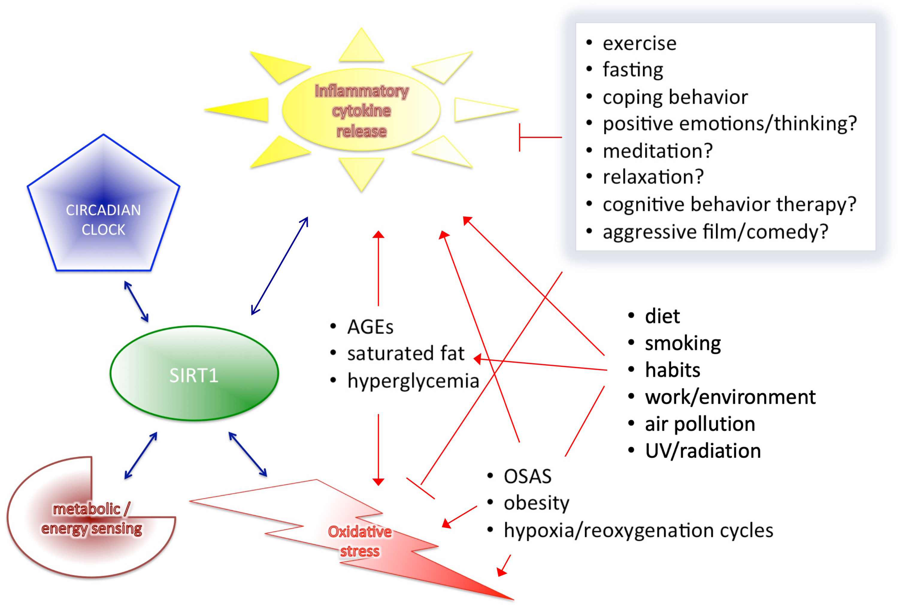
Fig. 3.

SIRT1 interplays with the four major components of health-regulation.

As reported, circadian rhythm alterations are frequent in metabolic syndrome, cancers and mood disorders [149, 150]. Food availability is predominant for peripheral clocks in the peripheral tissues, and is able to resynchronize them independently from the central clock. Thus, not surprising that a number of metabolic pathway regulator transcription factors (such as Rev-erb-alpha, ROR-alpha, PPAR) display tissue specific circadian expression in peripheral tissues (liver, white and brown adipose tissue, skeletal muscle). It has been identified that smoking is also able to disrupt circadian rhythm, decrease SIRT1 expression and is strongly associated with metabolic imbalance and inflammation as well [151, 152]. The CLOCK/NAD+-SIRT1 loop couples circadian oscillation to nutrient/energy metabolism at the CLOCK-BMAL1 transcription factor through SIRT1, which drives the cyclic expression of Bmal1, Per2 and Cry1 [149, 151, 153, 154]. Although smoking habits of patients are usually found in medical records, sleeping habits and its normalization are not well-followed and do not count into the optimal results of conventional treatment of metabolic and cardiovascular diseases or cancers. However, as our results suggest, it also has a significant impact on the effectiveness of a therapy.

Defense mechanisms related to inflammation are also important regulators of homeostasis (Fig. 3). They include SIRT1, AGER and pattern recognition receptors (PRR), like TLRs, and stop infections and other non-desired mechanisms in the human system [37, 82]. TLR is originally activated by bacterial lipids leading to subsequent release of IFNγ and IP-10 [155]. However, as we have shown, specific nutrient component, AGEs are also able to activate these mechanisms. Chronic exposure to such excessive nutrient oxidants, such as during prolonged hyperglycemia, leads to impairment of these native antioxidant defense mechanisms. Exhaustion of these system can be prevented by restriction of AGEs and/or the presence of polyunsaturated fatty acids in the diet, and similarly, by exercise or even specific treatment of OSAS patients, which are able to decrease inflammation and related oxidative stress as well by increased SIRT1 levels [35, 37, 38, 156, 157].

As we described earlier, oxidative stress is involved not only in immune system function but in aging, telomere attrition, DNA-damage, apoptosis and angiogenesis, and increases the level of MMP9, which is also known in cancer development, however, SIRT1 is able to block MMP-9 expression and regulates all these functions as well [158, 159, 160, 161]. Interestingly, a recent paper suggests that Treg cells, which are also the focus of recent cancer research, sustain and amplify tumor suppressor capacity in the microenvironment through oxidative stress [162]. However, it is promising that repeated exercise may reprogram these important regulatory mechanisms induced by oxidative stress in distant tissue microenvironments, i.e., in tumors or other tissues that are not directly involved in the training response, resulting in a cumulative antitumorigenic effect [136]. In addition, we found that SIRT1 inducers exert a similar effect as CR and exercises without side effects during the duration of the studies, which may be an alternative if the other two are inappropriate for some reason [137, 138, 139, 140, 141]. In tumors, SIRT1 expression varied by tumor types and subgroups. However, as we have found, the main drawback of all cancer papers is that mostly only the tumor samples and the surrounding normal tissues were examined. Unfortunately, these data do not allow us to compare these results with epidemiology papers that measured wider and systemic effect of SIRT1, rather than local tissue changes in cancer studies. Therefore, these data can give information for diagnostic, progression or local treatment purposes but do not explore the background and causal factors of tumor development. Higher levels of SIRT1 could reflect a necessity of higher defense in the body to eliminate a more aggressive tumor. However, data suggest that DNA damaging agents probably would not be sufficient in tumors overexpressing SIRT1 because SIRT1-regulated pathways bypass this by induction of survival [52]. Additionally, non-cancer related data detailed above suggest that higher SIRT1 expression is able to reduce MMP-9 either by downregulation of NFκB or by reducing the response to oxidative stress through several connected tumor-promoting processes. Therefore, SIRT1 may decrease metastatic progression under specific circumstances [68, 158] and could be a target for state specific therapies by regulating autophagy and apoptosis [101, 102].

In summary, in our review we focused on SIRT1 and related pathways as a molecular aspect of different lifestyles. Our findings support that interventions to prevent metabolic disorders can similarly prevent the development of cancer, as it has known relation to chronic inflammation and metabolic syndromes. With this review, we aimed to promote health science-related research and highlight the importance of beneficial lifestyle factors as effective tools for prevention and complementary treatment to reduce the development of diseases.

Abbreviations

ABCA1, ATP-binding cassette transporter A1; ADPR, Adenozin-diphosphate-ribose; AGEs, glycation end products; AGER1, AGE receptor 1; AROS, active regulator of SIRT1; CIMP-high, CpG island methylator phenotype; CLOCK, circadian locomotor output cycles kaput; COVID-19, coronavirus disease; CR, caloric restriction; DBC1, deleted in breast cancer 1; DFS, diseases free survival; EE, energy expenditure; EMT, epithelial to mesenchymal transition; eNOS, endothelial nitric oxide synthase; FXR, farnesoid X receptor; FEV1, forced expiratory volume in 1s; FOXO1, FOXO3, Forkhead box O 1 or 3; FVC, forced vital capacity; H2BC, HER2 overexpressed and/or amplified and ER and/or PR negative breast cancer; HDAC, histone deacetylase enzyme; HDL(-C), high-density lipoprotein(-C); HOMA-R, homeostasis model assessment for insulin resistance; HRBC, hormone receptor-positive breast cancer; IR, insulin resistane; IRI, insulin resistance index; IDC, invasive ductal carcinoma; LAGB, laparoscopic adjustable gastric banding; LDL(-C), low-density lipoprotein(-C); LNM, lymph node metastases; LXRα, oxysterol receptor; mTORC1, mammalian target-of-rapamycin comlpex-1; mTORC2, mammalian target-of-rapamycin comlpex-2; MSI-high, microsatellite instable-high; NAD+, Nicotinamid-adenin-dinucleotid; NO, Nitrogen-monoxide; NES, nuclear export sites; NLS, nuclear localization sites; NSCLC, non-small cell lung cancer; OSAS, obstructive sleep apnoea syndrome; OS, overall survival; PBMCs, peripheral blood monocytes; PUFA, polyunsaturated fatty acid; PCa, prostate carcinoma; PER1/2/3, circadian clock period proteins; PIN, prostatic intraepithelial neoplasia; PPARα, peroxisome proliferator-activated receptor-α; PPARγ, peroxisome proliferator-activated receptor-γ; RQ, respiratory quotient; PRR, pattern recognition receptors; SCCs, squamous cell carcinomas; SENP1, SUMO1/Sentrin specific peptidase 1; SOD, superoxide dismutase; T2DM, type 2 diabetes mellitus; TNBC, triple negative breast cancer; TNM stages, classification of malignancies (T, tumor, N, Node, M, metastases); WL, weight loss.

Author Contributions

ZN—design, data collection, visualization, writing manuscript, paper review, final approval; EK—paper review; IT—paper review.

Ethics Approval and Consent to Participate

Not applicable.

Acknowledgment

We would like to thank to Katherine McAllister, Robbie Carson and Sarah Osman at Queen’s University Belfast for their kind support at the beginning of the manuscript preparation.

Funding

This work was supported by the predoctoral research fellowship of the National Research, Development and Innovation Office of Hungary obtained for Eva Kiss (ÚNKP-21-4-I-SE-19).

Conflict of Interest

The authors declare no conflict of interest.

Publisher’s Note: IMR Press stays neutral with regard to jurisdictional claims in published maps and institutional affiliations.

References
[1]
Zhao L, Cao J, Hu K, He X, Yun D, Tong T, et al. Sirtuins and their Biological Relevance in Aging and Age-Related Diseases. Aging and Disease. 2020; 11: 927.
[2]
Kaeberlein M, McVey M, Guarente L. The SIR2/3/4 complex and SIR2 alone promote longevity in Saccharomyces cerevisiae by two different mechanisms. Genes & Development. 1999; 13: 2570–2580.
[3]
Frye RA. Phylogenetic Classification of Prokaryotic and Eukaryotic Sir2-like Proteins. Biochemical and Biophysical Research Communications. 2000; 273: 793–798.
[4]
Lin J, Lin VC, Rau K, Shieh P, Kuo D, Shieh J, et al. Resveratrol Modulates Tumor Cell Proliferation and Protein Translation via SIRT1-Dependent AMPK Activation. Journal of Agricultural and Food Chemistry. 2010; 58: 1584–1592.
[5]
Gehm BD, McAndrews JM, Chien PY, Jameson JL. Resveratrol, a polyphenolic compound found in grapes and wine, is an agonist for the estrogen receptor. Proceedings of the National Academy of Sciences of the United States of America. 1997; 94: 14138–14143.
[6]
Jang M, Cai L, Udeani GO, Slowing KV, Thomas CF, Beecher CWW, et al. Cancer Chemopreventive Activity of Resveratrol, a Natural Product Derived from Grapes. Science. 1997; 275: 218–220.
[7]
Bradamante S, Barenghi L, Villa A. Cardiovascular protective effects of resveratrol. Cardiovascular Drug Reviews. 2004; 22: 169–188.
[8]
Lagouge M, Argmann C, Gerhart-Hines Z, Meziane H, Lerin C, Daussin F, et al. Resveratrol Improves Mitochondrial Function and Protects against Metabolic Disease by Activating SIRT1 and PGC-1α. Cell. 2006; 127: 1109–1122.
[9]
Wang Q, Xu J, Rottinghaus GE, Simonyi A, Lubahn D, Sun GY, et al. Resveratrol protects against global cerebral ischemic injury in gerbils. Brain Research. 2002; 958: 439–447.
[10]
Martins IJ. COVID-19 Infection and Anti-Aging Gene Inactivation. Acta Scientific Nutritional Health. 2020; 4: 1–2.
[11]
Inoue H, Jiang XF, Katayama T, Osada S, Umesono K, Namura S. Brain protection by resveratrol and fenofibrate against stroke requires peroxisome proliferator-activated receptor alpha in mice. Neuroscience Letters. 2003; 352: 203–206.
[12]
Penumathsa SV, Thirunavukkarasu M, Koneru S, Juhasz B, Zhan L, Pant R, et al. Statin and resveratrol in combination induces cardioprotection against myocardial infarction in hypercholesterolemic rat. Journal of Molecular and Cellular Cardiology. 2007; 42: 508–516.
[13]
Laveti D, Kumar M, Hemalatha R, Sistla R, Naidu V, Talla V, et al. Anti-Inflammatory Treatments for Chronic Diseases: a Review. Inflammation & Allergy-Drug Targets. 2013; 12: 349–361.
[14]
Bryant T. Remarks on some Cases of Inflammation of the Breast Simulating Cancer. British Medical Journal. 1868; 2: 608–609.
[15]
Vaag A, Petersen JS. Future breakthroughs in medical treatment of age-related conditions. Ugeskr Laeger. 2009; 171: 794–796.
[16]
La Vecchia C, Giordano SH, Hortobagyi GN, Chabner B. Overweight, Obesity, Diabetes, and Risk of Breast Cancer: Interlocking Pieces of the Puzzle. The Oncologist. 2011; 16: 726–729.
[17]
Cao L, Yao G, Hu X, Chen L, Ye C. Advances in studies on metabolic syndrome and breast cancer. Zhonghua Wai Ke Za Zhi. 2015; 53: 966–969.
[18]
Russo GI, Vanella L, Castelli T, Cimino S, Reale G, Urzì D, et al. Heme oxygenase levels and metaflammation in benign prostatic hyperplasia patients. World Journal of Urology. 2016; 34: 1183–1192.
[19]
Zhang H, Xue R, Zhu S, Fu S, Chen Z, Zhou R, et al. M2-specific reduction of CD1d switches NKT cell-mediated immune responses and triggers metaflammation in adipose tissue. Cellular & Molecular Immunology. 2018; 15: 506–517.
[20]
Shoelson SE. Inflammation and insulin resistance. Journal of Clinical Investigation. 2006; 116: 1793–1801.
[21]
Bierhaus A, Humpert PM, Morcos M, Wendt T, Chavakis T, Arnold B, et al. Understanding RAGE, the receptor for advanced glycation end products. Journal of Molecular Medicine. 2005; 83: 876–886.
[22]
Ramasamy R, Yan SF, Schmidt AM. The RAGE Axis and Endothelial Dysfunction: Maladaptive Roles in the Diabetic Vasculature and beyond. Trends in Cardiovascular Medicine. 2005; 15: 237–243.
[23]
Weatherill AR, Lee JY, Zhao L, Lemay DG, Youn HS, Hwang DH. Saturated and Polyunsaturated Fatty Acids Reciprocally Modulate Dendritic Cell Functions Mediated through TLR4. The Journal of Immunology. 2005; 174: 5390–5397.
[24]
Rajkumar RP. Cross-National Variations in COVID-19 Mortality: The Role of Diet, Obesity and Depression. Diseases. 2021; 9: 36.
[25]
Kitada M, Kume S, Takeda-Watanabe A, Tsuda S, Kanasaki K, Koya D. Calorie restriction in overweight males ameliorates obesity-related metabolic alterations and cellular adaptations through anti-aging effects, possibly including AMPK and SIRT1 activation. Biochimica et Biophysica Acta (BBA) - General Subjects. 2013; 1830: 4820–4827.
[26]
Pedersen SB, Ølholm J, Paulsen SK, Bennetzen MF, Richelsen B. Low Sirt1 expression, which is upregulated by fasting, in human adipose tissue from obese women. International Journal of Obesity. 2008; 32: 1250–1255.
[27]
Wijngaarden MA, Bakker LE, van der Zon GC, t Hoen PA, van Dijk KW, Jazet IM, et al. Regulation of skeletal muscle energy/nutrient-sensing pathways during metabolic adaptation to fasting in healthy humans. American Journal of Physiology-Endocrinology and Metabolism. 2014; 307: E885–E895.
[28]
Moschen AR, Wieser V, Gerner RR, Bichler A, Enrich B, Moser P, et al. Adipose tissue and liver expression of SIRT1, 3, and 6 increase after extensive weight loss in morbid obesity. Journal of Hepatology. 2013; 59: 1315–1322.
[29]
Inamori T, Goda T, Kasezawa N, Yamakawa-Kobayashi K. The combined effects of genetic variation in the SIRT1 gene and dietary intake of n-3 and n-6 polyunsaturated fatty acids on serum LDL-C and HDL-C levels: a population based study. Lipids in Health and Disease. 2013; 12: 4.
[30]
Dumke CL, Mark Davis J, Angela Murphy E, Nieman DC, Carmichael MD, Quindry JC, et al. Successive bouts of cycling stimulates genes associated with mitochondrial biogenesis. European Journal of Applied Physiology. 2009; 107: 419–427.
[31]
Civitarese AE, Carling S, Heilbronn LK, Hulver MH, Ukropcova B, Deutsch WA, et al. Calorie restriction increases muscle mitochondrial biogenesis in healthy humans. PLoS Medicine. 2007; 4: e76.
[32]
Rappou E, Jukarainen S, Rinnankoski-Tuikka R, Kaye S, Heinonen S, Hakkarainen A, et al. Weight Loss is Associated with Increased NAD+/SIRT1 Expression but Reduced PARP Activity in White Adipose Tissue. The Journal of Clinical Endocrinology & Metabolism. 2016; 101: 1263–1273.
[33]
Guerra B, Guadalupe-Grau A, Fuentes T, Ponce-González JG, Morales-Alamo D, Olmedillas H, et al. SIRT1, AMP-activated protein kinase phosphorylation and downstream kinases in response to a single bout of sprint exercise: influence of glucose ingestion. European Journal of Applied Physiology. 2010; 109: 731–743.
[34]
Mendham AE, Duffield R, Coutts AJ, Marino F, Boyko A, Bishop DJ. Rugby-specific small-sided games training is an effective alternative to stationary cycling at reducing clinical risk factors associated with the development of type 2 diabetes: a randomized, controlled trial. PLoS ONE. 2015; 10: e0127548.
[35]
Chen W, Liaw S, Lin C, Chiu C, Lin M, Chang F. Effect of Nasal CPAP on SIRT1 and Endothelial Function in Obstructive Sleep Apnea Syndrome. Lung. 2015; 193: 1037–1045.
[36]
Siedlinski M, Boer JMA, Smit HA, Postma DS, Boezen HM. Dietary factors and lung function in the general population: wine and resveratrol intake. European Respiratory Journal. 2012; 39: 385–391.
[37]
Uribarri J, Cai W, Pyzik R, Goodman S, Chen X, Zhu L, et al. Suppression of native defense mechanisms, SIRT1 and PPARgamma, by dietary glycoxidants precedes disease in adult humans; relevance to lifestyle-engendered chronic diseases. Amino Acids. 2014; 46: 301–309.
[38]
Cheng Y, Kao C, Ma H, Hung C, Wang C, Liu D, et al. SIRT1-related inhibition of pro-inflammatory responses and oxidative stress are involved in the mechanism of nonspecific low back pain relief after exercise through modulation of Toll-like receptor 4. Journal of Biochemistry. 2015; 158: 299–308.
[39]
Kilic U, Gok O, Erenberk U, Dundaroz MR, Torun E, Kucukardali Y, et al. A remarkable age-related increase in SIRT1 protein expression against oxidative stress in elderly: SIRT1 gene variants and longevity in human. PLoS ONE. 2015; 10: e0117954.
[40]
Abe N, Uchida S, Otsuki K, Hobara T, Yamagata H, Higuchi F, et al. Altered sirtuin deacetylase gene expression in patients with a mood disorder. Journal of Psychiatric Research. 2011; 45: 1106–1112.
[41]
Garaulet M, Esteban Tardido A, Lee YC, Smith CE, Parnell LD, Ordovas JM. SIRT1 and CLOCK 3111T> C combined genotype is associated with evening preference and weight loss resistance in a behavioral therapy treatment for obesity. International Journal of Obesity. 2012; 36: 1436–1441.
[42]
Sung H, Ferlay J, Siegel RL, Laversanne M, Soerjomataram I, Jemal A, et al. Global Cancer Statistics 2020: GLOBOCAN Estimates of Incidence and Mortality Worldwide for 36 Cancers in 185 Countries. CA: a Cancer Journal for Clinicians. 2021; 71: 209–249.
[43]
Anand P, Kunnumakkara AB, Sundaram C, Harikumar KB, Tharakan ST, Lai OS, et al. Cancer is a Preventable Disease that Requires Major Lifestyle Changes. Pharmaceutical Research. 2008; 25: 2200–2200.
[44]
Sahar S, Sassone-Corsi P. The Epigenetic Language of Circadian Clocks. Circadian Clocks. 2013; 587: 29–44.
[45]
Mayo JC, Sainz RM, González Menéndez P, Cepas V, Tan D, Reiter RJ. Melatonin and sirtuins: a “not-so unexpected” relationship. Journal of Pineal Research. 2017; 62: e12391.
[46]
Kumar A, Chauhan S. How much successful are the medicinal chemists in modulation of SIRT1: a critical review. European Journal of Medicinal Chemistry. 2016; 119: 45–69.
[47]
Liu T, Liu PY, Marshall GM. The Critical Role of the Class III Histone Deacetylase SIRT1 in Cancer. Cancer Research. 2009; 69: 1702–1705.
[48]
Flick F, Luscher B. Regulation of sirtuin function by posttranslational modifications. Frontiers in Pharmacology. 2012; 3: 29.
[49]
Kozako T, Suzuki T, Yoshimitsu M, Arima N, Honda S, Soeda S. Anticancer agents targeted to sirtuins. Molecules. 2014; 19: 20295–20313.
[50]
Radak Z, Koltai E, Taylor AW, Higuchi M, Kumagai S, Ohno H, et al. Redox-regulating sirtuins in aging, caloric restriction, and exercise. Free Radical Biology and Medicine. 2013; 58: 87–97.
[51]
Joshi P, Quach OL, Giguere SS, Cristea IM. A Functional Proteomics Perspective of DBC1 as a Regulator of Transcription. Journal of Proteomics & Bioinformatics. 2013; Suppl 2: 002.
[52]
Rajendran R, Garva R, Krstic-Demonacos M, Demonacos C. Sirtuins: Molecular Traffic Lights in the Crossroad of Oxidative Stress, Chromatin Remodeling, and Transcription. Journal of Biomedicine and Biotechnology. 2011; 2011: 368276.
[53]
Dai Y, Faller DV. Transcription Regulation by Class III Histone Deacetylases (HDACs)-Sirtuins. Translational Oncogenomics. 2008; 3: 53–65.
[54]
Amirkhah R, Farazmand A, Irfan-Maqsood M, Wolkenhauer O, Schmitz U. The role of microRNAs in the resistance to colorectal cancer treatments. Cellular and Molecular Biology. 2015; 61: 17–23.
[55]
Chang H, Guarente L. SIRT1 and other sirtuins in metabolism. Trends in Endocrinology & Metabolism. 2014; 25: 138–145.
[56]
Finkel T, Deng C, Mostoslavsky R. Recent progress in the biology and physiology of sirtuins. Nature. 2009; 460: 587–591.
[57]
Picard F, Kurtev M, Chung N, Topark-Ngarm A, Senawong T, Machado de Oliveira R, et al. Sirt1 promotes fat mobilization in white adipocytes by repressing PPAR-γ. Nature. 2004; 429: 771–776.
[58]
Canto C, Auwerx J. PGC-1alpha, SIRT1 and AMPK, an energy sensing network that controls energy expenditure. Current Opinion in Lipidology. 2009; 20: 98–105.
[59]
Mootha VK, Handschin C, Arlow D, Xie X, St Pierre J, Sihag S, et al. Erralpha and Gabpa/b specify PGC-1alpha-dependent oxidative phosphorylation gene expression that is altered in diabetic muscle. Proceedings of the National Academy of Sciences of the United States of America. 2004; 101: 6570–6575.
[60]
Zeng L, Chen R, Liang F, Tsuchiya H, Murai H, Nakahashi T, et al. Silent information regulator, Sirtuin 1, and age-related diseases. Geriatrics & Gerontology International. 2009; 9: 7–15.
[61]
Roth J, Szulc AL, Danoff A. Energy, evolution, and human diseases: an overview. The American Journal of Clinical Nutrition. 2011; 93: 875S–883S.
[62]
Lin J, Wu H, Tarr PT, Zhang CY, Wu Z, Boss O, et al. Transcriptional co-activator PGC-1 alpha drives the formation of slow-twitch muscle fibres. Nature. 2002; 418: 797–801.
[63]
Kelley DE, Mandarino LJ. Fuel selection in human skeletal muscle in insulin resistance: a reexamination. Diabetes. 2000; 49: 677–683.
[64]
Hom FG, Goodner CJ. Insulin dose-response characteristics among individual muscle and adipose tissues measured in the rat in vivo with 3[H]2-deoxyglucose. Diabetes. 1984; 33: 153–159.
[65]
Mootha VK, Lindgren CM, Eriksson KF, Subramanian A, Sihag S, Lehar J, et al. PGC-1alpha-responsive genes involved in oxidative phosphorylation are coordinately downregulated in human diabetes. Nature Genetics. 2003; 34: 267–273.
[66]
Tanner CJ, Barakat HA, Dohm GL, Pories WJ, MacDonald KG, Cunningham PRG, et al. Muscle fiber type is associated with obesity and weight loss. American Journal of Physiology-Endocrinology and Metabolism. 2002; 282: E1191–E1196.
[67]
Mensink M, Hesselink MK, Russell AP, Schaart G, Sels JP, Schrauwen P. Improved skeletal muscle oxidative enzyme activity and restoration of PGC-1 alpha and PPAR beta/delta gene expression upon rosiglitazone treatment in obese patients with type 2 diabetes mellitus. International Journal of Obesity. 2007; 31: 1302–1310.
[68]
Ge Y, Zhou M, Chen C, Wu X, Wang X. Role of AMPK mediated pathways in autophagy and aging. Biochimie. 2022; 195: 100–113.
[69]
Garcia D, Shaw RJ. AMPK: Mechanisms of Cellular Energy Sensing and Restoration of Metabolic Balance. Molecular Cell. 2017; 66: 789–800.
[70]
Hardie DG, Ashford MLJ. AMPK: Regulating Energy Balance at the Cellular and whole Body Levels. Physiology. 2014; 29: 99–107.
[71]
Fulco M, Sartorelli V. Comparing and contrasting the roles of AMPK and SIRT1 in metabolic tissues. Cell Cycle. 2008; 7: 3669–3679.
[72]
Maya-Monteiro C, Bozza P. Leptin and mTOR: Partners in metabolism and inflammation. Cell Cycle. 2008; 7: 1713–1717.
[73]
Vachharajani VT, Liu T, Wang X, Hoth JJ, Yoza BK, McCall CE. Sirtuins Link Inflammation and Metabolism. Journal of Immunology Research. 2016; 2016: 8167273.
[74]
Martins IJ. COVID-19 and Cardiovascular Disease in the Global Chronic Disease Epidemic. Journal of Clinical and Medical Research. 2022; 4: 1–2.
[75]
Lee CCE, Ali K, Connell D, Mordi IR, George J, Lang EM, et al. COVID-19-Associated Cardiovascular Complications. Diseases. 2021; 9: 47.
[76]
Liu TF, Vachharajani VT, Yoza BK, McCall CE. NAD+-dependent Sirtuin 1 and 6 Proteins Coordinate a Switch from Glucose to Fatty Acid Oxidation during the Acute Inflammatory Response. Journal of Biological Chemistry. 2012; 287: 25758–25769.
[77]
Martins IJ. Single Gene Inactivation with Implications to Diabetes and Multiple Organ Dysfunction Syndrome. Journal of Clinical Epigenetics. 2017; 3: 24.
[78]
Liu TF, Yoza BK, El Gazzar M, Vachharajani VT, McCall CE. NAD+-dependent SIRT1 Deacetylase Participates in Epigenetic Reprogramming during Endotoxin Tolerance. Journal of Biological Chemistry. 2011; 286: 9856–9864.
[79]
Liu TF, Brown CM, El Gazzar M, McPhail L, Millet P, Rao A, et al. Fueling the flame: bioenergy couples metabolism and inflammation. Journal of Leukocyte Biology. 2012; 92: 499–507.
[80]
El Gazzar M, Yoza BK, Chen X, Hu J, Hawkins GA, McCall CE. G9a and HP1 couple histone and DNA methylation to TNFalpha transcription silencing during endotoxin tolerance. Journal of Biological Chemistry. 2008; 283: 32198–32208.
[81]
Martin H. Role of PPAR-gamma in inflammation. Prospects for therapeutic intervention by food components. Mutation Research. 2009; 669: 1–7.
[82]
Uribarri J, Cai W, Ramdas M, Goodman S, Pyzik R, Chen X, et al. Restriction of Advanced Glycation End Products Improves Insulin Resistance in Human Type 2 Diabetes: Potential Role of AGER1 and SIRT1. Diabetes Care. 2011; 34: 1610–1616.
[83]
Battisti E, Albanese A, Guerra L, Argnani L, Giordano N. Alpha lipoic acid and superoxide dismutase in the treatment of chronic low back pain. European Journal of Physical and Rehabilitation Medicine. 2013; 49: 659–664.
[84]
Borenstein DG. Epidemiology, etiology, diagnostic evaluation, and treatment of low back pain. Current Opinion in Rheumatology. 1999; 11: 151.
[85]
Cui J, Shen Y, Li R. Estrogen synthesis and signaling pathways during aging: from periphery to brain. Trends in Molecular Medicine. 2013; 19: 197–209.
[86]
Horstman AM, Dillon EL, Urban RJ, Sheffield-Moore M. The Role of Androgens and Estrogens on Healthy Aging and Longevity. The Journals of Gerontology Series a: Biological Sciences and Medical Sciences. 2012; 67: 1140–1152.
[87]
Wang N, Luo Z, Jin M, Sheng W, Wang H, Long X, et al. Exploration of age-related mitochondrial dysfunction and the anti-aging effects of resveratrol in zebrafish retina. Aging. 2019; 11: 3117–3137.
[88]
Chen C, Zhou M, Ge Y, Wang X. SIRT1 and aging related signaling pathways. Mechanisms of Ageing and Development. 2020; 187: 111215.
[89]
Yu Y, Zhao Y, Teng F, Li J, Guan Y, Xu J, et al. Berberine Improves Cognitive Deficiency and Muscular Dysfunction via Activation of the AMPK/SIRT1/PGC-1a Pathway in Skeletal Muscle from Naturally Aging Rats. the Journal of Nutrition, Health & Aging. 2018; 22: 710–717.
[90]
Zarzuelo MJ, López-Sepúlveda R, Sánchez M, Romero M, Gómez-Guzmán M, Ungvary Z, et al. SIRT1 inhibits NADPH oxidase activation and protects endothelial function in the rat aorta: Implications for vascular aging. Biochemical Pharmacology. 2013; 85: 1288–1296.
[91]
Lin JY, Kuo WW, Baskaran R, Kuo CH, Chen YA, Chen WS, et al. Correction for: Swimming exercise stimulates IGF1/PI3K/Akt and AMPK/SIRT1/PGC1alpha survival signaling to suppress apoptosis and inflammation in aging hippocampus. Aging. 2020; 12: 16663–16664.
[92]
Lin JY, Kuo WW, Baskaran R, Kuo CH, Chen YA, Chen WS, et al. Swimming exercise stimulates IGF1/PI3K/Akt and AMPK/SIRT1/PGC1alpha survival signaling to suppress apoptosis and inflammation in aging hippocampus. Aging. 2020; 12: 6852–6864.
[93]
Dai S, Mo Y, Wang Y, Xiang B, Liao Q, Zhou M, et al. Chronic Stress Promotes Cancer Development. Frontiers in Oncology. 2020; 10: 1492.
[94]
Afrisham R, Paknejad M, Soliemanifar O, Sadegh-Nejadi S, Meshkani R, Ashtary-Larky D. The Influence of Psychological Stress on the Initiation and Progression of Diabetes and Cancer. International Journal of Endocrinology and Metabolism. 2019; 17: e67400.
[95]
Booth L, Brunet A. The Aging Epigenome. Molecular Cell. 2016; 62: 728–744.
[96]
Nemeth Z, Takacs I, Molnar B. The epigenetic changes of DNA and their research methods. Orvosi Hetilap. 2022; 34: 1371-xxxx.
[97]
Kim J, Kim Y, Fang C, Russell R, Kim J, Fan W, et al. Differential Regulation of Distinct Vps34 Complexes by AMPK in Nutrient Stress and Autophagy. Cell. 2013; 152: 290–303.
[98]
Kim J, Kundu M, Viollet B, Guan K. AMPK and mTOR regulate autophagy through direct phosphorylation of Ulk1. Nature Cell Biology. 2011; 13: 132–141.
[99]
Tasdemir E, Maiuri MC, Galluzzi L, Vitale I, Djavaheri-Mergny M, D’Amelio M, et al. Regulation of autophagy by cytoplasmic p53. Nature Cell Biology. 2008; 10: 676–687.
[100]
Maddocks ODK, Vousden KH. Metabolic regulation by p53. Journal of Molecular Medicine. 2011; 89: 237–245.
[101]
Gonfloni S, Iannizzotto V, Maiani E, Bellusci G, Ciccone S, Diederich M. P53 and Sirt1: Routes of metabolism and genome stability. Biochemical Pharmacology. 2014; 92: 149–156.
[102]
Jiang Y, Botchway BOA, Hu Z, Fang M. Overexpression of SIRT1 Inhibits Corticosterone-Induced Autophagy. Neuroscience. 2019; 411: 11–22.
[103]
Gabay O, Sanchez C. Epigenetics, sirtuins and osteoarthritis. Joint Bone Spine. 2012; 79: 570–573.
[104]
He Q, Li Z, Wang Y, Hou Y, Li L, Zhao J. Resveratrol alleviates cerebral ischemia/reperfusion injury in rats by inhibiting NLRP3 inflammasome activation through Sirt1-dependent autophagy induction. International Immunopharmacology. 2017; 50: 208–215.
[105]
Wellman AS, Metukuri MR, Kazgan N, Xu X, Xu Q, Ren NSX, et al. Intestinal Epithelial Sirtuin 1 Regulates Intestinal Inflammation during Aging in Mice by Altering the Intestinal Microbiota. Gastroenterology. 2017; 153: 772–786.
[106]
Brown WM. Exercise-associated DNA methylation change in skeletal muscle and the importance of imprinted genes: a bioinformatics meta-analysis. British Journal of Sports Medicine. 2015; 49: 1567–1578.
[107]
Wang J, Lan X, Hsu P, Hsu H, Huang K, Parvin J, et al. Genome-wide analysis uncovers high frequency, strong differential chromosomal interactions and their associated epigenetic patterns in E2-mediated gene regulation. BMC Genomics. 2013; 14: 70.
[108]
Ibáñez-Sanz G, Díez-Villanueva A, Alonso MH, Rodríguez-Moranta F, Pérez-Gómez B, Bustamante M, et al. Risk Model for Colorectal Cancer in Spanish Population Using Environmental and Genetic Factors: Results from the MCC-Spain study. Scientific Reports. 2017; 7: 43263.
[109]
Jang KY, Kim KS, Hwang SH, Kwon KS, Kim KR, Park HS, et al. Expression and prognostic significance of SIRT1 in ovarian epithelial tumours. Pathology. 2009; 41: 366–371.
[110]
Uzelac B, Krivokuca A, Brankovic-Magic M, Magic Z, Susnjar S, Milovanovic Z, et al. Expression of SIRT1, SIRT3 and SIRT6 Genes for Predicting Survival in Triple-Negative and Hormone Receptor-Positive Subtypes of Breast Cancer. Pathology & Oncology Research. 2020; 26: 2723–2731.
[111]
Zu G, Ji A, Zhou T, Che N. Clinicopathological significance of SIRT1 expression in colorectal cancer: a systematic review and meta analysis. International Journal of Surgery. 2016; 26: 32–37.
[112]
Chen X, Sun K, Jiao S, Cai N, Zhao X, Zou H, et al. High levels of SIRT1 expression enhance tumorigenesis and associate with a poor prognosis of colorectal carcinoma patients. Scientific Reports. 2014; 4: 7481.
[113]
Cheng F, Su L, Yao C, Liu L, Shen J, Liu C, et al. SIRT1 promotes epithelial–mesenchymal transition and metastasis in colorectal cancer by regulating Fra-1 expression. Cancer Letters. 2016; 375: 274–283.
[114]
Shen Z, Wang B, Jiang K, Ye C, Cheng C, Yan Y, et al. Downregulation of miR-199b is associated with distant metastasis in colorectal cancer via activation of SIRT1 and inhibition of CREB/KISS1 signaling. Oncotarget. 2016; 7: 35092–35105.
[115]
Osama A, Sabry D, Hassany SM, Abdelmoneim SS, Sabry A. SIRT-1expression is associated with expression of NANOG in patients with colorectal adenocarcinoma. Cancer Biomarkers. 2016; 17: 155–163.
[116]
Kriegl L, Vieth M, Kirchner T, Menssen A. Up-regulation of c-MYC and SIRT1 expression correlates with malignant transformation in the serrated route to colorectal cancer. Oncotarget. 2012; 3: 1182–1193.
[117]
Nosho K, Shima K, Irahara N, Kure S, Firestein R, Baba Y, et al. SIRT1 histone deacetylase expression is associated with microsatellite instability and CpG island methylator phenotype in colorectal cancer. Modern Pathology. 2009; 22: 922–932.
[118]
Yu D, Jiang S, Pan Z, Cheng W, Zhang W, Yao X, et al. Expression and clinical significance of Sirt1 in colorectal cancer. Oncology Letters. 2016; 11: 1167–1172.
[119]
Aires V, Brassart B, Carlier A, Scagliarini A, Mandard S, Limagne E, et al. A role for peroxisome proliferator-activated receptor gamma in resveratrol-induced colon cancer cell apoptosis. Molecular Nutrition & Food Research. 2014; 58: 1785–1794.
[120]
Chen X, Hokka D, Maniwa Y, Ohbayashi C, Itoh T, Hayashi Y. Sirt1 is a tumor promoter in lung adenocarcinoma. Oncology Letters. 2014; 8: 387–393.
[121]
Grbesa I, Pajares MJ, Martinez-Terroba E, Agorreta J, Mikecin AM, Larrayoz M, et al. Expression of sirtuin 1 and 2 is associated with poor prognosis in non-small cell lung cancer patients. PLoS ONE. 2015; 10: e0124670.
[122]
Noh SJ, Baek HA, Park HS, Jang KY, Moon WS, Kang MJ, et al. Expression of SIRT1 and cortactin is associated with progression of non-small cell lung cancer. Pathology - Research and Practice. 2013; 209: 365–370.
[123]
Zhang T, Rong N, Chen J, Zou C, Jing H, Zhu X, et al. SIRT1 expression is associated with the chemotherapy response and prognosis of patients with advanced NSCLC. PLoS ONE. 2013; 8: e79162.
[124]
Tseng R, Lee C, Hsu H, Tzao C, Wang Y. Distinct HIC1-SIRT1-p53 Loop Deregulation in Lung Squamous Carcinoma and Adenocarcinoma Patients. Neoplasia. 2009; 11: 763–W11.
[125]
Leng S, Picchi MA, Liu Y, Thomas CL, Willis DG, Bernauer AM, et al. Genetic variation in SIRT1 affects susceptibility of lung squamous cell carcinomas in former uranium miners from the Colorado plateau. Carcinogenesis. 2013; 34: 1044–1050.
[126]
Cao YW, Li YC, Wan GX, Du XM, Li F. Clinicopathological and prognostic role of SIRT1 in breast cancer patients: a meta-analysis. International Journal of Clinical and Experimental Medicine. 2015; 8: 616–624.
[127]
Cao Y, Li W, Wan G, Li Y, Du X, Li Y, et al. Correlation and prognostic value of SIRT1 and Notch1 signaling in breast cancer. Journal of Experimental & Clinical Cancer Research. 2014; 33: 97.
[128]
Simic P, Williams E, Bell E, Gong J, Bonkowski M, Guarente L. SIRT1 Suppresses the Epithelial-to-Mesenchymal Transition in Cancer Metastasis and Organ Fibrosis. Cell Reports. 2013; 3: 1175–1186.
[129]
Chung YR, Kim H, Park SY, Park IA, Jang JJ, Choe J, et al. Distinctive role of SIRT1 expression on tumor invasion and metastasis in breast cancer by molecular subtype. Human Pathology. 2015; 46: 1027–1035.
[130]
Igci M, Kalender ME, Borazan E, Bozgeyik I, Bayraktar R, Bozgeyik E, et al. High-throughput screening of Sirtuin family of genes in breast cancer. Gene. 2016; 586: 123–128.
[131]
Chung SY, Jung YY, Park IA, Kim H, Chung YR, Kim JY, et al. Oncogenic role of SIRT1 associated with tumor invasion, lymph node metastasis, and poor disease-free survival in triple negative breast cancer. Clinical & Experimental Metastasis. 2016; 33: 179–185.
[132]
Jin M, Hyun CL, Park IA, Kim JY, Chung YR, Im S, et al. SIRT1 induces tumor invasion by targeting epithelial mesenchymal transition-related pathway and is a prognostic marker in triple negative breast cancer. Tumor Biology. 2016; 37: 4743–4753.
[133]
Baptista T, Graça I, Sousa EJ, Oliveira AI, Costa NR, Costa-Pinheiro P, et al. Regulation of histone H2a.Z expression is mediated by sirtuin 1 in prostate cancer. Oncotarget. 2013; 4: 1673–1685.
[134]
Hoffmann MJ, Engers R, Florl AR, Otte AP, Muller M, Schulz WA. Expression changes in EZH2, but not in BMI-1, SIRT1, DNMT1 or DNMT3B are associated with DNA methylation changes in prostate cancer. Cancer Biology & Therapy. 2007; 6: 1399–1408.
[135]
Jung-Hynes B, Nihal M, Zhong W, Ahmad N. Role of Sirtuin Histone Deacetylase SIRT1 in Prostate Cancer. A target for prostate cancer management via its inhibition? Journal of Biological Chemistry. 2009; 284: 3823–3832.
[136]
Koelwyn GJ, Quail DF, Zhang X, White RM, Jones LW. Exercise-dependent regulation of the tumour microenvironment. Nature Reviews Cancer. 2017; 17: 620–632.
[137]
Timmers S, Konings E, Bilet L, Houtkooper R, van de Weijer T, Goossens G, et al. Calorie Restriction-like Effects of 30 Days of Resveratrol Supplementation on Energy Metabolism and Metabolic Profile in Obese Humans. Cell Metabolism. 2011; 14: 612–622.
[138]
van der Meer AJ, Scicluna BP, Moerland PD, Lin J, Jacobson EW, Vlasuk GP, et al. The Selective Sirtuin 1 Activator SRT2104 Reduces Endotoxin-Induced Cytokine Release and Coagulation Activation in Humans. Critical Care Medicine. 2015; 43: e199–e202.
[139]
Libri V, Brown AP, Gambarota G, Haddad J, Shields GS, Dawes H, et al. A pilot randomized, placebo controlled, double blind phase I trial of the novel SIRT1 activator SRT2104 in elderly volunteers. PLoS ONE. 2012; 7: e51395.
[140]
Venkatasubramanian S, Noh RM, Daga S, Langrish JP, Joshi NV, Mills NL, et al. Cardiovascular Effects of a Novel SIRT1 Activator, SRT2104, in otherwise Healthy Cigarette Smokers. Journal of the American Heart Association. 2013; 2: e000042
[141]
Westerberg G, Chiesa JA, Andersen CA, Diamanti D, Magnoni L, Pollio G, et al. Safety, pharmacokinetics, pharmacogenomics and QT concentration-effect modelling of the SirT1 inhibitor selisistat in healthy volunteers. British Journal of Clinical Pharmacology. 2015; 79: 477–491.
[142]
Hardie DG, Carling D, Carlson M. The AMP-activated/SNF1 Protein Kinase Subfamily: Metabolic Sensors of the Eukaryotic Cell? Annual Review of Biochemistry. 1998; 67: 821–855.
[143]
Fujii N, Hayashi T, Hirshman MF, Smith JT, Habinowski SA, Kaijser L, et al. Exercise Induces Isoform-Specific Increase in 5′AMP-Activated Protein Kinase Activity in Human Skeletal Muscle. Biochemical and Biophysical Research Communications. 2000; 273: 1150–1155.
[144]
Gibala MJ, McGee SL, Garnham AP, Howlett KF, Snow RJ, Hargreaves M. Brief intense interval exercise activates AMPK and p38 MAPK signaling and increases the expression of PGC-1alpha in human skeletal muscle. Journal of Applied Physiology. 2009; 106: 929–934.
[145]
Wojtaszewski JF, Mourtzakis M, Hillig T, Saltin B, Pilegaard H. Dissociation of AMPK activity and ACCbeta phosphorylation in human muscle during prolonged exercise. Biochemical and Biophysical Research Communications. 2002; 298: 309–316.
[146]
Jing E, Gesta S, Kahn CR. SIRT2 Regulates Adipocyte Differentiation through FoxO1 Acetylation/Deacetylation. Cell Metabolism. 2007; 6: 105–114.
[147]
Wong S, Weber J. Deacetylation of the retinoblastoma tumour suppressor protein by SIRT1. Biochemical Journal. 2007; 407: 451–460.
[148]
Ruderman N, Prentki M. AMP kinase and malonyl-CoA: targets for therapy of the metabolic syndrome. Nature Reviews Drug Discovery. 2004; 3: 340–351.
[149]
Duez H, Staels B. Rev-erb-alpha: an integrator of circadian rhythms and metabolism. Journal of Applied Physiology. 2009; 107: 1972–1980.
[150]
Takahashi JS, Hong H, Ko CH, McDearmon EL. The genetics of mammalian circadian order and disorder: implications for physiology and disease. Nature Reviews Genetics. 2008; 9: 764–775.
[151]
Eckel-Mahan K, Sassone-Corsi P. Metabolism and the Circadian Clock Converge. Physiological Reviews. 2013; 93: 107–135.
[152]
Hwang JW, Sundar IK, Yao H, Sellix MT, Rahman I. Circadian clock function is disrupted by environmental tobacco/cigarette smoke, leading to lung inflammation and injury via a SIRT1-BMAL1 pathway. FASEB Journal. 2014; 28: 176–194.
[153]
Bao J, Sack MN. Protein deacetylation by sirtuins: delineating a post-translational regulatory program responsive to nutrient and redox stressors. Cellular and Molecular Life Sciences. 2010; 67: 3073–3087.
[154]
Gery S, Koeffler HP. Circadian rhythms and cancer. Cell Cycle. 2010; 9: 1097–1103.
[155]
Hu F, Li Y, Zheng L, Shi L, Liu H, Zhang X, et al. Toll-like receptors expressed by synovial fibroblasts perpetuate Th1 and th17 cell responses in rheumatoid arthritis. PLoS ONE. 2014; 9: e100266.
[156]
Xue B, Yang Z, Wang X, Shi H. Omega-3 polyunsaturated fatty acids antagonize macrophage inflammation via activation of AMPK/SIRT1 pathway. PLoS ONE. 2012; 7: e45990.
[157]
Yao H, Rahman I. Perspectives on translational and therapeutic aspects of SIRT1 in inflammaging and senescence. Biochemical Pharmacology. 2012; 84: 1332–1339.
[158]
Nakamaru Y, Vuppusetty C, Wada H, Milne JC, Ito M, Rossios C, et al. A protein deacetylase SIRT1 is a negative regulator of metalloproteinase‐9. The FASEB Journal. 2009; 23: 2810–2819.
[159]
Barnes PJ. Histone acetylation and deacetylation: importance in inflammatory lung diseases. European Respiratory Journal. 2005; 25: 552–563.
[160]
Ota H, Eto M, Ogawa S, Iijima K, Akishita M, Ouchi Y. SIRT1/eNOS axis as a potential target against vascular senescence, dysfunction and atherosclerosis. Journal of Atherosclerosis and Thrombosis. 2010; 17: 431–435.
[161]
Rajendrasozhan S, Yang SR, Kinnula VL, Rahman I. SIRT1, an antiinflammatory and antiaging protein, is decreased in lungs of patients with chronic obstructive pulmonary disease. American Journal of Respiratory and Critical Care Medicine. 2008; 177: 861–870.
[162]
Maj T, Wang W, Crespo J, Zhang H, Wang W, Wei S, et al. Oxidative stress controls regulatory T cell apoptosis and suppressor activity and PD-L1-blockade resistance in tumor. Nature Immunology. 2017; 18: 1332–1341.
Share
Back to top