


 , Shi Xu 1,2,*
1 Department of Burn and Plastic Surgery, Shenzhen Longhua District Central Hospital, Affiliated Central Hospital of Shenzhen Longhua District, Guangdong Medical University, 518110 Shenzhen, Guangdong, China
2 Department of Medical Laboratory, Shenzhen Longhua District Central Hospital, Affiliated Central Hospital of Shenzhen Longhua District, Guangdong Medical University, 518110 Shenzhen, Guangdong, China
†These authors contributed equally.
Academic Editors: Marcus Franz and Alexander Pfeil
Abstract
Objective: The aim of this study was to explore the effect of
concentrated growth factor (CGF) on the wound healing potential of human
epidermal cells (HaCaT) in vitro and in vivo. Methods:
CGF was extracted from venous blood using the centrifugal separation method. The
CGF-conditioned medium was prepared from CGF gel immersed in Dulbecco’s Modified
Eagle medium. Crystal violet staining and wound healing assay were used to
evaluate the proliferation and migration of HaCaT cells, respectively.
Lipopolysaccharide (LPS) was used to test the anti-inflammatory function of CGF.
An ELISA kit was employed to detect the concentration of growth factors and
interleukins in CGF medium. mRNA and protein levels of angiogenic biomarkers
(Angiopoietin-1 (ANGPT-1), vascular endothelial growth factor-A (VEGF-A) and Angiopoietin-2 (ANGPT-2) ) were determined by quantitative polymerase chain
reaction (qPCR) and Western blot, respectively. A dorsal excisional wound model
was recruited to test the wound healing effect of CGF in mice. Results:
Three-day treatment of HaCaT cells with CGF significantly promoted cell
proliferation, which was followed by an increase in Vascular Endothelial Growth Factor (VEGF) and Fibroblast Growth Factor (FGF) levels in the
medium. Cytokines (IL-6, IL-8 and TNF-
Keywords
- concentrated growth factor
- wound healing
- RAS signaling pathway
- HaCaT cell
Based on the healing theory of a slightly-humid environment and guided by the TIME mode, the clinical treatment of chronic wounds using debridement technology, negative pressure wound therapy, and multifunctional dressings has significantly improved wound healing rate, reduced amputation rate as well as fatality rates [1]. Despite great progress having been made in skin wound care in the past decades, many challenges still remain in clinical practice. With the acceleration of population aging process and the increasing incidence of diabetes as well as kinds of ulcers, chronic wounds exert huge pressure on public health services in both developed and developing countries [2]. It has been estimated that 2.4–4.5 million people suffer from chronic wounds, which cost over 31.7 billion dollars annually in the United States [3]. Therefore, novel biomaterials and active growth factors have been developed as potential and effective alternatives for skin wound healing in the clinic.
As the first physiological line of defense, skin is constantly exposed to potential injury. Wound healing is a conservative, complicated and multicellular biological process that retains skin integrity after trauma [4]. It is a sequential yet overlapping multiphase process consisting of hemostasis, inflammation, proliferation, and remodeling [5]. Chronic wounds generally are stalled in the inflammation phase, during which macrophages fail to turn from a pro-inflammatory M1 phenotype to an anti-inflammatory M2 phenotype [6]. The overexpressions of pro-inflammatory cytokines secreted by macrophages induce the influx of neutrophils, which further release metalloproteinases and elastases [3]. These abnormalities indeed impede wound healing activity and several mechanisms account for the dysregulated tissue repair including loss of endogenous extracellular matrix (ECM), impaired growth factor activity and bacterial infection [3]. Consequently, growth factors promoting tissue repair have been established in numerous studies [7].
Different growth factors have been found to be effective in promoting the wound healing process, including epidermal growth factor (EGF), fibroblast growth factor (FGF), vascular endothelial growth factor-A (VEGF-A), and platelet-derived growth factor (PDGF) [8, 9]. These growth factors are key to wound closure, because they participate in the regulation of cell communication and cellular responses which trigger the proliferation, migration, and differentiation of damaged cells, as well as events that occur during neovascularization, in the ECM and inflammatory response homeostasis [10].
In this study, we used concentrated growth factor (CGF), which belongs to the third generation of blood extractions following platelet-rich plasma (PRP) and platelet-rich fibrin (PRF). It has been well known for some time that PRP can be used therapeutically to promote wound healing in several scenarios [11, 12, 13]. It has been demonstrated in several studies that CGF plays an important role in the healing of difficult wounds, which is thought to be related to the high level of growth factors that CGF contains [14, 15]. However, the underlying mechanism is still unclear. To address this issue, the HaCaT cell line was selected and subsequently subjected to CGF exposure to test its effective roles in wound healing in vivo and in vitro, which may provide a potential therapeutic alternative to chronic wound healing.
This study was performed according to the principles recommended for the experimentation on humans and animals determined by the Institutional Review Board of Guangdong Medical University. Ethics committee approval was also obtained (No. GDY2102255). All enrolled subjects were informed about the procedures and objectives of the study before signing a consent form.
An immortalized human epidermal cell line (HaCaT) was purchased from the
American Type Culture Collection (Manassas, VA, USA). It was cultured in Dulbecco's modified Eagle medium (DMEM)
medium (Life Technologies, Carlsbad, CA, USA) enriched with 10% fetal bovine
serum (FBS) (Gibco, Australia) in an incubator which provides a humidified atmosphere
containing 5% CO
Venous blood was collected from four healthy adult volunteers of East Asian origin consisting of three males and one female. One sterile additive-free Vacuette tube containing autologous venous blood (9 mL) was centrifuged immediately in a fixed-program device (Silfradent, Forli, Italy). CGF clots were suspended in the middle layer in the Vacuette tube, and placed on gauze to eliminate excess serum and transferred to another tube for freezing. The frozen CGF clots were minced, homogenized, and placed in a refrigerator (–80 °C) for 1 hr, following with centrifugation (3000 g, 10 min) at room temperature. The supernatants, dissolved in 9 mL FBS-free DMEM medium, were considered as 100% CGF medium and stored at –80 °C for future usage. The final CGF concentrations (2%, 5% and 10%) in medium, which was determined in a previous study [16], was calculated based on the CGF volume that was added to the total volume of the culture medium without FBS and administered to the experimental arms. The control group only received DMEM medium.
We collected, washed and then resuspended the treated cells in NP-40 lysis
buffer (Beyotime Biotechnology, Cat# P0013F, Shanghai, China) containing a protease inhibitor to
prevent degradation for 1 hr. A tissue protein extraction reagent kit (Thermo,
Cat# 78510, CLD, USA) was employed to extract the excision wound model samples. After
complete reaction, samples were centrifuged at 13,000 rpm, 4 °C for 30
min and supernatants were transferred to new protease-free tubes. Protein
concentration was measured employing the Bradford Protein Assay Kit (Bio-Rad,
Berkeley, CA, USA). Sodium dodecyl sulfate polyacrylamide gel electrophoresis
(SDS–PAGE) with an 8%–15% gradient was used to load protein samples (30–60
| Name | Species | Manufacture | Cat# | Molecular weight (kDa) | Dilution factor |
| RAS | Rabbit | Cell Signaling Technology | 3965 | 21 | 1:1000 |
| p-C-RAF | Rabbit | Cell Signaling Technology | 9427 | 74 | 1:500 |
| C-RAF | Rabbit | Cell Signaling Technology | 9422 | 74 | 1:1000 |
| p-ERK | Rabbit | Cell Signaling Technology | 4370 | 44 | 1:1000 |
| ERK | Rabbit | Cell Signaling Technology | 4695 | 44 | 1:1000 |
| CD31 | Rabbit | Cell Signaling Technology | 3528 | 130 | 1:500 |
| ANGPT-1 | Rabbit | Abcam | ab183701 | 70 | 1:1000 |
| ANGPT-2 | Rabbit | Abcam | ab36014 | 66 | 1:1000 |
| VEGF-A | Rabbit | Abcam | Ab46154 | 26 | 1:1000 |
| Mouse | Beyotime Technology | AA128 | 42 | 1:2000 |
The cells were cultured in a 96-well plate at a moderate density (approximately
10
The TRIzol reagent bought from Invitrogen was used for RNA extraction
from harvested samples. Briefly, the TRIzol reagent and chloroform were added
orderly and vortexed vigorously for thorough reaction. After incubation for 15
min, samples were centrifuged at 12,000 g for 15 min at 4 °C. The upper
colorless aqueous phase was transferred to an RNase-free tube and isopropanol
(200
IL-6-F: 5’-CCGCTCGAGAGGAGCCCAGCTATGAACTC-3’,
IL-6-R: 5’-CCGGAATTCGACCAGAAGAAGGAATGCCC-3’,
IL-8-F: 5’-GAATGGGTTTGCTAGAATGTGATA-3’,
IL-8-R: 5’-CAGACTAGGGTTGCCAGATTTAAC-3’,
TNF-
TNF-
GAPDH-F: 5’-GGTGGTCTCCTCTGACTTCAACA-3’,
GAPDH-R: 5’-AGCTTGACAAAGTGGTCGTTGAG-3’
HaCaT cells were cultured in six-well plates at a moderate cell density (5
Around 1
Knockdown RAS was performed by recruiting a lentiviral particles kit
purchased from Santa Cruz (Cat# sc-29340-V, DAL, USA). The step was carried out according
to the manufacture’s protocol. In brief, the cells were pretreated with polybrene
(5
BALB/c-nu nude mice (Laboratory Animal Centre of Guangdong Province, Guangzhou, China) were assigned into two groups randomly. The first group (n = 8) was received saline (0.9%) only. The second group (n = 8) was given CGF gel. The animal research study was conducted in compliance with guidelines approved by the Animal Use and Care Administrative Advisory Committee at Shenzhen Longhua Central Hospital, Guangdong Medical University. The full-thickness excision wound model was generated in this study for the evaluation of wound healing performance of the CGF gel. All mice were anesthetized with isoflurane, and the wound area was sterilized with 70% ethanol. A 10 mm biopsy punch was used to cut out a full-thickness skin with a 10 mm diameter. Images of the wound were captured by a professional camera immediately. The mice were housed individually to avoid additional effects on the wound. Changes in the wound site were recorded on days 2, 4, 6, and 8. Wounds areas were selected after anesthesia for histological analysis. The wound healing rate was measured using the following equation:
Wound healing (%) = (A
(A
The levels of interleukin (IL)-6, IL-8, and tumor necrosis factor
(TNF)-
All data were obtained from at least three to four independent experiments and
were expressed as the mean
HaCaT cells were exposed to different concentrations of CGF (0, 2%, 5% and 10%) for 3 days, and crystal violet staining was used to test cell viability. As shown in Fig. 1A,B, CGF promoted HaCaT cell growth in a dose-dependent fashion. CGF contained mounts of growth factors and the levels of VEGF and FGF in the CGF medium were significantly up-regulated upon CGF treatment (Fig. 1C,D). A total of 5% CGF was considered as appropriate concentration to perform the following study.
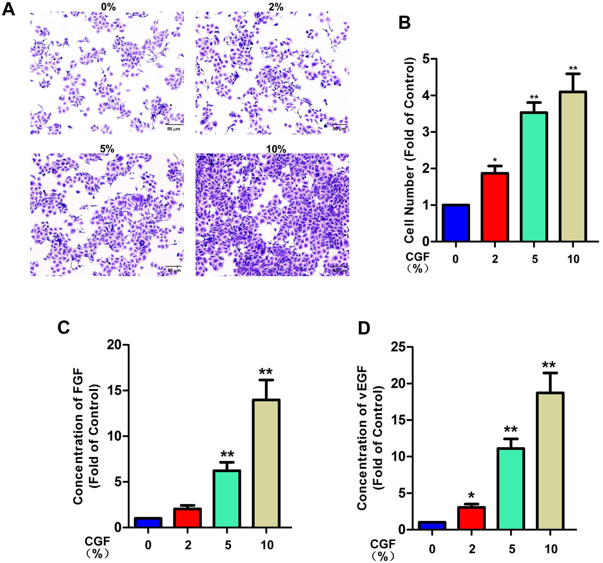 Fig. 1.
The promoting effect of CGF on the proliferation of HaCaT
cells. (A) Crystal violet staining was used to test the proliferation of HaCaT
cells upon CGF exposure for 3 days. (B) The comparison of cell numbers in each
group. (C,D) ELISA kits were used to test FGF and VEGF in the medium after cells
were exposed to different concentrations of CGF for 3 days. Data are displayed as
the mean
Cell migration is one of the key aspects in revascularization. The wound healing
assay indicated that HaCaT cells had a higher migration rate after CGF treatment
for 24 hr and 48 hr. The wound healing rate of HaCaT cells treated with CGF for
24 hr and 48 hr was increased sharply to 47.2
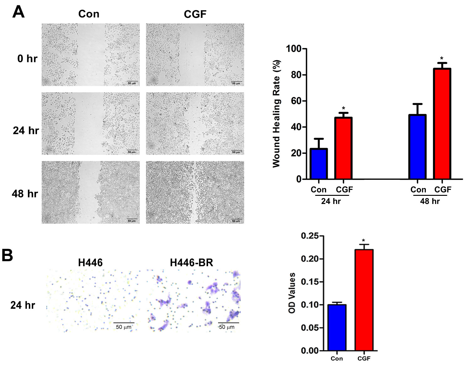 Fig. 2.
CGF stimulated the migration and invasion ability of HaCaT
cells. (A) Wound healing assay was employed to assess the migration of HaCaT
cells after CGF at different time points (0, 24, 48 hr). (B) Transwell assay was
recruited to determine the invasion ability of HaCaT cells at 24 hr after CGF
treatment. OD values for the crystal violet-stained cells were analyzed using
Image J software. All data are displayed as the mean
LPS (Escherichia coli O55:B5) is a well-known tool to activate cellular inflammation. As displayed in Fig. 3, the level of cytokines was significantly accumulated after LPS treatment for 1 and 3 days, and CGF could weakly inhibit the concentrations of cytokines after 1 and 3 days. The effect of LPS and CGF on HaCaT cell proliferation is demonstrated in Supplementary Fig. 1.
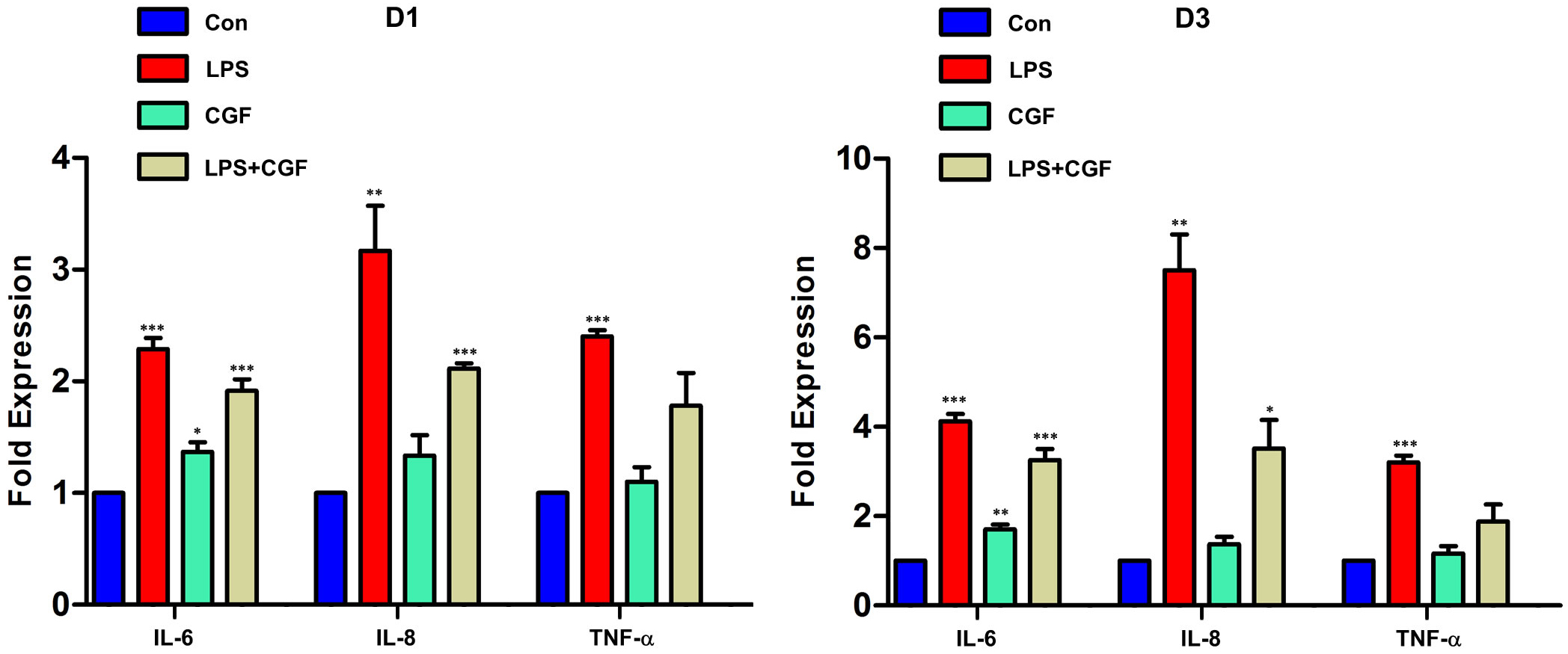 Fig. 3.
Effects of CGF on the production of proinflammatory cytokines in
LPS-stimulated HaCaT cells. HaCaT cells were exposed with or without LPS (1
HaCaT cells were exposed to different concentrations of CGF for 3 days. RAS,
p-C-RAF and p-ERK were up-regulated in a dose-dependent fashion, as displayed in
Fig. 4A,B. HaCaT cells were transfected with RAS lentiviral particles to knock
down the endogenous expression of RAS. The wound healing assay indicated that
silencing RAS could remarkably block the migration capacity of HaCaT cells. In
detail, the wound healing rate of HaCaT dropped sharply from
46.1
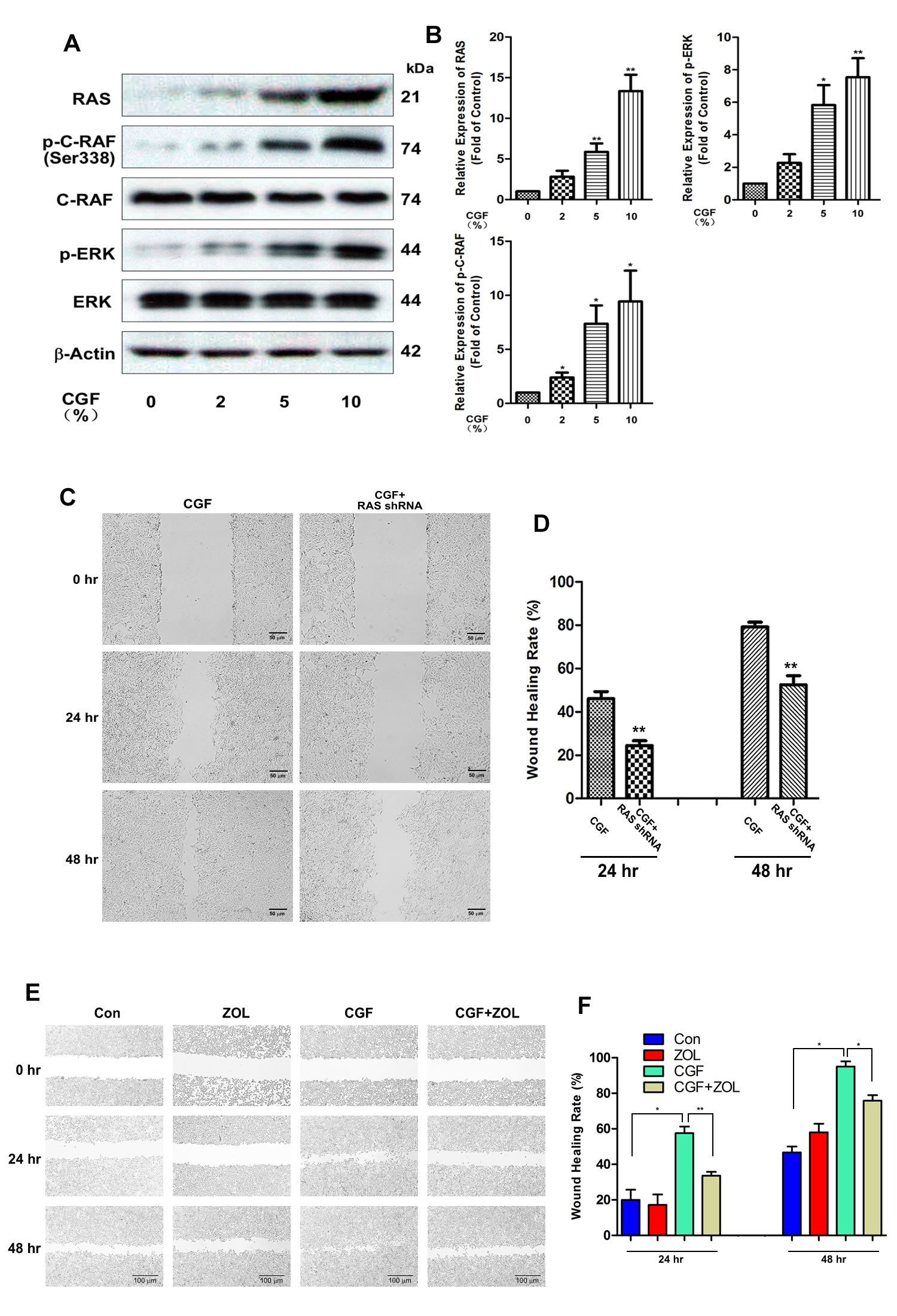 Fig. 4.
The RAS cell signaling pathway was induced upon CGF treatment.
(A) CGF-treated HaCaT cells were harvested at 3 days. RAS, p-C-RAF, C-RAF, p-ERK,
ERK, and
Our results above indicated that CGF treatment promoted cellular migration ability, which is an essential factor in angiogenesis. We found that angiogenic biomarkers (CD31, Angiopoietin-1 (ANGPT-1) and VEGF-A) were up-regulated in a dose-dependent fashion upon CGF treatment, meanwhile ANGPT-2 was down-regulated after CGF exposure in protein level (Fig. 5A,B). Similarly, the mRNA levels of these biomarkers also displayed similar trends (Fig. 5C–F).
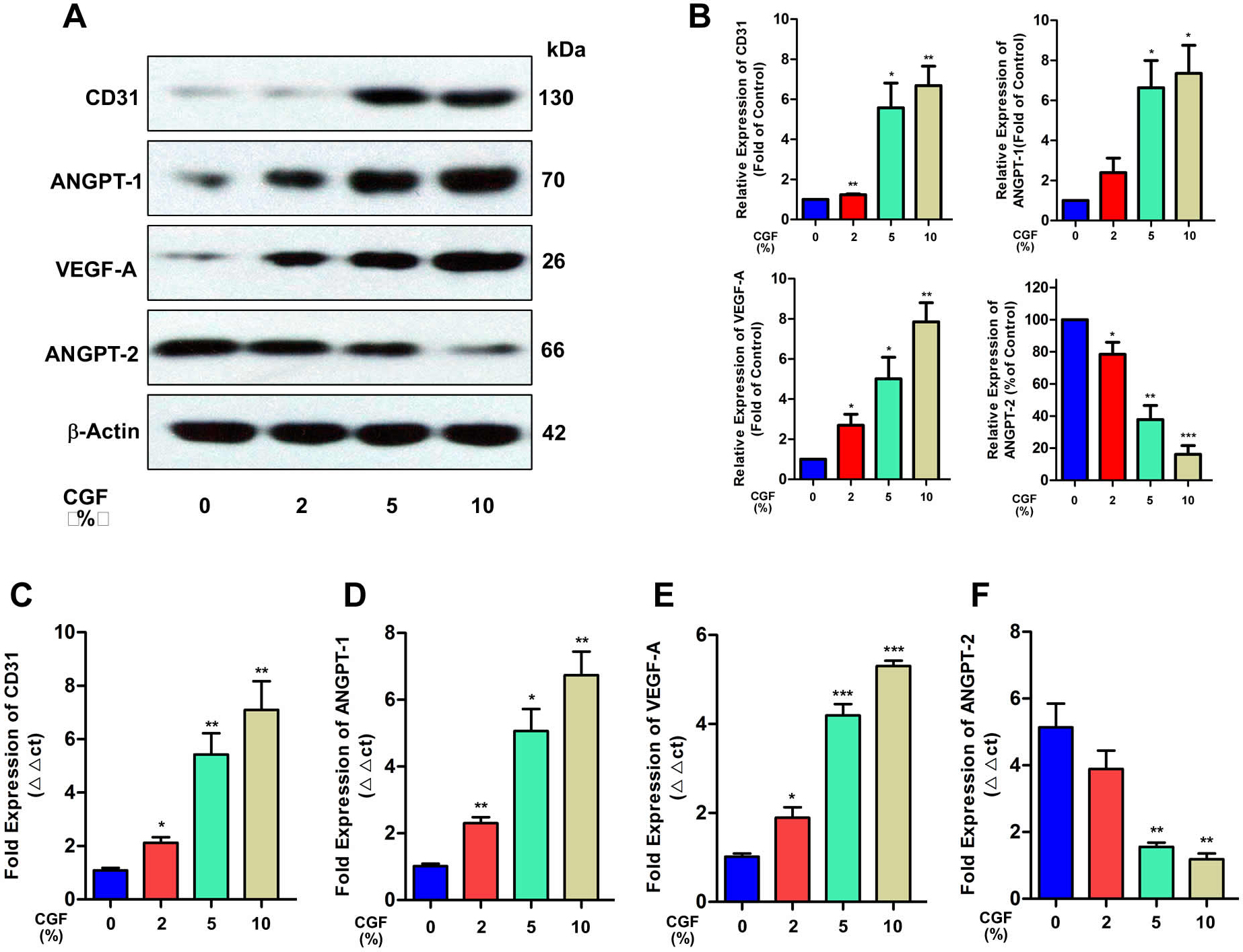 Fig. 5.
 Angiogenic biomarkers were activated upon CGF treatment. (A)
CD31, ANGPT-1, VEGF-A, and ANGPT-2 were measured using western blottingafter
cells were treated with different concentrations of CGF for 3 days. (B)
Quantification of CD31, ANGPT-1, VEGF-A and ANGPT-2 in western blot were
displayed. (C–F) qPCR was used to evaluate the expressions of CD31, ANGPT-1,
VEGF-A, and ANGPT-2 in cells treated with different concentrations of CGF (0%,
2%, 5%, and 10%) for 3 days. All data are shown as the mean
Wound closure was significantly improved in mice receiving CGF gel treatment
compared with that in mock-treated mice as displayed in Fig. 6A. Additionally,
the wound closure rate of mice was increased after CGF treatment from
18.6
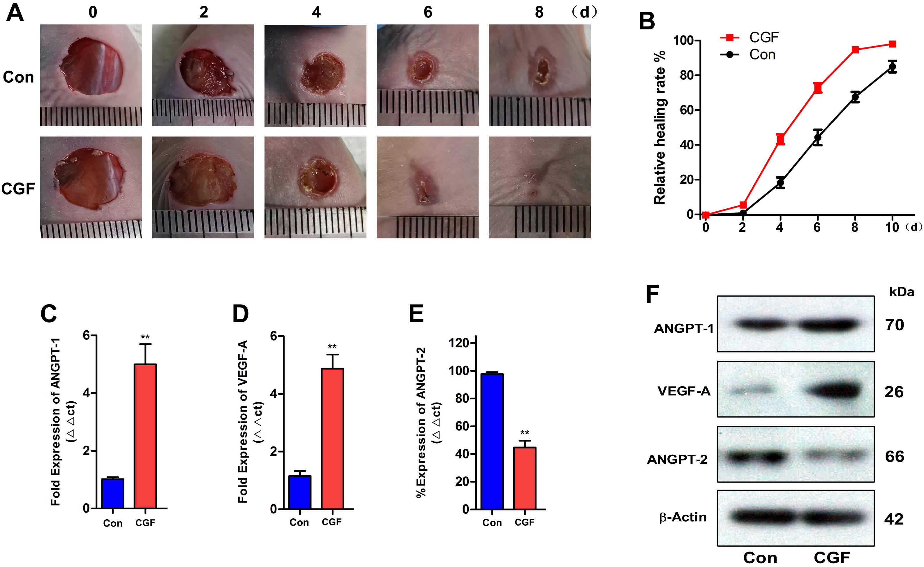 Fig. 6.
The in vivo effect of CGF in a wound mouse model. (A)
Photographs of skin wounds treated with CGF on Days 0, 2, 4, 6, and 8. (B)
Quantitative analysis of the wound closure area in the CGF and control groups
over time. Eight mice were recruited for each treatment group. (C–E) qPCR was
used to evaluate the expressions of ANGPT-1, VEGF-A, and ANGPT-2. (F) Western
blot was used to test the expressions of ANGPT-1, VEGF-A and ANGPT-2.
In this study, concentrated growth factor (CGF) was shown to effectively facilitate the proliferation as well as migration ability of HaCaT cells in vitro and excision wound model in vivo. The underlying mechanisms were elucidated to mainly involve activating the RAS signaling pathway and increasing the wound healing potential. Thus, this study provides novel findings on the therapeutic effects of CGF in wound healing.
CGF is considered as a novel natural biomaterial, which contains high levels of
platelets, cytokines, as well as growth factors to promote wound healing
activity; however little information has been obtained from skin wound healing
studies [18]. Although CGF contains more growth factors compared with PRP and
PRF, the best advantage of CGF might be the unique structure that provides a soft
texture with elasticity that allows malleable shaping to form a three-dimensional
structure consisting of fibrin to fill variable wound defects [19, 20]. Several
studies have demonstrated the characteristics of CGF by using scanning electron
microscopy (SEM) analysis, which revealed that CGF had a fiber-like appearance
with a 0.1–1.0
In the present study, LPS alone was displayed to benefit the proliferation of
HaCaT cells, and the up-regulated level in the combination arm was even higher,
suggesting CGF could still promote the proliferation HaCaT cells under
LPS-stimulated conditions (Supplementary Fig. 1). Meanwhile, CGF
contains varying amounts of platelets, cytokines, fibrins, and growth factors
such as VEGF, FGF, EGF, PDGF, insulin-like growth factor (IGF) and transforming
growth factor beta 1 (TGF-
Moderate inflammation is essential in the wound healing process, and both CGF
and LPS could lead to the production of proinflammatory cytokines and chemokines
including IL-6, IL-8, and TNF-
As a classic cell signaling pathway, the RAS/RAF/MEK/ERK (MAPK) cascade is vital for inter and intra-cellular communication, regulating fundamental cellular functions including proliferation, migration, survival, senescence, and differentiation [27]. The RAS family includes H-RAS, N-RAS, and K-RAS subtypes. Under normal conditions, RAS binds to GDP in an inactivated state. Subject to external stimulation with the guanine nucleotide exchange factor (GEF), RAS binds to guanosine triphosphate (GTP), thus becoming activated. GTP-RAS dimers/nanoclusters phosphorylate RAF, leading to the activation of downstream MEK/ERK. The activated MAPK cascade generates a response to regulate downstream cell signals [27]. EGF is one of the most important components in CGF, and triggers the RAS/RAF/MEK/ERK cascade through binding to its receptor located on the cell membrane. EGF signaling has a direct effect on promoting the migratory capacity of keratinocytes [28]. EGF encapsulation in gelatin-alginate coacervates could enhance efficacy in treating chronic diabetic wounds [29]. The FGFR1/ERK signaling pathway accounts for Kanglexin accelerating diabetic wound healing by promoting angiogenesis [30]. In our present study, we found that the RAS/RAF/MEK/ERK signaling pathway was activated upon CGF exposure, and the RAS inhibitor zoledronic acid stalled the migration of HaCaT cells after CGF treatment, which is consistent with our hypothesis. Therefore, the RAS/RAF/MEK/ERK cascade appears to play a critical role in CGF treatment for chronic wound healing and deserves in-depth study in the future.
As a fundamental physiological activity, wound healing could be divided into several stages including immediate hemostasis, acute inflammation, proliferation and maturation [31]. Angiogenesis is a vital accompanying process in the proliferation phase to form new blood vessels, which provide essential nutrients and oxygen for cell proliferation. Currently, novel therapeutic strategies for accelerating angiogenesis in healing cutaneous wounds include single and/or dual growth factors, gene and stem cell therapy, drug and drug-like compounds, and skin substitutes [32]. Intensive studies have reported that endogenous growth factors play a key function in benefiting angiogenesis and are essential for effective wound healing [33]. However, akin to our present study, delivery of exogenous growth factors has produced positive results in wound healing and angiogenesis in an animal model of full-thickness skin defect. Most of growth factor-based drugs for wound closure have not displayed clear benefits for promoting wound healing in clinical trials. One postulation for the failure of these trials is that patients acquire resistance to angiogenic growth factors in the context of disease [32, 34]. These mixed results from clinical trials have hindered the application of growth factors. CGF, obtained from a patient’s autologous venous blood, is characterized by rich growth factors and fibrin, which have concert effect in treatment thus leading to beneficial outcomes [14, 35].
It is necessary to unravel the in vivo healing effects of CGF using a full-thickness excisional wound model, which offers a systemic effect with the microenvironment of the wound bed being present under the CGF gel. The CGF gel was observed to accelerate the wound closure rate compared with the normal control group. In line with our in vitro study, angiogenic biomarkers were activated in the CGF treatment arm. Not only promoting wound healing, CGF can benefit skin appearance, promote pathological changes, and alter the elastic fiber structure of the skin, thus effectively delaying skin senescence in a mouse model [36]. Similarly, CGF along with nanofat could significantly improve the content of collagen and elastin, and has important function in fighting skin aging. And CGF combined with nanofat may provide a more reasonable alternative to skin anti-aging treatment [19].
Although we demonstrated that CGF has encouraging potential clinical applications, our study had several limitations. First, there is no conclusive data on the specific components of CGF mixtures. Moreover, the underlying interaction mechanism of growth factors in CGF has not been examined. Besides, high quality clinical randomized controlled studies are urgently needed to establish guidelines for the application of CGF as well as other blood extracts in wound healing.
CGF displayed a great enhancing wound healing effect in vitro and in vivo via activating the RAS cell signaling pathway. However, more studies will be required to validate the efficacy and safety of CGF in wound healing in the clinic.
CGF, concentrated growth factor; LPS, lipopolysaccharide; PRP, platelet rich
plasma; PRF, platelets rich fibrin; TGF-
The data generated in this study are available from the corresponding author upon reasonable request.
CK, and SX designed the research study. YueL and YanL conducted the experimental assay. YueL, YanL, CZ, and WL were responsible for writing of manuscript. SX contributed to the revised manuscript. All authors approved the final submitted manuscript.
This study was performed according to the principles recommended for the experimentation on humans and animals determined by the Institutional Review Board of Guangdong Medical University. Ethics committee approval was also obtained (No. GDY2102255).
Not applicable.
This research was supported by the Social Public Research Projects of Shenzhen Longhua District (Grant No. LHQJCYJ202102), Regional Joint Fund of Guangdong Province (Grant No. 2021A1515111063), Medical Scientific Research Foundation of Guangdong Province (Grant No. A2021253) and Shenzhen Longhua District Science and Innovation Bureau Fund for Medical Institutions (Grant No. 2021001 and 2021005).
The authors declare no conflict of interest. SX is serving as one of the Editorial Board members of this journal. We declare that SX had no involvement in the peer review of this article and has no access to information regarding its peer review. Full responsibility for the editorial process for this article was delegated to MF.
Supplementary material associated with this article can be found, in the online version, at https://doi.org/10.31083/j.fbl2712319.