IMR Press / FBL / Volume 26 / Issue 5 / DOI: 10.52586/4922
Open Access Review
Comparative review on left-handed Z-DNA
Show Less
1 Department of Zoology, Acharya Prafulla Chandra College, New Barrakpur, Kolkata, 700131 West Bengal, India
2 Department of Botany, Acharya Prafulla Chandra College, New Barrakpur, Kolkata, 700131 West Bengal, India
3 Department of Zoology, University of Calcutta, 700019 Kolkata, India
4 Department of Botany, Dinabandhu Andrews College, Garia, Kolkata, 700084 West Bengal, India
*Correspondence: jsarkar80@gmail.com (Joy Sarkar)
These authors contributed equally.
Front. Biosci. (Landmark Ed) 2021, 26(5), 29–35; https://doi.org/10.52586/4922
Submitted: 28 September 2020 | Accepted: 25 November 2020 | Published: 30 April 2021
Copyright: © 2021 The Author(s). Published by BRI.
This is an open access article under the CC BY 4.0 license (https://creativecommons.org/licenses/by/4.0/).
Abstract

Being polymorphic, deoxyribonucleic acid is worthy of raise a variety of structure like right-handed B to left-handed Z conformation. In left-handed contour of DNA consecutive nucleotides substitute between syn-arrangement and anti-arrangement, through the chain. 2D gel electrophoresis comprising d(PCpG)n of topo isomers of a plasmid inserts d(pCpG)n, in this ‘n’ ranges among 8 to 21, indicate the change of B-Z DNA. The high denseness of salt is required for conversion of B configuration d(CG)n toward Z configuration. The rate of B to Z transition is measured by “Cytosine Analogues” and “Fluorescence Spectroscopy”. h-ZαADAR1 that a Z-DNA’s binding domain, binds and stabilizes one part in Z configuration and therefore the remaining half in B deoxyribonucleic acid configuration. At halfway point, it creates B-Z junction. “Stacking” is the main reason for the B-Z DNA junction construction. Upregulation of ADAM-12, related with Z-DNA is said to a cause for cancer, arthritis, and hypertrophy. Z-DNA forming sequence (ZFS) conjointly generates massive - scale deletion in cells from mammals.

Keywords
Alzheimer’s disease
Wang model
Z-DNA-binding protein
Z-DNA-forming sequence
Review
2. Introduction

In 1979, a left-handed crystal deoxyribonucleic acid structure was published, which convey a unique zigzag, sugar-phosphate backbone, it’s named as Z conformation of deoxyribonucleic acid (Z-DNA) and it’s all biological relevance had yet to be established [1, 2]. It was already known that normal right-handed B conformation can assume a diverse number of configurations, under certain torsional stress [3]. Z configuration exists in high energy state than the common B-DNA configuration. This conformation has negative super helicity which soothes the structure. In contrast to B form with anti-conformation, in Z-DNA convey anti-conformation and syn-conformations alternately by rotating around glycosyl bonds, along with the chain [4]. Under bound condition non-B-DNA structure like cruciform, triplex, hairpin, etc. are formed by collapsible monotonous DNA sequence. This unusual structure has effects on several biological progressions [5]. Super helicity is the most significant inducer for Z contour in usual DNA. Non-super helical, natural DNA holds practically no Z-DNA, but other hand the same DNA under extreme negative super helicity, as in “form V” may have as much as 35–40% of its sequence in Z arrangement [6]. Except for Z-DNA, X-ray fiber diffraction outlines were framed and differentiates several conformations of DNA. Most DNA enters the A-DNA conformation which’s per turn contain 11 bp through right-handed helix [7]. The single-crystal method resolute the complementary structure, oligo deoxy nucleosides, d(GGTATACG) and d(IODO-CCGG) [7, 8] (Table 1).

Table 1.Comparable information between A-DNA, B-DNA, Z-DNA [2, 9].
Parameter A-DNA B-DNA Z-DNA
Helix sense Right-handed Right-handed Left-handed
Axial raise [in Armstrong] 2.55 3.4 3.7
Helix pitch 28 34 35
Base pair tilt 20 -6 7
Rotation per residues 33 36 -30
Diameter of helix [in angstrom] 23 20 18
Glycosidic bond configuration Anti Anti Anti
da, dT, dC, dG Anti Anti Syn
Inserted phosphate phosphate distance [in Armstrong] 5.9 7.0 7.0
da, dT, dC, dG 5.9 7.0 5.9
Suger pucker C3’-endo C2’-endo C2’-endo
da, dT, dC, dG C3’-endo C2’-endo C3’-endo

Existence of B-Z transition and Z-DNA is further deep-rooted by the specific ZBP discovery [10]. In vitro, Z-DNA was postulated for identification of proteins that bind with it in a structure-precise manner, act as a cis-element and aid in biological development. RNA Double Strand adenosine deaminase 1 is a type of the ZBP [11]. This ADAR1 has a Zα domain capable of transform B into Z conformation and create the junction [12, 13]. Formation of Z-DNA is induced by a unique sequence motif. Sometimes, it presents frequently adjacent with the start site of transcription and induce the transcription [14, 15, 16]. The junction between B-Z is formed with the help of ZBP. Formation of this portion carries out flipping over of bases, stacking of bases, and infringement of one base pair [17]. In another study also verified that normal B form also transfers into Z form by elevation of salt of aggregation [18, 19]. In humans, Z-DNA first came into consideration through the autoimmune disease Lupus erythematosus [20]. Z-DNA formation sequence (ZFS) is found to be associated with immune retorts and infection genome uncertainty. The Z configuration is also evidenced to be linked with large scale deletion in the cells of mammals [21, 22]. It also controls the genes transcription regulation of c-myc and CRH of human [23, 24].

3. Z-DNA structure

The optical investigation originally proposed the Z-DNA. The result of the experiment exhibited that a 4 mL NaCl solution contains a polymer which consists of discontinuous cytosine and guanine residue and formed a nearly inverted circular dichroism gamut [25]. Until 1979, the invention of Z-DNA remained unknown. Original atomic steadfastness exposed that it was not the same right-handed B-DNA which was invented by James D. Watson & H.C. Crick in 1953. Despite that, this new left-handed helical structure named as Z deoxyribonucleic acid. This Z form consists of extremely immunogenic antibodies to recognize the configuration, unlike B form of DNA [26]. There have some familiar features of B form with the d(Cg)3 system. The antiparallel double-helical structure holds Watson-Crick base pairing between the base of Guanine and Cytosine. The left-handed helicity oligomers have six base pairs with significant regularity. Balance correlated hexamers stack on one other so closely in an endless polymer of alternating cytosine guanine residues sequence [2].

Various conformational topographies differ the Z-DNA from the B-DNA (Fig. 1). The double-helical Hexa-nucleoside Penta Phosphate molecules allied with the crystal. Crystal of Z arrangement contains discontinuous cytosine and guanine residues’-DNA is dinucleotide while B-DNA is mononucleotide with anti-configuration. All deoxycytidine has anti-configuration whereas all the deoxyguanosine has anti syn-configuration.
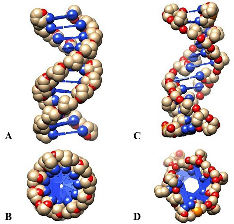
Fig. 1.

Evaluation of the structure between Z and B-DNA configuration. (A, B) Z-DNA configuration showing left-handed elongated spiral with a lateral view and a polar view. (C, D) B-DNA configuration showing right-handed elongated spiral with a lateral view and a polar view.

In Z arrangement the base pair is lifted from the center, so the guanine imidazole ring is originated at the edge, but in case of B-DNA those bases are at the center. In B configuration 34Å pitch with 10.5 bp is present where Z configuration convey 44.6Å pitch with 12 bp per turn [9]. Six levels of base pairing have been seen in the d(Cg)3 structure because of C1 base pair with G12, G2, C11 and so on. Z-DNA is not slanted with each other straight, but they remain linked to a literal translation of 7Å relative to each other so that it can shear the appearance from one another with a little rotation throughout the chain. Despite being stacked on other bases the guanine is loaded upon the oxygen atoms of prior deoxyribose residues. The backbone of sugar-phosphate is constant for both the Z form and B form. In B configuration the minor-grooves are above the base pairs. But in Z form minor-grooves exist below the base pair [2].

4. B-DNA to Z-DNA transition

Earliest Harvey model is used for examining B-Z transition. This model defines the procedure which is engrossed by another longitudinal DNA conscious models. Base pairs opening was another early observed mechanism regarding this model before the Wang model. At the present portrait of Zipper Model demonstrated that Z-DNA contains high energy joint that grows through the DNA polymer until the full B-Z trans polymer gets transferred into Z-DNA. Though there are some problems in the model because it does not disclose many vibrant structural details, so it has limited applications in thermodynamics. There are several facts regarding the transition of B-Z such as the high concentration of salt in the solution which balance the Z-DNA due to massive reduction in electrostatic repulsion in the pillar of phosphate. Negative helicity of deoxyribonucleic acid needs energy that can also uncoil B form to configure the Z form. Z-DNA can also be stabilized by transcription.

Maruyama and colleague establish the B-DNA to Z-DNA transition communed by a method called “cationic graft copolymer” where the Poly (L-lysine)-graft dextran (PLL-g-Dex), begins with two-step method including the creation of a clear intermediate [27]. Amid DNA phosphate group electrostatic repulsion reduce by the cationic backbone of the copolymer and the transition is a result of these 2 factors. The most plausible Z form created negative supercoiling, utilizing B-DNA occurs during several metabolisms like Transcription and replication processes [28]. For reducing the transition stress, unusual such DNA as Z-DNA is formed [6, 29]. Lee et al. (1992) used “Magnetic-tweezers” and FRET combinedly to examine at molecule level of negative supercoiling [30]. Magnetic tweezers are a very useful technique for investigating wind/unwinding procedure of twisted DNA through precisely controlling infinite tension [28, 31]. Therefore, B-Z change can be active by tiny negative super helicity and approximately one Pico Newton Tension. This outcome suggests that in tension Z arrangement is formed more easily in vivo [32]. Methanol, Ethanol, Ethylene Glycol (Dehydrating agent) balance the Z-DNA configuration. Due to adjacent clustering counteractions all over the DNA, though more strong ionic properties, thus it provides additional mutually repelling phosphate groups [6]. Antibodies and ZBP can bind the Z form of DNA selectively. This conformation has triggering capability. The Qu group had been reported that Alzheimer amyloid protein brings about the Z-B transition. Forming the Z-form is correlated with Alzheimer’s disease [33, 34]. Bae et al. analyzed to transition from B-Z conformational change occurs by Z-DNA binding protein unravel the detailed binding machinery and whether the protein industriously initiates Z-DNA’s or passively traps transitionally performed Z form. Therefore, it proved that the conformational selection mechanism stabilized the Z-DNAs by alternating the “induced fit” mechanism. A chemical modification also stabilizes Z-DNA transformation [4]. Bulky group’s introduction precise in a certain base also steady the growth of Z arrangement by increasing static hindrance.

5. B-DNA and Z-DNA hybrid junction

Double-stranded adenosine deaminase RNA is an enzyme of the deaminase family which edited the appearance of the ds-mRNA by converting adenosine to inosine and creating diversity between RNA and Protein [11]. It is noted as a naturally stirring protein with obvious specificity for methylated and hemi-brominated DNA contains discontinuous deoxy guanosine-deoxycytidine residues [13]. ADAR1 carry two binding motifs for Z-DNA, Zα and Zβ[11].

A few numbers of investigations were completed to show the interface between the solution of DNA and Zα ADAR1 domain. If the DNA solution is interacting with dodecamer (d(CG)6) it produces the B-DNA circular-dichroism spectrum. When Zα ADAR1 is mixed into the solution the spectrum progressively altered, which mirrored Z conformation. This demonstrated that the Zα domain is equipped for alleviating the dodecamer in the Z configuration. Brownian motion or Pedesis is the reason for this twist of dodecamer fragment. After this conformational change, DNA binds with the Zα domain to prevent the reappearance of B-DNA conformation [12, 13].

Kim et al. in 2005 developed a DNA duplex with 15 bp and with two hanging nucleotides [17]. This DNA duplex is co-crystallized with the ZαADAR1 domain (amino acids 140-202). So, Z-DNA is tightly bound with the binding domain of Z DNA, h-ZαADAR1. After the binding, it stabilizes one half in the Z configuration and remaining part in B form. In the centre portion, a B-Z junction is created [17]. At this DNA duplex, eight bases stabilized with normal Z-DNA conformation [2]. The remaining six bases are maintaining the typical B conformation [35]. On the link point, A-T bases are disrupted from each other and make a sharp turn, which obliged an inversion in the way of the backbone. This creates a bent at the intersection point of B-Z DNA. The disrupted A, T bases adopted anti-conformation. Base A is extended out from the helix and T is slanted analogous to the spiral. But first base-pair from the Z-DNA after the junction creates a long rise distance which clearly showed the stacked A-T the bases within the B-DNA conformation. Stacking is the main stabilizing factor for the junction, and it is proved that one bp extruding by breaking can cause reversion of the handedness of the duplex. Other than A-T bases, it is equally possible for other G-C bases to be extruded [17]. Thermodynamic examinations of the melting of oligomers holding the junction show that the edifice of the hybrid junction from B-DNA declines the melting free energy by 0.5 kcal/mol [36]. This B-Z configurational change and syn-conformation of both bases are done by base ‘flipping over’. A torsional strain breaks and causes base extrusion. This extruded base is allowed to flip over and reorganization the bp, which creates a ZIP-like movement in two direction. This movement for the limitation of the ZFS with an extruded base at the intersection. Base-pair disruption, expulsion, and reconstruction are lengthening the Z-DNA segment through an additional negative torsional strain of chromatin [17].

Another investigation also proved that B-Z DNA junction can be produced by oligomeric sequences in the aqueous solution at 3 M or high salt concentration. The 5.5 M NaCl with a 95 mM combination induces the A-T sequence into the Z-DNA conformation [18]. This study re-establishes that when NiCl2 is added in the salt solution, it creates a striking change in Raman Spectra, indicating A-T bases are adopting the Z conformation [19].

6. Z-DNA in human disease

In living body, Z-DNA can form and role as a dynamic component in various genome’s metabolic courses under certain biological circumstances [21]. Z-DNA is used in many precise activators or repressors enrolment for directive gene countenance, genome uncertainty control [22]. Another study proved that in cells of mammal’s ZFS fetch genetic uncertainty. Repair mechanism can proceed with the Z-DNA development in the mammal’s body, which creates a large genomic alteration. These sorts of changes are relevant to the breakage and translocation near ZFS in human lymphoma and leukaemia [9]. In humans, Z-DNA links with the transcription of the c-myc genes, which means when the Z-DNA development is turned off the cell gives a signal as a result, c-myc transcription also starts to down-regulate [23]. In the same way, Z-DNA development is also associated with the corticotropin-releasing hormone (CRH) gene transcription [37]. On the other hand, the human body also shows the activation of the Nrf2 gene which is relevant to the HO-1 gene’s promoter, which allied with Z-DNA development [24]. A few numbers of immunoglobulin-related genes (example-ETV6) are enriched by the Z-DNA sequence. But in blood cancer, these genes are related to translocation of the chromosome [22]. Interferonopathies disease like Aicardi-Goutières Syndrome is caused by Mutation, which reduces p150 Z-binding with impaired enzymatic activity. This is induced by dsRNAs and most commonly these dsRNAs derive from Alu retroelement. The Z-DNA and Z-RNA both are essential for limiting Alu retroelement intrusion of primate genomes [38]. Z-DNA provides a base for therapeutically reducing the chances of Arthritis, Cancer, and cardiac hypertrophy. This role is believed to be arbitrated by the downregulation of ADAM-12. It was observed that ADAM-12 protein expression is raised when there are pieces evidence of arthritis, cancer, and cardiac hypertrophy. Whereas ADAM-12 expression level Is exceptionally low in certain adult tissue. The regulation of ADAM-12 is related to the highly conserved region containing a stretch of dinucleotide repeat sequence and known as negative regulatory element (NRE), which serves as a repressor of ADAM-12 expression. There is a certain Z-DNA binding protein-like MeCP2. It modulates the ADAM-12 repression by recruiting NF1 transcriptional factors. Loss of ZFS leads to a low level of MeCP2 which results in metastatic breast cancer [22, 39]. Apart from this, HIF1α induced Z-DNA development in the microsatellite of slc11a1 gene promoter. It was also perceived to control its definite allele expression in patients of rheumatoid arthritis, tuberculosis [40]. Z-DNA also has an immunogenic character and it can prevent systemic lupus erythematosus. But in the patient’s sera of these diseases, some anti-Z-DNA antibody are found. Two kinds of antibody are found, first-one responsible for denaturation of both B and Z form and second-one is Z-DNA specific [20, 41]. Z arrangement also induce conformation instability by acting as a site for cancer-related genes like scl, bcl2, and c-myc [9]. B-Z junction is a site where CAG trinucleotide repeat instability happened. X fragile chromosome and skeletal dysplasia associated with CGG repeats and GAC trinucleotides repeat respectively [42, 43, 44]. In a study, typical left-handed Z-DNA was originated in brains of severe AD affected patients. Similarly, the moderately affected patients showed the existence of B-Z intermediate conformation in their brain DNA. Immunohistochemical data has proved that the total amount of Z form is one-seventh than B arrangements in human’s genome [45, 46]. It was also observed that some genes, related to Alzheimer’s like presenilin-1, presenilin-2, APOE (Apolipoprotein E), etc. are overexpressed in patients and has an important appeal in Alzheimer’s pathogenesis. Z-DNA existing in the brains of Alzheimer’s patients are far more vulnerable to hydroxyl radical-induced damage of DNA, in comparison to A-DNAs or B-DNAs. This was due to the occurrence of more exposed bases and patients with severe Alzheimer’s showed the existence of both Z-DNA and damaged DNA of similar types [47]. This finding has again been confirmed from another study which showed that Z-DNA became sensitive to hydrolytic enzyme DNase I, on incubation with Aβ protein for a certain period [34]. This results in alteration of Z arrangement back into normal B form. These transition of Z form to normal B form is verified as quicker process when an interaction of Aβ is made, in the existence of ethylene glycol also [48].

7. Conclusions

Z-DNA is a double-helical structure that preserves antiparallel backbone of sugar-phosphate chains with Watson Crick pairing. Despite that, it has a contour which is fundamentally dissimilar from B configuration of DNA. Two-dimensional Gel Electrophoresis offers us a powerful method to examine the super helicity-induced physical revolution in the DNA. Besides this, B-Z conversion is also designated here. One of a reasons for transition is a cause of free unfavourable energy. Affected advances are unrestricted from the uniting effect of genomics, human genetics, biophysics, and molecular studies on non-B-DNA configurations through mutation causing agents, intricate in Genetic diseases. Autoimmune processes may be suspected in all clinical conditions where specific anti-Z-DNA antibodies are found, but for further investigation, larger population is wanted to prove such an immunological hypothesis. Future prominence will challenge to tune the acceptance of the non-B-DNA configurations at a definite location of genes to correlate this behavior extra thoroughly with the generation reposition terminuses. Also, the analysis to recognize the kind of non-B-DNA structures that obtain certain sort of mutations and the fascinated enzyme on the evolution of therapeutics, to ameliorate the disturbing corollaries of these disorders.

8. Author contributions

PC and RR conceptualize this review article. RR analyzed and interpreted the information regarding Z-DNA structure and B-Z DNA transition. PC performed a study on B-Z DNA hybrid junction formation and effects of Z-DNA on human disease and was a major contributor in writing the manuscript. AC developed the figure based on available data. PC prepared the final draft of the manuscript under the supervision of JS. All authors read and approved the final manuscript.

9 Ethics approval and consent to participate

The work reported here in the manuscript is original and free from any plagiarism. All the data in the article are real and authentic. All the co-authors have read and agree to publish all the items listed above.

10. Acknowledgment

Reetabrita Roy and Pallab Chakraborty contributed equally to this article.

11. Funding

We don’t have any funding support from any organizational or institutional level. On behalf of all listed authors, the corresponding author declares that there is not any sort of financial and non-financial conflict of interest in the subject materials mentioned in this manuscript.

12. Conflict of interest

The authors declare no conflict of interest.

Abbreviations

Aβ protein, Amyloid β-protein; AD, Alzheimer’s disease; ADAM, a disintegrin and metalloproteinase; ADAR1, Adenosine Deaminase Acting On RNA; APOE, apolipoprotein E; CRH, corticotrophin-releasing hormone; DNA, Deoxyribonucleic acid; FRET, fluorescence resonance energy transfer; HO-1, heme oxygenase-1; IODO, 3-Iodo-L-tyrosine; MeCP2, methyl CpG binding protein 2; mRNA, messenger RNA; NF1, neurofibromatosis type 1; NRE, negative regulatory element; PLL-g-Dex, Poly (L-lysine) - graft dextran; RNA, Ribonucleic acid; ZBP, Z-DNA binding protein; ZFS, Z-DNA forming sequence.

References
[1]
de Rosa M, de Sanctis D, Rosario AL, Archer M, Rich A, Athanasiadis A, et al. Crystal structure of a junction between two Z-DNA helices. Proceedings of the National Academy of Sciences of the United States of America. 2010; 107: 9088–9092.
[2]
Wang AH, Quigley GJ, Kolpak FJ, Crawford JL, van Boom JH, van der Marel G, et al. Molecular structure of a left-handed double helical DNA fragment at atomic resolution. Nature. 1979; 282: 680–686.
[3]
Kohwi-Shigematsu T, Manes T, Kohwi Y. Unusual conformational effect exerted by Z-DNA upon its neighboring sequences. Proceedings of the National Academy of Sciences of the United States of America. 1987; 84: 2223–2227.
[4]
Rich A, Zhang S. Z-DNA: the long road to biological function. Nature Reviews Genetics. 2003; 4: 566–572.
[5]
Choi J, Majima T. Conformational changes of non-B DNA. Chemical Society Reviews. 2011; 40: 5893–5909.
[6]
Johnston BH. Generation and detection of Z-DNA. Methods in Enzymology. 1992; 211: 127–158.
[7]
Heinemann U, Alings C, Hahn M. Crystallographic studies of DNA helix structure. Biophysical Chemistry. 1994; 50: 157–167.
[8]
Cruz P, Bubienko E, Borer PN. A model for base overlap in RNA. Nature. 1982; 298: 198–200.
[9]
Rich A, Nordheim A, Wang AHJ. The chemistry and biology of left-handed Z-DNA. Annual Review of Biochemistry. 1984; 53: 791–846.
[10]
Kimura T, Kawai K, Fujitsuka M, Majima T. Detection of the G-quadruplex-TMPyP4 complex by 2-aminopurine modified human telomeric DNA. Chemical Communications. 2006; 4: 401–402.
[11]
Wang G, Vasquez KM. Z-DNA, an active element in the genome. Frontiers in Bioscience. 2007; 12: 4424–4438.
[12]
Kim Y, Lowenhaupt K, Maas S, Herbert A, Schwartz T, Rich A. The zab domain of the human RNA editing enzyme ADAR1 recognizes Z-DNA when surrounded by B-DNA. Journal of Biological Chemistry. 2000; 275: 26828–26833.
[13]
Berger I, Winston W, Manoharan R, Schwartz T, Alfken J, Kim Y, et al. Spectroscopic characterization of a DNA-binding domain, Zα, from the editing enzyme, dsRNA adenosine deaminase: evidence for left-handed Z-DNA in the Zα-DNA Complex. Biochemistry. 1998; 37: 13313–13321.
[14]
Champ PC, Maurice S, Vargason JM, Camp T, Ho PS. Distributions of Z-DNA and nuclear factor I in human chromosome 22: a model for coupled transcriptional regulation. Nucleic Acids Research. 2004; 32: 6501–6510.
[15]
Liu R, Liu H, Chen X, Kirby M, Brown PO, Zhao K. Regulation of CSF1 promoter by the SWI/SNF-like BAF complex. Cell. 2001; 106: 309–318.
[16]
Oh D, Kim Y, Rich A. Z-DNA-binding proteins can act as potent effectors of gene expression in vivo. Proceedings of the National Academy of Sciences. 2002; 99: 16666–16671.
[17]
Ha SC, Lowenhaupt K, Rich A, Kim Y, Kim KK. Crystal structure of a junction between B-DNA and Z-DNA reveals two extruded bases. Nature. 2005; 437: 1183–1186.
[18]
Ridoux JP, Liquier J, Taillandier E. Raman spectroscopy of Z-form poly[d(a-T)].poly[d(a-T)]. Biochemistry. 1988; 27: 3874–3878.
[19]
Dai Z, Thomas GA, Evertsz E, Peticolas WL. The length of a junction between the B and Z conformations in DNA is three base pairs or less. Biochemistry. 1989; 28: 6991–6996.
[20]
Lafer EM, Valle RP, Möller A, Nordheim A, Schur PH, Rich A, et al. Z-DNA-specific antibodies in human systemic lupus erythematosus. Journal of Clinical Investigation. 1983; 71: 314–321.
[21]
Kha DT, Wang G, Natrajan N, Harrison L, Vasquez KM. Pathways for double-strand break repair in genetically unstable Z-DNA-forming sequences. Journal of Molecular Biology. 2010; 398: 471–480.
[22]
Ravichandran S, Subramani VK, Kim KK. Z-DNA in the genome: from structure to disease. Biophysical Reviews. 2019; 11: 383–387.
[23]
Wittig B, Wölfl S, Dorbic T, Vahrson W, Rich A. Transcription of human c-myc in permeabilized nuclei is associated with formation of Z-DNA in three discrete regions of the gene. EMBO Journal. 1992; 11: 4653–4663.
[24]
Maruyama A, Mimura J, Harada N, Itoh K. Nrf2 activation is associated with Z-DNA formation in the human HO-1 promoter. Nucleic Acids Research. 2013; 41: 5223–5234.
[25]
Herbert A, Rich A. Left-handed Z-DNA: structure and function. Structural biology and functional genomics. Genetica. 1999; 106: 37–47.
[26]
Lafer EM, Möller A, Nordheim A, Stollar BD, Rich A. Antibodies specific for left-handed Z-DNA. Proceedings of the National Academy of Sciences of the United States of America. 1981; 78: 3546–3550.
[27]
Shimada N, Kano A, Maruyama A. Design of cationic graft copolymers as a potential inducer of B-Z transition. Nucleic Acids Symposium Series. 2009; 53: 251–252.
[28]
Yang X, Li Z, Polyakova T, Dejneka A, Zablotskii V, Zhang X. Effect of static magnetic field on DNA synthesis: the interplay between DNA chirality and magnetic field left-right asymmetry. FASEB BioAdvances. 2020; 2: 254–263.
[29]
Murchie AI, Lilley DM. Supercoiled DNA and cruciform structures. Methods in Enzymology. 1992; 211: 158–180.
[30]
Lee S, Kwak C, Shim J, Kim J, Choi S, Kim HF, et al. A cellular model of memory reconsolidation involves reactivation-induced destabilization and restabilization at the sensorimotor synapse in Aplysia. Proceedings of the National Academy of Sciences of the United States of America. 2012; 109: 14200–14205.
[31]
Strick TR, Allemand JF, Bensimon D, Bensimon A, Croquette V. The elasticity of a single supercoiled DNA molecule. Science. 1996; 271: 1835–1837.
[32]
Sanford DG, Stollar BD. Assay of anti-DNA antibodies. Methods in Enzymology. 1992; 212: 355–371.
[33]
Breslow JL, Ross D, McPherson J, Williams H, Kurnit D, Nussbaum AL, et al. Isolation and characterization of cDNA clones for human apolipoprotein A-I. Proceedings of the National Academy of Sciences of the United States of America. 1982; 79: 6861–6865.
[34]
Geng J, Zhao C, Ren J, Qu X. Alzheimer’s disease amyloid beta converting left-handed Z-DNA back to right-handed B-form. Chemical Communications. 2010; 46: 7187–7189.
[35]
Herbert A, Lowenhaupt K, Spitzner J, Rich A. Chicken double-stranded RNA adenosine deaminase has apparent specificity for Z-DNA. Proceedings of the National Academy of Sciences of the United States of America. 1995; 92: 7550–7554.
[36]
Sheardy RD, Levine N, Marotta S, Suh D, Chaires JB. A thermodynamic investigation of the melting of B-Z junction forming DNA oligomers. Biochemistry. 1994; 33: 1385–1391.
[37]
Wölfl S, Martinez C, Rich A, Majzoub JA. Transcription of the human corticotropin-releasing hormone gene in NPLC cells is correlated with Z-DNA formation. Proceedings of the National Academy of Sciences of the United States of America. 1996; 93: 3664–3668.
[38]
Herbert A. Z-DNA and Z-RNA in human disease. Communications Biology. 2019; 2: 7.
[39]
Ray BK, Dhar S, Shakya A, Ray A. Z-DNA-forming silencer in the first exon regulates human ADAM-12 gene expression. Proceedings of the National Academy of Sciences of the United States of America. 2011; 108: 103–108.
[40]
Bayele HK, Peyssonnaux C, Giatromanolaki A, Arrais-Silva WW, Mohamed HS, Collins H, et al. HIF-1 regulates heritable variation and allele expression phenotypes of the macrophage immune response gene SLC11a1 from a Z-DNA forming microsatellite. Blood. 2007; 110: 3039–3048.
[41]
Allinquant B, Malfoy B, Schuller E, Leng M. Presence of Z-DNA specific antibodies in Crohn’s disease, polyradiculoneuritis and amyotrophic lateral sclerosis. Clinical and Experimental Immunology. 1984; 58: 29–36.
[42]
Khan N, Kolimi N, Rathinavelan T. Twisting right to left: A…A mismatch in a CAG trinucleotide repeat overexpansion provokes left-handed Z-DNA conformation. PLoS Computational Biology. 2015; 11: e1004162.
[43]
Vorlícková M, Kejnovská I, Tumová M, Kypr J. Conformational properties of DNA fragments containing GAC trinucleotide repeats associated with skeletal displasias. European Biophysics Journal. 2001; 30: 179–185.
[44]
Renčiuk D, Kypr J, Vorlíčková M. CGG repeats associated with fragile X chromosome form left-handed Z-DNA structure. Biopolymers. 2011; 95: 174–181.
[45]
Soyer-Gobillard MO, Géraud ML, Coulaud D, Barray M, Théveny B, Révet B, et al. Location of B- and Z-DNA in the chromosomes of a primitive eukaryote dinoflagellate. Journal of Cell Biology. 1990; 111: 293–304.
[46]
Gagna CE, Lambert WC, Kuo HR, Farnsworth PN. Localization of B-DNA and Z-DNA in terminally differentiating fiber cells in the adult lens. Journal of Histochemistry and Cytochemistry. 1997; 45: 1511–1521.
[47]
Michalik V, Spotheim Maurizot M, Charlier M. Calculation of hydroxyl radical attack on different forms of DNA. Journal of Biomolecular Structure & Dynamics. 1995; 13: 565–575.
[48]
Zacharias W, Larson JE, Klysik J, Stirdivant SM, Wells RD. Conditions which cause the right-handed to left-handed DNA conformational transitions. Evidence for several types of left-handed DNA structures in solution. Journal of Biological Chemistry. 1982; 257: 2775–2782.
Share
Back to top