IMR Press / FBL / Volume 26 / Issue 11 / DOI: 10.52586/5022
Open Access Review
Discovery of novel peptide neurotoxins from sea anemone species
Show Less
1 Key Laboratory of Tropical Translational Medicine of Ministry of Education, Hainan Key Laboratory for Research and Development of Tropical Herbs, Hainan Medical University, 571199 Haikou, Hainan, China
2 Institute for Molecular Bioscience, The University of Queensland, St. Lucia, Brisbane, QLD 4072, Australia
*Correspondence: a.jin@imb.uq.edu.au (Ai-Hua Jin); gaobingmiao@hainmc.edu.cn (Bingmiao Gao)
Front. Biosci. (Landmark Ed) 2021, 26(11), 1256–1273; https://doi.org/10.52586/5022
Submitted: 10 June 2021 | Revised: 21 October 2021 | Accepted: 25 October 2021 | Published: 30 November 2021
Copyright: © 2021 The Author(s). Published by BRI.
This is an open access article under the CC BY 4.0 license (https://creativecommons.org/licenses/by/4.0/).
Abstract

As primitive metazoa, sea anemones are rich in various bioactive peptide neurotoxins. These peptides have been applied to neuroscience research tools or directly developed as marine drugs. To date, more than 1100 species of sea anemones have been reported, but only 5% of the species have been used to isolate and identify sea anemone peptide neurotoxins. There is an urgent need for more systematic discovery and study of peptide neurotoxins in sea anemones. In this review, we have gathered the currently available methods from crude venom purification and gene cloning to venom multiomics, employing these techniques for discovering novel sea anemone peptide neurotoxins. In addition, the three-dimensional structures and targets of sea anemone peptide neurotoxins are summarized. Therefore, the purpose of this review is to provide a reference for the discovery, development, and utilization of sea anemone peptide neurotoxins.

Keywords
Sea anemone
Peptide neurotoxins
Transcriptomics
Proteomics
Multiomics
Three-dimensional structure
2. Introduction

Sea anemones (Actiniaria), sometimes called the flowers of the sea, are among the oldest surviving orders of venomous animals that belong to the phylum Cnidaria [1]. Fossil data and genomics evidence suggest that their origin was prior to the Ediacaran period ~750 million years ago [2]. Within the class Anthozoa, sea anemones form the hexacorallian order Actiniaria. Actiniaria are divided into two extant suborders: Anenthemonae and Enthemonae. Anenthemonae is a suborder with fewer species, containing members of the families Actinernidae, Edwardsiidae, and Halcuriidae [3]. The model organism Nematostella vectensis is the most familiar and well-studied member of this Edwardsiidae family [4]. Enthemonae contains the preponderant majority of species and anatomical diversity within Actiniaria, further subdivided into the superfamilies Actinioidea, Actinostoloidea, and Metridiodea [5]. Sea anemones have strong adaptability and can be distributed in various marine environments from the intertidal zone to the abyssal sea and from tropical waters to polar seas [6]. Fig. 1 shows the representative sea anemone species worldwide; some of these sea anemones have a large biomass and play important ecological roles [7]. Sea anemones have a simple nervous system and lack the lowest brain base or central information processing mechanism. Without true muscle tissue and visual capacity, they rely on nematocysts on their tentacles to release venom for predation, defense, and intraspecific competition [8, 9]. Nematocytes present in all cnidarians produce highly complex venom-filled organelles known as nematocysts [10]. Nematocysts are the primary venom delivery apparatus of cnidarians, composed of a capsule containing an inverted tubule capable, which are forcefully everted and inject venom into the target organism when stimulated mechanically or chemically [10, 11].
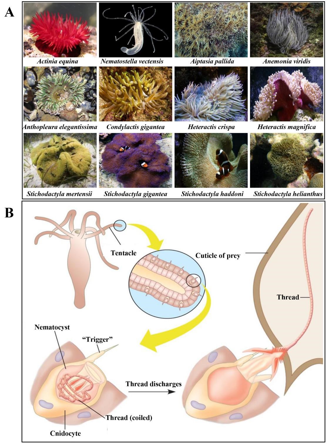
Fig. 1.

Representative sea anemone species worldwide.

Previous studies have shown that sea anemone toxins contain complex mixtures of proteins, peptides, and nonproteinaceous compounds (Table 1, Ref. [5, 12, 13, 14, 15, 16, 17, 18, 19, 20, 21, 22, 23, 24, 25, 26, 27, 28, 29, 30, 31]) [32, 19, 33]. Typically, the main peptide/protein compounds found in sea anemone venom can be divided into three groups: (1) phospholipase A2 enzymes (PLA2) that catalyze the hydrolysis of phospholipids and participate in inflammatory reactions [34, 35]; (2) cytolysins, which mainly form pores on the cell membrane and cause cell lysis [15]; and (3) peptide neurotoxins that act on voltage-gated sodium (NaV) channel, voltage-gated potassium (KV) channel, acid-sensing ion channel (ASIC), and other ion channels [24, 36, 37, 38, 39, 40]. These peptide neurotoxins thus have specific biological activities such as autoimmune, analgesic, and central nervous system inhibitory effects [41, 42, 43, 44].

Table 1.Representative families and pharmacological targets in sea anemone venoms [5].
Type Structure family Pharmacological group Ref.
Nonproteinaceous compounds Purine Adenosine receptor [12]
- 5-HT3 receptor [13]
Proteins Enzymes CYP74 Unknown [14]
PLA2 PLA2 [15]
Type III cytolysins
Endonuclease D Unknown [5]
Serine protease S1 Unknown [5]
Cytotoxins Actinoporins Type II cytolysins [16]
CRISP Unknown [17]
WSC domain proteins Unknown [18]
Peptide neurotoxins ATX III NaV type 3 [19]
β-defensin-like ASIC [20]
KV type 3 [21]
NaV type 1 [5]
NaV type 2
NaV type 4
BBH ASIC [5]
KV type 4 [22]
EGF-like EGF activity [23]
TRPV1 [24]
ICK ASIC [25]
KV type 5
Kunitz-domain KV type 2 [26]
TRPV1 [27]
Protease inhibitor [28]
PHAB KV type 6 [29]
SCRiPs TRPA1 [30]
ShK KV type 1 [31]
Note: 5-HT3, 5-hydroxytryptamine 3; CYP74, Cytochrome P450 proteins 74; PLA2, Phospholipase type A2; CRISP, Cysteine-rich proteins; WSC domain, Cell wall integrity and stress response component domain; ATX III, Anemonia sulcate toxin III; ASIC, Acid-sensing ion channel; BBH, Boundless β-hairpin; EGF-like, Epidermal growth factor-like; ICK, Inhibitor cystine-knot; TRPV1, Receptor potential channel type V1; PHAB, Proline-hinged asymmetric β-hairpin; SCRiPs, Small cysteine-rich peptides; TRPA1, Transient receptor potential channel type A1.

According to the latest published data in the WoRMS database, 1162 species of sea anemones have been recorded worldwide, and high-throughput transcriptome sequencing shows that there are more than 100 different peptide sequences in each sea anemone to date [5, 45]. In particular, 612 putative protein and peptide sequences were discovered from the sea anemone Anthopleura elegantissima[46]. Owing to the small overlap of sea anemone toxins among different sea anemone species and significant interspecies variation (e.g., Stichodactylidae family), there are an estimated 1,200,000 natural peptides that are produced by sea anemones [47]. However, only 5% of the species and approximately 378 toxins from sea anemones are annotated in UniProtKB (https://www.uniprot.org/). Sea anemones are a treasure trove for relatively undeveloped bioactive and therapeutic compounds. Therefore, the discovery of novel sea anemone peptide neurotoxins from sea anemone resources using combined omics methods and high-throughput biological assays is essential for the development of sea anemone peptide neurotoxin drugs.

3. Isolation of peptide neurotoxins from sea anemone venom

Traditional isolation and purification of sea anemone toxin are usually performed directly from the crude venom. At present, there are three main methods for extracting sea anemone crude venom: homogenization, milking, and electrical stimulation methods [48, 49, 50]. A total of 43 sea anemone peptide neurotoxins have been successfully isolated from different types of sea anemones by traditional crude venom purification, and these peptide neurotoxins are summarized in Table 2 (Ref. [23, 26, 30, 48, 51, 52, 53, 54, 55, 56, 57, 58, 59, 60, 61, 62, 63, 64, 65, 66, 67, 68, 69, 70, 71, 72, 73, 74, 75, 76]). Among the sea anemone peptide neurotoxins found by traditional crude venom isolates, the most common cysteine pattern is CXC-C-C-CC, and these toxins mainly act on the NaV channel but also on ASIC, the KV channel, and others [58, 59, 60, 61, 62]. Examples of neurotoxins with the same cysteine pattern (CXC-C-C-CC) include APETx1, APETx2, and Anthopleurin-A. These toxins act on the KV channel, ASIC, and NaVchannel, respectively [58, 60, 77]. Therefore, the activity of the sea anemone peptide neurotoxins is affected not only by the cysteine pattern but also by the amino acid sequence. In addition to the main CXC-C-C-CC pattern, there are many other cysteine patterns such as C-C-C-C, C-C-C-C-C-C, C-CC-C-C-C-C, C-C-CC-C-C-C-C, C-C-C-C-C-C-C-C, and C-C-C-C-C-C-C-CCC, which are found in sea anemone peptide neurotoxins. The cysteine patterns of sea anemone peptide neurotoxins can act on targets such as the KV channel and TRPA1, and some patterns do not have known targets [30, 73, 74].

Table 2. Sea anemone peptide neurotoxins isolated from sea anemone venom.
Method Name Species Sequence Target/Activity Ref.
Homogenization δ-TLTX-Ca1a Cryptodendrum adhaesivum VACKCDDDGPDVRSATFTGTVDLGSCNSGWEKCASYYTVIADCCRKPRG NaV [51]
δ-TLTX-Ta1a Thalassianthus aster VACKCDDDGPDIRSATLTGTVDLGSCDEGWEKCASYYTVIADCCRRPRS NaV [51]
gigantoxin III Stichodactyla gigantea AACKCDDDGPDIRSATLTGTVDLGSCNEGWEKCASFYTILADCCRRPR NaV [23]
gigantoxin II Stichodactyla gigantea GVPCRCDSGPHVRGNTLTGTVWVFGCPSGWHKCQKGSSTCCKQ NaV [23]
CgNa Condylactis gigantea GVPCRCDSDGPTVHGNTLSGTVWVGSCASGWHKCNDEYNIAYECCKE NaV [52]
AdE-1 Aiptasia diaphana GIPCRCDKNSDELNGEQSYMNGNCGDGWKKCRSVNAIFNCCQRV NaV [53]
Av2 Anemonia viridis GVPCLCDSDGPSVRGNTLSGIIWLAGCPSGWHNCKKHGPTIGWCCKQ NaV [54]
Av1 Anemonia viridis GAACLCKSDGPNTRGNSMSGTIWVFGCPSGWNNCEGRAIIGYCCKQ NaV [54]
Anthopleurin-A Anthopleura xanthogrammica GVSCLCDSDGPSVRGNTLSGTLWLYPSGCPSGWHNCKAHGPTIGWCCKQ NaV [55]
Anthopleurin-B Anthopleura xanthogrammica GVPCLCDSDGPRPRGNTLSGILWFYPSGCPSGWHNCKAHGPNIGWCCKK NaV [56]
Bg II Bunodosoma granulifera GASCRCDSDGPTSRGNTLTGTLWLIGRCPSGWHNCRGSGPFIGYCCKQ NaV [57]
Bg III Bunodosoma granulifera GASCRCDSDGPTSRGDTLTGTLWLIGRCPSGWHNCRGSGPFIGYCCKQ NaV [57]
APETx1 Anthopleura elegantissima GTTCYCGKTIGIYWFGTKTCPSNRGYTGSCGYFLGICCYPVD NaV, KV 11.1 [58]
APETx4 Anthopleura elegantissima GTTCYCGKTIGIYWFGKYSCPTNRGYTGSCPYFLGICCYPVD KV 10.1 [59]
APETx2 Anthopleura elegantissima GTACSCGNSKGIYWFYRPSCPTDRGYTGSCRYFLGTCCTPAD ASIC3 [60]
π-AnmTX Hcr 1b-2 Heteractis crispa GTPCKCHGYIGVYWFMLAGCPNGYGYNLSCPYFLGICCVKK ASIC1a, ASIC3 [61]
π-AnmTX Hcr 1b-3 Heteractis crispa GTPCKCHGYIGVYWFMLAGCPDGYGYNLSCPYFLGICCVKK ASIC1a [61]
π-AnmTX Hcr 1b-4 Heteractis crispa GTPCDCYGYTGVYWFMLSRCPSGYGYNLSCHYFMGICCVKR ASIC1a [61]
PhcrTx2 Phymanthus crucifer ALPCRCEGKTEYGDKWIFHGGCPNDYGYNDRCFMKPGSVCCYPKYE unknown [62]
Am II Antheopsis maculata ALLSCRCEGKTEYGDKWLFHGGCPNNYGYNYKCFMKPGAVCCYPQN unknown [63]
Am I Antheopsis maculata NVAVPPCGDCYQQVGNTCVRVPSLCPS NaV [63]
ShK Stichodactyla helianthus RSCIDTIPKSRCTAFQCKHSMKYRLSFCRKTCGTC KV 1.3 [64]
AsKs Bunadosoma granulifa ACKDNFAAATCKHVKENKNCGSQKYATNCAKTCGKC KV 1.2 [65]
AETX K Anemonia erythraea ACKDYLPKSECTQFRCRTSMKYKYTNCKKTCGTC KV 1 [66]
BgK Bunodosoma granulifera VCRDWFKETACRHAKSLGNCRTSQKYRANCAKTCELC KV1-3, KV 1.6, KCa 3.1 [67]
AeK Hteractis magnifica GCKDNFSANTCKHVKANNNCGSQKYATNCAKTCGKC KV 1 [68]
APEKTx1 Anthopleura elegantissima INSICLLPKKQGFCRARFYYNSSTRRCEMFYYGGCGGNANNFNTLEECEKVCLGYGEAWKAP KV 1.1 [26]
HCRG1 Heteractis crispa RGICSEPKVVGPCKAGLRRFYYDSETGECKPFIYGGCKGNKNNFETLHACRGICRA Serine protease inhibitor [69]
HCRG2 Heteractis crispa RGICLEPKVVGPCKARIRRFYYDSETGKCTPFIYGGCGGNGNNFETLHACRGICRA Serine protease inhibitor [69]
acrorhagin II Stichodactyla gigantea TDCRFVGAKCTKANNPCVGKVCNGYQLYCPADDDHCIMKLTFIP crab toxicity [70]
gigantoxin I Stichodactyla gigantea DVGVACTGQYASSFCLNGGTCRYIPELGEYYCICPGDYTGHRCEQMSV EGF activity [23]
Av3 Anemonia viridis RSCCPCYWGGCPWGQNCYPEGCSGPKV NaV [54]
acrorhagin I Actinia equina SSTPDGTWVKCRHDCFTKYKSCQMSDSCHDEQSCHQCHVKHTDCVNTGCP crab toxicity [70]
Electrical stimulation δ-AITX-Bca1a Bunodosoma capense CLCNSDGPSVRGNTLSGILWLAGCPSGWHNCKKHKPTIGWCCK NaV [71]
BcIII Bunodosoma caissarum GVACRCDSDGPTSRGNTLTGTLWLTGGCPSGWHNCRGSGP FIGYCCKK NaV [48]
BcIV Bunodosoma caissarum GLPCDCHGHTGTYWLNYYSKCPKGYGYTGRCRYLVGSCCYK NaV [72]
AbeTx1 Actinia bermudensis RCKTCSKGRCRPKPNCG KV [73]
BcsTx1 Bunodosoma caissarum ACIDRFPTGTCKHVKKGGSCKNSQKYRINCAKTCGLCH KV [74]
BcsTx2 Bunodosomacaissarum ACKDGFPTATCQHAKLVGNCKNSQKYRANCAKTCGPC KV [74]
BcsTx3 Bunodosoma caissarum GCKGKYEECTRDSDCCDEKNRSGRKLRCLTQCDEGGCLKYRQCLFYGGLQ KV [75]
τ-AnmTx Ueq 12-1 Urticina eques CYPGQPGCGHCSRPNYCEGARCESGFHDCGSDHWCDASGDRCCCA TRPA1 [30]
Milking CGTX-II Bunodosoma cangicum GVACRCDSDGPTVRGDSLSGTLWLTGGCPSGWHNCRGSGPFIGYCCKK NaV 1.1, NaV 1.5, NaV 1.6 [76]
CGTX-III Bunodosoma cangicum GVACRCDSDGPTVRGDSLSGTLWLTGGCPSGWHNCRGSGPFIGYCCKK NaV 1.1 [76]
Note: C is marked in red font to highlight cysteine.
3.1 Homogenization method

The earliest report of the purification of sea anemone peptide neurotoxin was in the 1960s [78]. At that time, several to dozens of sea anemones were collected and homogenized to obtain enough venom for the isolation of one or a few sea anemone peptide neurotoxins [19, 79, 80, 81]. The crude venom obtained using homogenization was separated and purified by gel chromatography and reverse phase high performance liquid chromatography (HPLC) [53]. A total of 11 kg of Condylactis gigantea from the Caribbean Sea near Havana was collected by Standker et al. [52] and homogenized to obtain crude venom. The venom was then further isolated and purified to obtain the CgNa toxin [52]. Similar studies include the sea anemone peptides Av1-3 (previously named ATX I-III) with neurotoxic activity that were isolated from 5.5 kg of wet Anemonia viridis (previously named Anemonia sulcata) [54].

To date, 33 sea anemone peptide neurotoxins have been isolated by extracting venom from different types of sea anemones using the homogenization method (Table 2). Among these, important and typical sea anemone peptide neurotoxins include ShK, CgNa, AdE-1, Anthopleurin-A, and Anthopleurin-B. The most frequently studied sea anemone toxin is the ShK toxin from the giant sun anemone (Stichodactyla helianthus) [64]. This peptide has the ability to block the Kv1.3 channels of T lymphocytes, inhibiting their activation and therefore acting as a therapeutic to treat autoimmune diseases [82, 83]. New analogues of ShK, with good selectivity for Kv1.3 channels, have also been developed [84, 85, 86]. An analogue of this peptide (ShK-186) has been developed into the first-in-class clinical candidate dalazatide, and phase I clinical trials have been completed for the treatment of psoriasis [87, 88]. Dalazatide (formerly known as ShK-186) is being advanced as a treatment for various autoimmune diseases including type 1 diabetes, inflammatory bowel diseases, body myositis, lupus, psoriasis, multiple sclerosis, psoriatic arthritis, rheumatoid arthritis, and ANCA vasculitis [1, 89].

3.2 Milking method

Isolation of toxins is usually performed by extracting toxins from homogenates of whole animals or frozen–thawed sea anemones or by isolation of the nematocysts followed by purification using various methods [90, 91]. The milking technique was first applied by Barnes to directly collect pure venom from jellyfish, where nematocysts are discharged through an amnion membrane [92]. Sencic and Macek described a new and simpler purification procedure of sea anemone toxins from the venom obtained using a new milking method different from that reported by Barnes [92]. They obtained two lethal and hemolytic peptide toxins, caritoxins I and II, from the sea anemone Actinia cari using the milking method [50].

Milking involves the gentle squeezing of sea anemones to collect their secretions, which is similar to stimulating sea anemones to release venom in the natural environment [93]. This method has the advantages of not harming the sea anemone, obtaining purer venom, and being able to extract the venom repeatedly. Therefore, milking has become an effective method to extract sea anemone venom, after homogenization and freeze-thawing methods. Zaharenko et al. [76] obtained the cangitoxin (CGTX) analogue CGTX-II directly from sea anemone venom by milking followed by two chromatographic steps. CGTC-II inhibited NaV1.1-1.6 channel action by delaying the inactivation of the NaV channel, which may be an interesting tool to study its interaction with these channels.

3.3 Electrical stimulation method

Electrical stimulation is often used to extract animal venom from inland poisonous animals such as spiders, scorpions, and wasps [94, 95, 96]. Malpezzi et al. [48] applied electrical stimulation to extract sea anemone Stichodactyla helianthus (formerly Stoichactis helianthus) venom for the first time and successfully isolated BcI, BcII, and BcIII. The main action of BcIII on Nav channels is a slowing of the inactivation process of the sodium current, with no significant effects on the activation kinetics. It is important to emphasize that the method used to obtain the venom in the present paper has many advantages: because it results in less contamination with other compounds from the sea anemone body, it simplifies the purification procedures and keeps the animals alive, allowing them to be reused to obtain more venom or return them to the sea.

BcIV, AbeTx1, BcsTx1-3, δ-AITX-Bca1a, and Ueq 12-1 were isolated by electrical stimulation from sea anemones Bunodosoma caissarum, Actinia bermudensis, Bunodosoma caissarum, Urticina eques, and Bunodosoma capense, respectively [30, 71, 72, 73, 74, 75]. BcIV and δ-AITX-Bca1a have the same cysteine pattern (CXC-C-C-CC), and both are typical NaV channel toxins [97]. Ueq 12-1 contains 10 cysteine residues with an unusual distribution and represents a new group of sea anemone peptides whose primary and spatial structurestructures are unique among the range of known sea anemone peptides. Since Ueq 12-1 showed moderate antibacterial activity against Gram-positive bacteria and enhanced activity against TRPA1, Ueq 12-1 is considered to be a potential analgesic drug with antibacterial properties [30].

In general, the extraction method of these venoms is time-consuming and labor-intensive. It is difficult to obtain a high amount of venom to isolate and purify sea anemone peptide neurotoxins, especially for rare sea anemone species. Therefore, more effective venom separation and extraction methods are needed to speed up the discovery of novel sea anemone peptide neurotoxins. This section does not represent a full-scale explanation of all the techniques used and improvements since the 1960s but instead provides a brief overview of the most commonly used techniques for sea anemone venom extractions for reference purposes.

4. Discovery of sea anemone peptide neurotoxins by molecular biology
4.1 Discovery of sea anemone peptide neurotoxins by gene cloning

Gene cloning was used to discover novel sea anemone peptide neurotoxins at the end of the 20th century [98]. The advantage of gene cloning is that the peptide neurotoxin gene can be amplified from a small number of sea anemone tentacles by PCR. This method overcomes the limitation of crude toxin purification method in large demand for sea anemone samples, and has attracted the attention of scientific researchers [99]. Primer design is a key step in any experiment using PCR to target and amplify known nucleotide sequences of interest [100]. Rationally designed primers can not only improve PCR amplification efficiency but can also screen target sequences with high specificity [101]. Primers were designed and synthesized based on the conserved sequence in the signal region or the relatively conserved introns in the pro-region or untranslated region of the 3’- or 5’-UTR of a specific known sea anemone peptide neurotoxin precursor (Fig. 2A) [102]. Usually, cDNA is prepared by reverse transcription from total RNA extracted from sea anemones [98]. Total cDNA was used as a template, and PCR amplification was performed with specific primers to perform 3’- and 5’-RACE [102]. The PCR products were purified by electrophoresis on agarose gel, ligated to the plasmid vector, and transferred to Escherichia coli for replication, proliferation, and sequencing (Fig. 2B) [103].
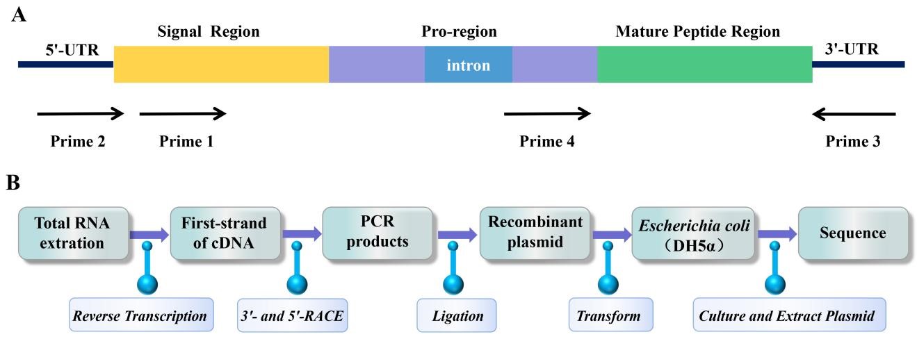
Fig. 2.

PCR amplification strategy and cloning process of sea anemone gene. (A) PCR amplification strategy to clone sea anemone toxin precursor genes from genomic DNA. (B) The process of gene cloning sea anemone peptide neurotoxins.

A total of nine types of sea anemone peptide neurotoxins were obtained by gene cloning, as shown in Table 3 (Ref. [40, 51, 70, 104, 105, 106, 107]). The most common cysteine pattern in peptide neurotoxins obtained by gene cloning is CXC-C-C-CC, and its main target is the NaVchannel, which is consistent with the results of venom isolation. However, other types of cysteine patterns obtained by this method, such as C-C-C-C, C-C-C-C-C-C, C-C-C-C-CC, C-C-C-C-C-C-C-C, and C-C-C-C-C-C-C-CCC, are more novel than the cysteine patterns of peptide neurotoxins isolated from venom. Although gene cloning can identify novel cysteine patterns of sea anemone peptide neurotoxins, the discovered toxin families are limited by the design of the primers [70]. This is because the design and synthesis of primers for gene cloning are based on known sequences of superfamilies. Therefore, only a few sea anemone peptide neurotoxins have been found through gene cloning in recent years, and it is difficult to discover new superfamilies using this technique. For example, a new actinoporin Hct-S4 was isolated from the tropical sea anemone Heteractis crispa, and 18 new isoforms were cloned with primers designed based on its N-terminus conserved sequence. These isoforms and Hct-S4 belong to the sphingomyelin-inhibited α-pore forming toxin (α-PFT) family [108]. Compared with the traditional isolation and purification of sea anemone peptide neurotoxins, gene cloning protects sea anemone resources because one sample is sufficient to complete the whole process of sea anemone peptide neurotoxin gene cloning [74, 104]. However, compared with high-throughput transcriptomics and proteomics, the gene cloning technique is low-throughput [105, 109].

Table 3.Representative sea anemone peptide neurotoxins discovered by gene cloning.
Name Species Sequence Target/Activity Ref.
SHTX IV Stichodactyla haddoni AACKCDDDGPDIRSATLTGTVDFWNCNEGWEKCTAVYTAVASCCRKKKG NaV [104]
δ-TLTX-Hh1x Heterodactyla hemprichii VACKCDDDGPDIRSATLTGTVDLGSCNEGWEKCASYYTVVADCCRRRRS NaV [51]
Hk2a Anthopleura sp MGVACLCDSDGPSVRGNTLSGTLWLAGCPSGWHNCKAHGPTIGWCCKQ NaV [105]
Crassicorin-I Urticina crassicornis GASCDCHPFVGTYWFGISNCPSGHGYPKKCASFFGVCCVK antimicrobial activity [106]
acrorhagin Ia Actinia equina SLTPSSDIPWEKCRHDCFAKYMSCQMSDSCHNKPSCRQCQVTYAICVSTGCP crab toxicity [70]
HCRG21 Heteractis crispa RGICSEPKVVGPCTAYFRRFYFDSETGKCTPFIYGGCEGNGNNFETLRACRAICRA TRPA1 [40]
SHTX III Stichodactyla haddoni TEEMPALCHLQPDVPKCRGYFPRYYYNPEVGKCEQFIYGGCGGNKNNFVSFEACRATCIIPL KV [104]
Magnificamide Heteractis magnifica SEGTSCYIYHGVYGICKAKCAEDMKAMAGMGVCEGDLCCYKTPW α-amylase [107]
Note: C is marked in red font to highlight cysteine.
4.2 Discovery of sea anemone peptide neurotoxins by high-throughput sequencing

Although natural crude venom purification and gene cloning have greatly helped efforts to discover novel sea anemone peptide neurotoxins, most of the unknown sea anemone peptide neurotoxins have not yet been characterized. Therefore, there is an urgent need to develop more efficient, resource-conserving, and high-throughput methods. Transcriptomics, proteomics, and multiomics integration have opened a new era in the discovery of sea anemone toxins, rapidly accelerating the discovery of novel sea anemone peptide neurotoxins [1, 109].

The high throughput technologies used in venomics, especially transcriptomics, has produced large data sets that need bioinformatics support to fully explore their potential [110, 111, 112]. Modern bioinformatics tools have been recently developed to mine venoms, helping focus experimental research on the most potentially interesting neurotoxins [113]. The number of neurotoxins unraveled by high throughput multiomics approaches is very large, efficient computational approaches are required to mine this massive amount of data. Computational approaches that have been developed to help predict neurotoxins molecular targets, three-dimensional structures and functions, and identifying outstanding neurotoxins with potential new characteristics [114, 115, 116].

General databases, such as the Protein Data Bank (PDB) [117], UniProt [118], and NCBI Genbank/GenPept [119], play important roles in simplifying access to information about sea anemone neurotoxin sequences and three-dimensional structures. However, the information about sea anemone neurotoxins is not standardized in these resources, especially the naming of neurotoxins and pharmacological activities, and mining for sea anemone neurotoxins is difficult. A recently developed resource, VenomZone, is provided by the Swiss Institute of Bioinformatics (SIB), and has information about the venoms from six types of organisms, including sea anemones, cone snail, spider, Scorpions, bees and snakes. However, Specialized databases, from venomous animals, are slowly emerging. ISOB (Indigenous snake species of Bangladesh) [120], Arachnoserver [121], and Conoserver [122] provide information on venoms from respectively.

4.2.1 Transcriptomics technology

The term “transcriptome” was first used by Velculescu to analyze a set of genes expressed in the yeast genome in a scientific paper in 1997 [123]. In more than 30 years of development, sequencing technology has achieved considerable development, from the first to the third generation of sequencing technologies [124]. At present, the second-generation short-read-length sequencing technology still holds an absolute dominant position in the global sequencing market, but third-generation sequencing technology has also developed rapidly in the past few years [125]. With the rapid development of molecular biology and the decreasing cost of sequencing nucleic acids, especially massively parallel sequencing technologies, a large number of transcriptomic analyses of cone snail, snake, spider, scorpion, and a few other animal venom glands have been carried out [126, 127, 128, 129].

At present, high-throughput transcriptomics has been applied to 13 types of sea anemones: Anthopleura elegantissima, Anthopleura dowii, Aiptasia pallida, Anemonia sulcata, Anemonia viridis, Cnidopus japonicus, Exaiptasia pallida, Heteractis crispa, Oulactis sp., Megalactis griffithsi, Nematostella vectensis, Stichodactyla helianthus, and Stichodactyla haddoni [18, 46, 130, 131, 132, 133, 134, 135, 136, 137, 138]. There is a growing body of literature using transcriptomics data to study the symbiotic relationship between sea anemones and their symbionts or to study the mechanism of the evolutionary development of sea anemones [131, 139, 140]. Few studies have reported the transcriptome sequencing of sea anemone venom and identification of venom-related peptides and proteins, which can be used for their structural and functional analyses and venom evolution in the future (Table 4, Ref. [18, 46, 87, 109, 136, 135, 141, 142, 143]) [132, 136]. Tentacles are ideal for transcriptomics analysis because the number and level of transcripts encoded by sea anemone peptide neurotoxins from tentacles are much greater than those encoded by sea anemone peptide neurotoxins from other tissues [132]. The transcriptomics of sea anemone venom can describe the expression of sea anemone toxin and provide a useful method for rapid identification of putative sea anemone peptide sequences [46]. For example, Mitchell and his team used a transcriptomic strategy with Illumina RNA-seq sequencing platforms to study venom from the tentacles of the Oulactis sp. and compiled a venom-related component library of 398 putative venom-related peptides and proteins, including one putative actinoporin [136]. The venom composition across different tissues (tentacles, mesenterial filaments, and columns) in three species of sea anemone (Anemonia sulcata, Heteractis crispa, and Megalactis griffithsi) was used in a combined RNA-seq and bioinformatic approach by Macrander et al. [132]. Their tissue-specific transcriptome analyses showed that there are significant variations in the abundance of toxin-like genes across tissues and species, which provides a framework for the characterization of tissue-specific venom and other functionally important genes in this lineage of simple bodied animals in the future. In addition, Sebé-Pedrós et al. [144] performed whole-organism single-cell transcriptomics of Nematostella vectensis. Their study revealed cnidarian cell type complexity and provided insights into the evolution of animal cell-specific genomic regulation [144].

4.2.2 Proteomics technology

The first description of proteomics dates back to the early 1980s when Bravo and Celis developed protein separation by exceptionally effective 2D gel electrophoresis followed by Edman degradation sequencing to identify proteins and compare them to available protein sequence databases [145]. This, together with the wider availability of protein sequence databases, opened the door to the wide use of proteomics [146]. With the rapid development of analytical instruments and bioinformatics, proteomics has become a rapidly developing field and has shown to be applicable to many organisms and cell types [147, 148]. It has become an important tool for the study of animal venom, enabling detailed research of venom composition [149, 150, 151, 152].

Venom proteomics with modern mass spectrometry technology has proven to be an effective and high-throughput method for the discovery of novel sea anemone peptide neurotoxins [141, 153, 154]. To date, high-throughput proteomics have been used for sea anemones such as Bunodactis verrucosa, Nematostella vectensis, Stichodactyla duerden, Anthopleura dowii, and Stichodactyla haddoni, with an average of 321 proteins and peptide neurotoxins being identified in each sea anemone (Table 4) [18, 109, 141, 143, 153]. The first proteomic studies on the venom of sea anemone were performed by Zaharenko and colleagues, who investigated the peptide mass fingerprint and some novel peptides in the neurotoxic fraction of the sea anemone Bunodosoma cangicum venom. Their data showed that at least 81 molecules were eluted in the neurotoxic fraction and may be employed as active peptides for prey capture and defense [155]. In addition, a combination of offline RPC-MALDI-TOF and online nano-RPC-ESI-LTQ-Orbitrap proteomic techniques was used for Stichodactyla duerdeni by Cassoli and his team, which identified a total 67 proteins and peptides and revealed the presence of a novel O-linked glycopeptide [153]. Moreover, proteomics has also been applied to the model sea anemone Nematostella vectensis to more fully elucidate the molecular and cellular mechanisms underlying the repair of hair cells following trauma [156].

Table 4.Reported transcriptomic and proteomic data from various sea anemones.
Species Sequencing platforms Number of peptides and proteins found by transcriptomics MS instruments Number of peptides and proteins found by proteomics Ref.
Oulactis sp. Illumina HiSeq 1500 398 [136]
Exaiptasia pallida Illumina NextSeq 500 547 [135]
Anthopleura elegantissima Illumina HiSeq 65 [46]
Bunodactis verrucosa - - MALDI-TOF/TOF 412 [141]
Cnidopus japonicus - - LC-MS/MS 27 [142]
Nematostella vectensis - - LC/MS 1135 [143]
Stichodactyla duerdeni - - MALDI–TOF MS 67 [87]
Anthopleura dowii Illumina 261 LC-MS/MS 156 [109]
Stichodactyla haddoni Illumina NextSeq 500 508 LC-MS/MS 131 [18]
4.2.3 Multiomics integration technology

Multiomics has constructed a set of research strategies for sea anemone toxins based on integrated correlation analysis of transcriptomics and proteomics, which have been proven to be effective, and high-throughput methods to identify a large number of sea anemone toxin sequences (Fig. 3) [139, 141]. Both transcriptomics and proteomics benefit from the emergence and development of bioinformatics, especially the development of bioinformatics software, the improvement of algorithms, and the expansion of searchable databases [157, 158]. Bioinformatic tools such as BLAST, UniProt, PFAM, and others have frequently been used for venom transcriptomics and proteomics, playing an important role in raw data processing, sequence recognition, protein analysis, and superfamily classification [159].
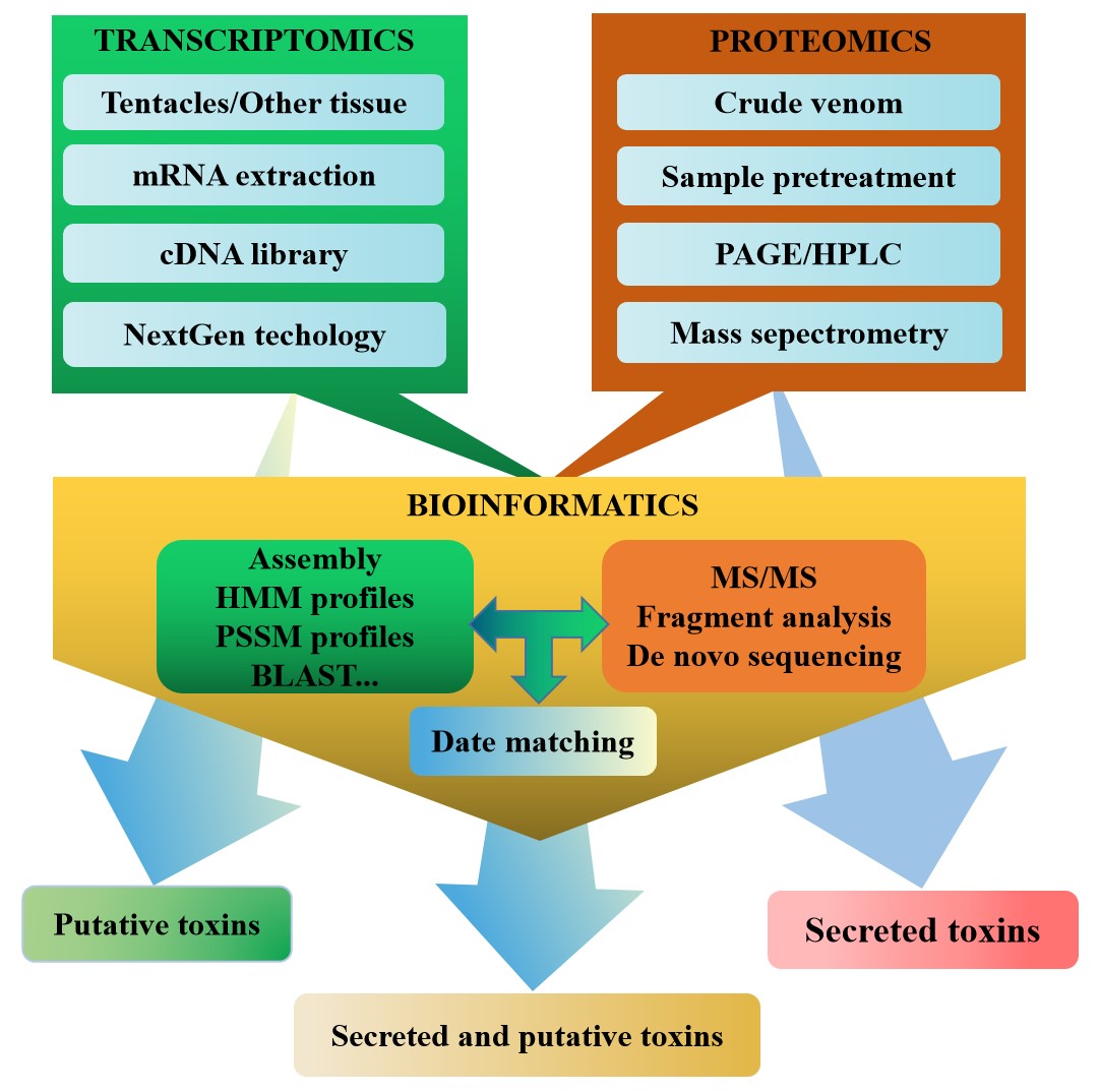
Fig. 3.

Multiomic approach to the discovery of sea anemone toxins.

Transcriptomics and proteomics are methods with limitations. Transcriptomics does not predicate post-transcriptional modifications or regulatory processes [28]. Proteomic techniques do not have the sensitivity to detect proteins with low abundance [29]. Therefore, multiomics technology combining transcriptomics and proteomics can effectively solve their respective limitations and can enrich the biological information from organisms. The multiomics techniques have enabled further exploration of the components of venomous animal species (scorpions, spiders, cone snails, and snakes) [160, 161, 162, 163], as well as sea anemone venom from a few species (Table 4) [18, 109]. Bruno Madio and colleagues used a combination of transcriptomics and proteomics to study Stichodactyla haddoni for the first time and identified 508 unique toxin transcripts, which were divided into 63 families. However, proteomic analysis of venom identified 52 toxins in these toxin families that might be false positives. In contrast, the combination of transcriptomic and proteomic data enabled positive identification of 23 families of putative toxins, 12 of which have no homology to known proteins or peptides [18]. In addition, Ramírez-Carreto and his colleagues also used a transcriptomic and proteomic analysis of the tentacles and mucus of the sea anemone Anthopleura dowii. Transcriptome analysis showed that 261 peptides were identified, while proteomic analysis identified 156 peptides. Some toxins identified in the tentacles and mucus proteome were not identified in the transcriptome [109]. In general, it was observed that the quantity and especially the diversity of probable toxins in the sea anemone transcriptome were far greater than those in the sea anemone proteome, which was similar to that in other venomous animals [164, 165].

Currently, transcriptomics and proteomics are considered to be effective, resource-saving, and high-throughput approaches for the discovery of novel sea anemone peptide neurotoxins. The multiomics approach is significantly more effective than the use of transcriptomics or proteomics alone. In the future, the application of multiomics technology in the development and utilization of sea anemone resources will accelerate the discovery of new sea anemone peptide neurotoxins and will continuously enrich and expand the database of sea anemone peptide neurotoxins.

5. Three-dimensional structures of sea anemone peptide neurotoxins

At present, three experimental methods, X-ray crystallography, nuclear magnetic resonance (NMR), and cryoelectron microscopy (cryo-EM), are used to determine the three-dimensional structure of sea anemone toxins [166, 167, 168]. The X-ray crystallography technique can obtain high-precision protein structures. However, many proteins, especially small molecular peptides, cannot be determined by this method due to the difficulty of preparing crystals for structural analysis [166]. The advantage of NMR and cryo-EM is that there is no need to prepare protein crystals [169, 170]. NMR is mainly used to determine the structure of small molecular peptides, while cryo-EM is mainly used to determine the structure of macromolecular proteins [171, 172]. Therefore, NMR has played a prominent role in determining the three-dimensional structures of sea anemone peptide toxins because it is often used to analyze the structure of small molecular peptides. The three-dimensional structures of sea anemone peptide toxins determined by Solution NMR are summarized in Table 5 (Ref. [22, 29, 30, 167, 173, 174, 175, 176, 20, 177, 178, 179, 180, 181, 182, 183]).

Table 5.Sea anemone peptide toxins with three-dimensional structures studied by Solution NMR.
Species Toxin Type Length (AA) PDB ID Ref.
Anemonia viridis BDS-1 KV channel 43 1BDS [173]
Bunodosoma granulifera BgK KV channel 37 1BGK [174]
Stichodactyla helianthus ShK KV channel 35 1ROO [175]
Actinia tenebrosa Ate1a KV channel 18 6AZA [29]
Oulactis sp OspTx2b KV channel 36 6BUC [167]
Anthopleura elegantissima APETx1 KV channel 42 1WQK [176]
Anthopleura elegantissima APETx2 KV channel 42 1WXN [20]
ASIC channel
Antopleura cascaia AcaTx1 KV channel 32 6NK9 To be published
ASIC channel published
Stichodactyla helianthus ShPI-1 Kunitz type 55 1SHP [177]
proteinase
inhibitor
Anemonia viridis ATX-IA NaV channel 46 1ATX [178]
Anemonia viridis ATX-III NaV channel 27 1ANS [179]
Anthopleura xanthogrammica Anthopleurin-A NaV channel 49 1AHL [180]
Anthopleura xanthogrammica Anthopleurin-B NaV channel 49 1APF [181]
Condylactis gigantea CgNa NaV channel 47 2H9X [182]
Stichodactyla helianthus Sh1 NaV channel 48 1SH1 [183]
Urticina grebelnyi Ugr 9-1 ASIC3 channel 29 2LZO [22]
Urticina eques Ueq 12-1 TRPA1 channel 45 5LAH [30]

The first three-dimensional protein structures of sea anemones were obtained in the 1980s by NMR analysis for Anthopleurin-A followed by ATX-IA [184, 178]. To date, nine unique structural folds have been identified based on the three-dimensional structure and/or cysteine-pattern: ShK, Kunitz-domain, ATX-III, PHAB, β-defensin-like, BBH, EGF-like, ICK, and SCRiPs. These structural types of sea anemone peptide toxins identified by NMR mainly act on the NaV, KV, ASIC, TRPA1, and TRPV1 channels, respectively (Table 1) [5].

Sea anemone peptide toxins acting on the Na
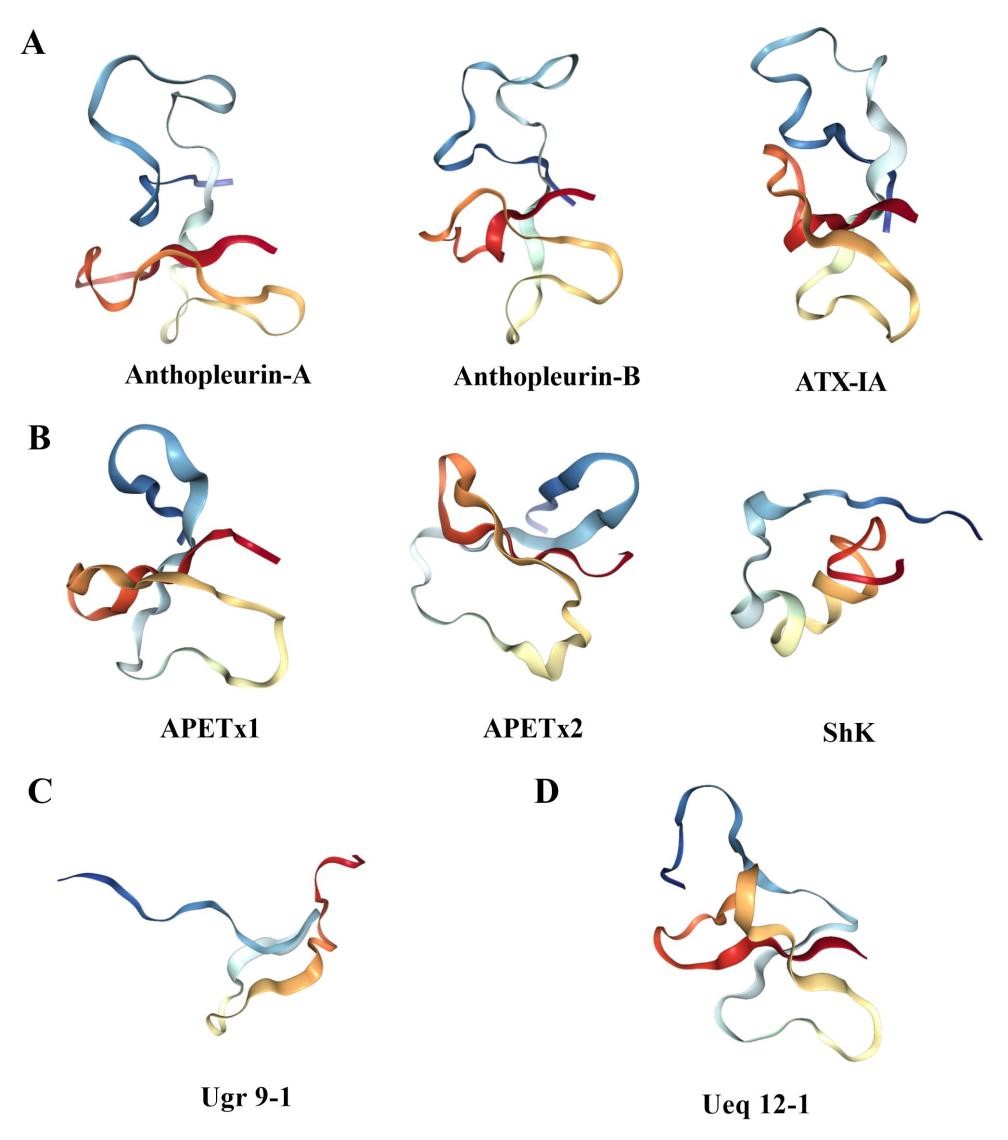
V channel include ATX-III, ATX-IA, Anthopleurin-A, Anthopleurin-B, CgNa, and Sh1 (Table 5). The ATX III fold is named after the first toxin described by the fold, namely, ATX III (Av3, δ- AITX-Avd2a, and Neurotoxin III) from Anemonia viridis. ATX III has a compact structure, including four reverse turns and two other chain reversals, but no regular ɑ-helix or β-sheet. In this molecule, several of the residues most affected by aggregation on the toxin surface form hydrophobic spots, which may form part of the NaV channel-binding surface [179]. The three-dimensional structures of Anthopleurin-A, Anthopleurin-B, and ATX-IA consist of an antiparallel β-sheet composed of four β-strands and a highly flexible loop (Fig. 4A) [178, 180, 181]. The highly flexible loop has been named the ‘Arg14 loop’ because Arg14 is the most conserved residue [38]. For example, the ATX-IA structure consists of a four-stranded β-sheet connected by two loops, and there is an additional flexible loop consisting of 11 residues [178]. Site-directed mutagenesis of Anthopleurin-B revealed that the flexibility of this loop is important for the binding and selectivity of these toxins to NaVchannels [185]. It is worth mentioning that Anthopleurin-A is selective for cardiac channels, while Anthopleurin-B has no selectivity for neuronal and cardiac sodium channels. This difference in selectivity between Anthopleurin-A and Anthopleurin-B is associated with the replacements at positions 12 and 49 [186].

Fig. 4.

Three dimensional structure of sea anemone toxin. (A) The structure of NaV channel toxins Anthopleurin-A, Anthopleurin-B, and ATX-IA. (B) The structure of KV channel toxins APETx1, APETx2, and ShK. (C) The structure of the ASIC3 channel toxin Ugr 9-1. (D) The structure of the TRPA1 channel toxin Ueq 12-1.

APETx1, APETx2, BDS-1, BgK, ShK, Ate1a, OspTx2b, AcaTx1, and ShPI-1 are examples of toxins that inhibit the KV channel (Table 5). APETx1, classified as belonging to the disulfide-rich all-β structural family, has a three-stranded antiparallel β-sheet, containing the only secondary structure and being the first Ether-a-go-go effector discovered to fold in this way and to contribute to the electrical activity of the heart [176]. The structures of the KV channel toxins APETx1 and APETx2 are structurally quite different from the ShK family of the KV channel toxins but similar to the NaV channel toxin Anthopleurin-A (Fig. 4B). This evidence clearly shows that sea anemones are capable of using different scaffolds (all-β in APETx1 vs. all-α in ShK) to block similar channels (hERG and KV1, respectively), while also using a common structural scaffold to create blockers of distinct targets, e.g., Anthopleurin-A, APETx1, and APETx2 act on the NaV channel, hERG, and ASIC channels, respectively [32].

The ASIC3 channel toxin Ugr 9-1 with an uncommon β-hairpin-like structure was isolated from the venom of the sea anemone Urticina grebelnyi. Ugr 9-1 does not share any sequence homology to another ASIC3 inhibitor, APETx2, previously isolated from the sea anemone Anthopleura elegantissima, but they are close structural homologues (Fig. 4C) [187]. NMR spectroscopy revealed that structure of Ugr 9-1 is stabilized by two S-S bridges, with three classical β-turns and a twisted β-hairpin without interstrand disulfide bonds [22]. Although the authors suggested that this represents a novel peptide spatial structure, which was suggested to be named BBH, other sea anemone toxins with a similar disulfide framework have in fact been reported previously [188, 189].

Ueq 12-1 isolated from sea anemones acts on the TRPA1 channel, and its three-dimensional structure has been determined by NMR spectroscopy, which represents a new stable disulfide fold, namely, SCRiPs (Fig. 4D). The three-dimensional structure of Ueq 12-1 shows that SCRiPs are organized into a peculiar W-shaped structure, the core of which is formed by a three-stranded antiparallel β-sheet, a small two-stranded parallel β-sheet, and one turn of a 3-10 helix. The surface of the peptide is polar without pronounced clusters of positively or negatively charged side chains [30].

6. Conclusions

In this review, we described the discovery methods for novel peptide neurotoxins from various sea anemones. The traditional methods of crude venom purification and gene cloning can only discover a few known or novel sea anemone peptide neurotoxins in a low-throughput way. However, the transcriptomic, venom proteomic, and multiomic methods have high efficiency, resource-conserving, and high-throughput advantages and have opened a new era of novel sea anemone peptide neurotoxin discovery. Finally, the three-dimensional structure types and corresponding action targets of sea anemone peptide neurotoxins were summarized, which provide a theoretical basis for marine drug research and development.

7. Author contributions

BG conceived and designed the article; LY and JF collected the data; JF wrote the article, while BG and AHJ revised the article.

8. Ethics approval and consent to participate

Not applicable.

9. Acknowledgment

We thank Yang Tao for helping to improve and beautify the pictures. Thanks to all the peer reviewers for their opinions and suggestions.

10. Funding

This work is supported by the National Natural Science Foundation of China (82060686) and Hainan Provincial Natural Science Foundation of China (820RC636).

11. Conflict of interest

The authors declare no conflict of interest.

Abbreviations

PLA2, phospholipase A2 enzymes; NaV channel, voltage-gated sodium channel; KV channel, voltage-gated potassium channel; ASIC, acid-sensing ion channel; 5-HT3, 5-hydroxytryptamine 3; CYP74, Cytochrome P450 proteins 74; CRISP, Cysteine-rich proteins; WSC domain, Cell wall integrity and stress response component domain; ATX III, Anemonia sulcate toxin III; BBH, Boundless β-hairpin; EGF-like, Epidermal growth factor-like; ICK, Inhibitor cystine-knot; TRPV1, Receptor potential channel type V1; PHAB, Proline-hinged asymmetric β-hairpin; SCRiPs, Small cysteine-rich peptides; TRPA1, Transient receptor potential channel type A1; HPLC, high performance liquid chromatography; NMR, nuclear magnetic resonance; cryo-EM, cryoelectron microscopy; PDB, Protein Data Bank; SIB, Swiss Institute of Bioinformatics; ISOB, Indigenous snake species of Bangladesh.

References
[1]
Prentis PJ, Pavasovic A, Norton RS. Sea Anemones: Quiet Achievers in the Field of Peptide Toxins. Toxins. 2018; 10: 36.
[2]
Jouiaei M, Yanagihara AA, Madio B, Nevalainen TJ, Alewood PF, Fry BG. Ancient Venom Systems: A Review on Cnidaria Toxins. Toxins. 2015; 7: 2251–2271.
[3]
Rodriguez E, Barbeitos MS, Brugler MR, Crowley LM, Grajales A, Gusmao L, et al. Hidden among sea anemones: the first comprehensive phylogenetic reconstruction of the order Actiniaria (Cnidaria, Anthozoa, Hexacorallia) reveals a novel group of hexacorals. PLoS ONE. 2014; 9: e96998.
[4]
Layden MJ, Rentzsch F, Rottinger E. The rise of the starlet sea anemone Nematostella vectensis as a model system to investigate development and regeneration. Wiley Interdisciplinary Reviews: Developmental Biology. 2016; 5: 408–428.
[5]
Madio B, King GF, Undheim EAB. Sea Anemone Toxins: A Structural Overview. Marine Drugs. 2019; 17: 325.
[6]
Fautin DG, Malarky L, Soberón J. Latitudinal diversity of sea anemones (Cnidaria: Actiniaria). The Biological Bulletin. 2013; 224: 89–98.
[7]
Hoepner CM, Abbott CA, Burke da Silva K. The Ecological Importance of Toxicity: Sea Anemones Maintain Toxic Defence When Bleached. Toxins. 2019; 11: 266.
[8]
Grimmelikhuijzen CJ, Westfall JA. The nervous systems of cnidarians. Exs. 1995; 72: 7–24.
[9]
Watson GM, Venable S, Mire P. Rhythmic sensitization of nematocyst discharge in response to vibrational stimuli. The Journal of Experimental Zoology. 2000; 286: 262–269.
[10]
Beckmann A, Ozbek S. The nematocyst: a molecular map of the cnidarian stinging organelle. International Journal of Developmental Biology. 2012; 56: 577–582.
[11]
David CN, Ozbek S, Adamczyk P, Meier S, Pauly B, Chapman J, et al. Evolution of complex structures: minicollagens shape the cnidarian nematocyst. Trends in Genetics. 2008; 24: 431–438.
[12]
Cooper RA, de Freitas JC, Porreca F, Eisenhour CM, Lukas R, Huxtable RJ. The sea anemone purine, caissarone: adenosine receptor antagonism. Toxicon. 1995; 33: 1025–1031.
[13]
Ferreira Junior WA, Zaharenko AJ, Kazuma K, Picolo G, Gutierrez VP, de Freitas JC, et al. Peripheral 5-HT3 Receptors Are Involved in the Antinociceptive Effect of Bunodosine 391. Toxins. 2017; 10: 12.
[14]
Gorina SS, Toporkova YY, Mukhtarova LS, Grechkin AN. The CYP443C1 (CYP74 clan) Cytochrome of Sea Anemone Nematostella vectensis-the First Metazoan Enzyme Possessing Hydroperoxide Lyase/Epoxyalcohol Synthase Activity. Doklady Biochemistry and Biophysics. 2019; 486: 192–196.
[15]
Anderluh G, Macek P. Cytolytic peptide and protein toxins from sea anemones (Anthozoa: Actiniaria). Toxicon. 2002; 40: 111–124.
[16]
Suput D. In vivo effects of cnidarian toxins and venoms. Toxicon. 2009; 54: 1190–1200.
[17]
Sher D, Knebel A, Bsor T, Nesher N, Tal T, Morgenstern D, et al. Toxic polypeptides of the hydra–a bioinformatic approach to cnidarian allomones. Toxicon. 2005; 45: 865–879.
[18]
Madio B, Undheim EAB, King GF. Revisiting venom of the sea anemone Stichodactyla haddoni: Omics techniques reveal the complete toxin arsenal of a well-studied sea anemone genus. Journal of Proteomics. 2017; 166: 83–92.
[19]
Frazão B, Vasconcelos V, Antunes A. Sea anemone (Cnidaria, Anthozoa, Actiniaria) toxins: an overview. Marine Drugs. 2012; 10: 1812–1851.
[20]
Chagot B, Escoubas P, Diochot S, Bernard C, Lazdunski M, Darbon H. Solution structure of APETx2, a specific peptide inhibitor of ASIC3 proton-gated channels. Protein Science. 2005; 14: 2003–2010.
[21]
Zhang M, Liu XS, Diochot S, Lazdunski M, Tseng GN. APETx1 from sea anemone Anthopleura elegantissima is a gating modifier peptide toxin of the human ether-a-go-go- related potassium channel. Molecular Pharmacology. 2007; 72: 259–268.
[22]
Osmakov DI, Kozlov SA, Andreev YA, Koshelev SG, Sanamyan NP, Sanamyan KE, et al. Sea anemone peptide with uncommon β-hairpin structure inhibits acid-sensing ion channel 3 (ASIC3) and reveals analgesic activity. The Journal of Biological Chemistry. 2013; 288: 23116–23127.
[23]
Shiomi K, Honma T, Ide M, Nagashima Y, Ishida M, Chino M. An epidermal growth factor-like toxin and two sodium channel toxins from the sea anemone Stichodactyla gigantea. Toxicon. 2003; 41: 229–236.
[24]
Cuypers E, Peigneur S, Debaveye S, Shiomi K, Tytgat J. TRPV1 Channel as New Target for Marine Toxins: Example of Gigantoxin I, a Sea Anemone Toxin Acting Via Modulation of the PLA2 Pathway. Acta Chimica Slovenica. 2011; 58: 735–741.
[25]
Rodríguez AA, Salceda E, Garateix AG, Zaharenko AJ, Peigneur S, López O, et al. A novel sea anemone peptide that inhibits acid-sensing ion channels. Peptides. 2014; 53: 3–12.
[26]
Peigneur S, Billen B, Derua R, Waelkens E, Debaveye S, Béress L, et al. A bifunctional sea anemone peptide with Kunitz type protease and potassium channel inhibiting properties. Biochemical Pharmacology. 2011; 82: 81–90.
[27]
Andreev YA, Kozlov SA, Korolkova YV, Dyachenko IA, Bondarenko DA, Skobtsov DI, et al. Polypeptide modulators of TRPV1 produce analgesia without hyperthermia. Marine Drugs. 2013; 11: 5100–5115.
[28]
Andreev YA, Kozlov SA, Koshelev SG, Ivanova EA, Monastyrnaya MM, Kozlovskaya EP, et al. Analgesic compound from sea anemone Heteractis crispa is the first polypeptide inhibitor of vanilloid receptor 1 (TRPV1). The Journal of Biological Chemistry. 2008; 283: 23914–23921.
[29]
Madio B, Peigneur S, Chin YKY, Hamilton BR, Henriques ST, Smith JJ, et al. PHAB toxins: a unique family of predatory sea anemone toxins evolving via intra-gene concerted evolution defines a new peptide fold. Cellular and Molecular Life Sciences. 2018; 75: 4511–4524.
[30]
Logashina YA, Solstad RG, Mineev KS, Korolkova YV, Mosharova IV, Dyachenko IA, et al. New Disulfide-Stabilized Fold Provides Sea Anemone Peptide to Exhibit Both Antimicrobial and TRPA1 Potentiating Properties. Toxins. 2017; 9: 154.
[31]
Jin L, Wu Y. Molecular mechanism of the sea anemone toxin ShK recognizing the Kv1.3 channel explored by docking and molecular dynamic simulations. Journal of Chemical Information and Modeling. 2007; 47: 1967–1972.
[32]
Norton RS. Structures of sea anemone toxins. Toxicon. 2009; 54: 1075–1088.
[33]
Khan N, Niazi ZR, Khan NR, Shah K, Rehman K, Wahab A, et al. Simulation Studies and Dynamic Interaction of Venom Peptides with Ion Channels. Protein and Peptide Letters. 2018; 25: 652–662.
[34]
Nevalainen TJ, Peuravuori HJ, Quinn RJ, Llewellyn LE, Benzie JA, Fenner PJ, et al. Phospholipase A2 in cnidaria. Comparative Biochemistry and Physiology Part B, Biochemistry & Molecular Biology. 2004; 139: 731–735.
[35]
Dennis EA, Cao J, Hsu YH, Magrioti V, Kokotos G. Phospholipase A2 enzymes: physical structure, biological function, disease implication, chemical inhibition, and therapeutic intervention. Chemical Reviews. 2011; 111: 6130–6185.
[36]
Norton RS, Pennington MW, Wulff H. Potassium channel blockade by the sea anemone toxin ShK for the treatment of multiple sclerosis and other autoimmune diseases. Current Medicinal Chemistry. 2004; 11: 3041-3052.
[37]
Castañeda O, Harvey AL. Discovery and characterization of cnidarian peptide toxins that affect neuronal potassium ion channels. Toxicon. 2009; 54: 1119–1124.
[38]
Moran Y, Gordon D, Gurevitz M. Sea anemone toxins affecting voltage-gated sodium channels–molecular and evolutionary features. Toxicon. 2009; 54: 1089–1101.
[39]
Wanke E, Zaharenko AJ, Redaelli E, Schiavon E. Actions of sea anemone type 1 neurotoxins on voltage–gated sodium channel isoforms. Toxicon. 2009; 54: 1102–1111.
[40]
Monastyrnaya M, Peigneur S, Zelepuga E, Sintsova O, Gladkikh I, Leychenko E, et al. Kunitz-Type Peptide HCRG21 from the Sea Anemone Heteractis crispa Is a Full Antagonist of the TRPV1 Receptor. Marine Drugs. 2016; 14: 229.
[41]
Tejuca M, Anderluh G, Dalla Serra M. Sea anemone cytolysins as toxic components of immunotoxins. Toxicon. 2009; 54: 1206–1214.
[42]
Subramanian B, Sangappellai T, Rajak RC, Diraviam B. Pharmacological and biomedical properties of sea anemones Paracondactylis indicus, Paracondactylis sinensis, Heteractis magnifica and Stichodactyla haddoni from East coast of India. Asian Pacific Journal of Tropical Medicine. 2011; 4: 722–726.
[43]
Leichenko EV, Monastirnaya MM, Zelepuga EA, Tkacheva ES, Isaeva MP, Likhatskaya GN, et al. Hct-a is a new actinoporin family from the heteractis crispa sea anemone. Acta Naturae. 2014; 6: 89–98.
[44]
Sottorff I, Künzel S, Wiese J, Lipfert M, Preußke N, Sönnichsen FD, et al. Antitumor Anthraquinones from an Easter Island Sea Anemone: Animal or Bacterial Origin? Marine Drugs. 2019; 17: 154.
[45]
Xiao M, Brugler MR, Broe MB, Gusmão LC, Daly M, Rodríguez E. Mitogenomics suggests a sister relationship of Relicanthus daphneae (Cnidaria: Anthozoa: Hexacorallia: incerti ordinis) with Actiniaria. Scientific Reports. 2019; 9: 18182.
[46]
Macrander J, Brugler MR, Daly M. A RNA-seq approach to identify putative toxins from acrorhagi in aggressive and non-aggressive Anthopleura elegantissima polyps. BMC Genomics. 2015; 16: 221.
[47]
Moghadasi Z, Jamili S, Shahbazadeh D, Pooshang Bagheri K. Toxicity and Potential Pharmacological Activities in the Persian Gulf Venomous Sea Anemone, Stichodactyla haddoni. Iranian Journal of Pharmaceutical Research. 2018; 17: 940–955.
[48]
Malpezzi EL, de Freitas JC, Muramoto K, Kamiya H. Characterization of peptides in sea anemone venom collected by a novel procedure. Toxicon. 1993; 31: 853–864.
[49]
Norton RS, Bobek G, Ivanov JO, Thomson M, Fiala-Beer E, Moritz RL, et al. Purification and characterisation of proteins with cardiac stimulatory and haemolytic activity from the anemone Actinia tenebrosa. Toxicon. 1990; 28: 29–41.
[50]
Sencic L, Macek P. New method for isolation of venom from the sea anemone Actinia cari. Purification and characterization of cytolytic toxins. Comparative Biochemistry and Physiology B, Comparative Biochemistry. 1990; 97: 687–693.
[51]
Maeda M, Honma T, Shiomi K. Isolation and cDNA cloning of type 2 sodium channel peptide toxins from three species of sea anemones (Cryptodendrum adhaesivum, Heterodactyla hemprichii and Thalassianthus aster) belonging to the family Thalassianthidae. Comparative Biochemistry and Physiology Part B, Biochemistry & Molecular Biology. 2010; 157: 389–393.
[52]
Ständker L, Béress L, Garateix A, Christ T, Ravens U, Salceda E, et al. A new toxin from the sea anemone Condylactis gigantea with effect on sodium channel inactivation. Toxicon. 2006; 48: 211–220.
[53]
Nesher N, Shapira E, Sher D, Moran Y, Tsveyer L, Turchetti-Maia AL, et al. AdE-1, a new inotropic Na(+) channel toxin from Aiptasia diaphana, is similar to, yet distinct from, known anemone Na(+) channel toxins. The Biochemical Journal. 2013; 451: 81–90.
[54]
Béress L, Béress R, Wunderer G. Isolation and characterisation of three polypeptides with neurotoxic activity from Anemonia sulcata. FEBS Letters. 1975; 50: 311–314.
[55]
Norton TR, Shibata S, Kashiwagi M, Bentley J. Isolation and characterization of the cardiotonic polypeptide anthopleurin-A from the sea anemone Anthopleura xanthogrammica. Journal of Pharmaceutical Sciences. 1976; 65: 1368–1374.
[56]
Reimer NS, Yasunobu CL, Yasunobu KT, Norton TR. Amino acid sequence of the Anthopleura xanthogrammica heart stimulant, anthopleurin-B. The Journal of Biological Chemistry. 1985; 260: 8690–8693.
[57]
Goudet C, Ferrer T, Galan L, Artiles A, Batista CF, Possani LD, et al. Characterization of two Bunodosoma granulifera toxins active on cardiac sodium channels. British Journal of Pharmacology. 2001; 134: 1195–1206.
[58]
Diochot S, Loret E, Bruhn T, Béress L, Lazdunski M. APETx1, a new toxin from the sea anemone Anthopleura elegantissima, blocks voltage-gated human ether-a-go-go-related gene potassium channels. Molecular Pharmacology. 2003; 64: 59–69.
[59]
Moreels L, Peigneur S, Galan DT, De Pauw E, Béress L, Waelkens E, et al. APETx4, a Novel Sea Anemone Toxin and a Modulator of the Cancer-Relevant Potassium Channel K(V)10.1. Marine Drugs. 2017; 15: 287.
[60]
Diochot S, Baron A, Rash LD, Deval E, Escoubas P, Scarzello S, et al. A new sea anemone peptide, APETx2, inhibits ASIC3, a major acid-sensitive channel in sensory neurons. The EMBO Journal. 2004; 23: 1516–1525.
[61]
Kalina R, Gladkikh I, Dmitrenok P, Chernikov O, Koshelev S, Kvetkina A, et al. New APETx-like peptides from sea anemone Heteractis crispa modulate ASIC1a channels. Peptides. 2018; 104: 41–49.
[62]
Rodríguez AA, Garateix A, Salceda E, Peigneur S, Zaharenko AJ, Pons T, et al. PhcrTx2, a New Crab-Paralyzing Peptide Toxin from the Sea Anemone Phymanthus crucifer. Toxins. 2018; 10: 72.
[63]
Honma T, Hasegawa Y, Ishida M, Nagai H, Nagashima Y, Shiomi K. Isolation and molecular cloning of novel peptide toxins from the sea anemone Antheopsis maculata. Toxicon. 2005; 45: 33–41.
[64]
Castañeda O, Sotolongo V, Amor AM, Stöcklin R, Anderson AJ, Harvey AL, et al. Characterization of a potassium channel toxin from the Caribbean Sea anemone Stichodactyla helianthus. Toxicon. 1995; 33: 603–613.
[65]
Schweitz H, Bruhn T, Guillemare E, Moinier D, Lancelin JM, Beress L, et al. Kalicludines and kaliseptine. Two different classes of sea anemone toxins for voltage sensitive K+ channels. The Journal of Biological Chemistry. 1995; 270: 25121–25126.
[66]
Hasegawa Y, Honma T, Nagai H, Ishida M, Nagashima Y, Shiomi K. Isolation and cDNA cloning of a potassium channel peptide toxin from the sea anemone Anemonia erythraea. Toxicon. 2006; 48: 536–542.
[67]
Cotton J, Crest M, Bouet F, Alessandri N, Gola M, Forest E, et al. A potassium-channel toxin from the sea anemone Bunodosoma granulifera, an inhibitor for Kv1 channels. Revision of the amino acid sequence, disulfide-bridge assignment, chemical synthesis, and biological activity. European Journal of Biochemistry. 1997; 244: 192–202.
[68]
Minagawa S, Ishida M, Nagashima Y, Shiomi K. Primary structure of a potassium channel toxin from the sea anemone Actinia equina. FEBS Letters. 1998; 427: 149–151.
[69]
Gladkikh I, Monastyrnaya M, Zelepuga E, Sintsova O, Tabakmakher V, Gnedenko O, et al. New Kunitz-Type HCRG Polypeptides from the Sea Anemone Heteractis crispa. Marine Drugs. 2015; 13: 6038–6063.
[70]
Honma T, Minagawa S, Nagai H, Ishida M, Nagashima Y, Shiomi K. Novel peptide toxins from acrorhagi, aggressive organs of the sea anemone Actinia equina. Toxicon. 2005; 46: 768–774.
[71]
van Losenoord W, Krause J, Parker-Nance S, Krause R, Stoychev S, Frost CL. Purification and biochemical characterisation of a putative sodium channel agonist secreted from the South African Knobbly sea anemone Bunodosoma capense. Toxicon. 2019; 168: 147–157.
[72]
Oliveira JS, Zaharenko AJ, Ferreira WA, Jr., Konno K, Shida CS, Richardson M, et al. BcIV, a new paralyzing peptide obtained from the venom of the sea anemone Bunodosoma caissarum. A comparison with the Na+ channel toxin BcIII. Biochimica et Biophysica Acta. 2006; 1764: 1592–1600.
[73]
DJ BO, Peigneur S, Silva-Gonçalves LC, Arcisio-Miranda M, JE PWB, Tytgat J. AbeTx1 Is a Novel Sea Anemone Toxin with a Dual Mechanism of Action on Shaker-Type K+ Channels Activation. Marine Drugs. 2018; 16: 360.
[74]
Orts DJ, Peigneur S, Madio B, Cassoli JS, Montandon GG, Pimenta AM, et al. Biochemical and electrophysiological characterization of two sea anemone type 1 potassium toxins from a geographically distant population of Bunodosoma caissarum. Marine Drugs. 2013; 11: 655–679.
[75]
Orts DJ, Moran Y, Cologna CT, Peigneur S, Madio B, Praher D, et al. BcsTx3 is a founder of a novel sea anemone toxin family of potassium channel blocker. The FEBS Journal. 2013; 280: 4839–4852.
[76]
Zaharenko AJ, Ferreira WA, Jr., de Oliveira JS, Konno K, Richardson M, Schiavon E, et al. Revisiting cangitoxin, a sea anemone peptide: purification and characterization of cangitoxins II and III from the venom of Bunodosoma cangicum. Toxicon. 2008; 51: 1303–1307.
[77]
Shibata S, Norton TR, Izumi T, Matsuo T, Katsuki S. A polypeptide (AP-A) from sea anemone (Anthopleura xanthogrammica) with potent positive inotropic action. The Journal of Pharmacology and Experimental Therapeutics. 1976; 199: 298–309.
[78]
Shapiro BI. Purification of a toxin from tentacles of the anemone Condylactis gigantea. Toxicon. 1968; 5: 253–259.
[79]
Schweitz H, Vincent JP, Barhanin J, Frelin C, Linden G, Hugues M, et al. Purification and pharmacological properties of eight sea anemone toxins from Anemonia sulcata, Anthopleura xanthogrammica, Stoichactis giganteus, and Actinodendron plumosum. Biochemistry. 1981; 20: 5245–5252.
[80]
Macek P, Lebez D. Isolation and characterization of three lethal and hemolytic toxins from the sea anemone Actinia equina L. Toxicon. 1988; 26: 441–451.
[81]
Lin XY, Ishida M, Nagashima Y, Shiomi K. A polypeptide toxin in the sea anemone Actinia equina homologous with other sea anemone sodium channel toxins: isolation and amino acid sequence. Toxicon. 1996; 34: 57–65.
[82]
Pennington MW, Mahnir VM, Krafte DS, Zaydenberg I, Byrnes ME, Khaytin I, et al. Identification of three separate binding sites on SHK toxin, a potent inhibitor of voltage-dependent potassium channels in human T-lymphocytes and rat brain. Biochemical and Biophysical Research Communications. 1996; 219: 696–701.
[83]
Beeton C, Pennington MW, Norton RS. Analogs of the sea anemone potassium channel blocker ShK for the treatment of autoimmune diseases. Inflamm Allergy Drug Targets. 2011; 10: 313–321.
[84]
Chang SC, Huq R, Chhabra S, Beeton C, Pennington MW, Smith BJ, et al. N-Terminally extended analogues of the K+ channel toxin from Stichodactyla helianthus as potent and selective blockers of the voltage-gated potassium channel Kv1.3. The FEBS Journal. 2015; 282: 2247–2259.
[85]
Murray JK, Qian YX, Liu B, Elliott R, Aral J, Park C, et al. Pharmaceutical Optimization of Peptide Toxins for Ion Channel Targets: Potent, Selective, and Long-Lived Antagonists of Kv1.3. Journal of Medicinal Chemistry. 2015; 58: 6784–6802.
[86]
Pennington MW, Chang SC, Chauhan S, Huq R, Tajhya RB, Chhabra S, et al. Development of highly selective Kv1.3-blocking peptides based on the sea anemone peptide ShK. Marine Drugs. 2015; 13: 529–542.
[87]
Tarcha EJ, Olsen CM, Probst P, Peckham D, Munoz-Elias EJ, Kruger JG, et al. Safety and pharmacodynamics of dalazatide, a Kv1.3 channel inhibitor, in the treatment of plaque psoriasis: A randomized phase 1b trial. PLoS ONE. 2017; 12: e0180762.
[88]
Chandy KG, Norton RS. Peptide blockers of Kv1.3 channels in T cells as therapeutics for autoimmune disease. Current Opinion in Chemical Biology. 2017; 38: 97–107.
[89]
Wang X, Li G, Guo J, Zhang Z, Zhang S, Zhu Y, et al. Kv1.3 Channel as a Key Therapeutic Target for Neuroinflammatory Diseases: State of the Art and Beyond. Frontiers in Neuroscience 2019; 13: 1393.
[90]
Martins RD, Alves RS, Martins AM, Barbosa PS, Evangelista JS, Evangelista JJ, et al. Purification and characterization of the biological effects of phospholipase A(2) from sea anemone Bunodosoma caissarum. Toxicon. 2009; 54: 413–420.
[91]
Hu B, Guo W, Wang LH, Wang JG, Liu XY, Jiao BH. Purification and characterization of gigantoxin-4, a new actinoporin from the sea anemone Stichodactyla gigantea. International Journal of Biological Sciences. 2011; 7: 729–739.
[92]
Barnes JH. Extraction of cnidarian venom from living tentacle. Toxicon. 1967; 4: 292.
[93]
Razpotnik A, Krizaj I, Sribar J, Kordis D, Macek P, Frangez R, et al. A new phospholipase A2 isolated from the sea anemone Urticina crassicornis - its primary structure and phylogenetic classification. The FEBS Journal. 2010; 277: 2641–2653.
[94]
Gorio A, Hurlbut WP, Ceccarelli B. Acetylcholine compartments in mouse diaphragm. Comparison of the effects of black widow spider venom, electrical stimulation, and high concentrations of potassium. Journal of Cell Biology. 1978; 78: 716–733.
[95]
Yagmur EA, Ozkan O, Karaer KZ. Determination of the Median Lethal Dose and Electrophoretic Pattern of Hottentotta saulcyi (Scorpiones, Buthidae) Scorpion Venom. Journal of Arthropod-Borne Diseases 2015; 9: 238–245.
[96]
O’Connor R, Rosenbrook W, Jr., Erickson R. Hymenoptera: Pure Venom from Bees, Wasps, and Hornets. Science. 1963; 139: 420.
[97]
Shiomi K. Novel peptide toxins recently isolated from sea anemones. Toxicon. 2009; 54: 1112–1118.
[98]
Gallagher MJ, Blumenthal KM. Cloning and expression of wild-type and mutant forms of the cardiotonic polypeptide anthopleurin B. The Journal of Biological Chemistry. 1992; 267: 13958–13963.
[99]
Anderluh G, Pungercar J, Strukelj B, Macek P, Gubensek F. Cloning, sequencing, and expression of equinatoxin II. Biochemical and Biophysical Research Communications. 1996; 220: 437–442.
[100]
Li K, Brownley A. Primer design for RT-PCR. Methods in Molecular Biology. 2010; 630: 271–299.
[101]
Chuang LY, Cheng YH, Yang CH. Specific primer design for the polymerase chain reaction. Biotechnology Letters. 2013; 35: 1541–1549.
[102]
Fu Y, Li C, Dong S, Wu Y, Zhangsun D, Luo S. Discovery Methodology of Novel Conotoxins from Conus Species. Marine Drugs. 2018; 16: 417.
[103]
Gendeh GS, Young LC, de Medeiros CL, Jeyaseelan K, Harvey AL, Chung MC. A new potassium channel toxin from the sea anemone Heteractis magnifica: isolation, cDNA cloning, and functional expression. Biochemistry. 1997; 36: 11461–11471.
[104]
Honma T, Kawahata S, Ishida M, Nagai H, Nagashima Y, Shiomi K. Novel peptide toxins from the sea anemone Stichodactyla haddoni. Peptides. 2008; 29: 536–544.
[105]
Liu WH, Wang L, Wang YL, Peng LS, Wu WY, Peng WL, et al. Cloning and characterization of a novel neurotoxin from the sea anemone Anthopleura sp. Toxicon. 2003; 41: 793–801.
[106]
Kim CH, Lee YJ, Go HJ, Oh HY, Lee TK, Park JB, et al. Defensin-neurotoxin dyad in a basally branching metazoan sea anemone. The FEBS Journal. 2017; 284: 3320–3338.
[107]
Sintsova O, Gladkikh I, Kalinovskii A, Zelepuga E, Monastyrnaya M, Kim N, et al. Magnificamide, a β-Defensin-Like Peptide from the Mucus of the Sea Anemone Heteractis magnifica, Is a Strong Inhibitor of Mammalian α-Amylases. Marine Drugs. 2019; 17: 542.
[108]
Tkacheva ES, Leychenko EV, Monastyrnaya MM, Issaeva MP, Zelepuga EA, Anastuk SD, et al. New Actinoporins from sea anemone Heteractis crispa: cloning and functional expression. Biochemistry Biokhimiia. 2011; 76: 1131–1139.
[109]
Ramírez-Carreto S, Vera-Estrella R, Portillo-Bobadilla T, Licea-Navarro A, Bernaldez-Sarabia J, Rudiño-Piñera E, et al. Transcriptomic and Proteomic Analysis of the Tentacles and Mucus of Anthopleura dowii Verrill, 1869. Marine Drugs. 2019; 17: 436.
[110]
Ilyin SE, Bernal A, Horowitz D, Derian CK, Xin H. Functional informatics: convergence and integration of automation and bioinformatics. Pharmacogenomics. 2004; 5: 721–730.
[111]
Huang H, Hu ZZ, Arighi CN, Wu CH. Integration of bioinformatics resources for functional analysis of gene expression and proteomic data. Frontiers in Bioscience-Landmark. 2007; 12: 5071-5088.
[112]
Miller DJ, Wang Y, Kesidis G. Emergent unsupervised clustering paradigms with potential application to bioinformatics. Frontiers in Bioscience-Landmark. 2008; 13: 677-690.
[113]
Tan PT, Khan AM, Brusic V. Bioinformatics for venom and toxin sciences. Brief Bioinform. 2003; 4: 53–62.
[114]
Ojeda PG, Ramirez D, Alzate-Morales J, Caballero J, Kaas Q, Gonzalez W. Computational Studies of Snake Venom Toxins. Toxins. 2017; 10: 8.
[115]
Chen R, Chung SH. Computational Studies of Venom Peptides Targeting Potassium Channels. Toxins. 2015; 7: 5194–5211.
[116]
Mansbach RA, Travers T, McMahon BH, Fair JM, Gnanakaran S. Snails In Silico: A Review of Computational Studies on the Conopeptides. Marine Drugs. 2019; 17: 145.
[117]
Gutmanas A, Alhroub Y, Battle GM, Berrisford JM, Bochet E, Conroy MJ, et al. PDBe: Protein Data Bank in Europe. Nucleic Acids Research. 2014; 42: D285–291.
[118]
UniProt C. UniProt: a hub for protein information. Nucleic Acids Research. 2015; 43: D204–212.
[119]
Sayers EW, Cavanaugh M, Clark K, Ostell J, Pruitt KD, Karsch-Mizrachi I. GenBank. Nucleic Acids Research. 2020; 48: D84–D86.
[120]
Roly ZY, Hakim MA, Zahan AS, Hossain MM, Reza MA. ISOB: A Database of Indigenous Snake Species of Bangladesh with respective known venom composition. Bioinformation. 2015; 11: 107–114.
[121]
Pineda SS, Chaumeil PA, Kunert A, Kaas Q, Thang MWC, Le L, et al. ArachnoServer 3.0: an online resource for automated discovery, analysis and annotation of spider toxins. Bioinformatics. 2018; 34: 1074–1076.
[122]
Kaas Q, Yu R, Jin AH, Dutertre S, Craik DJ. ConoServer: updated content, knowledge, and discovery tools in the conopeptide database. Nucleic Acids Research. 2012; 40: D325–330.
[123]
Velculescu VE, Zhang L, Zhou W, Vogelstein J, Basrai MA, Bassett DE, Jr., et al. Characterization of the yeast transcriptome. Cell. 1997; 88: 243–251.
[124]
Heather JM, Chain B. The sequence of sequencers: The history of sequencing DNA. Genomics. 2016; 107: 1–8.
[125]
Mardis ER. Next-generation sequencing platforms. Annual Review of Analytical Chemistry. 2013; 6: 287–303.
[126]
Yao G, Peng C, Zhu Y, Fan C, Jiang H, Chen J, et al. High-Throughput Identification and Analysis of Novel Conotoxins from Three Vermivorous Cone Snails by Transcriptome Sequencing. Marine Drugs. 2019; 17: 193.
[127]
Rodrigues RS, Boldrini-Franca J, Fonseca FP, de la Torre P, Henrique-Silva F, Sanz L, et al. Combined snake venomics and venom gland transcriptomic analysis of Bothropoides pauloensis. Journal of Proteomics. 2012; 75: 2707–2720.
[128]
Hu Z, Chen B, Xiao Z, Zhou X, Liu Z. Transcriptomic Analysis of the Spider Venom Gland Reveals Venom Diversity and Species Consanguinity. Toxins. 2019; 11: 68.
[129]
Kazuma K, Masuko K, Konno K, Inagaki H. Combined Venom Gland Transcriptomic and Venom Peptidomic Analysis of the Predatory Ant Odontomachus monticola. Toxins. 2017; 9: 323.
[130]
Ayala-Sumuano JT, Licea-Navarro A, Rudino-Pinera E, Rodriguez E, Rodriguez-Almazan C. Sequencing and de novo transcriptome assembly of Anthopleura dowii Verrill (1869), from Mexico. Genom Data. 2017; 11: 92–94.
[131]
Sunagawa S, Wilson EC, Thaler M, Smith ML, Caruso C, Pringle JR, et al. Generation and analysis of transcriptomic resources for a model system on the rise: the sea anemone Aiptasia pallida and its dinoflagellate endosymbiont. BMC Genomics. 2009; 10: 258.
[132]
Macrander J, Broe M, Daly M. Tissue-Specific Venom Composition and Differential Gene Expression in Sea Anemones. Genome Biology and Evolution. 2016; 8: 2358–2375.
[133]
Urbarova I, Foret S, Dahl M, Emblem A, Milazzo M, Hall-Spencer JM, et al. Ocean acidification at a coastal CO2 vent induces expression of stress-related transcripts and transposable elements in the sea anemone Anemonia viridis. PLoS ONE. 2019; 14: e0210358.
[134]
Grafskaia EN, Polina NF, Babenko VV, Kharlampieva DD, Bobrovsky PA, Manuvera VA, et al. Discovery of novel antimicrobial peptides: A transcriptomic study of the sea anemone Cnidopus japonicus. Journal of Bioinformatics and Computational Biology. 2018; 16: 1840006.
[135]
Davey PA, Rodrigues M, Clarke JL, Aldred N. Transcriptional characterisation of the Exaiptasia pallida pedal disc. BMC Genomics. 2019; 20: 581.
[136]
Mitchell ML, Tonkin-Hill GQ, Morales RAV, Purcell AW, Papenfuss AT, Norton RS. Tentacle Transcriptomes of the Speckled Anemone (Actiniaria: Actiniidae: Oulactis sp.): Venom-Related Components and Their Domain Structure. Marine Biotechnology. 2020; 22: 207–219.
[137]
Elran R, Raam M, Kraus R, Brekhman V, Sher N, Plaschkes I, et al. Early and late response of Nematostella vectensis transcriptome to heavy metals. Molecular Ecology. 2014; 23: 4722–4736.
[138]
Rivera-de-Torre E, Martinez-Del-Pozo A, Garb JE. Stichodactyla helianthus’ de novo transcriptome assembly: Discovery of a new actinoporin isoform. Toxicon. 2018; 150: 105–114.
[139]
Warner JF, Guerlais V, Amiel AR, Johnston H, Nedoncelle K, Rottinger E. NvERTx: a gene expression database to compare embryogenesis and regeneration in the sea anemone Nematostella vectensis. Development. 2018; 145: dev162867.
[140]
Macrander JC, Dimond JL, Bingham BL, Reitzel AM. Transcriptome sequencing and characterization of Symbiodinium muscatinei and Elliptochloris marina, symbionts found within the aggregating sea anemone Anthopleura elegantissima. Marine Genomics. 2018; 37: 82–91.
[141]
Domínguez-Pérez D, Campos A, Alexei Rodríguez A, Turkina MV, Ribeiro T, Osorio H, et al. Proteomic Analyses of the Unexplored Sea Anemone Bunodactis verrucosa. Marine Drugs. 2018; 16: 42.
[142]
Babenko VV, Mikov AN, Manuvera VA, Anikanov NA, Kovalchuk SI, Andreev YA, et al. Identification of unusual peptides with new Cys frameworks in the venom of the cold-water sea anemone Cnidopus japonicus. Scientific Reports. 2017; 7: 14534.
[143]
Levitan S, Sher N, Brekhman V, Ziv T, Lubzens E, Lotan T. The making of an embryo in a basal metazoan: Proteomic analysis in the sea anemone Nematostella vectensis. Proteomics. 2015; 15: 4096–4104.
[144]
Sebe-Pedros A, Saudemont B, Chomsky E, Plessier F, Mailhe MP, Renno J, et al. Cnidarian Cell Type Diversity and Regulation Revealed by Whole-Organism Single-Cell RNA-Seq. Cell. 2018; 173: 1520-1534 e1520.
[145]
Bravo R, Celis JE. A search for differential polypeptide synthesis throughout the cell cycle of HeLa cells. Journal of Cell Biology. 1980; 84: 795–802.
[146]
Abd El-Aziz TM, Soares AG, Stockand JD. Advances in venomics: Modern separation techniques and mass spectrometry. Journal of Chromatography B: Analytical Technologies in the Biomedical and Life Sciences. 2020; 1160: 122352.
[147]
Aslam B, Basit M, Nisar MA, Khurshid M, Rasool MH. Proteomics: Technologies and Their Applications. Journal of Chromatographic Science. 2017; 55: 182–196.
[148]
Noordin R, Othman N. Proteomics technology - a powerful tool for the biomedical scientists. Malaysian Journal of Medical Sciences. 2013; 20: 1–2.
[149]
Tasoulis T, Isbister GK. A Review and Database of Snake Venom Proteomes. Toxins. 2017; 9: 290.
[150]
Ward MJ, Rokyta DR. Venom-gland transcriptomics and venom proteomics of the giant Florida blue centipede, Scolopendra viridis. Toxicon. 2018; 152: 121–136.
[151]
Zhang H, Fu Y, Wang L, Liang A, Chen S, Xu A. Identifying novel conopepetides from the venom ducts of Conus litteratus through integrating transcriptomics and proteomics. Journal of Proteomics. 2019; 192: 346–357.
[152]
Langenegger N, Nentwig W, Kuhn-Nentwig L. Spider Venom: Components, Modes of Action, and Novel Strategies in Transcriptomic and Proteomic Analyses. Toxins. 2019; 11: 611.
[153]
Cassoli JS, Verano-Braga T, Oliveira JS, Montandon GG, Cologna CT, Peigneur S, et al. The proteomic profile of Stichodactyla duerdeni secretion reveals the presence of a novel O-linked glycopeptide. Journal of Proteomics. 2013; 87: 89–102.
[154]
Naamati G, Fromer M, Linial M. Expansion of tandem repeats in sea anemone Nematostella vectensis proteome: A source for gene novelty? BMC Genomics. 2009; 10: 593.
[155]
Zaharenko AJ, Ferreira WA, Jr., Oliveira JS, Richardson M, Pimenta DC, Konno K, et al. Proteomics of the neurotoxic fraction from the sea anemone Bunodosoma cangicum venom: Novel peptides belonging to new classes of toxins. Comparative Biochemistry and Physiology Part D, Genomics & Proteomics. 2008; 3: 219–225.
[156]
Tang PC, Watson GM. Proteomic identification of hair cell repair proteins in the model sea anemone Nematostella vectensis. Hearing Research. 2015; 327: 245–256.
[157]
Oldrati V, Arrell M, Violette A, Perret F, Sprüngli X, Wolfender JL, et al. Advances in venomics. Molecular BioSystems. 2016; 12: 3530–3543.
[158]
Xie B, Huang Y, Baumann K, Fry BG, Shi Q. From Marine Venoms to Drugs: Efficiently Supported by a Combination of Transcriptomics and Proteomics. Marine Drugs. 2017; 15: 103.
[159]
Kaas Q, Craik DJ. Bioinformatics-Aided Venomics. Toxins. 2015; 7: 2159–2187.
[160]
Abdel-Rahman MA, Quintero-Hernandez V, Possani LD. Venom proteomic and venomous glands transcriptomic analysis of the Egyptian scorpion Scorpio maurus palmatus (Arachnida: Scorpionidae). Toxicon. 2013; 74: 193–207.
[161]
Jiang L, Zhang D, Zhang Y, Peng L, Chen J, Liang S. Venomics of the spider Ornithoctonus huwena based on transcriptomic versus proteomic analysis. Comparative Biochemistry and Physiology Part D, Genomics & Proteomics. 2010; 5: 81–88.
[162]
Himaya SW, Jin AH, Dutertre S, Giacomotto J, Mohialdeen H, Vetter I, et al. Comparative Venomics Reveals the Complex Prey Capture Strategy of the Piscivorous Cone Snail Conus catus. Journal of Proteome Research. 2015; 14: 4372–4381.
[163]
Campos PF, Andrade-Silva D, Zelanis A, Paes Leme AF, Rocha MM, Menezes MC, et al. Trends in the Evolution of Snake Toxins Underscored by an Integrative Omics Approach to Profile the Venom of the Colubrid Phalotris mertensi. Genome Biology and Evolution. 2016; 8: 2266–2287.
[164]
Ponce D, Brinkman DL, Potriquet J, Mulvenna J. Tentacle Transcriptome and Venom Proteome of the Pacific Sea Nettle, Chrysaora fuscescens (Cnidaria: Scyphozoa). Toxins. 2016; 8: 102.
[165]
Xie B, Li X, Lin Z, Ruan Z, Wang M, Liu J, et al. Prediction of Toxin Genes from Chinese Yellow Catfish Based on Transcriptomic and Proteomic Sequencing. International Journal of Molecular Sciences. 2016; 17: 556.
[166]
Dang B, Chhabra S, Pennington MW, Norton RS, Kent SBH. Reinvestigation of the biological activity of d-allo-ShK protein. The Journal of Biological Chemistry. 2017; 292: 12599–12605.
[167]
Krishnarjuna B, Villegas-Moreno J, Mitchell ML, Csoti A, Peigneur S, Amero C, et al. Synthesis, folding, structure and activity of a predicted peptide from the sea anemone Oulactis sp. with an ShKT fold. Toxicon. 2018; 150: 50–59.
[168]
Mechaly AE, Bellomio A, Gil-Carton D, Morante K, Valle M, Gonzalez-Manas JM, et al. Structural insights into the oligomerization and architecture of eukaryotic membrane pore-forming toxins. Structure. 2011; 19: 181–191.
[169]
Carrillo M, Pandey S, Sanchez J, Noda M, Poudyal I, Aldama L, et al. High-resolution crystal structures of transient intermediates in the phytochrome photocycle. Structure. 2021; 29: 743–754 e744.
[170]
Zhang Y, Devries ME, Skolnick J. Structure modeling of all identified G protein-coupled receptors in the human genome. PLOS Computational Biology. 2006; 2: e13.
[171]
Veith K, Martinez Molledo M, Almeida Hernandez Y, Josts I, Nitsche J, Low C, et al. Lipid-like Peptides can Stabilize Integral Membrane Proteins for Biophysical and Structural Studies. Chembiochem. 2017; 18: 1735–1742.
[172]
Bohnuud T, Kozakov D, Vajda S. Evidence of conformational selection driving the formation of ligand binding sites in protein-protein interfaces. PLOS Computational Biology. 2014; 10: e1003872.
[173]
Driscoll PC, Gronenborn AM, Beress L, Clore GM. Determination of the three-dimensional solution structure of the antihypertensive and antiviral protein BDS-I from the sea anemone Anemonia sulcata: a study using nuclear magnetic resonance and hybrid distance geometry-dynamical simulated annealing. Biochemistry. 1989; 28: 2188–2198.
[174]
Dauplais M, Lecoq A, Song J, Cotton J, Jamin N, Gilquin B, et al. On the convergent evolution of animal toxins. Conservation of a diad of functional residues in potassium channel-blocking toxins with unrelated structures. The Journal of Biological Chemistry. 1997; 272: 4302–4309.
[175]
Tudor JE, Pallaghy PK, Pennington MW, Norton RS. Solution structure of ShK toxin, a novel potassium channel inhibitor from a sea anemone. Nature Structural Biology. 1996; 3: 317–320.
[176]
Chagot B, Diochot S, Pimentel C, Lazdunski M, Darbon H. Solution structure of APETx1 from the sea anemone Anthopleura elegantissima: a new fold for an HERG toxin. Proteins. 2005; 59: 380–386.
[177]
Antuch W, Berndt KD, Chavez MA, Delfin J, Wuthrich K. The NMR solution structure of a Kunitz-type proteinase inhibitor from the sea anemone Stichodactyla helianthus. European Journal of Biochemistry. 1993; 212: 675–684.
[178]
Widmer H, Billeter M, Wuthrich K. Three-dimensional structure of the neurotoxin ATX Ia from Anemonia sulcata in aqueous solution determined by nuclear magnetic resonance spectroscopy. Proteins. 1989; 6: 357–371.
[179]
Manoleras N, Norton RS. Three-dimensional structure in solution of neurotoxin III from the sea anemone Anemonia sulcata. Biochemistry. 1994; 33: 11051–11061.
[180]
Pallaghy PK, Scanlon MJ, Monks SA, Norton RS. Three-dimensional structure in solution of the polypeptide cardiac stimulant anthopleurin-A. Biochemistry. 1995; 34: 3782–3794.
[181]
Monks SA, Pallaghy PK, Scanlon MJ, Norton RS. Solution structure of the cardiostimulant polypeptide anthopleurin-B and comparison with anthopleurin-A. Structure. 1995; 3: 791–803.
[182]
Salceda E, Perez-Castells J, Lopez-Mendez B, Garateix A, Salazar H, Lopez O, et al. CgNa, a type I toxin from the giant Caribbean sea anemone Condylactis gigantea shows structural similarities to both type I and II toxins, as well as distinctive structural and functional properties(1). The Biochemical Journal. 2007; 406: 67–76.
[183]
Fogh RH, Kem WR, Norton RS. Solution structure of neurotoxin I from the sea anemone Stichodactyla helianthus. A nuclear magnetic resonance, distance geometry, and restrained molecular dynamics study. The Journal of Biological Chemistry. 1990; 265: 13016–13028.
[184]
Gooley PR, Blunt JW, Norton RS. Conformational heterogeneity in polypeptide cardiac stimulants from sea anemones. FEBS Letters. 1984; 174: 15–19.
[185]
Seibert AL, Liu J, Hanck DA, Blumenthal KM. Arg-14 loop of site 3 anemone toxins: effects of glycine replacement on toxin affinity. Biochemistry. 2003; 42: 14515–14521.
[186]
Khera PK, Benzinger GR, Lipkind G, Drum CL, Hanck DA, Blumenthal KM. Multiple cationic residues of anthopleurin B that determine high affinity and channel isoform discrimination. Biochemistry. 1995; 34: 8533–8541.
[187]
Andreev YA, Osmakov DI, Koshelev SG, Maleeva EE, Logashina YA, Palikov VA, et al. Analgesic Activity of Acid-Sensing Ion Channel 3 (ASIcapital ES, Cyrillic3) Inhibitors: Sea Anemones Peptides Ugr9-1 and APETx2 versus Low Molecular Weight Compounds. Marine Drugs. 2018; 16: 500.
[188]
Kozlov SA, Andreev Ia A, Murashev AN, Skobtsov DI, D’Iachenko I A, Grishin EV. [New polypeptide components from the Heteractis crispa sea anemone with analgesic activity]. Bioorganicheskaia Khimiia. 2009; 35: 789–798. (In Russian)
[189]
Nikolaev MV, Dorofeeva NA, Komarova MS, Korolkova YV, Andreev YA, Mosharova IV, et al. TRPV1 activation power can switch an action mode for its polypeptide ligands. PLoS ONE. 2017; 12: e0177077.
Share
Back to top