IMR Press / FBL / Volume 25 / Issue 11 / DOI: 10.2741/4886
Review
New Insights for nicotinamide: Metabolic disease, autophagy, and mTOR
Show Less
1 Cellular and Molecular Signaling, New York, New York 10022
Send correspondence to: Kenneth Maiese, Cellular and Molecular Signaling, New York, New York 10022, E-mail: wntin75@yahoo.com
Front. Biosci. (Landmark Ed) 2020, 25(11), 1925–1973; https://doi.org/10.2741/4886
Published: 1 June 2020
Abstract

Metabolic disorders, such as diabetes mellitus (DM), are increasingly becoming significant risk factors for the health of the global population and consume substantial portions of the gross domestic product of all nations. Although conventional therapies that include early diagnosis, nutritional modification of diet, and pharmacological treatments may limit disease progression, tight serum glucose control cannot prevent the onset of future disease complications. With these concerns, novel strategies for the treatment of metabolic disorders that involve the vitamin nicotinamide, the mechanistic target of rapamycin (mTOR), mTOR Complex 1 (mTORC1), mTOR Complex 2 (mTORC2), AMP activated protein kinase (AMPK), and the cellular pathways of autophagy and apoptosis offer exceptional promise to provide new avenues of treatment. Oversight of these pathways can promote cellular energy homeostasis, maintain mitochondrial function, improve glucose utilization, and preserve pancreatic β-cell function. Yet, the interplay among mTOR, AMPK, and autophagy pathways can be complex and affect desired clinical outcomes, necessitating further investigations to provide efficacious treatment strategies for metabolic dysfunction and DM.

Keywords
Alzheimer’s disease
AMPK
Apoptosis
Autophagy
Dementia
Diabetes Mellitus
Erythropoietin
Nicotinamide
mTOR
Oxidative Stress
PARP
Rapamycin
Sirtuin
SIRT1
Review
2. INTRODUCTION: THE GLOBAL IMPACT OF METABOLIC DISEASE AND DIABETES MELLITUS
2.1. Non-communicable diseases

Metabolic disorders, such as diabetes mellitus (DM), form a significant component of non-communicable diseases (NCDs). Of concern, NCDs are increasing in incidence throughout the world. According to the World Health Organization, approximately seventy percent of deaths that are recorded each year are the result of NCDs (1, 2). Wealthy as well as low income countries are affected. More than ten percent of the population less than sixty years of age is affected in high-income countries (1). Interestingly, of the over forty million people that die each year from NCDs, fifteen million individuals are younger with ages between thirty and sixty-nine years old. Yet, NCDs affect a greater proportion of the population in low and middle-income countries with at least one-third of the population under the age of sixty suffering from NCDs.

This rise in NCDs parallels the observed increase in life expectancy of the world’s population (3). The age of the world’s population continues to increase with new estimates of life expectancy approaching eighty years of age (4). With life expectancy marked by a one percent decrease in the age-adjusted death rate from the years 2000 through 2011 (5), the number of individuals over the age of sixty-five has been noted to double during the previous 50 years (6). Furthermore, the number of older individuals in large developing countries such as India and China also will increase from five to ten percent over the next several decades (7, 8). Multiple factors may account for the observed increased in lifespan for the world’s population. These include improvements in treatments for multiple disorders that involve endocrine disease, vascular disease, acute neurodegenerative disorders, and nutrition as well as improved access to preventive care (9-13).

2.2. Metabolic disease and diabetes mellitus

As an important NCD, DM is increasingly being recognized as a target to develop novel treatment strategies to reduce death and disability for the world’s population (14-16) (Table 1). Approximately eighty percent of adults with DM are living in low- and middle-income countries (17). More than $20,000 USD are required to care for each individual with DM per year. The care for patients with DM equals approximately $760 billion United States Dollars (USD) (17) and consumes more than seventeen percent of the Gross Domestic Product in the United States (US) as reported by the Centers for Medicare and Medicaid Services (CMS) (18). With the loss of function and disability that results from DM, an estimated sixty-nine billion USDs are consumed from reduced productivity linked to DM.

Table 1 Highlights of metabolic oversight by nicotinamide through autophagy and mTOR
1. As important NCDs, metabolic disorders and DM are increasingly being recognized as critical targets to develop novel treatment strategies to limit death and disability for the world’s population. 2. Although conventional therapies that include early diagnosis, nutritional modification of diet, and pharmacological treatments may slow disease progression, they cannot prevent the onset of future disease complications with metabolic disorders. 3. The vitamin nicotinamide, mTOR, mTORC1, mTORC2, AMPK, and the cellular pathways of autophagy and apoptosis offer innovative strategies to provide new treatment options for metabolic disorders. 4. Nicotinamide and the oversight of mTOR pathways that are associated with growth factors, such as EPO, and inhibitors of nicotinamide, such as SIRT1, can foster cellular energy homeostasis, improve glucose utilization, and preserve pancreatic b-cell function. 5. However, in order to optimize translation to positive clinical outcomes, a fine modulatory control is required for nicotinamide, AMPK, and autophagy pathways during metabolic disorders. Control of these complex pathways must account for parameters such as cellular levels of NAD+ generated by nicotinamide that can, under some scenarios, lead to reduced pancreatic b-cell function, insulin resistance, mitochondrial oxidative stress, and cell death. 6. With these observations, it is evident that targeting nicotinamide as an effective agent to treat metabolic disorders requires careful scrutiny of the fine balance in activity required for mTOR and autophagic pathways.
AMPK: AMP activated protein kinase; DM: diabetes mellitus; EPO: erythropoietin; mTOR: mechanistic target of rapamycin; mTORC1: mTOR Complex 1; mTORC2: mTOR Complex 2; NCD: non-communicable disease; SIRT1: silent mating type information regulation 2 homolog 1 (S. cerevisiae)

To add to these financial concerns for DM, the number of individuals with DM is expected to rise to seven hundred million individuals by the year 2045 according to the International Diabetes Federation (17). Currently, it is believed that close to five hundred million individuals have DM (7, 19-22). An additional four hundred million individuals also have some form of metabolic disease and are at risk for developing DM but remain undiagnosed at this time (17, 23-25).

Obesity in the general population is considered to be another risk factor for the development of DM. Obesity results in impaired glucose tolerance that leads to DM progression (26-28). As a result, impaired glucose tolerance and obesity increases the risk of developing DM in young individuals (29). Obesity with excess body fat also can affect stem cell proliferation, aging, inflammation, oxidative stress injury, and mitochondrial function (28, 30-35).

DM affects all systems of the body. For example, in the peripheral nervous system, at least seventy percent of individuals with DM can develop some degree of diabetic peripheral neuropathy. DM can lead to both autonomic neuropathy (36) and peripheral nerve disease (37, 38). Assessments of peripheral neuropathies can be challenging, since the disorder is chronic in nature, may be sub-clinical, and prior deficits may go undetected even after improved control over glucose homeostasis has been initiated. In the central nervous system, DM can cause insulin resistance and dementia in patients with Alzheimer’s disease (AD) (16, 39, 40). DM can affect multiple cellular pathways that lead to the progression of cognitive loss (7, 41-45). DM also has been linked to mental illness (46, 47), cerebral vascular injury (7, 24, 48-51), impairment of microglial activity (16, 39, 40), and can impact stem cell proliferation (7, 41-45). In addition, DM can result in endothelial dysfunction (3, 7, 52-54), cardiovascular disease (25, 26, 53, 55-61), retinal disease (62-64), and immune function disorders (65-70).

3. NOVEL AVENUES TO TARGET METABOLIC DIESEASE AND DIABETES MELLITUS

Given the significant death and disability that metabolic disease and DM can cause in the global population with a severe financial drain on world economies, it is clear that new avenues of therapeutic discovery are required. With conventional therapies, early diagnosis of DM and rapid treatment can offer some degree of protection and may inhibit the progression of DM (3, 21, 71-75). Yet, tight serum glucose control does not always resolve the complications from DM (29, 76). In addition, use of diet control treatments may be effective to prevent hyperglycemic events, but these strategies have potential risks that can decrease organ mass through processes that involve autophagy (77). As a result, new avenues for therapeutic strategies to address metabolic disorders are urgently needed. One novel strategy that offers exciting prospects involves the agent nicotinamide and the pathways associated with the mechanistic target of rapamycin (mTOR), mTOR Complex 1 (mTORC1), mTOR Complex 2 (mTORC2), AMP activated protein kinase (AMPK), and programmed cell death with autophagy and apoptosis (Table 1).

4. THE VITAMIN NICOTINAMIDE
4.1. Production and metabolism of nicotinamide

The vitamin nicotinamide is the amide form of vitamin B3 (niacin). It is obtained through synthesis in the body or as a dietary source and supplement, such as from animal sources or plants (78). Nicotinic acid is the other form of the water-soluble vitamin B3 (79) . The principal form of niacin in dietary plant sources is nicotinic acid that is rapidly absorbed through the gastrointestinal epithelium (80). Nicotinamide is generated through the conversion of nicotinic acid in the liver or through the hydrolysis of the coenzyme ß-nicotinamide adenine dinucleotide (NAD+) (Figure 1). Once nicotinamide is obtained in the body, it serves as the precursor for NAD+ (81, 82). It is also necessary for the synthesis of nicotinamide adenine dinucleotide phosphate (NADP+) (83). Initially, nicotinamide is changed to its mononucleotide form (NMN) with the enzyme nicotinic acid/nicotinamide adenylyltransferase yielding the dinucleotides NAAD+ and NAD+. NAAD+ converts to NAD+ through NAD+ synthase (84) or NAD+ can be synthesized through nicotinamide riboside kinase that phosphorylates nicotinamide riboside to NMN (85, 86).
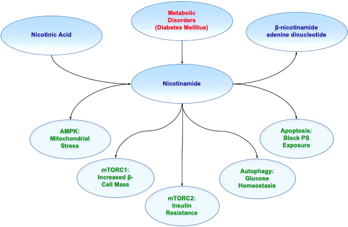
Figure 1

Nicotinamide and metabolic disease. Nicotinamide is generated through the conversion of nicotinic acid in the liver as one source. Once nicotinamide is obtained in the body, it serves as the precursor for NAD+. Nicotinamide through the mechanistic target of rapamycin (mTOR), mTOR Complex 1 (mTORC1), mTOR Complex 2 (mTORC2), AMP activated protein kinase (AMPK), and pathways of autophagy and apoptosis offer innovative strategies to treat metabolic disorders such as diabetes mellitus (DM). For example, during DM, AMP activated protein kinase (AMPK) can limit mitochondrial stress, mTOR Complex 1 (mTORC1),can increase pancreatic ß-cell mass, mTOR Complex 2 (mTORC2) can improve insulin resistance, autophagy can foster glucose homeostasis, and blockade of apoptosis can prevent cellular membrane phosphatidylserine (PS) externalization to promote cell longevity. Further appreciation and understanding of the complexities of these pathways can foster new strategies for translation into innovative treatments for metabolic disorders, such as DM.

4.2. Nicotinamide, normal physiology, and disease states

Nicotinamide through NAD+ has a critical physiological role in cellular metabolism and can be directly utilized by cells to synthesize NAD+ (82, 87, 88). Nicotinamide also participates in energy metabolism through the tricarboxylic acid cycle by utilizing NAD+ in the mitochondrial respiratory electron transport chain for the production of ATP, DNA synthesis, and DNA repair (89-91). Under some circumstances, nicotinamide can preserve mitochondrial function as a mechanism to enhance cellular survival (82, 92). Some studies suggest that the specific levels of NAD+ can be the critical factor for cell survival (88, 93, 94). Increased administration of nicotinamide may be useful against tumorigenesis (95) and lead to apoptotic cell death in cancer cells (96, 97).

If nicotinamide is depleted in the body, fatigue, loss of appetite, pigmented rashes of the skin, and oral ulcerations can result. More severe states of deficiency lead to pellagra that is characterized by cutaneous rashes, oral ulcerations, gastrointestinal difficulties, and cognitive loss (98). Pellagra can occur during conditions of low nicotinamide or the inability to absorb nicotinamide (99). As a result, the cellular pathways of nicotinamide are essential for energy metabolism and are directly tied to normal physiological processes as well as disease states that include inflammatory pathways (100), energy metabolism (72), vascular disease (101, 102), alcohol toxicity (103), and oxidative stress (88, 104, 105).

During periods of cellular injury that can involve oxidative stress (21, 106), reactive oxygen species can be scavenged by endogenous antioxidant systems that include nicotinamide, superoxide dismutase, glutathione peroxidase, catalase, and small molecule substances such as vitamins C, D, E, and K (107-113). However, nicotinamide can affect cellular survival during metabolic dysfunction and impact multiple systems of the body that are particularly affected by aging. Nicotinamide can foster protection during aging and mitochondrial dysfunction (79, 106, 114, 115), neuronal cell injury (103, 104, 116-119), vascular aging processes (29, 73, 101, 120), vascular demise and associated angiogenesis (101, 104, 117, 121-123), and neurodegenerative disorders such as AD (7, 87, 98, 124). Nicotinamide offers protection usually in a specific concentration range (81). Administration of nicotinamide in a range of 5.0 - 25.0 mmol/L can significantly protect neurons during oxidative stress injuries. This concentration range is similar to other injury paradigms in both animal models (125) and in cell culture models (82, 121, 126). It is important to note that elevated concentrations in some experimental models may not offer protection and can be detrimental (108, 127).

5. NICOTINAMIDE AND METABOLIC DYSFUNCTION
5.1. Nicotinamide and diabetes mellitus

Nicotinamide plays a significant role during metabolic dysfunction and DM (29, 72, 73) (Table 1). Nicotinamide may lower insulin resistance and glucose release in combination with other factors to prevent the onset and progression of DM (128-130). Nicotinamide may protect against skeletal muscle atrophy during DM (131) and may reduce inflammation of the brain during DM with the administration of niacin (132). Prior work also has shown that nicotinamide can maintain normal fasting blood glucose with streptozotocin-induced DM in animal models (133, 134) and inhibit oxidative stress pathways that lead to cell death and apoptosis (121, 135-138). Nicotinamide can significantly improve glucose utilization, prevent excessive lactate production, and improve electrophysiologic capacity in ischemic animal models (139). Oral nicotinamide administration (1200mg/m2/day) protects pancreatic β-cell function and prevents clinical disease in islet-cell antibody-positive first-degree relatives of type-1 DM (140). Nicotinamide administration (25mg/kg) in patients with recent onset type-1 DM combined with intensive insulin therapy for up to two years after diagnosis can significantly reduce HbA1c levels (141). Yet, the duration of nicotinamide administration may influence the efficacy of this agent since long-term administration has been reported to support glucose intolerance in some animal models (93). Prolonged exposure of nicotinamide in other studies was reported to result in impaired pancreatic β-cell function and cell growth (142, 143). Nicotinamide also may inhibit cytochromes P450 and hepatic metabolism (144).

5.2. Nicotinamide and maintenance of mitochondrial function

As previously noted during metabolic disease, nicotinamide is vital for maintaining important cellular energy homeostasis and mitochondrial function (78, 79, 82, 120). Nicotinamide can control mitochondrial function at the level of mitochondrial membrane pore formation to prevent the release of cytochrome c (82, 121, 136). Pretreatment of cells with either nicotinamide alone or in combination with the mitochondrial permeability transition pore inhibitor cyclosporin A prior to an injury paradigm can inhibit mitochondrial membrane depolarization (145, 146). Nicotinamide can block the chemical induction of mitochondrial membrane depolarization during exposure to either tert-butylhydroperoxide or atractyloside (104). There are other pathways that nicotinamide may use to maintain cellular metabolic homeostasis through the maintenance of mitochondrial membrane potential (121, 126). Nicotinamide can lead to the phosphorylation of Bad to prevent mitochondrial membrane depolarization and subsequent cytochrome c release (82, 121, 136), block the assembly of the mitochondrial permeability transition pore complex similar to the action of cyclosporin A (147), or stabilize cellular energy metabolism since the maintenance of mitochondrial membrane potential is an ATP facilitated process (148).

6. NICOTINAMIDE, PATHWAYS OF CELL DEATH, AND AUTOPHAGY
6.1. Apoptotic Cell Death

As a vital modulator of cell survival, nicotinamide oversees pathways of programmed cell death that involves apoptosis (149-151). The apoptotic pathway consists of two distinct phases. The early phase involves the loss of plasma membrane phosphatidylserine (PS) asymmetry (152-158). A later phase results in genomic DNA degradation (156, 159-163). Apoptosis is initiated through a cascade activation of nucleases and proteases that involve caspases (105, 106, 164-167). These processes can influence both the early phase of apoptosis with the loss of plasma membrane PS asymmetry and the later phase that leads to genomic DNA degradation. Loss of membrane PS asymmetry activates inflammatory cells to target, engulf, and remove injured cells (152, 157, 168, 169). If the engulfment by inflammatory cells can be prevented and cells that have membrane PS residues exposed are not removed, then functional cells expressing membrane PS residues can be rescued (62, 160, 170, 171). Yet, once the destruction of cellular DNA occurs, the process is usually not considered to be completely reversible (172).

Nicotinamide can address both phases of apoptotic cell injury. During different cellular injury paradigms, nicotinamide can prevent exposure of membrane PS residues to block inflammatory cell activation (81, 121, 126, 136). In particular, nicotinamide may reduce cardiovascular injury by blocking membrane PS exposure in vascular cells (82, 121), since membrane PS residue externalization in vascular cells can lead to hypercoagulation states (173) and cellular inflammation (174, 175). Nicotinamide may reverse a previously sustained insult (82, 104, 121, 126, 136, 176). Post-treatment strategies with nicotinamide that can follow apoptotic injury in “real-time” show that cellular injury can be reversed. Nicotinamide can reverse an initial progression of membrane PS inversion and prevent PS exposure (82, 126, 176, 177). These results support the hypothesis that if a cellular injury does not progress to DNA degradation, the injury can be reversible (82, 126, 176, 177).

In relation to apoptotic DNA degradation, nicotinamide prevents apoptotic DNA injury in vascular cells (104, 121), neurons (116, 124, 136, 178), keratinocytes (179), corneal endothelial cells (180, and photoreceptor cells {Kiuchi, 2002 #3733, 181). Nicotinamide also can inhibit DNA replication in some cell systems (182). Dependent upon the cellular conditions, nicotinamide may not be able to prevent DNA degradation (87). During periods of acidosis-induced cellular toxicity that involve decreased pH, nicotinamide cannot prevent cellular injury during intracellular acidification (126).

6.2. Autophagy

Autophagy is a process that recycles components of the cytoplasm in cells for tissue remodeling (183-185) (Table 1). As a result, it eliminates non-functional organelles (24, 149, 151, 186, 187). Macroautophagy recycles organelles and sequesters cytoplasmic proteins and organelles into autophagosomes. Autophagosomes subsequently combine with lysosomes for degradation and recycling (188, 189). Microautophagy consists of lysosomal membranes invagination for the sequestration and digestion of cytoplasmic components (8). Chaperone-mediated autophagy requires cytosolic chaperones to transport cytoplasmic components across lysosomal membranes (67, 190).

Similar to nicotinamide, autophagy is involved with clinical aging pathways (12, 105, 184). Studies with Drosophila show that neural aggregate accumulation observed with aging is linked to a reduction in the autophagy pathway. These neural aggregates lead to behavior impairments that can be resolved with the maintenance of autophagy pathways in neurons (191). Autophagy also is involved in a number of other disorders that may be tied to aging such as dementia (40, 192-196), AD (7, 12, 39, 40, 193, 197-201), Huntington’s disease (HD) (172, 202-204), and DM (21, 27, 39, 40, 62, 193, 205).

Nicotinamide has been tied to autophagic pathways, especially as an inhibitor of sirtuin pathways, such as those involved with silent mating type information regulation 2 homolog 1 (Saccharomyces cerevisiae) (SIRT1) (7, 16). Nicotinamide can lead to the induction of delayed autophagy and decreased survival in cancer cells (206). Mitochondrial autophagy can result in an increased NAD+/NADH ratio during nicotinamide administration (93, 105, 207) and in some cases induction of autophagy can affect mitochondrial mass (196, 208, 209). Chronic administration of nicotinamide can result in skeletal muscle autophagy (93). Nicotinamide has been shown to protect against palmitate-induced hepatotoxicity via SIRT1-dependent induction of autophagy (210).

7. NICOTINAMIDE AND THE MECHANISITIC TARGET OF RAPAMYCIN
7.1. Nicotinamide and mTOR

Through autophagy, nicotinamide has been shown to rely upon the pathways of the mechanistic target of rapamycin (mTOR) (Table 1). For example, nicotinamide can protect hypoxic myocardial cells through the induction of autophagy and the modulation of mTOR pathways (211). mTOR, a 289-kDa serine/threonine protein kinase, is increasingly being recognized as a critical pathway for nicotinamide given the ability of this vitamin to control cellular metabolism and the programmed death pathways of autophagy and apoptosis. mTOR also is known as the mammalian target of rapamycin and the FK506-binding protein 12-rapamycin complex-associated protein 1 (203, 212) and is encoded by a single gene FRAP1 (213-215). The target of rapamycin (TOR) was initially discovered in Saccharomyces cerevisiae with the genes TOR1 and TOR2 (216). Using rapamycin-resistant TOR mutants, TOR1 and TOR2 are now known to encode the Tor1 and Tor2 isoforms in yeast (217). The compound rapamycin is a macrolide antibiotic in Streptomyces hygroscopicus that blocks TOR and mTOR activity (24).

mTOR serves as the principal component of the protein complexes mTOR Complex 1 (mTORC1) and mTOR Complex 2 (mTORC2) (218-220) (Figure 2). Rapamycin prevents mTORC1 activity by binding to immunophilin FK-506-binding protein 12 (FKBP12) that attaches to the FKBP12 -rapamycin-binding domain (FRB) at the carboxy (C) -terminal of mTOR to interfere with the FRB domain of mTORC1 (221). The mechanism of how rapamycin blocks mTORC1 activity with the interaction of the domain of FRB is not entirely clear. One pathway may involve allosteric changes on the catalytic domain as well as the inhibition of phosphorylation of protein kinase B (Akt) and p70 ribosomal S6 kinase (p70S6K) (222). mTORC1 is more sensitive to inhibition by rapamycin than mTORC2, but chronic administration of rapamycin can inhibit mTORC2 activity as a result of the disruption of the assembly of mTORC2.
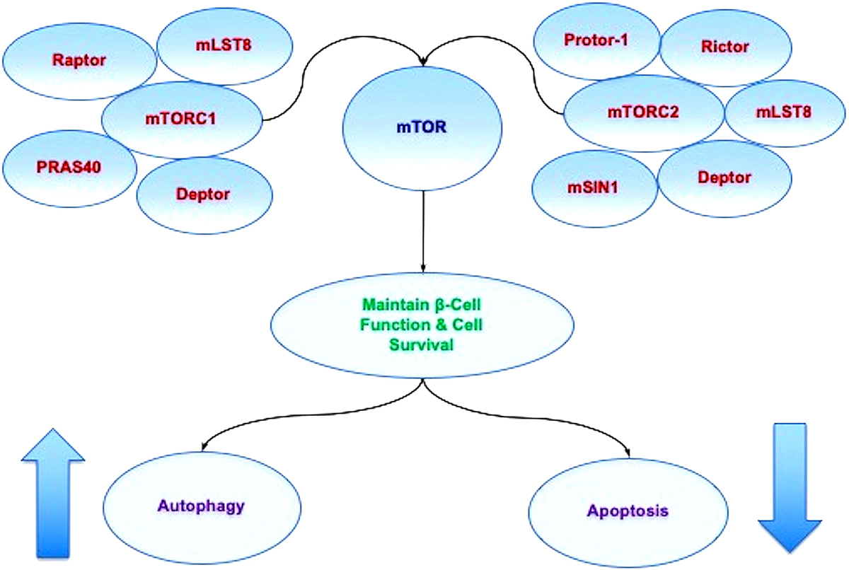
Figure 2

mTOR oversight of autophagy and apoptosis. mTOR is the principal component of the protein complexes mTOR Complex 1 (mTORC1) and mTOR Complex 2 (mTORC2). mTORC1 is composed of Raptor, the proline rich Akt substrate 40 kDa (PRAS40), Deptor (DEP domain-containing mTOR interacting protein), and mammalian lethal with Sec13 protein 8, termed mLST8 (mLST8) (214). mTORC2 is composed of Rictor, mLST8, Deptor, the mammalian stress-activated protein kinase interacting protein (mSIN1), and the protein observed with Rictor-1 (Protor-1). Autophagy activity can be controlled through mTOR since activation of autophagy occurs during the inhibition of mTOR. As an example, mTOR inhibition also may be required for maintaining a balance between pancreatic -cell proliferation and cell size. Yet, mTOR activation can be beneficial at time s since mTOR activation protects pancreatic -cells against cholesterol-induced apoptosis, reduces glucolipotoxicity, and results in increased neuronal cell survival in cellular models of diabetes mellitus. These observations demonstrate that a fine balance in activity is required for mTOR, autophagic, and apoptotic pathways.

7.2. mTOR as a component of mTORC1

mTORC1 is composed of Raptor, the proline rich Akt substrate 40 kDa (PRAS40), Deptor (DEP domain-containing mTOR interacting protein), and mammalian lethal with Sec13 protein 8, termed mLST8 (mLST8) (216). mTORC1 binds to its constituents through the protein Ras homologue enriched in brain (Rheb) that phosphorylates the Raptor residue serine863 and other residues that include serine859, serine855, serine877, serine696, and threonine706 (223). The inability to phosphorylate serine863 limits mTORC1 activity, as shown using a site-direct mutation of serine863 (224). mTOR can control Raptor activity which can be blocked by rapamycin (224). Deptor, an inhibitor, blocks mTORC1 activity by binding to the FAT domain (FKBP12 -rapamycin-associated protein (FRAP), ataxia-telangiectasia (ATM), and the transactivation/transformation domain-associated protein) of mTOR. If the activity of Deptor is suppressed, Akt, mTORC1, and mTORC2 activities are increased (225). PRAS40 blocks mTORC1 activity by preventing the association of p70 ribosomal S6 kinase (p70S6K) and the eukaryotic initiation factor 4E (eIF4E)-binding protein 1 (4EBP1) with Raptor (188, 226). mTORC1 is active once PRAS40 is phosphorylated by Akt. This releases PRAS40 from Raptor to sequester PRAS40 in the cytoplasm of the cell with the docking protein 14-3-3 (227-231). mLST8, in contrast to Deptor and PRAS40, promotes mTOR kinase activity. This involves the binding of p70S6K and 4EBP1 to Raptor (232). In relation to cellular metabolism, mLST8 also controls insulin signaling through the mammalian forkhead transcription factor FoxO3 (67, 233). mLST8 is also necessary for Akt and protein kinase C-α (PKCα) phosphorylation and is required for Rictor to associate with mTOR (233).

7.3. mTOR as a component of mTORC2

mTORC2 is composed of Rictor, mLST8, Deptor, the mammalian stress-activated protein kinase interacting protein (mSIN1), and the protein observed with Rictor-1 (Protor-1) (188, 213). mTORC2 oversees cytoskeleton remodeling through PKCα and cell migration through the Rac guanine nucleotide exchange factors P-Rex1 and P-Rex2 and through Rho signaling (234). mTORC2 promotes activity of protein kinases that includes glucocorticoid induced protein kinase 1 (SGK1), a member of the protein kinase A/protein kinase G/protein kinase C (AGC) family of protein kinases. Protor-1, a Rictor-binding subunit of mTORC2, activates SGK1 (235, 236). The kinase domain of mTOR phosphorylates mSIN1 and blocks lysosomal degradation of this protein. Rictor and mSIN1 also phosphorylate Akt at serine473 and foster threonine308 phosphorylation by phosphoinositide-dependent kinase 1 (PDK1) to enhance cell survival.

7.4. mTOR and AMP activated protein kinase

Both mTORC1 and mTORC2 are tied to metabolic cellular regulation. These pathways are closely linked to the AMP activated protein kinase (AMPK) (16, 56, 237, 238) (Table 1). mTORC1 is associated with metabolic cellular pathways and can stimulate lipogenesis and fat storage (15, 239), may increase pancreatic ß-cell mass (240), and can improve glucose homeostasis (241). In the presence of other cellular pathways and systems such as with SIRT1, mTORC1 activity may require inhibition to preserve glucose homeostasis (242). mTORC2 also may be necessary to maintain glucose homeostasis (15), since loss of this pathway can promote severe hyperglycemia (243). Impairment in mTORC2 signaling can result in oxidative damage and insulin resistance (244). mTORC2 signaling also plays a significant role for the maintenance of pancreatic β-cell proliferation and mass (245).

In regards to AMPK, AMPK blocks mTORC1 activity through the hamartin (tuberous sclerosis 1)/tuberin (tuberous sclerosis 2) (TSC1/TSC2) complex that inhibits mTORC1 (246, 247) (Figure 3). Control of the TSC1/TSC2 complex also is controlled though phosphoinositide 3-kinase (PI 3-K), Akt, and its phosphorylation of TSC2. Extracellular signal-regulated kinases (ERKs), protein p90 ribosomal S6 kinase 1 (RSK1), and glycogen synthase kinase -3β (GSK-3β) can modulate the activity of the TSC1/TSC2 complex as well. TSC2 functions as a GTPase-activating protein (GAP) that converts G protein Rheb (Rheb-GTP) into the inactive GDP-bound form (Rheb-GDP). Once Rheb-GTP is active, Rheb-GTP associates with Raptor to control the binding of 4EBP1 to mTORC1 and increase mTORC1 activity (248). AMPK can phosphorylate TSC2 to increase GAP activity to change Rheb-GTP into the inactive Rheb-GDP and block mTORC1 activity (249).
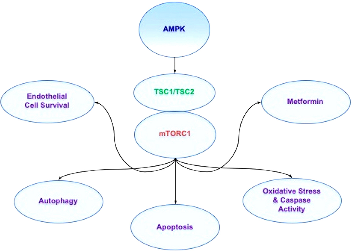
Figure 3

AMPK affects multiple pathways through mTOR signaling. AMP activated protein kinase (AMPK) inhibits mTOR Complex 1 (mTORC1) activity through the hamartin (tuberous sclerosis 1)/tuberin (tuberous sclerosis 2) (TSC1/TSC2) complex. During periods of hyperglycemia, AMPK activity increases basal autophagy activity and maintains endothelial cell survival. In addition, AMPK can regulate apoptosis and autophagy during vascular disease and oxidative stress cell injury. Agents used to treat diabetes mellitus, such as biguanides and metformin, rely upon AMPK to restore cellular function. Through metformin, AMPK can be activated, leads to the induction of autophagy, and protects against cell apoptosis. Metformin also has been shown to limit lipid peroxidation in the brain and spinal cord and decrease caspase activity during toxic insults.

8. NICOTINAMIDE, mTOR, AMPK, AND METABOLIC DISORDERS
8.1. mTOR and metabolic function

mTOR pathways are intimately tied to cellular metabolic function (24, 250). mTOR activation protects pancreatic β-cells against cholesterol-induced apoptosis (251), reduces glucolipotoxicity (252), and results in increased neuronal cell survival in cellular models of DM (253). mTOR activity controls insulin signaling in experimental models of AD and maintains astrocyte viability (254), promotes the differentiation of adipocytes (255), preserves endothelial cell function during hyperglycemia (54), and maintains glucose homeostasis (241). mTOR also may offer protection as a component of the Mediterranean diet as a focus on obesity in the population. The diet has been reported to reduce Aβ toxicity in astrocytes through enhanced Akt activity by consumption of polyphenol of olives and olive oil that ultimately could prevent the onset or progression of AD (254). In patients with metabolic syndrome, mTOR activation has been found to be diminished, suggesting that loss of mTOR may lead to insulin resistance and the increased risk of vascular thrombosis (256). In addition, growth factors that offer cellular protection against oxidative stress, such as insulin growth factor-1 (IGF-1) (257, 258) and erythropoietin (EPO) (259-270), may employ mTOR pathways as well (16, 227, 271-276). For example, EPO utilizes components of the mTOR pathway, such as PRAS40 and Akt, to enhance cell survival (227, 272, 273, 276) and limit toxic cellular environments (275, 277-279). EPO also plays a significant role as a potential cellular protectant during aging (280) and DM with the modulation of mTOR pathways, in part, to maintain cellular survival (24, 62, 73, 74, 175, 281-286).

8.2. Nicotinamide and the downstream pathways of mTOR

Nicotinamide and mTOR downstream pathways also play a critical role during metabolic dysfunction (Table 1). For example, mTOR pathway activation with p70S6K and 4EBP1 can improve insulin secretion in pancreatic β-cells and increase resistance to β-cell streptozotocin toxicity and obesity in mice (240). Both p70S6K and 4EBP1 in the mTOR pathway are also utilized by nicotinamide to protect against radiation-induced apoptosis (179).

Interestingly, nicotinamide has a close relationship with poly (ADP-ribose) polymerase (PARP) (81, 87) that is also tied to mTOR and Akt (287, 288). PARP is a nuclear protein that binds to DNA strand breaks and cleaves NAD+ into nicotinamide and ADP-ribose. PARP catalyzes the synthesis of poly (ADP-ribose) from its substrate NAD+, which can foster the process of DNA repair (289). Nicotinamide can prevent PARP degradation and allow for DNA repair through the direct inhibition of caspase 3 (121, 126, 136). However, elevated concentrations of nicotinamide can lead to PARP degradation and apoptotic injury (290). Under some circumstances, a reduction in PARP activity may enhance neuronal cell survival, such as during cerebral ischemia (72). Prevention of NAD+ depletion during enhanced PARP activity also has been demonstrated to prevent cellular lysis during oxidative stress. In experimental studies of AD, PARP is present in the frontal and temporal cortex more frequently than in controls, suggesting that increased levels of functional PARP enzyme are present to potentially lead to the depletion of NAD+ stores (291). Reduction of PARP activity may be protective against oxidative stress and Aß (124). Furthermore, recent work illustrates that inhibition of the mTOR and PARP axis has protective effects on photoreceptors against visible-light-induced parthanatos (288), a form of programmed cell death that is distinct from other cell death processes such as necrosis and apoptosis and a PARP-1 dependent cell death.

Nicotinamide also can protect hypoxic cardiomyocytes and reduce intracellular mitochondrial stress through activation of the AMPK pathway (292). During periods of reduced dietary intake that may increase lifespan (116), AMPK activation can shift to beneficial oxidative metabolism (293) and has been shown to limit ischemic brain damage in diabetic animal models (294). AMPK activation also can strengthen memory retention in models of AD and DM (237), may assist with the elimination of ß-amyloid (Aß) in the brain (295), facilitate tau clearance (201), and limit chronic inflammation in the nervous system (12, 247, 296). AMPK can limit insulin resistance (297) and protect endothelial progenitor cells during periods of hyperglycemia (56) similar to the vascular protective properties of nicotinamide (104, 121, 298).

AMPK controls metabolic pathways through apoptosis and autophagy (Figure 3). During periods of hyperglycemia, AMPK activity can increase basal autophagy activity (149, 172) and maintain endothelial cell survival (54, 299). AMPK can regulate apoptosis and autophagy during coronary artery disease (300), endothelial dysfunction during hyperglycemia (54), and oxidative stress cell injury (301, 302). Anti-senescence activity also can be fostered through mTOR inhibition, AMPK activation, and the acceleration of autophagic flux (303). In addition to nicotinamide, other agents rely upon the ability of AMPK to modulate cellular metabolism. Agents used to treat DM, such as biguanides and metformin, use mTOR, AMPK, and autophagy to restore cellular function. Metformin inhibits mTOR activity, promotes autophagy, and may function at times in an AMPK-independent manner (304). Other reports note that through metformin, AMPK is activated, restores the induction of autophagy, and protects against diabetic cardiac cell apoptosis (305). Metformin also has been shown to limit lipid peroxidation in the brain and spinal cord and decrease caspase activity during toxic insults (306). Such results may be associated with the ability of autophagic pathways to limit oxidative stress under some circumstances (27, 307).

8.3. Nicotinamide and the Necessary Modulation of Autophagy with mTOR

Interestingly, similar to nicotinamide having optimal dose concentrations for beneficial biological outcomes, autophagy also requires specific modulation of activity (Table 1). Under some circumstances, inhibition of autophagy may be required to achieve a benefit. In some experimental models of AD, autophagy activation can be one factor that leads to neuronal cell death (200). Increased activity of autophagy also can result in significant loss of cardiac and liver tissue in diabetic rats during attempts to achieve glycemic control through diet modification (77). Advanced glycation end products (AGEs), agents that can result in complications during DM, have also been shown to result in the activation of autophagy and vascular smooth muscle proliferation that can lead to atherosclerosis (308) and cardiomyopathy (309). Autophagy activation also can injure endothelial progenitor cells, lead to mitochondrial oxidative stress (310), and block angiogenesis (311) during periods of elevated glucose levels. Chronic inflammatory conditions, such as lichen planus, also have been linked to mTOR inhibition and autophagy activation (312). Interneuron progenitor growth in the brain relies upon mTOR activity with the inhibition of autophagy (313). During ischemic stroke in rodents, inhibition of autophagy may also limit infarct size and rescue cerebral neurons (314).

One mechanism for the modulation of autophagy activity may be through mTOR given that activation of autophagy occurs during the inhibition of mTOR (16, 315, 316). At times, inhibition of mTOR during DM can be beneficial and provide protection such as during cerebral ischemia (294). mTOR inhibition also may be necessary for maintaining a balance between pancreatic β-cell proliferation and cell size (245). Yet, other studies suggest that loss of mTOR activity can be detrimental. During mTOR inhibition with rapamycin, reduced β-cell function, insulin resistance, and decreased insulin secretion can promote the progression of DM (317). Decreased activity of mTOR also has been shown to increase mortality in a mouse model of DM (318). Translocation of glucose transporters to the plasma membrane in skeletal muscle can be blocked in the absence of mTOR activity (319). These studies may suggest that there may be a biological feedback for mTOR, such as through AMPK inhibition, to prevent excessive mTOR activity and allow for the activation of autophagy. For example, if mTOR activity is allowed to go unchecked with the down-regulation of AMPK activity during experimental studies, mTOR and p70S6K can lead to glucose intolerance by inhibiting the insulin receptor substrate 1 (IRS-1) (320).

Nicotinamide may require a close modulation of autophagy and mTOR to offer protection during disorders such as DM. Nicotinamide can offer cellular protection through autophagy (211) that requires inhibition of mTOR activity to allow for the activation of autophagy. Dysregulation of autophagy can potentially lead to the progression of cognitive loss with AD and the induction of DM (39). At least 33 autophagy-related genes (Atg) that have been identified in yeast with 40 autophagy-related genes involved in autophagosomes formation (185) that can affect multiple disorders including DM (15, 67, 321). Atg1, Atg13 (also known as Apg13), and Atg17 are associated with the PI 3-K, Akt, and TOR pathways (149). Autophagy haploinsufficiency with deletion of Atg7 gene in mouse models of obesity leads to increased insulin resistance with elevated lipids and inflammation (322). Loss of autophagic proteins Atg7, Atg5, and LC3 also can be responsible for diabetic nephropathy (323). Autophagy can be beneficial at the cellular level. Autophagy can remove misfolded proteins and eliminate non-functioning mitochondria to maintain β-cell function and prevent the onset of DM (324). Exercise in mice has been shown to promote the induction of autophagy and regulate glucose homeostasis (325). Autophagy also can improve insulin sensitivity during high fat diets in mice (297) and may be protective to microglia during acute glucose fluctuations (193).

It is important to recognize that in a number of circumstances, autophagy and apoptosis are closely linked to alter cellular survival. Inhibition of mTOR activity with activation of autophagy can reduce markers of senescence and aging in the skin of patients (326). Blockade of mTOR also can prevent aging and cell injury with extension of cell longevity through AMPK in endothelial cells (303). Additional work suggests that a balance is required during embryogenesis for autophagy and apoptosis that can affect body axis formation (327). Inhibition of mTOR pathways can trigger autophagy activation and affect tumor cell growth through the inhibition of apoptosis (328, 329). Yet, dependent upon the balance between autophagy and apoptotic pathways, other studies have shown that with nicotinamide one can block apoptosis in human colon cancer cells through the activation of autophagy (207). These studies support the premise that the balance between autophagy and apoptosis requires careful modulation to foster a desired biological outcome with nicotinamide.

9. FUTURE PERSPECTIVES FOR NICOTINAMIDE

Metabolic disorders, such as DM, pose a significant risk for the global population and contribute to the large percentage of deaths that are the result of NCDs (1, 2) (Table 1). Of even greater concern is the observation that of the over forty million people that die each year from NCDs, fifteen million individuals are between the ages of thirty and sixty-nine years old. In addition, NCDs affect a greater proportion of the population in low and middle-income countries. As the world’s population continues to age, life expectancy has been increasing that has occurred in parallel to the rise in NCDs (Figure 1).

As significant NCDs, metabolic disorders and DM are seen as critical targets to address and develop innovative strategies to limit death and disability for the global population afflicted by these disorders. Almost eighty percent of adults with DM reside in low- and middle-income countries and the care for patients with DM consumes more than seventeen percent of the Gross Domestic Product in the US (18). Furthermore, DM is believed to affect seven hundred million people by the year 2045 (17). DM affects all systems of the body that can involve the nervous system, cardiovascular system, and immune system. DM also alters new tissue generation and repair. Current conventional therapies for metabolic disorders and DM such as early recognition of disease onset, treatment with nutrition, and administration of pharmacological care can offer some degree of protection and may slow the progression of DM. However, treatments that lead to tight serum glucose control do not always resolve the eventual complications from DM and may pose potential risks that can decrease organ mass through autophagy. Given these concerns, innovative and novel strategies for metabolic disorders that involve DM are highly warranted for rapid development. Nicotinamide and the pathways associated with mTOR, mTORC1, mTORC2, AMPK, autophagy, and apoptosis fill this void to offer new avenues for the treatment of metabolic disorders.

Nicotinamide offers a partially unique perspective as a cellular protectant since it can foster cellular survival against oxidative stress not only during aging processes (72, 105, 114) and life span extension (29, 92, 116), but also during metabolic dysfunction (72, 88, 128, 131, 330). Nicotinamide can reverse a previously sustained insult by limiting the onset and progression of membrane PS exposure and apoptotic cell death. During DM, nicotinamide maintains cellular energy homeostasis and mitochondrial function, lowers insulin resistance and glucose release, protects against skeletal muscle atrophy, reduces inflammation of the brain, protects pancreatic β-cell function, improves glucose utilization, and prevents excessive lactate production. Nicotinamide employs mTOR pathways such that it can prevent myocardial cell death during hypoxia through the oversight of mTOR and autophagy. As a result, nicotinamide relies upon the inverse relationship between mTOR and autophagy pathways to control cellular survival. Similar to nicotinamide, mTOR activation can protect pancreatic β-cells and maintain glucose homeostasis. Through autophagy activation, the mTOR related pathway of AMPK can increase basal autophagy activity to prevent cellular apoptosis during DM. In addition, nicotinamide has been shown to protect hypoxic cardiomyocytes and reduce intracellular mitochondrial stress through activation of AMPK and autophagic pathways. Chronic administration of nicotinamide can activate skeletal muscle autophagy as a potential protective response to lipotoxicity.

However, there are a number of hurdles to consider for the targeting of nicotinamide, mTOR, and autophagy for the treatment of metabolic disorders. Nicotinamide appears to offer cellular protection during metabolic dysfunction in specific concentration ranges. Nicotinamide may protect against skeletal muscle atrophy during DM and can reduce inflammation of the brain during diabetes with the administration of niacin. Yet, the duration of nicotinamide administration may influence the efficacy of this agent since long-term administration has been reported to support glucose intolerance in some animal models and prolonged exposure of nicotinamide can result in impaired β-cell function and cell growth (142, 143). The acute and chronic levels of NAD+ that are generated may be vital to determine cellular survival. Similar to nicotinamide, autophagy may require critical modulation of activity. Nicotinamide can protect against palmitate-induced hepatotoxicity via SIRT1-dependnet induction of autophagy. In addition, autophagy can improve insulin sensitivity during high fat diets and may protect microglia during acute glucose fluctuations (193). Yet, autophagy activation also can injure endothelial progenitor cells, lead to mitochondrial oxidative stress, and block angiogenesis during periods of elevated glucose levels. mTOR, as a pathway that is inhibited during autophagy activation, may be a critical factor for nicotinamide to maintain cell viability during metabolic dysfunction. In some studies, loss of mTOR activity can be detrimental and result in reduced β-cell function, insulin resistance, and decreased insulin secretion that foster the progression of DM. In other scenarios, inhibition of mTOR during DM can be beneficial and provide protection such as during cerebral ischemia and may be necessary for maintaining a balance between pancreatic β-cell proliferation and cell size. Given these observations, it becomes clear that targeting nicotinamide as an effective agent to treat metabolic disorders may require careful scrutiny of the fine balance in activity required for mTOR and autophagic pathways. Additional investigations are warranted and would be highly advantageous to examine the intricate pathways of the vitamin nicotinamide and how they can be effectively utilized to provide innovative clinical strategies for metabolic dysfunction and DM.

10. ACKNOWLEDGMENTS

This research was supported by the following grants to Kenneth Maiese: American Diabetes Association, American Heart Association, NIH NIEHS, NIH NIA, NIH NINDS, and NIH ARRA.

Abbreviations
Abbreviation Expansion
AD

Alzheimer's disease

AMPK

AMP activated protein kinase

Atg

Autophagic related genes

Deptor

DEP domain-containing mTOR interacting protein

DM

Diabetes mellitus

EPO

Erythropoietin

4EBP1

Eukaryotic initiation factor 4E (eIF4E-binding protein 1

ERKs

Extracellular signal-regulated kinases

TSC1/TSC2

Hamartin (tuberous sclerosis 1/tuberin (tuberous sclerosis 2

IRS-1

Insulin receptor substrate 1

mLST8

Mammalian lethal with Sec13 protein 8

termed

mLST8

mTOR

Mechanistic target of rapamycin

mTORC1

mTOR Complex 1

mTORC2

mTOR Complex 2

NCDs

Non-communicable diseases

p70S6K

p70 ribosomal S6 kinase

PARP

poly (ADP-ribose) polymerase (PARP)

PS

Phosphatidylserine

PDK1

Phosphoinositide-dependent kinase 1

PRAS40

Proline rich Akt substrate 40 kDa

Akt

Protein kinase B

SIRT1

Silent mating type information regulation 2 homolog 1 (Saccharomyces cerevisiae:

US

United States

USD

United States dollars

References
[1]
Organization World Health Description of the global burden of NCDs, their risk factors and determinants Global status report on noncommunicable diseases 2010(April) 2011 1 176
[2]
Organization World Health Global action plan on the public health response to dementia 2017-2025 2017 1 44
[3]
SirtuinsK. MaieseDeveloping Innovative Treatments for Aged-Related Memory Loss and Alzheimer's DiseaseCurr Neurovasc Res2018154DOI: 10.2174/1567202616666181128120003
[4]
Maiese K Cutting through the Complexities of mTOR for the Treatment of Stroke Curr Neurovasc Res 2014 11 2 177 186
[5]
Minino A. M Death in the United States, 2011 NCHS Data Brief 2013 115 1 8
[6]
Hayutin A Global demographic shifts create challenges and opportunities PREA Quarterly, (Fall) 2007 46 53
[7]
MaieseKSIRT1 and stem cells: In the forefront with cardiovascular disease, neurodegeneration and cancerWorld J Stem Cells20157223542DOI: 10.4252/wjsc.v7.i2.235
[8]
Maiese K Programming apoptosis and autophagy with novel approaches for diabetes mellitus Curr Neurovasc Res 2015 12 2 173 88
[9]
Diamanti-KandarakisEDatilloMMacutDDuntasL. HGonosEGoulisDKanaka-GantenbeinCKapetanouMKoukkouE. GLambrinoudakiIMichalakiMNader-EftekhariSPasqualiRPeppaMTzanelaMVassilatouEVryonidouAMechanisms in Endocrinology: Aging and Anti-aging Endocrinology: A Combo - Endocrinology OverviewEur J Endocrinol20171766R283R308DOI: 10.1530/eje-16-1061
[10]
FannD. YNgG. YPohLArumugamT. VPositive effects of intermittent fasting in ischemic strokeExp Gerontol20178993102DOI: 10.1016/j.exger.2017.01.014
[11]
LushchakOStrilbytskaOPiskovatskaVStoreyK. BKoliadaAVaisermanAThe role of the TOR pathway in mediating the link between nutrition and longevityMech Ageing Dev2017164127138DOI: 10.1016/j.mad.2017.03.005
[12]
MaieseKMoving to the Rhythm with Clock (Circadian) Genes, Autophagy, mTOR, and SIRT1 in Degenerative Disease and CancerCurr Neurovasc Res2017143299304DOI: 10.2174/1567202614666170718092010
[13]
StefanatosRSanzAThe role of mitochondrial ROS in the aging brainFEBS Lett20185925743758DOI: 10.1002/1873-3468.12902
[14]
KlimontovV. VBulumbaevaD. MFazullinaO. NLykovA. PBgatovaN. POrlovN. BKonenkovV. IPfeifferA. F. HPivovarova-RamichORudovichNCirculating Wnt1-inducible signaling pathway protein-1 (WISP-1/CCN4) is a novel biomarker of adiposity in subjects with type 2 diabetesJ Cell Commun Signal2019DOI: 10.1007/s12079-019-00536-4
[15]
MaieseKNovel nervous and multi-system regenerative therapeutic strategies for diabetes mellitus with mTORNeural Regen Res201611337285DOI: 10.4103/1673-5374.179032
[16]
MaieseKImpacting dementia and cognitive loss with innovative strategies: mechanistic target of rapamycin, clock genes, circular non-coding ribonucleic acids, and Rho/RockNeural Regen Res2019145773774DOI: 10.4103/1673-5374.249224
[17]
Federation International Diabetes Diabetes. IDF Diabetes Atlas(9th Edition) 2019
[18]
MedicareCenters forServicesMedicaid National Health Expenditure Projections 2018-2027. www.cms.gov2019
[19]
HaldarS. RChakrabartyAChowdhurySHaldarASenguptaSBhattacharyyaMOxidative stress-related genes in type 2 diabetes: association analysis and their clinical impactBiochem Genet2015534-693119DOI: 10.1007/s10528-015-9675-z
[20]
JiaGAroorA. RMartinez-LemusL. AReviewJ. R. Sowers: Invited Over-nutrition, mTOR Signaling and Cardiovascular Diseases.Am J Physiol Regul Integr Comp Physiol201430710R1198206DOI: 10.1152/ajpregu.00262.2014
[21]
MaieseKNew Insights for Oxidative Stress and Diabetes MellitusOxid Med Cell Longev, 2015(2015:875961)2015DOI: 10.1155/2015/875961
[22]
YeQFuJ. FPaediatric type 2 diabetes in China-Pandemic, progression, and potential solutionsPediatr Diabetes2018191DOI: 10.1111/pedi.12517
[23]
Harris M. I Eastman R. C Early detection of undiagnosed diabetes mellitus: a US perspective Diabetes Metab Res Rev 2000 16 4 230 6
[24]
mTORK. MaieseDriving apoptosis and autophagy for neurocardiac complications of diabetes mellitusWorld J Diabetes20156221724DOI: 10.4239/wjd.v6.i2.217
[25]
Maiese K Chong Z. Z Shang Y. C Mechanistic insights into diabetes mellitus and oxidative stress Curr Med Chem 2007 14 16 1729 38
[26]
BarchettaICiminiF. ACiccarelliGBaroniM. GCavalloM. GSick fat: the good and the bad of old and new circulating markers of adipose tissue inflammationJ Endocrinol Invest2019DOI: 10.1007/s40618-019-01052-3
[27]
MaieseKErythropoietin and diabetes mellitusWorld J Diabetes2015614125973DOI: 10.4239/wjd.v6.i14.1259
[28]
WangA. RYanX. QZhangCDuC. QLongW. JZhanDRenJLuoX. PCharacterization of Wnt1-inducible Signaling Pathway Protein-1 in Obese Children and AdolescentsCurr Med Sci2018385868874DOI: 10.1007/s11596-018-1955-5
[29]
Maiese K Chong Z. Z Shang Y. C Hou J Novel Avenues of Drug Discovery and Biomarkers for Diabetes Mellitus J Clin Pharmacol 2011 51 2 128 52
[30]
CerneaMTangWGuanHYangKWisp1 mediates Bmp3-stimulated mesenchymal stem cell proliferationJ Mol Endocrinol20165613946DOI: 10.1530/jme-15-0217
[31]
CurjuricIImbodenMBridevauxP. OGerbaseM. WHaunMKeidelDKumarAPonsMRochatTSchikowskiTSchindlerCEckardsteinA. vonKronenbergFProbst-HenschN. MCommon SIRT1 variants modify the effect of abdominal adipose tissue on aging-related lung function declineAge (Dordr)201638352DOI: 10.1007/s11357-016-9917-y
[32]
HillJ. HSoltCFosterM. TObesity associated disease risk: the role of inherent differences and location of adipose depotsHorm Mol Biol Clin Investig2018DOI: 10.1515/hmbci-2018-0012
[33]
LiuZGanLZhangTRenQSunCMelatonin alleviates adipose inflammation through elevating alpha-ketoglutarate and diverting adipose-derived exosomes to macrophages in miceJ Pineal Res201864112455DOI: 10.1111/jpi.12455
[34]
Maiese K Picking a bone with WISP1 (CCN4): new strategies against degenerative joint disease J Transl Sci 2016 1 3 83 85
[35]
MehtaJRayalamSWangXCytoprotective Effects of Natural Compounds against Oxidative StressAntioxidants (Basel)2018710DOI: 10.3390/antiox7100147
[36]
AlbieroMPoncinaNTjwaMCiciliotSMenegazzoLCeolottoGKreutzenbergS. Vigili deMouraRGiorgioMPelicciPAvogaroAFadiniG. PDiabetes causes bone marrow autonomic neuropathy and impairs stem cell mobilization via dysregulated p66Shc and Sirt1Diabetes2014634135365DOI: 10.2337/db13-0894
[37]
GomesM. BNegratoC. AAlpha-lipoic acid as a pleiotropic compound with potential therapeutic use in diabetes and other chronic diseasesDiabetol Metab Syndr20146180DOI: 10.1186/1758-5996-6-80
[38]
Gomez-BrouchetABlaesNMouledousLFourcadeOTackIFrancesBGirolamiJ. PMinvilleVBeneficial effects of levobupivacaine regional anaesthesia on postoperative opioid induced hyperalgesia in diabetic miceJ Transl Med2015131208DOI: 10.1186/s12967-015-0575-0
[39]
CaberlottoLNguyenT. PLauriaMPriamiCRimondiniRMaioliSCedazo-MinguezASitaGMorroniFCorsiMCarboniLCross-disease analysis of Alzheimer's disease and type-2 Diabetes highlights the role of autophagy in the pathophysiology of two highly comorbid diseasesSci Rep2019913965DOI: 10.1038/s41598-019-39828-5
[40]
SuMNaderiKSamsonNYoussefIFulopLBozsoZLarocheSDelatourBDavisSMechanisms Associated with Type 2 Diabetes as a Risk Factor for Alzheimer-Related PathologyMol Neurobiol2019DOI: 10.1007/s12035-019-1475-8
[41]
MaieseKChongZ. ZShangY. CHouJFoxO proteins: cunning concepts and considerations for the cardiovascular systemClin Sci (Lond)20091163191203DOI: 10.1042/CS20080113
[42]
TangP. CNgY. FHoSGydaMChanS. WResveratrol and cardiovascular health--promising therapeutic or hopeless illusion?Pharmacol Res20149088115DOI: 10.1016/j.phrs.2014.08.001
[43]
XiangLMittwedeP. NClemmerJ. SGlucose Homeostasis and Cardiovascular Alterations in DiabetesCompr Physiol201554181539DOI: 10.1002/cphy.c150001
[44]
XuY. JTappiaP. SNekiN. SDhallaN. SPrevention of diabetes-induced cardiovascular complications upon treatment with antioxidantsHeart Fail Rev201419111321DOI: 10.1007/s10741-013-9379-6
[45]
YaoTFujimuraTMurayamaKOkumuraKSekoYOxidative Stress-Responsive Apoptosis Inducing Protein (ORAIP) Plays a Critical Role in High Glucose-Induced Apoptosis in Rat Cardiac Myocytes and Murine Pancreatic beta-CellsCells201764DOI: 10.3390/cells6040035
[46]
HadamitzkyMHerringAKirchhofJBendixIHaightM. JKeyvaniKLuckemannLUnteroberdorsterMSchedlowskiMRepeated systemic treatment with rapamycin affects behavior and amygdala protein expression in ratsInt J Neuropsychopharmacol2018DOI: 10.1093/ijnp/pyy017
[47]
IgnacioZ. MReusG. ZArentC. OAbelairaH. MPitcherM. RQuevedoJNew perspectives on the involvement of mTOR in depression as well as in the action of antidepressant drugsBr J Clin Pharmacol201682512801290DOI: 10.1111/bcp.12845
[48]
RosaM. DiChitotriosidaseL. MalaguarneraA New Inflammatory Marker in Diabetic ComplicationsPathobiology20168342119DOI: 10.1159/000443932
[49]
Maiese K Chong Z. Z Hou J Shang Y. C Erythropoietin and oxidative stress Curr Neurovasc Res 2008 5 2 125 42
[50]
TulsulkarJNadaS. ESlotterbeckB. DMcInerneyM. FShahZ. AObesity and hyperglycemia lead to impaired post-ischemic recovery after permanent ischemia in miceObesity (Silver Spring)201524241723DOI: 10.1002/oby.21388
[51]
XiaoF. HHeY. HLiQ. GWuHLuoL. HKongQ. PA genome-wide scan reveals important roles of DNA methylation in human longevity by regulating age-related disease genesPLoS One2015103e0120388DOI: 10.1371/journal.pone.0120388
[52]
ArildsenLAndersenJ. VWaagepetersenH. SNissenJ. B. DSheykhzadeMHypermetabolism and impaired endothelium-dependent vasodilation in mesenteric arteries of type 2 diabetes mellitus db/db miceDiab Vasc Dis Res20191661479164119865885DOI: 10.1177/1479164119865885
[53]
DingSZhuYLiangYHuangHXuYZhongCCircular RNAs in Vascular Functions and DiseasesAdv Exp Med Biol20181087287297DOI: 10.1007/978-981-13-1426-1_23
[54]
PalP. BSonowalHShuklaKSrivastavaS. KRamanaK. VAldose reductase regulates hyperglycemia-induced HUVEC death via SIRT1/AMPK-alpha1/mTOR pathwayJ Mol Endocrinol2019DOI: 10.1530/jme-19-0080
[55]
AlexandruNPopovDGeorgescuAPlatelet dysfunction in vascular pathologies and how can it be treatedThromb Res2012129211626DOI: 10.1016/j.thromres.2011.09.026
[56]
ChiuS. CChaoC. YChiangE. ISyuJ. NRodriguezR. LTangF. YN-3 polyunsaturated fatty acids alleviate high glucose-mediated dysfunction of endothelial progenitor cells and prevent ischemic injuries both in vitro and in vivoJ Nutr Biochem201742172181DOI: 10.1016/j.jnutbio.2017.01.009
[57]
Maiese K Disease onset and aging in the world of circular RNAs J Transl Sci 2016 2 6 327 329
[58]
MaieseKChongZ. ZShangY. CHouJRogue proliferation versus restorative protection: where do we draw the line for Wnt and forkhead signaling?Expert Opin Ther Targets200812790516DOI: 10.1517/14728222.12.7.9.05
[59]
MaieseKLiFChongZ. ZShangY. CThe Wnt signaling pathway: Aging gracefully as a protectionist?Pharmacol Ther200811815881DOI: S0163-7258(08)00017-X
[60]
Perez-HernandezNVargas-AlarconGPosadas-SanchezRMartinez-RodriguezNTovilla-ZarateC. ARodriguez-CortesA. APerez-MendezOBlachman-BraunRRodriguez-PerezJ. MPHACTR1 Gene Polymorphism Is Associated with Increased Risk of Developing Premature Coronary Artery Disease in Mexican PopulationInt J Environ Res Public Health2016138DOI: 10.3390/ijerph13080803
[61]
ThackerayJ. TRadziukJHarperM. ESuuronenE. JAscahK. JBeanlandsR. SDasilvaJ. NSympathetic nervous dysregulation in the absence of systolic left ventricular dysfunction in a rat model of insulin resistance with hyperglycemiaCardiovasc Diabetol20111075DOI: 10.1186/1475-2840-10-75
[62]
Maiese K Novel applications of trophic factors, Wnt and WISP for neuronal repair and regeneration in metabolic disease Neural Regen Res 2015 10 4 518 528
[63]
MishraMDuraisamyA. JKowluruR. ASirt1- A Guardian of the Development of Diabetic RetinopathyDiabetes2018DOI: 10.2337/db17-0996
[64]
PonnalaguMSubramaniMJayadevCShettyRDasDRetinal pigment epithelium-secretome: A diabetic retinopathy perspectiveCytokine201795126135DOI: 10.1016/j.cyto.2017.02.013
[65]
KellD. BPretoriusENo effects without causes: the Iron Dysregulation and Dormant Microbes hypothesis for chronic, inflammatory diseasesBiol Rev Camb Philos Soc2018DOI: 10.1111/brv.12407
[66]
LinXBerberineN. ZhangPathways to protect neuronsPhytother Res2018DOI: 10.1002/ptr.6107
[67]
Maiese K FoxO Transcription Factors and Regenerative Pathways in Diabetes Mellitus Curr Neurovasc Res 2015 12 4 404 413
[68]
Maiese K Chong Z. Z Shang Y. C Wang S Translating cell survival and cell longevity into treatment strategies with SIRT1 Rom J Morphol Embryol 2011 52 4 1173 85
[69]
WoodhamsLAl-SalamiHThe roles of bile acids and applications of microencapsulation technology in treating Type 1 diabetes mellitusTher Deliv201786401409DOI: 10.4155/tde-2017-0010
[70]
ZhaoYScottN. AFynchSElkerboutLWongW. WMasonK. DStrasserAHuangD. CKayT. WThomasH. EAutoreactive T cells induce necrosis and not BCL-2-regulated or death receptor-mediated apoptosis or RIPK3-dependent necroptosis of transplanted islets in a mouse model of type 1 diabetesDiabetologia2015581140148DOI: 10.1007/s00125-014-3407-5
[71]
GuoTLiuTSunYLiuXXiongRLiHLiZZhangZTianZTianYSonodynamic therapy inhibits palmitate-induced beta cell dysfunction via PINK1/Parkin-dependent mitophagyCell Death Dis2019106457DOI: 10.1038/s41419-019-1695-x
[72]
KlimovaNKristianTMulti-targeted Effect of Nicotinamide Mononucleotide on Brain Bioenergetic MetabolismNeurochem Res2019DOI: 10.1007/s11064-019-02729-0
[73]
MaieseKChongZ. ZShangY. CWangSNovel directions for diabetes mellitus drug discoveryExpert Opin Drug Discov2013813548DOI: 10.1517/17460441.2013.736485
[74]
OthmanM. A. MRajabEAlMubarakAAlNaisarMBahzadNKamalAErythropoietin Protects Against Cognitive Impairment and Hippocampal Neurodegeneration in Diabetic MiceBehav Sci (Basel)201891DOI: 10.3390/bs9010004
[75]
YelumalaiSGiribabuNKarimKOmarS. ZSallehN. BIn vivo administration of quercetin ameliorates sperm oxidative stress, inflammation, preserves sperm morphology and functions in streptozotocin-nicotinamide induced adult male diabetic ratsArch Med Sci2019151240249DOI: 10.5114/aoms.2018.81038
[76]
CocaS. GIsmail-BeigiFHaqNKrumholzH. MParikhC. RRole of intensive glucose control in development of renal end points in type 2 diabetes mellitus: systematic review and meta-analysis intensive glucose control in type 2 diabetesArch Intern Med2012172107619DOI: 10.1001/archinternmed.2011.2230
[77]
LeeJ. HLeeJ. HJinMHanS. DChonG. RKimI. HKimSKimS. YChoiS. BNohY. HDiet control to achieve euglycemia induces significant loss of heart and liver weight via increased autophagy compared with ad libitum diet in diabetic ratsExp Mol Med201446e111DOI: 10.1038/emm.2014.52
[78]
MaieseKChongZ. ZHouJShangY. CThe vitamin nicotinamide: translating nutrition into clinical careMolecules2009149344685DOI: 14093446
[79]
BraidyNLiuYNAD+ therapy in age-related degenerative disorders: A benefit/risk analysisExp Gerontol2020110831DOI: 10.1016/j.exger.2020.110831
[80]
RexAFinkHPharmacokinetic aspects of reduced nicotinamide adenine dinucleotide (NADH) in ratsFront Biosci200813373541DOI: 2962
[81]
Li F Chong Z. Z Maiese K Navigating novel mechanisms of cellular plasticity with the NAD+ precursor and nutrient nicotinamide Front Biosci 2004 9 2500 2520
[82]
Maiese K Nicotinamide Z. Z. Chong necessary nutrient emerges as a novel cytoprotectant for the brain Trends Pharmacol Sci 2003 24 5 228 32
[83]
Jackson T. M Rawling J. M Roebuck B. D Kirkland J. B Large supplements of nicotinic acid and nicotinamide increase tissue NAD+ and poly(ADP-ribose) levels but do not affect diethylnitrosamine-induced altered hepatic foci in Fischer-344 rats J Nutr 1995 125 6 1455 61
[84]
WojcikMSeidleH. FBieganowskiPBrennerCGlutamine-dependent NAD+ synthetase. How a two-domain, three-substrate enzyme avoids wasteJ Biol Chem20062814433395402DOI: M607111200
[85]
KhanJ. AForouharFTaoXTongLNicotinamide adenine dinucleotide metabolism as an attractive target for drug discoveryExpert Opin Ther Targets2007115695705DOI: 10.1517/14728222.11.5.6.95
[86]
KhanJ. AXiangSTongLCrystal structure of human nicotinamide riboside kinaseStructure2007158100513DOI: S0969-2126(07)00251-1
[87]
Li F Chong Z. Z Maiese K Cell Life Versus Cell Longevity: The Mysteries Surrounding the NAD(+) Precursor Nicotinamide Curr Med Chem 2006 13 8 883 95
[88]
MaieseKTriple play: Promoting neurovascular longevity with nicotinamide, WNT, and erythropoietin in diabetes mellitusBiomed Pharmacother2008624218232DOI: S0753-3322(08)00026-7
[89]
Magni G Amici A Emanuelli M Orsomando G Raffaelli N Ruggieri S Enzymology of NAD+ homeostasis in man Cell Mol Life Sci 2004 61 1 19 34
[90]
Lin S. J Guarente L Nicotinamide adenine dinucleotide, a metabolic regulator of transcription, longevity and disease Curr Opin Cell Biol 2003 15 2 241 6
[91]
Hageman G. J Stierum R. H Niacin, poly(ADP-ribose) polymerase-1 and genomic stability Mutat Res 2001 475 1-2 45 56
[92]
CantoCHoutkooperR. HPirinenEYounD. YOosterveerM. HCenYFernandez-MarcosP. JYamamotoHAndreuxP. ACettour-RosePGademannKRinschCSchoonjansKSauveA. AAuwerxJThe NAD(+) Precursor Nicotinamide Riboside Enhances Oxidative Metabolism and Protects against High-Fat Diet-Induced ObesityCell Metab201215683847DOI: 10.1016/j.cmet.2012.04.022
[93]
QiZXiaJXueXHeQJiLDingSLong-term treatment with nicotinamide induces glucose intolerance and skeletal muscle lipotoxicity in normal chow-fed mice: compared to diet-induced obesityJ Nutr Biochem2016363141DOI: 10.1016/j.jnutbio.2016.07.005
[94]
ShearD. ADixonC. EBramlettH. MMondelloSDietrichW. DDeng-BryantYSchmidK. EWangK. KHayesR. LPovlishockJ. TKochanekP. MTortellaF. CNicotinamide Treatment in Traumatic Brain Injury: Operation Brain Trauma TherapyJ Neurotrauma201633652337DOI: 10.1089/neu.2015.4115
[95]
Jayaram H. N Kusumanchi P Yalowitz J. A NMNAT Expression and its Relation to NAD Metabolism Curr Med Chem 2011 18 13 1962 72
[96]
FengYWangYJiangCFangZZhangZLinXSunLJiangWNicotinamide induces mitochondrial-mediated apoptosis through oxidative stress in human cervical cancer HeLa cellsLife Sci2017DOI: 10.1016/j.lfs.2017.06.003
[97]
KulkarniC. ABrookesPCellular Compartmentation and the Redox/Non-Redox Functions of NAD<sup>+</sup>Antioxid Redox Signal2019DOI: 10.1089/ars.2018.7722
[98]
BayrakdarE. TuruncArmaganGUyanikgilYKanitLKoyluEYalcinAEx vivo protective effects of nicotinamide and 3-aminobenzamide on rat synaptosomes treated with Abeta(1-42)Cell Biochem Funct2014DOI: 10.1002/cbf.3049
[99]
WilliamsA. CHillL. JRamsdenD. BNicotinamide, NAD(P)(H), and Methyl-Group Homeostasis Evolved and Became a Determinant of Ageing Diseases: Hypotheses and Lessons from PellagraCurr Gerontol Geriatr Res20122012302875DOI: 10.1155/2012/302875
[100]
YanezMJhanjiMMurphyKGowerR. MSajishMJabbarzadehENicotinamide Augments the Anti-Inflammatory Properties of Resveratrol through PARP1 ActivationSci Rep20199110219DOI: 10.1038/s41598-019-46678-8
[101]
CsicsarATarantiniSYabluchanskiyABalasubramanianPKissTFarkasEBaurJ. AUngvariZ. IRole of endothelial NAD+ deficiency in age-related vascular dysfunctionAm J Physiol Heart Circ Physiol2019DOI: 10.1152/ajpheart.00039.2019
[102]
Lespay-RebolledoCTapia-BustosABustamanteDMoralesPHerrera-MarschitzMThe Long-Term Impairment in Redox Homeostasis Observed in the Hippocampus of Rats Subjected to Global Perinatal Asphyxia (PA) Implies Changes in Glutathione-Dependent Antioxidant Enzymes and TIGAR-Dependent Shift Towards the Pentose Phosphate Pathways: Effect of NicotinamideNeurotox Res2019DOI: 10.1007/s12640-019-00064-4
[103]
IeraciAHerreraD. GNicotinamide Inhibits Ethanol-Induced Caspase-3 and PARP-1 Over-activation and Subsequent Neurodegeneration in the Developing Mouse CerebellumCerebellum2018DOI: 10.1007/s12311-017-0916-z
[104]
Chong Z. Z Lin S. H Maiese K The NAD+ precursor nicotinamide governs neuronal survival during oxidative stress through protein kinase B coupled to FOXO3a and mitochondrial membrane potential J Cereb Blood Flow Metab 2004 24 7 728 43
[105]
MaieseKThe bright side of reactive oxygen species: lifespan extension without cellular demiseJ Transl Sci201623185187DOI: 10.15761/jts.1000138
[106]
Chong Z. Z Li F Maiese K Oxidative stress in the brain: Novel cellular targets that govern survival during neurodegenerative disease Prog Neurobiol 2005 75 3 207 46
[107]
KaramzadNMalekiVCarson-ChahhoudKAziziSSahebkarAGargariB. PA systematic review on the mechanisms of vitamin K effects on the complications of diabetes and pre-diabetesBiofactors2019DOI: 10.1002/biof.1569
[108]
MahmoudY. IMahmoudA. ARole of nicotinamide (vitamin B3) in acetaminophen-induced changes in rat liver: Nicotinamide effect in acetaminophen-damaged liverExp Toxicol Pathol2016DOI: 10.1016/j.etp.2016.05.003
[109]
WangJSunPBaoYDouBSongDLiYVitamin E renders protection to PC12 cells against oxidative damage and apoptosis induced by single-walled carbon nanotubesToxicol In vitro20122613241DOI: 10.1016/j.tiv.2011.10.004
[110]
WohlrabJKreftDNiacinamide - mechanisms of action and its topical use in dermatologySkin Pharmacol Physiol20142763115DOI: 10.1159/000359974
[111]
XuPSauveA. AVitamin B3, the nicotinamide adenine dinucleotides and agingMech Ageing Dev2010131428798DOI: 10.1016/j.mad.2010.03.006
[112]
YuanHWanJLiLGePLiHZhangLTherapeutic benefits of the group B3 vitamin nicotinamide in mice with lethal endotoxemia and polymicrobial sepsisPharmacol Res201265332837DOI: 10.1016/j.phrs.2011.11.014
[113]
ZhouCNaLShanRChengYLiYWuXSunCDietary Vitamin C Intake Reduces the Risk of Type 2 Diabetes in Chinese Adults: HOMA-IR and T-AOC as Potential MediatorsPLoS One2016119e0163571DOI: 10.1371/journal.pone.0163571
[114]
Castro-PortuguezRSutphinG. LKynurenine pathway, NAD(+) synthesis, and mitochondrial function: Targeting tryptophan metabolism to promote longevity and healthspanExp Gerontol2020110841DOI: 10.1016/j.exger.2020.110841
[115]
Chong Z. Z Li F Maiese K Stress in the brain: novel cellular mechanisms of injury linked to Alzheimer's disease Brain Res Brain Res Rev 2005 49 1 1 21
[116]
BalanVMillerG. SKaplunLBalanKChongZ. ZLiFKaplunAVanBerkumM. FArkingRFreemanD. CMaieseKTzivionGLife span extension and neuronal cell protection by Drosophila nicotinamidaseJ Biol Chem200828341278109DOI: M804681200
[117]
Chong Z. Z Maiese K Enhanced Tolerance against Early and Late Apoptotic Oxidative Stress in Mammalian Neurons through Nicotinamidase and Sirtuin Mediated Pathways Curr Neurovasc Res 2008 5 3 159 70
[118]
PetersonT. CHoaneM. RMcConomyKFarinFBammlerTMacDonaldJKantorEAndersonGA Combination Therapy of Nicotinamide and Progesterone Improves Functional Recovery Following Traumatic Brain InjuryJ Neurotrauma2014DOI: 10.1089/neu.2014.3530
[119]
ZhangYWangJChenGFanDDengMInhibition of Sirt1 promotes neural progenitors toward motoneuron differentiation from human embryonic stem cellsBiochem Biophys Res Commun201140426104DOI: 10.1016/j.bbrc.2010.12.014
[120]
PoljsakBMilisavIThe NAD(+)-depletion theory of ageing: NAD(+) as the link between oxidative stress, inflammation, caloric restriction, exercise, DNA repair, longevity and health spanRejuvenation Res2016DOI: 10.1089/rej.2015.1767
[121]
Chong Z. Z Lin S. H Maiese K Nicotinamide Modulates Mitochondrial Membrane Potential and Cysteine Protease Activity during Cerebral Vascular Endothelial Cell Injury J Vasc Res 2002 39 2 131 47
[122]
ItzhakiOGreenbergEShalmonBKubiATrevesA. JShapira-FrommerRAviviCOrtenbergRBen-AmiESchachterJBesserM. JMarkelGNicotinamide inhibits vasculogenic mimicry, an alternative vascularization pathway observed in highly aggressive melanomaPLoS One201382e57160DOI: 10.1371/journal.pone.0057160
[123]
MikhedYDaiberAStevenSMitochondrial Oxidative Stress, Mitochondrial DNA Damage and Their Role in Age-Related Vascular DysfunctionInt J Mol Sci20151671591815953DOI: 10.3390/ijms160715918
[124]
BayrakdarE. TuruncUyanikgilYKanitLKoyluEYalcinANicotinamide treatment reduces the levels of oxidative stress, apoptosis, and PARP-1 activity in Abeta(1-42)-induced rat model of Alzheimer's diseaseFree Radic Res201448214658DOI: 10.3109/10715762.2013.857018
[125]
Kiuchi K Yoshizawa K Shikata N Matsumura M Tsubura A Nicotinamide prevents N-methyl-N-nitrosourea-induced photoreceptor cell apoptosis in Sprague-Dawley rats and C57BL mice Exp Eye Res 2002 74 3 383 92
[126]
Lin S. H Vincent A Shaw T Maynard K. I Maiese K Prevention of nitric oxide-induced neuronal injury through the modulation of independent pathways of programmed cell death J Cereb Blood Flow Metab 2000 20 9 1380 91
[127]
NaiaLRosenstockT. ROliveiraA. MOliveira-SousaS. ICaldeiraG. LCarmoCLacoM. NHaydenM. ROliveiraC. RRegoA. CComparative Mitochondrial-Based Protective Effects of Resveratrol and Nicotinamide in Huntington's Disease ModelsMol Neurobiol2016DOI: 10.1007/s12035-016-0048-3
[128]
Ahangarpour A Akbari F. Ramezani Ali Moghadam H. Fathi Effect of C-peptide Alone or in Combination with Nicotinamide on Glucose and Insulin Levels in Streptozotocin-Nicotinamide-Induced Type 2 Diabetic Mice Malays J Med Sci 2014 21 4 12 7
[129]
FolwarcznaJJanasACegielaUPytlikMSliwinskiLMatejczykMNowackaARudyKKrivosikovaZStefikovaKGajdosMCaffeine at a Moderate Dose Did Not Affect the Skeletal System of Rats with Streptozotocin-Induced DiabetesNutrients2017911DOI: 10.3390/nu9111196
[130]
GhasemiAKhalifiSJediSStreptozotocin-nicotinamide-induced rat model of type 2 diabetes (review)Acta Physiol Hung2014101440820DOI: 10.1556/APhysiol.101.2014.4.2
[131]
GuoSChenQSunYChenJNicotinamide protects against skeletal muscle atrophy in streptozotocin-induced diabetic miceArch Physiol Biochem20191255470477DOI: 10.1080/13813455.2019.1638414
[132]
LeeH. JYangS. JSupplementation with Nicotinamide Riboside Reduces Brain Inflammation and Improves Cognitive Function in Diabetic MiceInt J Mol Sci20192017DOI: 10.3390/ijms20174196
[133]
Reddy S Bibby N. J Wu D Swinney C Barrow G Elliott R. B A combined casein-free-nicotinamide diet prevents diabetes in the NOD mouse with minimum insulitis Diabetes Res Clin Pract 1995 29 2 83 92
[134]
Hu Y Wang Y Wang L Zhang H Zhao B Zhang A Li Y Effects of nicotinamide on prevention and treatment of streptozotocin-induced diabetes mellitus in rats Chin Med J (Engl) 1996 109 11 819 22
[135]
Chlopicki S Swies J Mogielnicki A Buczko W Bartus M Lomnicka M Adamus J Gebicki J 1-Methylnicotinamide (MNA), a primary metabolite of nicotinamide, exerts anti-thrombotic activity mediated by a cyclooxygenase-2/prostacyclin pathway Br J Pharmacol 2007 152 2 230 9
[136]
Chong Z. Z Lin S. H Li F Maiese K The sirtuin inhibitor nicotinamide enhances neuronal cell survival during acute anoxic injury through Akt, Bad, PARP, and mitochondrial associated "anti-apoptotic" pathways Curr Neurovasc Res 2005 2 4 271 85
[137]
Hara N Yamada K Shibata T Osago H Hashimoto T Tsuchiya M Elevation of cellular NAD levels by nicotinic acid and involvement of nicotinic acid phosphoribosyltransferase in human cells J Biol Chem 2007 282 34 24574 82
[138]
Ieraci A Herrera D. G Nicotinamide Protects against Ethanol-Induced Apoptotic Neurodegeneration in the Developing Mouse Brain PLoS Med 2006 3 4 e101
[139]
Tam D Tam M Maynard K. I Nicotinamide modulates energy utilization and improves functional recovery from ischemia in the in vitro rabbit retina Ann N Y Acad Sci 2005 1053 258 68
[140]
OlmosP. RHodgsonM. IMaizAManriqueMValdesM. D. DeFonceaRAcostaA. MEmmerichM. VVelascoSMunizO. POyarzunC. AClaroJ. CBastiasM. JToroL. ANicotinamide protected first-phase insulin response (FPIR) and prevented clinical disease in first-degree relatives of type-1 diabeticsDiabetes Res Clin Pract200671332033DOI: S0168-8227(05)00328-1
[141]
Crino A Schiaffini R Ciampalini P Suraci M. C Manfrini S Visalli N Matteoli M. C Patera P Buzzetti R Guglielmi C Spera S Costanza F Fioriti E Pitocco D Pozzilli P A two year observational study of nicotinamide and intensive insulin therapy in patients with recent onset type 1 diabetes mellitus J Pediatr Endocrinol Metab 2005 18 8 749 54
[142]
Liu H. K Green B. D Flatt P. R McClenaghan N. H McCluskey J. T Effects of long-term exposure to nicotinamide and sodium butyrate on growth, viability, and the function of clonal insulin secreting cells Endocr Res 2004 30 1 61 8
[143]
Reddy S Salari-Lak N Sandler S Long-term effects of nicotinamide-induced inhibition of poly(adenosine diphosphate-ribose) polymerase activity in rat pancreatic islets exposed to interleukin-1 beta Endocrinology 1995 136 5 1907 12
[144]
Gaudineau C Auclair K Inhibition of human P450 enzymes by nicotinic acid and nicotinamide Biochem Biophys Res Commun 2004 317 3 950 6
[145]
Schinder A. F Olson E. C Spitzer N. C Montal M Mitochondrial dysfunction is a primary event in glutamate neurotoxicity J Neurosci 1996 16 19 6125 33
[146]
Walter D. H Haendeler J Galle J Zeiher A. M Dimmeler S Cyclosporin A inhibits apoptosis of human endothelial cells by preventing release of cytochrome C from mitochondria Circulation 1998 98 12 1153 7
[147]
Halestrap A. P Woodfield K. Y Connern C. P Oxidative stress, thiol reagents, and membrane potential modulate the mitochondrial permeability transition by affecting nucleotide binding to the adenine nucleotide translocase J Biol Chem 1997 272 6 3346 54
[148]
Piana G. La Marzulli D Consalvo M. I Lofrumento N. E Cytochrome c-induced cytosolic nicotinamide adenine dinucleotide oxidation, mitochondrial permeability transition, and apoptosis Arch Biochem Biophys 2003 410 2 201 11
[149]
KlionskyD. JAbdelmohsenKAbeAAbedinM. JAbeliovichHArozenaA. AcevedoAdachiHAdamsC. MAdamsP. DAdeliKAdhihettyP. JAdlerS. GAgamGAgarwalRAghiM. KAgnelloMAgostinisPAguilarP. VAguirre-GhisoJAiroldiE. MAit-Si-AliSAkematsuTAkporiayeE. TAl-RubeaiMAlbaicetaG. MAlbaneseCAlbaniDAlbertM. LAldudoJAlgulHAlirezaeiMAllozaIAlmasanAAlmonte-BecerilMAlnemriE. SAlonsoCAltan-BonnetNAltieriD. CAlvarezSAlvarez-ErvitiLAlvesSAmadoroGAmanoAAmantiniCAmbrosioSAmelioIAmerA. OAmessouMAmonAAnZAnaniaF. AAndersenS. UAndleyU. PAndreadiC. KAndrieu-AbadieNAnelAAnnD. KAnoopkumar-DukieSAntonioliMAokiHApostolovaNAquilaSAquilanoKArakiKAramaEArandaAArayaJArcaroAAriasEArimotoHAriosaA. RArmstrongJ. LArnouldTArsovIAsanumaKAskanasVAsselinEAtarashiRAthertonS. SAtkinJ. DAttardiL. DAubergerPAuburgerGAurelianLAutelliRAvaglianoLAvantaggiatiM. LAvrahamiLAwaleSAzadNBachettiTBackerJ. MBaeD. HBaeJ. SBaeO. NBaeS. HBaehreckeE. HBaekS. HBaghdiguianSBagniewska-ZadwornaABaiHBaiJBaiX. YBaillyYBalajiK. NBalduiniWBallabioABalzanRBanerjeeRBanhegyiGBaoHBarbeauBBarrachinaM. DBarreiroEBartelBBartolomeABasshamD. CBassiM. TBast Jr.R. CBasuABatistaM. TBatokoHBattinoMBauckmanKBaumgarnerB. LBayerK. UBealeRBeaulieuJ. FBeck Jr.G. RBeckerCBeckhamJ. DBedardP. ABednarskiP. JBegleyT. JBehlCBehrendsCBehrensG. MBehrnsK. EBejaranoEBelaidABelleudiFBenardGBerchemGBergamaschiDBergamiMBerkhoutBBerliocchiLBernardABernardMBernassolaFBertolottiABessA. SBesteiroSBettuzziSBhallaSBhattacharyyaSBhutiaS. KBiagoschCBianchiM. WBiard-PiechaczykMBillesVBincolettoCBingolBBirdS. WBitounMBjedovIBlackstoneCBlancLBlancoG. ABlomhoffH. KBoada-RomeroEBocklerSBoesMBoesze-BattagliaKBoiseL. HBolinoABomanABonaldoPBordiMBoschJBotanaL. MBottiJBouGBoucheMBouchecareilhMBoucherM. JBoultonM. EBouretS. GBoyaPBoyer-GuittautMBozhkovP. VBradyNBragaV. MBrancoliniCBrausG. HPedroJ. M. Bravo-SanBrennanL. ABresnickE. HBrestPBridgesDBringerM. ABriniMBritoG. CBrodinBBrookesP. SBrownE. JBrownKBroxmeyerH. EBruhatABrumP. CBrumellJ. HBrunetti-PierriNBryson-RichardsonR. JBuchSBuchanA. MBudakHBulavinD. VBultmanS. JBultynckGBumbasirevicVBurelleYBurkeR. EBurmeisterMButikoferPCaberlottoLCadwellKCahovaMCaiDCaiJCaiQCalatayudSCamougrandNCampanellaMCampbellG. RCampbellMCampelloSCandauRCaniggiaICantoniLCaoLCaplanA. BCaragliaMCardinaliCCardosoS. MCarewJ. SCarletonL. ACarlinC. RCarloniSCarlssonS. RCarmona-GutierrezDCarneiroL. ACarnevaliOCarraSCarrierACarrollBCasasCCasasJCassinelliGCastetsPCastro-ObregonSCavalliniGCeccheriniICecconiFCederbaumA. ICenaVCenciSCerellaCCerviaDCetrulloSChaachouayHChaeH. JChaginA. SChaiC. YChakrabartiGChamilosGChanE. YChanM. TChandraDChandraPChangC. PChangR. CChangT. YChathamJ. CChatterjeeSChauhanSCheYCheethamM. ECheluvappaRChenC. JChenGChenG. CChenGChenHChenJ. WChenJ. KChenMChenMChenPChenQChenQChenS. DChenSChenS. SChenWChenW. JChenW. QChenWChenXChenY. HChenY. GChenYChenYChenYChenY. JChenY. QChenYChenZChenZChengAChengC. HChengHCheongHCherrySChesneyJCheungC. HChevetEChiH. CChiS. GChiacchieraFChiangH. LChiarelliRChiarielloMChieppaMChinL. SChiongMChiuG. NChoD. HChoS. GChoW. CChoY. YChoY. SChoiA. MChoiE. JChoiE. KChoiJChoiM. EChoiS. IChouT. FChouaibSChoubeyDChoubeyVChowK. CChowdhuryKChuC. TChuangT. HChunTChungHChungTChungY. LChwaeY. JCianfanelliVCiarciaRCiechomskaI. ACirioloM. RCironeMClaerhoutSClagueM. JClariaJClarkeP. GClarkeRClementiECleyratCCnopMCocciaE. MCoccoTCodognoPCoersJCohenE. EColecchiaDColettoLCollN. SColucci-GuyonECominciniSCondelloMCookK. LCoombsG. HCooperC. DCooperJ. MCoppensICorasanitiM. TCorazzariMCorbalanRCorcelle-TermeauECorderoM. DCorral-RamosCCortiOCossarizzaACostelliPCostesSCotmanS. LCoto-MontesACottetSCouveECoveyL. RCowartL. ACoxJ. SCoxonF. PCoyneC. BCraggM. SCravenR. JCrepaldiTCrespoJ. LCriolloACrippaVCruzM. TCuervoA. MCuezvaJ. MCuiTCutillasP. RCzajaM. JCzyzyk-KrzeskaM. FDagdaR. KDahmenUDaiCDaiWDaiYDalbyK. NValleL. DallaDalmassoGD'AmelioMDammeMDarfeuille-MichaudADargemontCDarley-UsmarV. MDasarathySDasguptaBDashSDassC. RDaveyH. MDavidsL. MDavilaDDavisR. JDawsonT. MDawsonV. LDazaPBellerocheJ. deFigueiredoP. deFigueiredoR. C. deFuenteJ. de laMartinoL. DeMatteisA. DeMeyerG. R. DeMilitoA. DeSantiM. DeSouzaW. deTataV. DeZioD. DeDebnathJDechantRDecuypereJ. PDeeganSDehayBBelloB. DelReD. P. DelDelage-MourrouxRDelbridgeL. MDeldicqueLDelorme-AxfordEDengYDengjelJDenizotMDentPDerC. JDereticVDerrienBDeutschEDevarenneT. PDevenishR. JBartolomeoS. DiDanieleN. DiDomenicoF. DiNardoA. DiPaolaS. DiPietroA. DiRenzoL. DiDiAntonioADiaz-ArayaGDiaz-LaviadaIDiaz-MecoM. TDiaz-NidoJDickeyC. ADicksonR. CDiederichMDigardPDikicIDinesh-KumarS. PDingCDingW. XDingZDiniLDistlerJ. HDiwanADjavaheri-MergnyMDmytrukKDobsonR. CDoetschVDokladnyKDokudovskayaSDonadelliMDongX. CDongXDongZDonohue Jr.T. MDoranK. SD'OraziGDorn 2ndG. WDosenkoVDridiSDruckerLDuJDuL. LDuLToitA. duDuaPDuanLDuannPDubeyV. KDuchenM. RDuchosalM. ADuezHDugailIDumitV. IDuncanM. CDunlopE. ADunn Jr.W. ADupontNDupuisLDuranR. VDurcanT. MDuvezin-CaubetSDuvvuriUEapenVEbrahimi-FakhariDEchardAEckhartLEdelsteinC. LEdingerA. LEichingerLEisenbergTEisenberg-LernerAEissaN. TEl-DeiryW. SEl-KhouryVElazarZEldar-FinkelmanHElliottC. JEmanueleEEmmeneggerUEngedalNEngelbrechtA. MEngelenderSEnserinkJ. MErdmannRErenpreisaJEriREriksenJ. LErmanAEscalanteREskelinenE. LEspertLEsteban-MartinezLEvansT. JFabriMFabriasGFabriziCFacchianoAFaergemanN. JFaggioniAFairlieW. DFanCFanDFanJFangSFantoMFanzaniAFarkasTFaureMFavierF. BFearnheadHFedericiMFeiEFelizardoT. CFengHFengYFengYFergusonT. AFernandezA. FFernandez-BarrenaM. GFernandez-ChecaJ. CFernandez-LopezAFernandez-ZapicoM. EFeronOFerraroEFerreira-HalderC. VFesusLFeuerRFieselF. CFilippi-ChielaE. CFilomeniGFimiaG. MFingertJ. HFinkbeinerSFinkelTFioritoFFisherP. BFlajoletMFlamigniFFloreyOFlorioSFlotoR. AFoliniMFolloCFonE. AFornaiFFortunatoFFraldiAFrancoRFrancoisAFrancoisAFrankelL. BFraserI. DFreyNFreyssenetD. GFrezzaCFriedmanS. LFrigoD. EFuDFuentesJ. MFueyoJFujitaniYFujiwaraYFujiyaMFukudaMFuldaSFuscoCGabryelBGaestelMGaillyPGajewskaMGaladariSGaliliGGalindoIGalindoM. FGalliciottiGGalluzziLGalluzziLGalyVGammohNGandySGanesanA. KGanesanSGanleyI. GGannageMGaoF. BGaoFGaoJ. XNannigL. GarciaVescoviE. GarciaGarcia-MaciaMGarcia-RuizCGargA. DGargP. KGarginiRGassenN. CGaticaDGattiEGavardJGavathiotisEGeLGePGeSGeanP. WGelmettiVGenazzaniA. AGengJGenschikPGernerLGestwickiJ. EGewirtzD. AGhavamiSGhigoEGhoshDGiammarioliA. MGiampieriFGiampietriCGiatromanolakiAGibbingsD. JGibelliniLGibsonS. BGinetVGiordanoAGiorginiFGiovannettiEGirardinS. EGispertSGiulianoSGladsonC. LGlavicAGleaveMGodefroyNGogal Jr.R. MGokulanKGoldmanG. HGolettiDGoligorskyM. SGomesA. VGomesL. CGomezHGomez-ManzanoCGomez-SanchezRGoncalvesD. AGoncuEGongQGongoraCGonzalezC. BGonzalez-AlegrePGonzalez-CaboPGonzalez-PoloR. AGopingI. SGorbeaCGorbunovN. VGoringD. RGormanA. MGorskiS. MGoruppiSGoto-YamadaSGotorCGottliebR. AGozesIGozuacikDGrabaYGraefMGranatoG. EGrantG. DGrantSGravinaG. LGreenD. RGreenhoughAGreenwoodM. TGrimaldiBGrosFGroseCGroulxJ. FGruberFGrumatiPGruneTGuanJ. LGuanK. LGuerraBGuillenCGulshanKGunstJGuoCGuoLGuoMGuoWGuoX. GGustA. AGustafssonA. BGutierrezEGutierrezM. GGwakH. SHaasAHaberJ. EHadanoSHagedornMHahnD. RHalaykoA. JHamacher-BradyAHamadaKHamaiAHamannAHamasakiMHamerIHamidQHammondE. MHanFHanWHandaJ. THanoverJ. AHansenMHaradaMHarhaji-TrajkovicLHarperJ. WHarrathA. HHarrisA. LHarrisJHaslerUHasselblattPHasuiKHawleyR. GHawleyT. SHeCHeC. YHeFHeGHeR. RHeX. HHeY. WHeY. YHeathJ. KHebertM. JHeinzenR. AHelgasonG. VHenselMHenskeE. PHerCHermanP. KHernandezAHernandezCHernandez-TiedraSHetzCHiesingerP. RHigakiKHilfikerSHillB. GHillJ. AHillW. DHinoKHofiusDHofmanPHoglingerG. UHohfeldJHolzM. KHongYHoodD. AHoozemansJ. JHoppeTHsuCHsuC. YHsuL. CHuDHuGHuH. MHuHHuM. CHuY. CHuZ. WHuaFHuaYHuangCHuangH. LHuangK. HHuangK. YHuangSHuangSHuangW. PHuangY. RHuangYHuangYHuberT. BHuebbePHuhW. KHulmiJ. JHurG. MHurleyJ. HHusakZHussainS. NHussainSHwangJ. JHwangSHwangT. IIchiharaAImaiYImbrianoCInomataMIntoTIovaneVIovannaJ. LIozzoR. VIpN. YIrazoquiJ. EIribarrenPIsakaYIsakovicA. JIschiropoulosHIsenbergJ. SIshaqMIshidaHIshiiIIshmaelJ. EIsidoroCIsobeK. IIsonoEIssazadeh-NavikasSItahanaKItakuraEIvanovA. IIyerA. KIzquierdoJ. MIzumiYIzzoVJaattelaMJaberNJacksonD. JJacksonW. TJacobT. GJacquesT. SJagannathCJainAJanaN. RJangB. KJaniAJanjiBJannigP. RJanssonP. JJeanSJendrachMJeonJ. HJessenNJeungE. BJiaKJiaLJiangHJiangHJiangLJiangTJiangXJiangXJiangXJiangYJiangYJimenezAJinCJinHJinLJinMJinSJinwalU. KJoE. KJohansenTJohnsonD. EJohnsonG. VJohnsonJ. DJonaschEJonesCJoostenL. AJordanJJosephA. MJosephBJoubertA. MJuDJuJJuanH. FJuenemannKJuhaszGJungH. SJungJ. UJungY. KJungbluthHJusticeM. JJuttenBKaakoushN. OKaarnirantaKKaasikAKabutaTKaefferBKagedalKKahanaAKajimuraSKakhlonOKaliaMKalvakolanuD. VKamadaYKambasKKaminskyyV. OKampingaH. HKandouzMKangCKangRKangT. CKankiTKannegantiT. DKannoHKanthasamyA. GKantorowMKaparakis-LiaskosMKapuyOKarantzaVKarimM. RKarmakarPKaserAKaushikSKawulaTKaynarA. MKeP. YKeZ. JKehrlJ. HKellerK. EKemperJ. KKenworthyA. KKeppOKernAKesariSKesselDKettelerRKettelhutI. DKhambuBKhanM. MKhandelwalV. KKhareSKiangJ. GKigerA. AKiharaAKimA. LKimC. HKimD. RKimD. HKimE. KKimH. YKimH. RKimJ. SKimJ. HKimJ. CKimJ. HKimK. WKimM. DKimM. MKimP. KKimS. WKimS. YKimY. SKimYKimchiAKimmelmanA. CKimuraTKingJ. SKirkegaardKKirkinVKirshenbaumL. AKishiSKitajimaYKitamotoKKitaokaYKitazatoKKleyR. AKlimeckiW. TKlinkenbergMKluckenJKnaevelsrudHKnechtEKnuppertzLKoJ. LKobayashiSKochJ. CKoechlin-RamonatxoCKoenigUKohY. HKohlerKKohlweinS. DKoikeMKomatsuMKominamiEKongDKongH. JKonstantakouE. GKoppB. TKorcsmarosTKorhonenLKorolchukV. IKoshkinaN. VKouYKoukourakisM. IKoumenisCKovacsA. LKovacsTKovacsW. JKoyaDKraftCKraincDKramerHKravic-StevovicTKrekWKretz-RemyCKrickRKrishnamurthyMKriston-ViziJKroemerGKruerM. CKrugerRKtistakisN. TKuchitsuKKuhnCKumarA. PKumarAKumarAKumarDKumarDKumarRKumarSKunduMKungH. JKunoAKuoS. HKuretJKurzTKwokTKwonT. KKwonY. TKyrmiziISpadaA. R. LaLafontFLahmTLakkarajuALamTLamarkTLancelSLandowskiT. HLaneD. JLaneJ. DLanziCLapaquettePLapierreL. RLaporteJLaukkarinenJLaurieG. WLavanderoSLavieLLaVoieM. JLawB. YLawH. KLawK. BLayfieldRLazoP. ACamL. LeRochK. G. LeStunffH. LeLeardkamolkarnVLecuitMLeeB. HLeeC. HLeeE. FLeeG. MLeeH. JLeeHLeeJ. KLeeJLeeJ. HLeeJ. HLeeMLeeM. SLeeP. JLeeS. WLeeS. JLeeS. JLeeS. YLeeS. HLeeS. SLeeS. JLeeSLeeY. RLeeY. JLeeY. HLeeuwenburghCLefortSLegouisRLeiJLeiQ. YLeibD. ALeibowitzGLekliILemaireS. DLemastersJ. JLembergM. KLemoineALengSLenzGLenziPLermanL. OBarbatoD. LettieriLeuJ. ILeungH. YLevineBLewisP. ALezoualc'hFLiCLiFLiF. JLiJLiKLiLLiMLiMLiQLiRLiSLiWLiWLiXLiYLianJLiangCLiangQLiaoYLiberalJLiberskiP. PLiePLiebermanA. PLimH. JLimK. LLimKLimaR. TLinC. SLinC. FLinFLinFLinF. CLinKLinK. HLinP. HLinTLinW. WLinY. SLinYLindenRLindholmDLindqvistL. MLingorPLinkermannALiottaL. ALipinskiM. MLiraV. ALisantiM. PLitonP. BLiuBLiuCLiuC. FLiuFLiuH. JLiuJLiuJ. JLiuJ. LLiuKLiuLLiuLLiuQLiuR. YLiuSLiuSLiuWLiuX. DLiuXLiuX. HLiuXLiuXLiuXLiuYLiuYLiuZLiuZLiuzziJ. PLizardGLjujicMLodhiI. JLogueS. ELokeshwarB. LLongY. CLonialSLoosBLopez-OtinCLopez-VicarioCLorenteMLorenziP. LLorinczPLosMLotzeM. TLovatP. ELuBLuBLuJLuQLuS. MLuSLuYLucianoFLuckhartSLucocqJ. MLudovicoPLugeaALukacsN. WLumJ. JLundA. HLuoHLuoJLuoSLuparelloCLyonsTMaJMaYMaYMaZMachadoJMachado-SantelliG. MMacianFMacIntoshG. CMacKeiganJ. PMacleodK. FMacMickingJ. DMacMillan-CrowL. AMadeoFMadeshMMadrigal-MatuteJMaedaAMaedaTMaegawaGMaellaroEMaesHMagarinosMMaieseKMaitiT. KMaiuriLMaiuriM. CMakiC. GMalliRMalorniWMaloyanAMami-ChouaibFManNManciasJ. DMandelkowE. MMandellM. AManfrediA. AManieS. NManzoniCMaoKMaoZMaoZ. WMarambaudPMarconiA. MMareljaZMarfeGMargetaMMargittaiEMariMMarianiF. VMarinCMarinelliSMarinoGMarkovicIMarquezRMartelliA. MMartensSMartinK. RMartinS. JMartinSMartin-AcebesM. AMartin-SanzPMartinand-MariCMartinetWMartinezJMartinez-LopezNMartinez-OutschoornUMartinez-VelazquezMMartinez-VicenteMMartinsW. KMashimaHMastrianniJ. AMatareseGMatarresePMateoRMatobaSMatsumotoNMatsushitaTMatsuuraAMatsuzawaTMattsonM. PMatusSMaugeriNMauvezinCMayerAMaysingerDMazzoliniG. DMcBrayerM. KMcCallKMcCormickCMcInerneyG. MMcIverS. CMcKennaSMcMahonJ. JMcNeishI. AMechta-GrigoriouFMedemaJ. PMedinaD. LMegyeriKMehrpourMMehtaJ. LMeiYMeierU. CMeijerA. JMelendezAMelinoGMelinoSMeloE. J. deMenaM. AMeneghiniM. DMenendezJ. AMenezesRMengLMengL. HMengSMenghiniRMenkoA. SMenna-BarretoR. FMenonM. BMeraz-RiosM. AMerlaGMerliniLMerlotA. MMerykAMeschiniSMeyerJ. NMiM. TMiaoC. YMicaleLMichaeliSMichielsCMigliaccioA. RMihailidouA. SMijaljicaDMikoshibaKMilanEMiller-FlemingLMillsG. BMillsI. GMinakakiGMinassianB. AMingX. FMinibayevaFMininaE. AMinternJ. DMinucciSMiranda-VizueteAMitchellC. HMiyamotoSMiyazawaKMizushimaNMnichKMograbiBMohseniSMoitaL. FMolinariMMolinariMMollerA. BMollereauBMollinedoFMongilloMMonickM. MMontagnaroSMontellCMooreD. JMooreM. NMora-RodriguezRMoreiraP. IMorelEMorelliM. BMorenoSMorganM. JMorisAMoriyasuYMorrisonJ. LMorrisonL. AMorselliEMoscatJMoseleyP. LMostowySMotoriEMottetDMottramJ. CMoussaC. EMpakouV. EMukhtarHLevyJ. M. MulcahyMullerSMunoz-MorenoRMunoz-PinedoCMunzCMurphyM. EMurrayJ. TMurthyAMysorekarI. UNabiI. RNabissiMNaderG. ANagaharaYNagaiYNagataKNagelkerkeANagyPNaiduS. RNairSNakanoHNakatogawaHNanjundanMNapolitanoGNaqviN. INardacciRNarendraD. PNaritaMNascimbeniA. CNatarajanRNavegantesL. CNawrockiS. TNazarkoT. YNazarkoV. YNeillTNeriL. MNeteaM. GNetea-MaierR. TNevesB. MNeyP. ANezisI. PNguyenH. TNguyenH. PNicotA. SNilsenHNilssonPNishimuraMNishinoINiso-SantanoMNiuHNixonR. ANjarV. CNodaTNoegelA. ANolteE. MNorbergENorgaK. KNoureiniS. KNotomiSNotterpekLNowikovskyKNukinaNNurnbergerTO'DonnellV. BO'DonovanTO'DwyerP. JOehmeIOesteC. LOgawaMOgretmenBOguraYOhY. JOhmurayaMOhshimaTOjhaROkamotoKOkazakiTOliverF. JOllingerKOlssonSOrbanD. POrdonezPOrhonIOroszLO'RourkeE. JOrozcoHOrtegaA. LOrtonaEOsellameL. DOshimaJOshimaSOsiewaczH. DOtomoTOtsuKOuJ. JOuteiroT. FOuyangD. YOuyangHOverholtzerMOzbunM. AOzdinlerP. HOzpolatBPacelliCPaganettiPPageGPagesGPagniniUPajakBPakS. CPakos-ZebruckaKPakpourNPalkovaZPalladinoFPallaufKPalletNPalmieriMPaludanS. RPalumboCPalumboSPampliegaOPanHPanWPanaretakisTPandeyAPantazopoulouAPapackovaZPapademetrioD. LPapassideriIPapiniAParajuliNPardoJParekhV. VParentiGParkJ. IParkJParkO. KParkerRParlatoRParysJ. BParzychK. RPasquetJ. MPasquierBPasumarthiK. BPatschanDPattersonCPattingreSPattisonSPauseAPavenstadtHPavoneFPedrozoZPenaF. JPenalvaM. APendeMPengJPennaFPenningerJ. MPensalfiniAPepeSPereiraG. JPereiraP. CCruzV. Perez-de laPerez-PerezM. EPerez-RodriguezDPerez-SalaDPerierCPerlAPerlmutterD. HPerrottaIPervaizSPesonenMPessinJ. EPetersG. JPetersenMPetracheIPetrofB. JPetrovskiGPhangJ. MPiacentiniMPierdominiciMPierrePPierrefite-CarleVPietrocolaFPimentel-MuinosF. XPinarMPinedaBPinkas-KramarskiRPintiMPintonPPiperdiBPiretJ. MPlataniasL. CPlattaH. WPloweyE. DPoggelerSPoirotMPolcicPPolettiAPoonA. HPopelkaHPopovaBPoprawaIPouloseS. MPoultonJPowersS. KPowersTPozuelo-RubioMPrakKPrangeRPrescottMPriaultMPrinceSProiaR. LProikas-CezanneTProkischHPromponasV. JPrzyklenkKPuertollanoRPugazhenthiSPuglielliLPujolAPuyalJPyeonDQiXQianW. BQinZ. HQiuYQuZQuadrilateroJQuinnFRabenNRabinowichHRadognaFRagusaM. JRahmaniMRainaKRamanadhamSRameshRRamiARandall-DemlloSRandowFRaoHRaoV. ARasmussenB. BRasseT. MRatovitskiE. ARautouP. ERayS. KRazaniBReedB. HReggioriFRehmMReichertA. SReinTReinerD. JReitsERenJRenXRennaMReuschJ. ERevueltaJ. LReyesLRezaieA. RRichardsR. IRichardsonD. RRichettaCRiehleM. ARihnB. HRikihisaYRileyB. ERimbachGRippoM. RRitisKRizziFRizzoERoachP. JRobbinsJRobergeMRocaGRoccheriM. CRochaSRodriguesC. MRodriguezC. ICordobaS. R. deRodriguez-MuelaNRoelofsJRogovV. VRohnT. TRohrerBRomanelliDRomaniLRomanoP. SRonceroM. IRosaJ. LRoselloARosenK. VRosenstielPRost-RoszkowskaMRothK. ARoueGRouisMRouschopK. MRuanD. TRuanoDRubinszteinD. CRucker 3rdE. BRudichARudolfERudolfRRueggM. ARuiz-RoldanCRupareliaA. ARusminiPRussD. WRussoG. LRussoGRussoRRustenT. ERyabovolVRyanK. MRyterS. WSabatiniD. MSacherMSachseCSackM. NSadoshimaJSaftigPSagi-EisenbergRSahniSSaikumarPSaitoTSaitohTSakakuraKSakoh-NakatogawaMSakurabaYSalazar-RoaMSalomoniPSalujaA. KSalvaterraP. MSalvioliRSamaliASanchezA. MSanchez-AlcazarJ. ASanchez-PrietoRSandriMSanjuanM. ASantaguidaSSantambrogioLSantoniGSantosC. N. DosSaranSSardielloMSargentGSarkarPSarkarSSarriasM. RSarwalM. MSasakawaCSasakiMSassMSatoKSatoMSatrianoJSavarajNSaveljevaSSchaeferLSchaibleU. EScharlMSchatzlH. MSchekmanRScheperWSchiaviASchipperH. MSchmeisserHSchmidtJSchmitzISchneiderB. ESchneiderE. MSchneiderJ. LSchonE. ASchonenbergerM. JSchonthalA. HSchorderetD. FSchroderBSchuckSSchulzeR. JSchwartenMSchwarzT. LSciarrettaSScottoKScovassiA. IScreatonR. AScreenMSecaHSedejSSegatoriLSegevNSeglenP. OSegui-SimarroJ. MSegura-AguilarJSekiESellCSelliezISemenkovichC. FSemenzaG. LSenUSerraA. LSerrano-PueblaASesakiHSetoguchiTSettembreCShackaJ. JShajahan-HaqA. NShapiroI. MSharmaSSheHShenC. JShenC. CShenH. MShenSShenWShengRShengXShengZ. HShepherdT. GShiJShiQShiQShiYShibutaniSShibuyaKShidojiYShiehJ. JShihC. MShimadaYShimizuSShinD. WShinoharaM. LShintaniMShintaniTShioiTShirabeKShiri-SverdlovRShirihaiOShoreG. CShuC. WShuklaDSibirnyA. ASicaVSigurdsonC. JSigurdssonE. MSijwaliP. SSikorskaBSilveiraW. ASilvente-PoirotSSilvermanG. ASimakJSimmetTSimonA. KSimonH. USimoneCSimonsMSimonsenASinghRSinghS. VSinghS. KSinhaDSinhaSSinicropeF. ASirkoASirohiKSishiB. JSittlerASiuP. MSivridisESkwarskaASlackRSlaninovaISlavovNSmailiS. SSmalleyK. SSmithD. RSoenenS. JSoleimanpourS. ASolhaugASomasundaramKSonJ. HSonawaneASongCSongFSongH. KSongJ. XSongWSooK. YSoodA. KSoongT. WSoontornniyomkijVSoriceMSotgiaFSoto-PantojaD. RSotthibundhuASousaM. JSpainkH. PSpanP. NSpangASparksJ. DSpeckP. GSpectorS. ASpiesC. DSpringerWClairD. SStacchiottiAStaelsBStangM. TStarczynowskiD. TStarokadomskyyPSteegbornCSteeleJ. WStefanisLSteffanJStellrechtC. MStenmarkHStepkowskiT. MSternS. TStevensCStockwellB. RStokaVStorchovaZStorkBStratouliasVStravopodisD. JStrnadPStroheckerA. MStromA. LStromhaugPStulikJSuY. XSuZSubausteC. SSubramaniamSSueC. MSuhS. WSuiXSuksereeSSulzerDSunF. LSunJSunJSunS. YSunYSunYSunYSundaramoorthyVSungJSuzukiHSuzukiKSuzukiNSuzukiTSuzukiY. JSwansonM. SSwantonCSwardKSwarupGSweeneyS. TSylvesterP. WSzatmariZSzegezdiESzlosarekP. WTaegtmeyerHTafaniMTaillebourgETaitS. WTakacs-VellaiKTakahashiYTakatsSTakemuraGTakigawaNTalbotN. JTamagnoETamburiniJTanC. PTanLTanM. LTanMTanY. JTanakaKTanakaMTangDTangDTangGTanidaITanjiKTannousB. ATapiaJ. ATasset-CuevasITatarMTavassolyITavernarakisNTaylorATaylorG. STaylorG. ATaylorJ. PTaylorM. JTchetinaE. VTeeA. RTeixeira-ClercFTelangSTencomnaoTTengB. BTengR. JTerroFTettamantiGTheissA. LTheronA. EThomasK. JThomeM. PThomesP. GThorburnAThornerJThumTThummMThurstonT. LTianLTillATingJ. PTitorenkoV. ITokerLToldoSToozeS. ATopisirovicITorgersenM. LTorosantucciLTorrigliaATorrisiM. RTournierCTownsRTrajkovicVTravassosL. HTriolaGTripathiD. NTrisciuoglioDTroncosoRTrougakosI. PTruttmannA. CTsaiK. JTschanM. PTsengY. HTsukubaTTsungATsvetkovA. STuSTuanH. YTucciMTumbarelloD. ATurkBTurkVTurnerR. FTveitaA. ATyagiS. CUbukataMUchiyamaYUdelnowAUenoTUmekawaMUmemiya-ShirafujiRUnderwoodB. RUngermannCUreshinoR. PUshiodaRUverskyV. NUzcateguiN. LVaccariTVaccaroM. IVachovaLVakifahmetoglu-NorbergHValdorRValenteE. MValletteFValverdeA. MBergheG. Van denBoschL. Van DenBrinkG. R. van denGootF. G. van derKleiI. J. van derLaanL. J. van derDoornW. G. vanEgmondM. vanGolenK. L. vanKaerL. VanCampagneM. van LookerenVandenabeelePVandenbergheWVanhorebeekIVarela-NietoIVasconcelosM. HVaskoRVavvasD. GVega-NaredoIVelascoGVelentzasA. DVelentzasP. DVellaiTVellengaEVendelboM. HVenkatachalamKVenturaNVenturaSVerasP. SVerdierMVertessyB. GVialeAVidalMVieiraHVierstraR. DVigneswaranNVijNVilaMVillarMVillarV. HVillarroyaJVindisCViolaGViscomiM. TVitaleGVoglD. TVoitsekhovskajaO. VHaefenC. vonSchwarzenbergK. vonVothD. EVouret-CraviariVVuoriKVyasJ. MWaeberCWalkerC. LWalkerM. JWalterJWanLWanXWangBWangCWangC. YWangCWangCWangCWangDWangFWangFWangGWangH. JWangHWangH. GWangHWangH. DWangJWangJWangMWangM. QWangP. YWangPWangR. CWangSWangT. FWangXWangX. JWangX. WWangXWangXWangYWangYWangYWangY. JWangYWangYWangY. TWangYWangZ. NWappnerPWardCWardD. MWarnesGWatadaHWatanabeYWataseKWeaverT. EWeekesC. DWeiJWeideTWeihlC. CWeindlGWeisS. NWenLWenXWenYWestermannBWeyandC. MWhiteA. RWhiteEWhittonJ. LWhitworthA. JWielsJWildFWildenbergM. EWilemanTWilkinsonD. SWilkinsonSWillboldDWilliamsCWilliamsKWilliamsonP. RWinklhoferK. FWitkinS. SWohlgemuthS. EWollertTWolvetangE. JWongEWongG. WWongR. WWongV. KWoodcockE. AWrightK. LWuCWuDWuG. SWuJWuJWuMWuMWuSWuW. KWuYWuZXavierC. PXavierR. JXiaG. XXiaTXiaWXiaYXiaoHXiaoJXiaoSXiaoWXieC. MXieZXieZXilouriMXiongYXuCXuCXuFXuHXuHXuJXuJXuJXuLXuXXuYXuYXuZ. XXuZXueYYamadaTYamamotoAYamanakaKYamashinaSYamashiroSYanBYanBYanXYanZYanagiYYangD. SYangJ. MYangLYangMYangP. MYangPYangQYangWYangW. YYangXYangYYangYYangZYangZYaoM. CYaoP. JYaoXYaoZYaoZYasuiL. SYeMYedvobnickBYeganehBYehE. SYeyatiP. LYiFYiLYinX. MYipC. KYooY. MYooY. HYoonS. YYoshidaK. IYoshimoriTYoungK. HYuHYuJ. JYuJ. TYuJYuLYuW. HYuX. FYuZYuanJYuanZ. MYueB. YYueJYueZZacksD. NZacksenhausEZaffaroniNZagliaTZakeriZZecchiniVZengJZengMZengQZervosA. SZhangD. DZhangFZhangGZhangG. CZhangHZhangHZhangHZhangHZhangJZhangJZhangJZhangJZhangJ. PZhangLZhangLZhangLZhangLZhangM. YZhangXZhangX. DZhangYZhangYZhangYZhangYZhangYZhaoMZhaoW. LZhaoXZhaoY. GZhaoYZhaoYZhaoY. XZhaoZZhaoZ. JZhengDZhengX. LZhengXZhivotovskyBZhongQZhouG. ZZhouGZhouHZhouS. FZhouX. JZhuHZhuHZhuW. GZhuWZhuX. FZhuYZhuangS. MZhuangXZiparoEZoisC. EZoladekTZongW. XZorzanoAZughaierS. M Guidelines for the use and interpretation of assays for monitoring autophagy (3rd edition)Autophagy20161211222DOI: 10.1080/15548627.2015.1100356
[150]
MaieseKAutophagy to the RescueCurr Neurovasc Res2017143199DOI: 10.2174/1567202614666170724160119
[151]
MaieseKChongZ. ZShangY. CWangSTargeting disease through novel pathways of apoptosis and autophagyExpert Opin Ther Targets20121612120314DOI: 10.1517/14728222.2012.719499
[152]
HouJChongZ. ZShangY. CMaieseKEarly apoptotic vascular signaling is determined by Sirt1 through nuclear shuttling, forkhead trafficking, bad, and mitochondrial caspase activationCurr Neurovasc Res20107295112DOI: 10.2174/156720210791184899
[153]
Maiese K Vincent A. M Membrane asymmetry and DNA degradation: functionally distinct determinants of neuronal programmed cell death J Neurosci Res 2000 59 4 568 80
[154]
Schutters K Reutelingsperger C Phosphatidylserine targeting for diagnosis and treatment of human diseases Apoptosis 2010 15 9 1072 82
[155]
Shang Y. C Chong Z. Z Hou J Maiese K Wnt1, FoxO3a, and NF-kappaB oversee microglial integrity and activation during oxidant stress Cell Signal 2010 22 9 1317 29
[156]
TaveiraG. BMelloE. OSouzaS. BMonteiroR. MRamosA. CCarvalhoA. ORodriguesROkorokovL. AGomesV. MProgrammed cell death in yeast by thionin-like peptide from Capsicum annuum fruits involving activation of capases and extracelullar H(+) fluxBiosci Rep2018DOI: 10.1042/bsr20180119
[157]
WeiLSunCLeiMLiGYiLLuoFLiYDingLLiuZLiSXuPActivation of Wnt/beta-catenin Pathway by Exogenous Wnt1 Protects SH-SY5Y Cells Against 6-Hydroxydopamine ToxicityJ Mol Neurosci201349110515DOI: 10.1007/s12031-012-9900-8
[158]
WilliamsC. JDexterD. TNeuroprotective and symptomatic effects of targeting group III mGlu receptors in neurodegenerative diseaseJ Neurochem20141291420DOI: 10.1111/jnc.12608
[159]
Hou J Wang S Shang Y. C Chong Z. Z Maiese K Erythropoietin Employs Cell Longevity Pathways of SIRT1 to Foster Endothelial Vascular Integrity During Oxidant Stress Curr Neurovasc Res 2011 8 3 220 35
[160]
KimSKangI. HNamJ. BChoYChungD. YKimS. HKimJ. SChoY. DHongE. KSohnN. WShinJ. WAmeliorating the Effect of Astragaloside IV on Learning and Memory Deficit after Chronic Cerebral Hypoperfusion in RatsMolecules2015202190421DOI: 10.3390/molecules20021904
[161]
LiuS. LLinH. XLinC. YSunX. QYeL. PQiuFWenWHuaXWuX. QLiJSongL. BGuoLTIMELESS confers cisplatin resistance in nasopharyngeal carcinoma by activating the Wnt/beta-catenin signaling pathway and promoting the epithelial mesenchymal transitionCancer Lett2017402117130DOI: 10.1016/j.canlet.2017.05.022
[162]
LuYShenTYangHGuWRuthenium Complexes Induce HepG2 Human Hepatocellular Carcinoma Cell Apoptosis and Inhibit Cell Migration and Invasion through Regulation of the Nrf2 PathwayInt J Mol Sci2016175DOI: 10.3390/ijms17050775
[163]
Maiese K The Many Facets of Cell Injury: Angiogenesis to Autophagy Curr Neurovasc Res 2012 9 2 1 2
[164]
DaiCCiccotostoG. DCappaiRWangYTangSHoyerDSchneiderE. KVelkovTXiaoXRapamycin confers neuroprotection against colistin-induced oxidative stress, mitochondria dysfunction and apoptosis through the activation of autophagy and mTOR/Akt/CREB signaling pathwaysACS Chem Neurosci201794824837DOI: 10.1021/acschemneuro.7b00323
[165]
DingCZhangJLiBDingZChengWGaoFZhangYXuYZhangSCornin protects SHSY5Y cells against oxygen and glucose deprivationinduced autophagy through the PI3K/Akt/mTOR pathwayMol Med Rep20171718792DOI: 10.3892/mmr.2017.7864
[166]
GaoCYuHYanCZhaoWLiuYZhangDLiJLiuNX-linked inhibitor of apoptosis inhibits apoptosis and preserves the blood-brain barrier after experimental subarachnoid hemorrhageSci Rep2017744918DOI: 10.1038/srep44918
[167]
ParkAKohH. CNF-kappaB/mTOR-mediated autophagy can regulate diquat-induced apoptosisArch Toxicol2019DOI: 10.1007/s00204-019-02424-7
[168]
BaileyT. JFossumS. LFimbelS. MMontgomeryJ. EHydeD. RThe inhibitor of phagocytosis, O-phospho-L-serine, suppresses Muller glia proliferation and cone cell regeneration in the light-damaged zebrafish retinaExp Eye Res201091560112DOI: 10.1016/j.exer.2010.07.017
[169]
Shang Y. C Chong Z. Z Hou J Maiese K FoxO3a governs early microglial proliferation and employs mitochondrial depolarization with caspase 3, 8, and 9 cleavage during oxidant induced apoptosis Curr Neurovasc Res 2009 6 4 223 38
[170]
XinY. JYuanBYuBWangY. QWuJ. JZhouW. HQiuZTet1-mediated DNA demethylation regulates neuronal cell death induced by oxidative stressSci Rep201557645DOI: 10.1038/srep07645
[171]
YuTLiLChenTLiuZLiuHLiZErythropoietin attenuates advanced glycation endproducts-induced toxicity of schwann cells in vitroNeurochem Res2015404698712DOI: 10.1007/s11064-015-1516-2
[172]
MaieseKFoxO Proteins in the Nervous SystemAnal Cell Pathol (Amst)20152015569392DOI: 10.1155/2015/569392
[173]
Bombeli T Karsan A Tait J. F Harlan J. M Apoptotic vascular endothelial cells become procoagulant Blood 1997 89 7 2429 42
[174]
Chong Z. Z Kang J. Q Maiese K Angiogenesis and plasticity: role of erythropoietin in vascular systems J Hematother Stem Cell Res 2002 11 6 863 71
[175]
MaieseKChongZ. ZShangY. CRaves and risks for erythropoietinCytokine Growth Factor Rev2008192145155DOI: S1359-6101(08)00006-3
[176]
Lin S. H Chong Z. Z Nicotinamide K. Maiese A Nutritional Supplement that Provides Protection Against Neuronal and Vascular Injury J Med Food 2001 4 1 27 38
[177]
Maiese K Lin S Chong Z. Z Elucidating neuronal and vascular injury through the cytoprotective agent nicotinamide Curr Med Chem-Imm, Endoc. & Metab. Agents 2001 1 3 257 267
[178]
KohP. ONicotinamide attenuates the injury-induced decrease of hippocalcin in ischemic brain injuryNeurosci Lett2013545610DOI: 10.1016/j.neulet.2013.04.010
[179]
LinFXuWGuanCZhouMHongWFuLLiuDXuANiacin protects against UVB radiation-induced apoptosis in cultured human skin keratinocytesInt J Mol Med2012294593600DOI: 10.3892/ijmm.2012.886
[180]
ZhaoCLiWDuanHLiZJiaYZhangSWangXZhouQShiWNAD(+) precursors protect corneal endothelial cells from UVB-induced apoptosisAm J Physiol Cell Physiol2020DOI: 10.1152/ajpcell.00445.2019
[181]
PeresypkinaAPazhinskyADanilenkoLLugovskoySPokrovskiiMBeskhmelnitsynaESolovevNPobedaAKorokinMLevkovaEGubarevaVKorokinaLMartynovaOSoldatovVPokrovskiiVRetinoprotective Effect of 2-Ethyl-3-hydroxy-6-methylpyridine NicotinateBiology (Basel)202093DOI: 10.3390/biology9030045
[182]
LiW. YRenJ. HTaoN. NRanL. KChenXZhouH. ZLiuBLiX. SHuangA. LChenJThe SIRT1 inhibitor, nicotinamide, inhibits hepatitis B virus replication in vitro and in vivoArch Virol2015DOI: 10.1007/s00705-015-2712-8
[183]
PreauSAmblerMSigurtaAKleymanADysonAHillN. EBoulangerESingerMProtein recycling and limb muscle recovery after critical illness in slow- and fast-twitch limb muscleAm J Physiol Regul Integr Comp Physiol2019DOI: 10.1152/ajpregu.00221.2018
[184]
WangYLeW. D Autophagy and Ubiquitin-Proteasome System.Adv Exp Med Biol20191206527550DOI: 10.1007/978-981-15-0602-4_25
[185]
ZhangNZhaoYOther Molecular Mechanisms Regulating AutophagyAdv Exp Med Biol20191206261271DOI: 10.1007/978-981-15-0602-4_13
[186]
ChenCLuYSiuH. MGuanJZhuLZhangSYueJZhangLIdentification of Novel Vacuolin-1 Analogues as Autophagy Inhibitors by Virtual Drug Screening and Chemical SynthesisMolecules2017226DOI: 10.3390/molecules22060891
[187]
Rosa M. Di Distefano G Gagliano C Rusciano D Malaguarnera L Autophagy in Diabetic Retinopathy Curr Neuropharmacol 2016 14 8 810 825
[188]
MaieseKDriving neural regeneration through the mammalian target of rapamycinNeural Regen Res201491514137DOI: 10.4103/1673-5374.139453
[189]
WhiteC. RDattaGGiordanoSHigh-Density Lipoprotein Regulation of Mitochondrial FunctionAdv Exp Med Biol2017982407429DOI: 10.1007/978-3-319-55330-6_22
[190]
MoorsT. EHoozemansJ. JIngrassiaABeccariTParnettiLChartier-HarlinM. CBergW. D. van deTherapeutic potential of autophagy-enhancing agents in Parkinson's diseaseMol Neurodegener201712111DOI: 10.1186/s13024-017-0154-3
[191]
RatliffE. PMauntzR. EKotzebueR. WGonzalezAAchalMBarekatAFinleyK. ASparhawkJ. MRobinsonJ. EHerrD. RHarrisG. LJoinerW. JFinleyK. DAging and Autophagic Function Influences the Progressive Decline of Adult Drosophila BehaviorsPLoS One2015107e0132768DOI: 10.1371/journal.pone.0132768
[192]
CrinoP. BThe mTOR signalling cascade: paving new roads to cure neurological diseaseNat Rev Neurol201612737992DOI: 10.1038/nrneurol.2016.81
[193]
HsiehC. FLiuC. KLeeC. TYuL. EWangJ. YAcute glucose fluctuation impacts microglial activity, leading to inflammatory activation or self-degradationSci Rep201991840DOI: 10.1038/s41598-018-37215-0
[194]
HuMLiuZLvPWangHZhuYQiQXuJGaoLNimodipine activates neuroprotective signaling events and inactivates autophages in the VCID rat hippocampusNeurol Res20173910904909DOI: 10.1080/01616412.2017.1356157
[195]
Maiese K Forkhead transcription factors: new considerations for alzheimer’s disease and dementia J Transl Sci 2016 2 4 241 247
[196]
FactorsK. Maiese: Forkhead TranscriptionFormulating a FOXO Target for Cognitive LossCurr Neurovasc Res2017144415420DOI: 10.2174/1567202614666171116102911
[197]
ChengJNorthB. JZhangTDaiXTaoKGuoJWeiWThe emerging roles of protein homeostasis-governing pathways in Alzheimer's diseaseAging Cell2018175e12801DOI: 10.1111/acel.12801
[198]
HanKJiaNZhongYShangXS14G-humanin alleviates insulin resistance and increases autophagy in neurons of APP/PS1 transgenic mouseJ Cell Biochem2017DOI: 10.1002/jcb.26452
[199]
LiLThe Molecular Mechanism of Glucagon-Like Peptide-1 Therapy in Alzheimer's Disease, Based on a Mechanistic Target of Rapamycin PathwayCNS Drugs2017317535549DOI: 10.1007/s40263-017-0431-2
[200]
SaleemSBiswasS. CTribbles Pseudokinase 3 Induces Both Apoptosis and Autophagy in Amyloid-beta-induced Neuronal DeathJ Biol Chem2017292725712585DOI: 10.1074/jbc. M116.744730
[201]
ZhangZ. HWuQ. YZhengRChenCChenYLiuQHoffmannP. RNiJ. ZSongG. LSelenomethionine mitigates cognitive decline by targeting both tau hyperphosphorylation and autophagic clearance in an Alzheimer's disease mouse modelJ Neurosci201737924492462DOI: 10.1523/jneurosci.3229-16.2017
[202]
LeeJ. HTecedorLChenY. HMonteysA. MSowadaM. JThompsonL. MDavidsonB. LReinstating aberrant mTORC1 activity in Huntington's disease mice improves disease phenotypesNeuron201585230315DOI: 10.1016/j.neuron.2014.12.019
[203]
MaieseKThe mechanistic target of rapamycin (mTOR) and the silent mating-type information regulation 2 homolog 1 (SIRT1): oversight for neurodegenerative disordersBiochem Soc Trans2018462351360DOI: 10.1042/bst20170121
[204]
VidalR. LFigueroaACourtF. AThielenPMolinaCWirthCCaballeroBKiffinRSegura-AguilarJCuervoA. MGlimcherL. HHetzCTargeting the UPR transcription factor XBP1 protects against Huntington's disease through the regulation of FoxO1 and autophagyHum Mol Genet20122110224562DOI: 10.1093/hmg/dds040
[205]
TongJLaiYYaoY. AWangX. JShiY. SHouH. JGuJ. YChenFLiuX. BQiliqiangxin Rescues Mouse Cardiac Function by Regulating AGTR1/TRPV1-Mediated Autophagy in STZ-Induced Diabetes MellitusCell Physiol Biochem201847413651376DOI: 10.1159/000490822
[206]
HanJShiSMinLWuTXiaWYingWNAD(+) Treatment Induces Delayed Autophagy in Neuro2a Cells Partially by Increasing Oxidative StressNeurochem Res2011361222707DOI: 10.1007/s11064-011-0551-x
[207]
KimS. WLeeJ. HMoonJ. HNazimU. MLeeY. JSeolJ. WHurJEoS. KLeeJ. HParkS. YNiacin alleviates TRAIL-mediated colon cancer cell death via autophagy flux activationOncotarget201674435668DOI: 10.18632/oncotarget.5374
[208]
FerrucciMBiagioniFRyskalinLLimanaqiFGambardellaSFratiAFornaiFAmbiguous Effects of Autophagy Activation Following Hypoperfusion/IschemiaInt J Mol Sci2018199DOI: 10.3390/ijms19092756
[209]
WangB. HHouQLuY. QJiaM. MQiuTWangX. HZhangZ. XJiangYKetogenic diet attenuates neuronal injury via autophagy and mitochondrial pathways in pentylenetetrazol-kindled seizuresBrain Res2017DOI: 10.1016/j.brainres.2017.10.009
[210]
ShenCDouXMaYMaWLiSSongZNicotinamide protects hepatocytes against palmitate-induced lipotoxicity via SIRT1-dependent autophagy inductionNutr Res2017404047DOI: 10.1016/j.nutres.2017.03.005
[211]
LiWZhuLRuanZ. BWangM. XRenYLuWNicotinamide protects chronic hypoxic myocardial cells through regulating mTOR pathway and inducing autophagyEur Rev Med Pharmacol Sci2019231255035511DOI: 10.26355/eurrev_201906_18220
[212]
MaieseKNovel Treatment Strategies for the Nervous System: Circadian Clock Genes, Non-coding RNAs, and Forkhead Transcription FactorsCurr Neurovasc Res20181518191DOI: 10.2174/1567202615666180319151244
[213]
ChongZ. ZShangY. CWangSMaieseKShedding new light on neurodegenerative diseases through the mammalian target of rapamycinProg Neurobiol2012992128148DOI: 10.1016/j.pneurobio.2012.08.001
[214]
JeskoHStepienALukiwW. JStrosznajderR. PThe Cross-Talk Between Sphingolipids and Insulin-Like Growth Factor Signaling: Significance for Aging and NeurodegenerationMol Neurobiol201956535013521DOI: 10.1007/s12035-018-1286-3
[215]
Maiese K Molecules to Medicine with mTOR: Translating Critical Pathways into Novel Therapeutic Strategies Elsevier and Academic Press, ISBN 9780128027332 2016
[216]
MaieseKChongZ. ZShangY. CmTORS. Wangon target for novel therapeutic strategies in the nervous systemTrends Mol Med20131915160DOI: 10.1016/j.molmed.2012.11.001
[217]
Heitman J Movva N. R Hall M. N Targets for cell cycle arrest by the immunosuppressant rapamycin in yeast Science 1991 253 5022 905 9
[218]
Hwang S. K Kim H. H The functions of mTOR in ischemic diseases BMB Rep 2011 44 8 506 11
[219]
Erythropoietin and mTOR: A "One-Two Punch" for Aging-Related Disorders Accompanied by Enhanced Life Expectancy Curr Neurovasc Res 2016 13 4 329 340
[220]
Morentin P. B. Martinez de Martinez-Sanchez N Roa J Ferno J Nogueiras R Tena-Sempere M Dieguez C mTOR M. Lopez: Hypothalamic the rookie energy sensor Curr Mol Med 2014 14 1 3 21
[221]
MaieseKTaking aim at Alzheimer's disease through the mammalian target of rapamycinAnn Med2014468587596DOI: 10.3109/07853890.2014.941921
[222]
Xue Q Nagy J. A Manseau E. J Phung T. L Dvorak H. F Benjamin L. E Rapamycin inhibition of the Akt/mTOR pathway blocks select stages of VEGF-A164-driven angiogenesis, in part by blocking S6Kinase Arterioscler Thromb Vasc Biol 2009 29 8 1172 8
[223]
Foster K. G Acosta-Jaquez H. A Romeo Y Ekim B Soliman G. A Carriere A Roux P. P Ballif B. A Fingar D. C Regulation of mTOR complex 1 (mTORC1) by raptor Ser863 and multisite phosphorylation J Biol Chem 2010 285 1 80 94
[224]
Wang L Lawrence Jr. J. C Sturgill T. W Harris T. E Mammalian target of rapamycin complex 1 (mTORC1) activity is associated with phosphorylation of raptor by mTOR J Biol Chem 2009 284 22 14693 7
[225]
Peterson T. R Laplante M Thoreen C. C Sancak Y Kang S. A Kuehl W. M Gray N. S Sabatini D. M DEPTOR is an mTOR inhibitor frequently overexpressed in multiple myeloma cells and required for their survival Cell 2009 137 5 873 86
[226]
MallaRAshby Jr.C. RNarayananN. KNarayananBFaridiJ. STiwariA. KProline-rich AKT substrate of 40-kDa (PRAS40) in the pathophysiology of cancerBiochem Biophys Res Commun201546331616DOI: 10.1016/j.bbrc.2015.05.041
[227]
ChongZ. ZShangY. CWangSMaieseKPRAS40 Is an Integral Regulatory Component of Erythropoietin mTOR Signaling and CytoprotectionPLoS ONE201279e45456DOI: 10.1371/journal.pone.0045456
[228]
Fonseca B. D Smith E. M Lee V. H MacKintosh C Proud C. G PRAS40 is a target for mammalian target of rapamycin complex 1 and is required for signaling downstream of this complex J Biol Chem 2007 282 34 24514 24
[229]
Shang Y. C Chong Z. Z Wang S Maiese K WNT1 Inducible Signaling Pathway Protein 1 (WISP1) Targets PRAS40 to Govern beta-Amyloid Apoptotic Injury of Microglia Curr Neurovasc Res 2012 9 4 239 249
[230]
WangHZhangQWenQZhengYPhilipLJiangHLinJZhengWProline-rich Akt substrate of 40kDa (PRAS40): a novel downstream target of PI3k/Akt signaling pathwayCell Signal20122411724DOI: 10.1016/j.cellsig.2011.08.010
[231]
XiongXXieRZhangHGuLXieWChengMJianZKovacinaKZhaoHPRAS40 plays a pivotal role in protecting against stroke by linking the Akt and mTOR pathwaysNeurobiol Dis2014664352DOI: 10.1016/j.nbd.2014.02.006
[232]
Kim D. H Sarbassov D. D Ali S. M Latek R. R Guntur K. V Erdjument-Bromage H Tempst P Sabatini D. M GbetaL, a positive regulator of the rapamycin-sensitive pathway required for the nutrient-sensitive interaction between raptor and mTOR Mol Cell 2003 11 4 895 904
[233]
Guertin D. A Stevens D. M Thoreen C. C Burds A. A Kalaany N. Y Moffat J Brown M Fitzgerald K. J Sabatini D. M Ablation in mice of the mTORC components raptor, rictor, or mLST8 reveals that mTORC2 is required for signaling to Akt-FOXO and PKCalpha, but not S6K1 Dev Cell 2006 11 6 859 71
[234]
Jacinto E Loewith R Schmidt A Lin S Ruegg M. A Hall A Hall M. N Mammalian TOR complex 2 controls the actin cytoskeleton and is rapamycin insensitive Nat Cell Biol 2004 6 11 1122 8
[235]
Garcia-Martinez J. M Alessi D. R mTOR complex 2 (mTORC2) controls hydrophobic motif phosphorylation and activation of serum- and glucocorticoid-induced protein kinase 1 (SGK1) Biochem J 2008 416 3 375 85
[236]
Pearce L. R Sommer E. M Sakamoto K Wullschleger S Alessi D. R Protor-1 is required for efficient mTORC2-mediated activation of SGK1 in the kidney Biochem J 2011 436 1 169 79
[237]
DuL. LChaiD. MZhaoL. NLiX. HZhangF. CZhangH. BLiuL. BWuKLiuRWangJ. ZZhouX. WAMPK Activation Ameliorates Alzheimer's Disease-Like Pathology and Spatial Memory Impairment in a Streptozotocin-Induced Alzheimer's Disease Model in RatsJ Alzheimers Dis201543377584DOI: 10.3233/jad-140564
[238]
JiangTYuJ. TZhuX. CWangH. FTanM. SCaoLZhangQ. QGaoLShiJ. QZhangY. DTanLAcute metformin preconditioning confers neuroprotection against focal cerebral ischaemia by pre-activation of AMPK-dependent autophagyBr J Pharmacol201417113314657DOI: 10.1111/bph.12655
[239]
Chakrabarti P English T Shi J Smas C. M Kandror K. V Mammalian target of rapamycin complex 1 suppresses lipolysis, stimulates lipogenesis, and promotes fat storage Diabetes 2010 59 4 775 81
[240]
Hamada S Hara K Hamada T Yasuda H Moriyama H Nakayama R Nagata M Yokono K Upregulation of the mammalian target of rapamycin complex 1 pathway by Ras homolog enriched in brain in pancreatic beta-cells leads to increased beta-cell mass and prevention of hyperglycemia Diabetes 2009 58 6 1321 32
[241]
MallaRWangYChanW. KTiwariA. KFaridiJ. SGenetic ablation of PRAS40 improves glucose homeostasis via linking the AKT and mTOR pathwaysBiochem Pharmacol2015DOI: 10.1016/j.bcp.2015.04.016
[242]
Li Y Xu S Giles A Nakamura K Lee J. W Hou X Donmez G Li J Luo Z Walsh K Guarente L Zang M Hepatic overexpression of SIRT1 in mice attenuates endoplasmic reticulum stress and insulin resistance in the liver Faseb J 2011
[243]
TreinsCAlliouacheneSHassounaRXieYBirnbaumM. JPendeMThe combined deletion of S6K1 and Akt2 deteriorates glycaemic control in high fat dietMol Cell Biol2012DOI: 10.1128/mcb.00514-12
[244]
WangR. HKimH. SXiaoCXuXGavrilovaODengC. XHepatic Sirt1 deficiency in mice impairs mTorc2/Akt signaling and results in hyperglycemia, oxidative damage, and insulin resistanceJ Clin Invest201112111447790DOI: 10.1172/jci46243
[245]
GuYLindnerJKumarAYuanWMagnusonM. ARictor/mTORC2 is essential for maintaining a balance between beta-cell proliferation and cell sizeDiabetes201160382737DOI: 10.2337/db10-1194
[246]
MaieseKHarnessing the Power of SIRT1 and Non-coding RNAs in Vascular DiseaseCurr Neurovasc Res20171418288DOI: 10.2174/1567202613666161129112822
[247]
PeixotoC. AOliveiraW. H. deAraujoS. M. da RochaNunesA. K. SAMPK activation: Role in the signaling pathways of neuroinflammation and neurodegenerationExp Neurol2017DOI: 10.1016/j.expneurol.2017.08.013
[248]
Sato T Nakashima A Guo L Tamanoi F Specific activation of mTORC1 by Rheb G-protein in vitro involves enhanced recruitment of its substrate protein J Biol Chem 2009 284 19 12783 91
[249]
Inoki K Zhu T Guan K. L TSC2 mediates cellular energy response to control cell growth and survival Cell 2003 115 5 577 90
[250]
ChongZ. ZMaieseKMammalian Target of Rapamycin Signaling in Diabetic Cardiovascular DiseaseCardiovasc Diabetol201211145DOI: 10.1186/1475-2840-11-45
[251]
ZhouJWuJZhengFJinMLiHGlucagon-like peptide-1 analog-mediated protection against cholesterol-induced apoptosis via mammalian target of rapamycin activation in pancreatic betaTC-6 cells -1mTORbetaTC-6J Diabetes2015722319DOI: 10.1111/1753-0407.12177
[252]
MiaoX. YGuZ. YLiuPHuYLiLGongY. PShuHLiuYLiC. LThe human glucagon-like peptide-1 analogue liraglutide regulates pancreatic beta-cell proliferation and apoptosis via an AMPK/mTOR/P70S6K signaling pathwayPeptides201339719DOI: 10.1016/j.peptides.2012.10.006
[253]
LiuY. WZhangLLiYChengY. QZhuXZhangFYinX. XActivation of mTOR signaling mediates the increased expression of AChE in high glucose condition: in vitro and in vivo evidencesMol Neurobiol2015DOI: 10.1007/s12035-015-9425-6
[254]
CrespoM. CTome-CarneiroJPintadoCDavalosAVisioliFBurgos-RamosEHydroxytyrosol restores proper insulin signaling in an astrocytic model of Alzheimer's diseaseBiofactors2017434DOI: 10.1002/biof.1356
[255]
JungC. HLeeD. HAhnJLeeHChoiW. HJangY. JHaT. Ygamma-Oryzanol Enhances Adipocyte Differentiation and Glucose UptakeNutrients20157648514861DOI: 10.3390/nu7064851
[256]
Pasini E Flati V Paiardi S Rizzoni D Porteri E Aquilani R Assanelli D Corsetti G Speca S Rezzani R Ciuceis C. De Agabiti-Rosei E Intracellular molecular effects of insulin resistance in patients with metabolic syndrome Cardiovasc Diabetol 2010 9 46
[257]
ChenCXuYSongYIGF-1 gene-modified muscle-derived stem cells are resistant to oxidative stress via enhanced activation of IGF-1R/PI3K/AKT signaling and secretion of VEGFMol Cell Biochem20143861-216775DOI: 10.1007/s11010-013-1855-8
[258]
NagelGPeterR. SRosenbohmAKoenigWDupuisLRothenbacherDLudolphA. CAssociation of Insulin-like Growth Factor 1 Concentrations with Risk for and Prognosis of Amyotrophic Lateral Sclerosis - Results from the ALS Registry SwabiaSci Rep2020101736DOI: 10.1038/s41598-020-57744-x
[259]
Chong Z. Z Kang J. Q Maiese K Erythropoietin is a novel vascular protectant through activation of Akt1 and mitochondrial modulation of cysteine proteases Circulation 2002 106 23 2973 9
[260]
Chong Z. Z Kang J. Q Maiese K Apaf-1, Bcl-xL, Cytochrome c, and Caspase-9 Form the Critical Elements for Cerebral Vascular Protection by Erythropoietin J Cereb Blood Flow Metab 2003 23 3 320 30
[261]
CuiLGuoJZhangQYinJLiJZhouWZhangTYuanHZhaoJZhangLCarmichaelP. LPengSErythropoietin activates SIRT1 to protect human cardiomyocytes against doxorubicin-induced mitochondrial dysfunction and toxicityToxicol Lett20172752838DOI: 10.1016/j.toxlet.2017.04.018
[262]
LiQHanYDuJJinHZhangJNiuMQinJRecombinant Human Erythropoietin Protects Against Hippocampal Damage in Developing Rats with Seizures by Modulating Autophagy via the S6 Protein in a Time-Dependent MannerNeurochem Res2017DOI: 10.1007/s11064-017-2443-1
[263]
Maiese K Regeneration in the nervous system with erythropoietin Front Biosci (Landmark Ed) 2016 21 561 96
[264]
MaieseKWarming Up to New Possibilities with the Capsaicin Receptor TRPV1: mTOR, AMPK, and ErythropoietinCurr Neurovasc Res2017142184189DOI: 10.2174/1567202614666170313105337
[265]
MaieseKLiFChongZ. ZNew avenues of exploration for erythropoietinJama20052931905DOI: 293/1/90
[266]
ShiMFloresBLiPGillingsNMcMillanK. LYeJHuangL. JSidhuS. SZhongY. PGrompeM. TStreeterP. RMoeO. WHuM. CEffects of Erythropoietin Receptor Activity on Angiogenesis, Tubular Injury and Fibrosis in Acute Kidney Injury: A "U-Shaped" RelationshipAm J Physiol Renal Physiol, ajprenal2017003062017DOI: 10.1152/ajprenal.00306.2017
[267]
VinbergMHojmanPPedersenB. KKessingL. VMiskowiakK. WEffects of erythropoietin on body composition and fat-glucose metabolism in patients with affective disordersActa Neuropsychiatr201818DOI: 10.1017/neu.2018.16
[268]
WangSZhangCLiJNiyaziSZhengLXuMRongRYangCZhuTErythropoietin protects against rhabdomyolysis-induced acute kidney injury by modulating macrophage polarizationCell Death Dis201784e2725DOI: 10.1038/cddis.2017.104
[269]
XuTJinHLaoYWangPZhangSRuanHMaoQZhouLXiaoLTongPWuCAdministration of erythropoietin prevents bone loss in osteonecrosis of the femoral head in miceMol Med Rep201716687558762DOI: 10.3892/mmr.2017.7735
[270]
YuY. BSuK. HKouY. RGuoB. CLeeK. IWeiJLeeT. SRole of transient receptor potential vanilloid 1 in regulating erythropoietin-induced activation of endothelial nitric oxide synthaseActa Physiol (Oxf)20172192465477DOI: 10.1111/apha.12723
[271]
JangWKimH. JLiHJoK. DLeeM. KYangH. OThe Neuroprotective Effect of Erythropoietin on Rotenone-Induced Neurotoxicity in SH-SY5Y Cells Through the Induction of AutophagyMol Neurobiol201553638123821DOI: 10.1007/s12035-015-9316-x
[272]
LeeH. JKohS. HSongK. MSeolI. JParkH. KThe Akt/mTOR/p70S6K Pathway Is Involved in the Neuroprotective Effect of Erythropoietin on Hypoxic/Ischemic Brain Injury in a Neonatal Rat ModelNeonatology2016110293100DOI: 10.1159/000444360
[273]
MaieseKChongZ. ZShangY. CErythropoietinS. Wangnew directions for the nervous systemInt J Mol Sci20121391110229DOI: 10.3390/ijms130911102
[274]
Shang Y. C Chong Z. Z Wang S Maiese K Erythropoietin and Wnt1 Govern Pathways of mTOR, Apaf-1, and XIAP in Inflammatory Microglia Curr Neurovasc Res 2011 8 4 270 285
[275]
Shang Y. C Chong Z. Z Wang S Maiese K Prevention of beta-amyloid degeneration of microglia by erythropoietin depends on Wnt1, the PI 3-K/mTOR pathway, Bad, and Bcl-xL Aging (Albany NY) 2012 4 3 187 201
[276]
WangG. BNiY. LZhouX. PZhangW. FThe AKT/mTOR pathway mediates neuronal protective effects of erythropoietin in sepsisMol Cell Biochem20143851-212532DOI: 10.1007/s11010-013-1821-5
[277]
TazangiP. EsmaeiliMoosaviS. MShabaniMHaghaniMErythropoietin improves synaptic plasticity and memory deficits by decrease of the neurotransmitter release probability in the rat model of Alzheimer's diseasePharmacol Biochem Behav20151301521DOI: 10.1016/j.pbb.2014.12.011
[278]
LiY. PYangG. JJinLYangH. MChenJChaiG. SWangLErythropoietin attenuates Alzheimer-like memory impairments and pathological changes induced by amyloid beta42 in miceBrain Res2015161815967DOI: 10.1016/j.brainres.2015.05.031
[279]
SunJMartinJ. MVanderpoelVSumbriaR. KThe Promises and Challenges of Erythropoietin for Treatment of Alzheimer's DiseaseNeuromolecular Med2019DOI: 10.1007/s12017-019-08524-y
[280]
MaieseKPreserving Brain Function During Development and Aging with ErythropoietinCurr Neurovasc Res2019DOI: 10.2174/1567202616999190821143340
[281]
Chong Z. Z Hou J Shang Y. C Wang S Maiese K EPO Relies upon Novel Signaling of Wnt1 that Requires Akt1, FoxO3a, GSK-3beta, and beta-Catenin to Foster Vascular Integrity During Experimental Diabetes Curr Neurovasc Res 2011 8 2 103 20
[282]
Chong Z. Z Shang Y. C Maiese K Vascular injury during elevated glucose can be mitigated by erythropoietin and Wnt signaling Curr Neurovasc Res 2007 4 3 194 204
[283]
GradinaruDMarginaDIlieMBorsaCIonescuCPradaG. ICorrelation between erythropoietin serum levels and erythrocyte susceptibility to lipid peroxidation in elderly with type 2 diabetesActa Physiol Hung201510244008DOI: 10.1556/036.102.2015.4.7
[284]
HamedSBennettC. LDemiotCUllmannYTeotLDesmouliereAErythropoietin, a novel repurposed drug: an innovative treatment for wound healing in patients with diabetes mellitusWound Repair Regen20142212333DOI: 10.1111/wrr.12135
[285]
MontesanoABonfigliA. RLucaM. DeCroccoPGaragnaniPMarascoEPirazziniCGiulianiCRomagnoliFFranceschiCPassarinoGTestaROlivieriFRoseGErythropoietin (EPO) haplotype associated with all-cause mortality in a cohort of Italian patients with Type-2 DiabetesSci Rep20199110395DOI: 10.1038/s41598-019-46894-2
[286]
NiuH. SChangC. HNiuC. SChengJ. TLeeK. SErythropoietin ameliorates hyperglycemia in type 1-like diabetic ratsDrug Des Devel Ther201610187784DOI: 10.2147/dddt.s105867
[287]
Gallyas Jr.FSumegiBSzaboCRole of Akt Activation in PARP Inhibitor Resistance in CancerCancers (Basel)2020123DOI: 10.3390/cancers12030532
[288]
PanY. RSongJ. YFanBWangYCheLZhangS. MChangY. XHeCLiG. YmTOR may interact with PARP-1 to regulate visible light-induced parthanatos in photoreceptorsCell Commun Signal202018127DOI: 10.1186/s12964-019-0498-0
[289]
Satoh M. S Lindahl T Role of poly(ADP-ribose) formation in DNA repair Nature 1992 356 6367 356 8
[290]
Saldeen J Welsh N Nicotinamide-induced apoptosis in insulin producing cells is associated with cleavage of poly(ADP-ribose) polymerase Mol Cell Endocrinol 1998 139 1-2 99 107
[291]
Love S Barber R Wilcock G. K Increased poly(ADP-ribosyl)ation of nuclear proteins in Alzheimer's disease Brain 1999 122 Pt 2 247 53
[292]
LaiY. FWangLLiuW. YNicotinamide pretreatment alleviates mitochondrial stress and protects hypoxic myocardial cells via AMPK pathwayEur Rev Med Pharmacol Sci201923417971806DOI: 10.26355/eurrev_201902_17143
[293]
MorozNCarmonaJ. JAndersonEHartA. CSinclairD. ABlackwellT. KDietary restriction involves NAD -dependent mechanisms and a shift toward oxidative metabolismAging Cell201413610751085DOI: 10.1111/acel.12273
[294]
LiuPYangXHeiCMeliYNiuJSunTLiP. ARapamycin Reduced Ischemic Brain Damage in Diabetic Animals Is Associated with Suppressions of mTOR and ERK1/2 SignalingInt J Biol Sci2016128103240DOI: 10.7150/ijbs.15624
[295]
ZhaoHWangZ. CWangK. FChenX. YAbeta peptide secretion is reduced by Radix Polygalaeinduced autophagy via activation of the AMPK/mTOR pathwayMol Med Rep201512227712776DOI: 10.3892/mmr.2015.3781
[296]
MaieseKTargeting molecules to medicine with mTOR, autophagy and neurodegenerative disordersBr J Clin Pharmacol201682512451266DOI: 10.1111/bcp.12804
[297]
LiuYPalanivelRRaiEParkMGaborT. VScheidM. PXuASweeneyGAdiponectin stimulates autophagy and reduces oxidative stress to enhance insulin sensitivity during high fat diet feeding in miceDiabetes20146413648DOI: 10.2337/db14-0267
[298]
Stevens M. J Li F Drel V. R Abatan O. I Kim H Burnett D Larkin D Obrosova I. G Nicotinamide reverses neurological and neurovascular deficits in streptozotocin diabetic rats J Pharmacol Exp Ther 2007 320 1 458 64
[299]
WeikelK. ACacicedoJ. MRudermanN. BIdoYKnockdown of GSK3beta Increases Basal Autophagy and AMPK Signaling in Nutrient-laden Human Aortic Endothelial CellsBiosci Rep2016365piie00382DOI: 10.1042/bsr20160174
[300]
DongYChenHGaoJLiuYLiJWangJMolecular machinery and interplay of apoptosis and autophagy in coronary heart diseaseJ Mol Cell Cardiol20191362741DOI: 10.1016/j.yjmcc.2019.09.001
[301]
AfraH. ShokriZangooeiMMeshkaniRGhahremaniM. HIlbeigiDKhedriAShahmohamadnejadSKhaghaniSNourbakhshMHesperetin is a potent bioactivator that activates SIRT1-AMPK signaling pathway in HepG2 cellsJ Physiol Biochem2019DOI: 10.1007/s13105-019-00678-4
[302]
ZhaoDSunXLvSSunMGuoHZhaiYWangZDaiPZhengLYeMWangXSalidroside attenuates oxidized lowdensity lipoproteininduced endothelial cell injury via promotion of the AMPK/SIRT1 pathwayInt J Mol Med2019DOI: 10.3892/ijmm.2019.4153
[303]
ZhangHYangXPangXZhaoZYuHZhouHGenistein protects against ox-LDL-induced senescence through enhancing SIRT1/LKB1/AMPK-mediated autophagy flux in HUVECsMol Cell Biochem20194551DOI: 10.1007/s11010-018-3476-8
[304]
KalenderASelvarajAKimS. YGulatiPBruleSViolletBKempB. EBardeesyNDennisPSchlagerJ. JMaretteAKozmaS. CThomasGMetformin, independent of AMPK, inhibits mTORC1 in a rag GTPase-dependent mannerCell Metab2010115390401DOI: 10.1016/j.cmet.2010.03.014
[305]
HeCZhuHLiHZouM. HXieZDissociation of Bcl-2-Beclin1 complex by activated AMPK enhances cardiac autophagy and protects against cardiomyocyte apoptosis in diabetesDiabetes2013624127081DOI: 10.2337/db12-0533
[306]
OdaS. SMetformin Protects against Experimental Acrylamide Neuropathy in RatsDrug Dev Res2017DOI: 10.1002/ddr.21400
[307]
ZimmermanM. ABiggersC. DLiP. ARapamycin treatment increases hippocampal cell viability in an mTOR-independent manner during exposure to hypoxia mimetic, cobalt chlorideBMC Neurosci201819182DOI: 10.1186/s12868-018-0482-4
[308]
HuPLaiDLuPGaoJHeHERK and Akt signaling pathways are involved in advanced glycation end product-induced autophagy in rat vascular smooth muscle cellsInt J Mol Med20122946138DOI: 10.3892/ijmm.2012.891
[309]
LeeYHongYLeeS. RChangK. TAutophagy contributes to retardation of cardiac growth in diabetic ratsLab Anim Res201228299107DOI: 10.5625/lar.2012.28.2.9.9
[310]
MartinoLMasiniMNovelliMBeffyPBuglianiMMarselliLMasielloPMarchettiPTataV. DePalmitate activates autophagy in INS-1E beta-cells and in isolated rat and human pancreatic isletsPLoS ONE201275e36188DOI: 10.1371/journal.pone.0036188
[311]
Kim K. A Shin Y. J Akram M Kim E. S Choi K. W Suh H Lee C. H Bae O. N High glucose condition induces autophagy in endothelial progenitor cells contributing to angiogenic impairment Biol Pharm Bull 2014 37 7 1248 52
[312]
WangLWuWChenJLiYXuMCaiYmiR122 and miR199 synergistically promote autophagy in oral lichen planus by targeting the Akt/mTOR pathwayInt J Mol Med2019DOI: 10.3892/ijmm.2019.4068
[313]
KaMSmithA. LKimW. YMTOR controls genesis and autophagy of GABAergic interneurons during brain developmentAutophagy20170DOI: 10.1080/15548627.2017.1327927
[314]
YinBLiangHChenYChuKHuangLFangLMatroEJiangWLuoBEGB1212 post-treatment ameliorates hippocampal CA1 neuronal death and memory impairment induced by transient global cerebral ischemia/reperfusionAm J Chin Med2013416132941DOI: 10.1142/s0192415x13500894
[315]
DorvashMFarahmandniaMTavassolyIA Systems Biology Roadmap to Decode mTOR Control System in CancerInterdiscip Sci2020121111DOI: 10.1007/s12539-019-00347-6
[316]
MaieseKDissecting the Biological Effects of Isoflurane through the Mechanistic Target of Rapamycin (mTOR) and microRNAs (miRNAs)Curr Neurovasc Res2019165DOI: 10.2174/1567202616999191024151901
[317]
Fraenkel M Ketzinel-Gilad M Ariav Y Pappo O Karaca M Castel J Berthault M. F Magnan C Cerasi E Kaiser N Leibowitz G mTOR inhibition by rapamycin prevents beta-cell adaptation to hyperglycemia and exacerbates the metabolic state in type 2 diabetes Diabetes 2008 57 4 945 57
[318]
SataranatarajanKIkenoYBokovAFeliersDYalamanchiliHLeeH. JMariappanM. MTabatabai-MirHDiazVPrasadSJavorsM. AChoudhuryG. GhoshHubbardG. BBarnesJ. LRichardsonAKasinathB. SRapamycin Increases Mortality in db/db Mice, a Mouse Model of Type 2 DiabetesJ Gerontol A Biol Sci Med Sci2016717850857DOI: 10.1093/gerona/glv170
[319]
DeblonNBourgoinLVeyrat-DurebexCPeyrouMVinciguerraMCaillonAMaederCFournierMMontetXRohner-JeanrenaudFFotiMChronic mTOR inhibition by rapamycin induces muscle insulin resistance despite weight loss in ratsBr J Pharmacol20121657232540DOI: 10.1111/j.1476-5381.2011.01716.x
[320]
Kang S Chemaly E. R Hajjar R. J Lebeche D Resistin promotes cardiac hypertrophy via the AMP-activated protein kinase/mammalian target of rapamycin (AMPK/mTOR) and c-Jun N-terminal kinase/insulin receptor substrate 1 (JNK/IRS1) pathways J Biol Chem 2011 286 21 18465 73
[321]
WeckmanAIevaA. DiRotondoFSyroL. VOrtizL. DKovacsKCusimanoM. DAutophagy in the endocrine glandsJ Mol Endocrinol2014522R15163DOI: 10.1530/jme-13-0241
[322]
LimY. MLimHHurK. YQuanWLeeH. YCheonHRyuDKooS. HKimH. LKimJKomatsuMLeeM. SSystemic autophagy insufficiency compromises adaptation to metabolic stress and facilitates progression from obesity to diabetesNat Commun201454934DOI: 10.1038/ncomms5934
[323]
MaLFuRDuanZLuJGaoJTianLLvZChenZHanJJiaLWangLSirt1 is essential for resveratrol enhancement of hypoxia-induced autophagy in the type 2 diabetic nephropathy ratPathol Res Pract2016DOI: 10.1016/j.prp.2016.02.001
[324]
LiuZStanojevicVBrindamourL. JHabenerJ. FGLP1-derived nonapeptide GLP1(28-36)amide protects pancreatic beta-cells from glucolipotoxicityJ Endocrinol2012213214354DOI: 10.1530/joe-11-0328
[325]
HeCBassikM. CMoresiVSunKWeiYZouZAnZLohJFisherJSunQKorsmeyerSPackerMMayH. IHillJ. AVirginH. WGilpinCXiaoGBassel-DubyRSchererP. ELevineBExercise-induced BCL2-regulated autophagy is required for muscle glucose homeostasisNature201248173825115DOI: 10.1038/nature10758
[326]
ChungC. LLawrenceIHoffmanMElgindiDNadhanKPotnisMJinASershonCBinneboseRLorenziniASellCTopical rapamycin reduces markers of senescence and aging in human skin: an exploratory, prospective, randomized trialGeroscience2019416DOI: 10.1007/s11357-019-00113-y
[327]
XuGShenHNibonaEWuKKeXHafizM. A. AlLiangXZhongXZhouQQiCZhaoHFundc1 is necessary for proper body axis formation during embryogenesis in zebrafishSci Rep20199118910DOI: 10.1038/s41598-019-55415-0
[328]
MuNLeiYWangYWangYDuanQMaGLiuXSuLInhibition of SIRT1/2 upregulates HSPA5 acetylation and induces pro-survival autophagy via ATF4-DDIT4-mTORC1 axis in human lung cancer cellsApoptosis2019DOI: 10.1007/s10495-019-01559-3
[329]
ZhouLGaoWWangKHuangZZhangLZhangZZhouJNiceE. CHuangCBrefeldin A inhibits colorectal cancer growth by triggering Bip/Akt-regulated autophagyFaseb j, fj201801983R2019DOI: 10.1096/fj.201801983R
[330]
MaieseKShangY. CChongZ. ZHouJDiabetes mellitus: channeling care through cellular discoveryCurr Neurovasc Res2010715964DOI: BSP/CNR/E-Pub/00008
Share
Back to top