IMR Press / FBE / Volume 15 / Issue 1 / DOI: 10.31083/j.fbe1501005
Open Access Review
Is miR-223 Upregulation in Inflammatory Bowel Diseases a Protective Response?
Show Less
1 Research Department, Medlab Clinical, 2015 Sydney, Australia
2 Faculty of Medicine and Health, The University of Sydney, 2006 Sydney, Australia
*Correspondence: Jiezhongchen66@gmail.com (Jiezhong Chen); luis.vitetta@sydney.edu.au (Luis Vitetta)
Front. Biosci. (Elite Ed) 2023, 15(1), 5; https://doi.org/10.31083/j.fbe1501005
Submitted: 24 June 2022 | Revised: 9 November 2022 | Accepted: 11 November 2022 | Published: 6 February 2023
(This article belongs to the Special Issue MicroRNAs and Other Non-coding RNAs in Human Health)
Copyright: © 2023 The Author(s). Published by IMR Press.
This is an open access article under the CC BY 4.0 license.
Abstract

Inflammatory bowel diseases (IBD) are characterized by chronic inflammation and damage of colonocytes with etiology of genetic, epigenetic and environmental factors. MicroRNA-223 (miR-223) has been found to be increased in both IBD patients and animal colitis models. However, contentious opinions relevant to the roles of miR-223 in IBD have been reported. Notwithstading that most studies have described that miR-223 has anti-inflammatory effects, several reports have progressed a pro-inflammatory view. In this review, we summarise both the anti-inflammatory and pro-inflammatory effects of miR-223 on key molecules in inflammatory responses in both animal models and in patients diagnosed with IBD and objectively discuss the possible basis for the discrepancies.

Keywords
miR-223
IBD
inflammation
inflammasome
gut microbiota
1. Introduction

Inflammatory bowel diseases (IBD) including ulcerative colitis (UC) and Crohn’s disease (CD) are common and complex conditions that are difficult to cure. UC often starts from the rectum and extends to part or whole colon in a continuous manner with mucosal damage and clinical manifestations are blood diarrhea and abdominal pain [1]. CD is characterized by transmural damage, which is an immune-related abnormality and could occur in any part of the gastrointestinal tract [2, 3]. Multiple factors including genetic, epigenetic, environmental and intestinal microbiota dysregulation have been attributed to the causes of IBD [4]. The main characteristic of IBD is the chronic inflammation that ensues with increased levels of pro-inflammatory cytokines interleukin-6 (IL-6), IL-12, IL-17, IL-23, IL-1β, Tumor Necrosis Factor-α (TNF-α), and interferon-γ (IFN-γ) as well as decreased levels of anti-inflammatory IL-10 and TGF-β [4]. In IBD, activated macrophages and dendritic cells are stimulated to secret pro-inflammatory cytokines, which in turn activate pro-inflammatory immune cells type 1 T helper cells (Th1) and Th17 cells, whilst inhibiting anti-inflammatory immune cells Th2 and regulatory T cells.

As proinflammatory responese play crucial roles in the pathogenesis of IBD, anti-inflammatory therapies have been developed [5]. The standard regimens include 5-aminosalicylates, stroids and biologics. The commonly used biologics are anti-TNF-alpha infliximab, anti-integrin vedolizumab and anti-IL6/Janus-Kinase (JAK) signaling Torfacitinib [1]. These have been effective in some patients but response rate is still low, i.e., treatment outcomes are limited and efficacy is often suboptimal. IBD not only decreases quality of life but also increases the risk of colon cancer [6]. Therefore, the requisite is to further investigate the mechanisms of the disease as a prelude to searching for novel therapeutic regimens for curative treatments. Further understanding of each altered factor in IBD could facilitate the development of effective therapeutic approaches for IBD. Newly developing therapeutic approaches have been broad such as signalling pathway inhibitors, gut microbiota modulation and targeting microRNAs (miRs) [7, 8]. For examples, anti-IL-23 antibodies risankizumab, mirikizumab, ustekinumab, anti-Janus-Kinase1 (JAK1) signaling small molecule filgotinib have been in phase2/3 clinical trials [1, 9, 10, 11]. The interactions of these approaches could be intertricate through modulation of inflammatory responses.

The pathogenesis of IBD is still not well elucidated; as such the complexity presents a collective of interactions between genetic defects, immune dysregulation and environmental factors such as perturbations of the gut microbiota [12, 13]. It has been recognised that a dysregulated gut microbiota (gut dysbiosis) could be a crucial causal factor, which together with other factors initiates and or promotes inflammatory disease development. In IBD, the abundance of bacteria from the phyla Firmucutes is reduced while members from the phyla Proteobacteria and Bacteroidetes have an increased abundance [14, 15, 16]. Sokol et al. [14] reported that the Firmucutes to Bacteroidetes ratio was decreased in IBD patients. There was also decreased abundance of the beneficial bacterial genus Bifidobacteria [14] Machiels et al. [15] also identified that butyrate-producing bacteria Fecalibacterium prausnitzii and Roseburia hominis, both from the Firmucutes phyla, were decreased. Frank et al. [16] showed that Enterobacteriaceae family from the phylum Proteobacteria was outgrown. These changes could cause increased intestinal permeability and inflammation [17]. The mechanisms are associated with altered commensal bacterial metabolites such as bile acids, short-chain fatty acids and tryptophan metabolites [18]. Indeed, the decrease in abundance of butyrate-producing bacteria F. prausnitizii and R. hominis [19, 20, 21, 22] could cause decreased levels of butyrate, which is important in anti-inflammatory mechanism and intestinal permeability [23].

With the exception of the involvement of multiple pro-inflammatory signalling pathways, miR alterations have been extensively studied in IBD. Endogeneous miRs have a base length of 19–24 nucleotides that are noncoding single stranded RNAs, which regulate gene expression by binding to the 3’-untranslational regions (3’-UTRs) of mRNAs and degrade the mRNAs [24].

Many miRNAs have been found to be altered in IBD and are associated with the pathogenesis of IBD [25, 26]. To this, a review has reported on the expanding research evidence that supports that miRs promote post-transcriptional regulation of intestinal proinflammatory responses, an important step in IBD cellular turnover [27]. Wang and colleagues (2018) [28] review has highlighted studies that have shown that entities such as miR-142-3p, miR-320, miR-192, and miR-122 can target the Nucleotide-binding oligomerization domain-containing protein 2 (NOD2) gene [27], an IBD-relevant autophagy and immune system function gene. This genetic dependent effect modulates autophagy in IBD. Other entities such as miR-142-3p, miR-93, miR-106B, miR-30C, miR-130a, miR-346, and miR-20a were reported to regulate inflammation by targeting the autophagy gene (i.e., ATG16L1) via several different molecular pathways. For example, miR-196 can downregulate the IRGM gene [28], that codes for a protein that plays an important role in immunity [29]. In addition, miR-196 suppresses autophagy by inhibiting the accumulation of LC3II [28]. The review also reports that endoplasmic reticulum stress response molecular pathways with miR-665, miR-375, and miR-150 modulate autophagy by regulating the unfolded protein response with an important role in IBD intestinal fibrosis. Relevance to the release of anti– or pro–inflammatory cellular factors in autophagy-related pathways, miR-146b, miR-221-5p, miR-132, microRNA-223 (miR-223), miR-155, and miR-21 have been reported to regulate NF-κB or mTOR signaling activities [28] that in turn induce or inhibit cellular recycling in intestinal cells.

Given that IBD is an immune-mediated digestive/intestinal system inflammatory disease that includes Crohn’s disease (CD) and ulcerative colitis (UC), among the various miRs, miR-223 has a central role. MiR-223 is a crucial regulator of innate immunity, involving myeloid differentiation and neutrophil and macrophage functionality [30], while being highly expressed in neutrophils that effectively limits the activation and function of neutrophils in inflammatory diseases [30]. Uncontrolled immune cellular activation (i.e., myeloid activation) can lead to detrimental consequences in inflammatory disease. Hence, miR-223 serves as a negative feedback mechanism controlling excessive innate immune responses in the maintenance of myeloid cell homeostasis [30]. In addition to inflammatory diseases Yuan et al. [30] have also reported on the involvement of miR-223 in acute respiratory distress syndrome and IBD. An important highlight of current research efforts is the reported therapeutic benefit, that miR-223 has to dampen inflammatory targets as well as the potential treatment to control excessive innate immune responses during mucosal inflammations [30, 31, 32]. Therefore in this review, we summarize the current progress of the role of miR-223 in the pathogenesis of IBD in association with the gut microbiota and discuss the contentious findings.

2. Increased miR-223 Levels in IBD and Controversial Results on Its Effects on Colitis

The increased miR-223 levels in colonic tissues and blood samples have been well demonstrated in both IBD patients and animal models. Several clinical studies showed that many miRs were altered in the samples from patients with UC and CD. Among them miR-223 levels were highly increased [24, 33, 34, 35]. Wang et al. [35] investigated the blood levels of miR-223 in patients with UC and CD and found that miR-223 were increased in both UC and CD, which were correlated with inflammatory indicator C-reactive protein levels. Wu et al. [24] found that miR-223 levels in CD samples obtained from colonoscopic pinch biopsies were 8.6 folds of that in samples from healthy controls. The clinical findings of increased miR-223 in IBD have been confirmed in various animal colitis models. In the IL-10-/- mouse model which causes mild coitis, Schaefer et al. [36] found miR-223 in colon and peripheral blood leukocytes were increased. In a dextran sulfate sodium (DSS)-induced colitis model, increased miR-223 levels were correlated with increased colonic inflammation [37, 38].

Studies with animal colitis models have demonstrated that increased levels of miR-223 reported a protective effect on colitis. Zhang et al. [37] demonstrated that administration of miR-223 agomir alleviated DSS-induced colitis while antagomir aggravated the colitis. Neudecker et al. [38] reported that deficits of miR-223 aggravated DSS-induced colitis in mice that expressed an miR-223 gene mutation (miR-223-/y). Controversially, in a trinitrobenzene sulphonic acid (TNBS)-induced colitis mouse model, Wang et al. [39] reported that miR-223 levels were increased but inhibition of miR-223 by antagomir improved histological score with decreased colonic inflammation indicated by myeloperoxidase activity and decreased weight loss. The difference in the effects of miR-223 may be associated with the use of different IBD models. We will examine the effects of miR-223 on key inflammatory-associated molecules to explain the discrepancy.

3. Effects of miR-223 on the Key Inflammatory Components in IBD

Many studies have reported that the roles of increased miR-223 levels in the pathogenesis of IBD are mediated by the direct effects of miR-223 on key molecules in multiple signalling pathways. MiR-223 either promotes or inhibits inflammation, leading to controversial opinions about the roles of MiR-223 in IBD. In a recent review miR-223 was reported to be an effective regulator of immune cell differentiation as well as a regulator of inflammatory processes of molecular regulatory networks and treatments for inflammatory diseases this being especially and highly relevant for IBD [32].

NLRP3 Inflammasome

NLRP3 (Nucleotide-binding oligomerization domain, Leucine rich Repeat and Pyrin domain-containing protein 3) inflammasome is a defense mechanism to microbial infections [40]. It is activated by microbial-associated molecular patterns (MAMPs) and damage-associated molecular patterns (DAMPs) to elicite innate immune responses. MAMPs and DAMPs bind to and activate pattern recognition receptors (PRRs) such as toll-like receptors (TLRs) and nucleotide-binding oligomerization domain-containing-2 (NOD2), resulting in activation of the NF
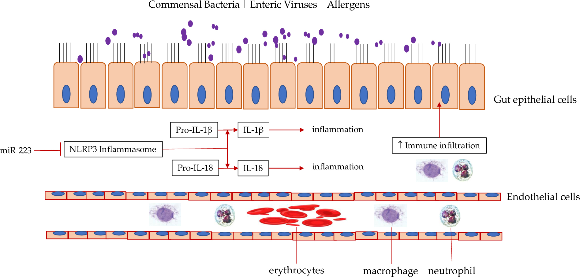
-κB pathway. Activation of NF-κB triggers increased expression of NLRP3 protein [41]. Activated NLRP3 converts inactive pro-caspase 1 into active caspase 1, which in turn cleaves pro-IL-1β and pro-IL-18 to their active forms (Fig. 1, Ref. [38]).

Fig. 1.

Effect of miR-223 on inflammasome in IBD. MiR-223 can block NLRP3 inflammasome, which is pro-inflammatory by converting pro-IL-1β into IL-1β and pro-IL-18 into IL-18, two proinflammatory cytokines. Inhibitors such as MCC950 can reduce NLRP3 inflammasome activity to decrease production of IL-1β and IL-18 [Adapted from J Exp Med 2017; 214: 1737–1752] [38]. Abbreviations: NLRP3, Nucleotide-binding oligomerization domain, Leucine rich Repeat and Pyrin domain-containing protein 3; IL-18, interleukin 18.

An uncontrolled and excessive activation of NLRP3 has been reported to be a major pathogenetic factor of many inflammatory diseases including IBD and knockout of NLRP3 improved these diseases [40, 42]. The anti-inflammatory effect of miR-223 could be explained by its inhibitory effect on NLRP3. MiR-223 reduces NLRP3 expression by binding to its mRNA 3’-UTR, leading to degradation of NLRP3 mRNAs [41]. Correspondingly, increased miR-223 reduces NLRP3 inflammasome and pro-inflammatory cytokines IL-1β and IL-18. In contrast, reduced miR-223 should increase inflammasome activity. Indeed, it has been shown that inflammasome and IL-1β production are enhanced in miR-223-/y mice treated with DSS, supporting overall protective effects of miR-223 in a DSS-induced colitis model [38]. Consequently, miR-223 is required for maintaining the inflammatory tone within an appropriate degree of response. The overall anti-inflammatory effect of miR-223 in other disease states (e.g., lung inflammation, rheumatoid arthritis) provides further evidence [32]. A recent study showed that miR-223 in neutrophils was a key factor for resolution of liver inflammation after the injury factor disappeared [43]. MiR-223 also protected neurons from inflammation-caused damage in an encephalomyelitis mouse model [44]. Furthermore, miR-223 mimics together with miR-142 delivered by microvesicles through intratracheal instillation suppressed lung inflammation triggered by the endotoxin lipopolysaccharides (LPS) [37]. The associated mechanism was shown to be inhibition of NLRP3 expression.

Inhibition of inflammasome in IBD provides a therapeutic effect. Isoflavone formononetin can inhibit the NLRP3 inflammasome to reduce intestinal inflammation in a DSS-induced colitis mouse model [24]. Rev-ErbA proteins are members of the nuclear receptor family of intracellular transcription factors. As such these proteins regulate the colon clock, and have been found to suppress NLRP3 via inhibition of NF-κB [24]. Knockout of Rev-ErbA exacerbated DSS colitis and its activation has been shown to attenuate DSS-induced colitis [45]. A small molecule selective inhibitor MCC950 has been developed to target NLRP3, leading to decreased IL-1β in experimental autoimmune encephamyolitis, CAPS and Muckle-Wells syndrome [46]. MCC950 has been shown to attenuate experimental cryopyrin-associated periodic syndrome and experimental autoimmune encephalomyelitis in rodents, the diseases with activation of NLRP3 [46]. MCC950 could be also effective in IBD as its pathogenesis is also associated with a proinflammatory response. Therefore, increased miR-223 could have a protective effect by inhibiting the NLRP3 inflammasome.

4. NF-κB Pathway

NF-κB pathway is a major mediator of inflammation [47]. Prolonged activation of NF-κB causes chronic inflammation and cancer. It has been well demonstrated to be involved in the pathogenesis of IBD [48, 49]. TLRs activated by MAMPs can increase NF-κB activation. Inflammatory factor like TNF-α can also activate NF-κB through TNF receptor. Activation of NF-κB results in increased production of proinflammatory cytokines including IL-6, IL-1β and TNF-α as well as Cox-2 and nitric oxide. In animal models including TNBS-induced colitis and IL-10-deficient mice, intravenous administration p65 anti-sense oligonucleotides reduced intestinal inflammation [50].

The effects of miR-223 on NF
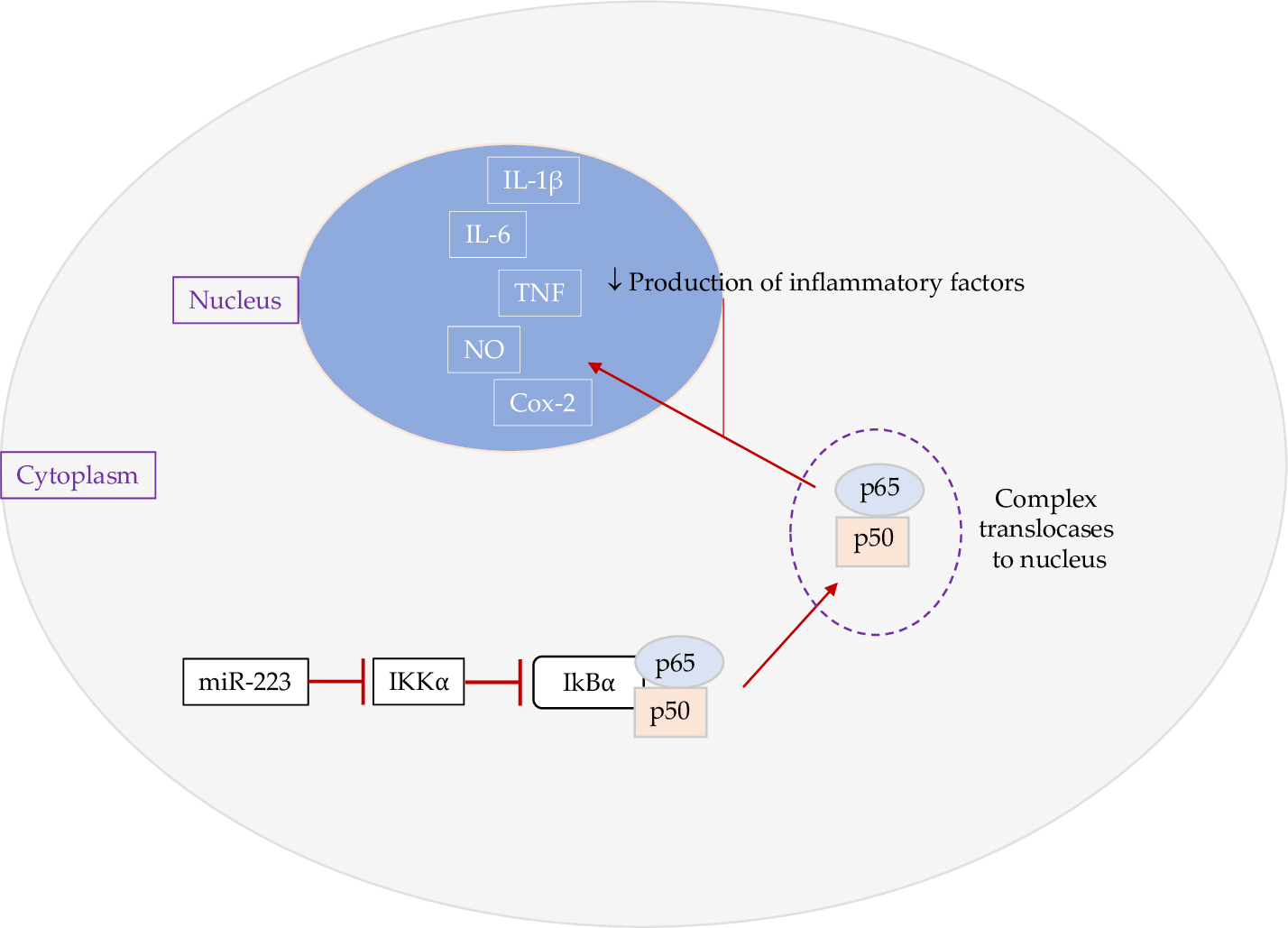
-κB are multiple. NF-κB is composed of p65 and p50 subunits which are suppressed by the inhibitory subunit IκBα (NF-κB polypeptide gene enhancer in B cells inhibitor alpha) (Fig. 2). IKKα (IκB kinase alpha), a negative regulator of IκBα, has been identified as a target of miR-223 [51]. By comparing 30 UC patients and 20 non-IBD controls, Valmiki et al. [50] miR-223 levels were 8.63 folds of that of controls while IKKα was 40% less in UC patients than that in controls. Transfection of miR-223 into LPS-stimulated HT-29 cells resulted in 4.16-fold decrease in IKKα mRNA levels. The decreased effect of IKKα on IκBα should lead to activation of NF-κB. In another study, NF-κB activators Traf6/Tab1 (TNF receptor-associated factor 6/TGF-beta activated kinase 1) and Cullin 1a/b were identified as targets of miR-223 [52]. Transfection of miR-223 into HUVECs blocked nuclear translocation of NF-κB as well as inhibition of p38 MAPK, JNK and ERK phosphorylation [53]. These studies suggest that miR-223 could reduce proinflammatory status in IBD through inhibiting NF-κB pathway.

Fig. 2.

Effect of miR-223 on NF-κB pathway. MiR-223 targets IKKα, Traf6/Tab1 and Cul1a/b, leading to increased NF-κB suppressor IκBα and thus decreased NF-κB nuclear translocation and reduced production of proinflammatory factors. Abbreviations: NF-κB, nuclear factor kappa light chain enhancer of activated B cells; IκBα, NF-κB polypeptide gene enhancer in B cells inhibitor alpha; IKKα, IκBα; Traf6/Tab1, TNF-α receptor-associated factor 6/TGF-beta activated kinase 1; Cul1a/b, cullin 1a/b.

5. IL-6/STAT3 Pathway

IL-6/STAT3 (signal transducer and activator of transcription 3) pathway has been demonstrated to play key roles in the pathogenesis of IBD [54]. It increases macrophage and T cell survivial, proliferation and differentiation and thus increases the production of pro-inflammatory cytokines. Therefore, the IL-6/STAT3 pathway has been targeted for the treatment of IBD. So far, Torfacitinib, an inhibitor of IL-6 downstream signalling molecule JAK, has been approved by the FDA for the treatment of IBD [55, 56].

MiR-223 has been shown to inhibit the IL-6/STAT3 pathway in a mouse colitis model induced by DSS [37]. MiR-223 agomir decreased MPO, TNF-α, IL-6 and IL-17 levels while increased IL-10 levels. In contrast, antagomir increased these proinflammatory cytokines and decreased IL-10. Agomir decreased gp130, pSTAT3, Bcl-2 and Bcl-xl while antagomir increased them. A study has demonstrated that miR-223 can inhibit IL-6/STAT3 pathway in cell culture of vascular smooth muscle cells; miR-223 targeted 3’-UTR of STAT3 mRNA rather than IL-6 mRNA [57]. The inhibitory effects of miR-223 on mRNAs of the components of IL-6/STAT3 pathway could be examined in T cells, macrophages and other cells that are more relavant to colitis.

6. The Phosphatidylinositol 3-kinase (PI3K)/Protein Kinase B (Akt) (PI3K/Akt) Signaling Pathway

The PI3K/Akt pathway has been extensively studied and reported to participate in the regulation of multiple downstream cellular processes [58]. The downstream activation signaling of corresponding effector molecules serve important fuctional activities in the cell cycle, in cellular growth and proliferation. Increased activation of the PI3K/Akt pathway is proinflammatory and oncogenic. It down-regulates anti-inflammatory cytokines such as IL-10 and upregulates pro-inflammatory cytokines such as IL-12, IL-6 and IL-17. Moreover we note that the effect on IL-10 has been shown to exert immunosuppressive functions to reduce tissue damage caused by excess and uncontrolled inflammatory effector responses, especially in microbial host defences [59]; an important effect during the resolution phase of infection/inflammation that is directed at maintaining homeostasis to intestinal bacteria. Several studies showed that the activation of the pathway in IBD and the target therapy against the pathway has been tested in animal models [60, 61, 62].

In TNBS-induced colitis, proinflammatory cytokines IL-6, TNF-α and IL-1β are increased while anti-inflammatory cytokines such as IL-10 is decreased [61]. These changes are PI3K/Akt pathway dependent. Inhibition of the activity of the PI3K/Akt pathway decreased the proinflammatory cytokines and increased IL-10 production. Further activation of the PI3K/Akt pathway by IGF-1 increases proinflammatory cytokines and decreased IL-10. Inhibition of the PI3K/Akt also increases ZO-1 and tight junction function [61].

MiR-223 has been shown to be increased in DSS-caused inflammation and AOM/DSS-induced colorectal cancer [63]. The miR-223 levels in colon cancer cells were inversely correlated with the activity of PI3K/Akt. The target mRNA was identified as IGFR (insulin-like growth factor 1 receptor), an upsteam signalling molecule of the PI3K/Akt pathway. As the activation of the PI3K/Akt pathway plays a key role in promoting colitis-associated colorectal cancer [64, 65], inhibition of the pathway by increased miR-223 could have protective effect. Importantly, this study compared the miR-223 levels in myeloids and epithelial cells, which provides initial clue for the cell-context effect of miR-223.

7. FOXO

FOXO proteins belong to the Forkhead family of transcription factors, which regulate many physiological processes. MiR-223 has been shown to inhibit FOXO in colonocytes, leading to increased activity of NF
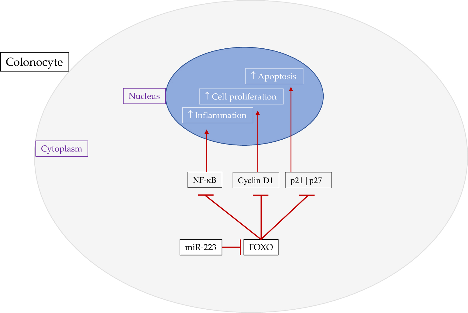
-κB (Fig. 3) [66]. Pro-inflammatory cytokines secreted by colonocytes are also increased. Therefore, miR-223 has been considered to be pro-inflammatory and proposed for the therapeutic targeting in IBD. Knockout of FOXO3 is causal for chronic inflammation and increased severity of azoxymethane-induced colon cancer [67]. Transcriptomic analysis has shown that FOXO3-defiecient animals have increased inflammatory microenviroments such as increased infiltration of macrophages and neutrophils. These results provide evidence that inhibition of FOXO3 activity by miR-223 could be involved in inflammation in IBD.

Fig. 3.

Effect of miR-223 on FOXO. MiR-223 targets FOXO in colonocytes, resulting in increased NF-κB activation and inflammation. Blockade of FOXO can increase cell proliferation through increased cyclin D1 and decreased apoptosis through decreased p21, p27 proteins.

Knockout of FOXO4 aggravated TNBS-induced colitis with increased CCL5, TNF-α, IFN-γ and CD4+ T cell infiltration [68]. Intestinal permeability was increased with ZO-1 and claudin-1 decreased. However, there may be benficial effects of FOXO3 inhibition on colonocytes which has not been investigated in IBD. FOXO3 inhibition could incease colonocyte proliferation and decrease colonocyte apoptosis [69]. It has been well documented that FOXO3 can inhibit cell proliferation through downregulation of cyclin D [70]. FOXO3 also enhances p27kip to cause cell cycle arrest [71] Inhibition of FOXO3 may be helpful for reducing inflammation-caused damage on colonocytes in IBD.

8. CLDN8

Elevated miR-223 can decrease intestinal barrier integrity through inhibition of claudin8 (CLDN8), a tight junction backbone protein, in TNBS-induced colitis and thus increase translocation of LPS from the lumen to the intestinal mucosa, leading to LPS-induced systemic inflammation [39]. MiR-223 regulates CLDN by targeting its mRNA 3’-UTR to decrease CLDN8 protein expression [39]. Wang et al. [39] revealed that inhibition of miR-223 increased CLDN8 and thus facilitated tight junction formation and increased intestinal barrier integrity. In Caco-2 cells, siRNA silencing of CLDN8 decreased trans-epithelial electrical resistance while over-expression of the protein increased tight junction integrity and function [72]. A recent study confirmed that mast cell-derived miR-223 exosomes reduced the expression of tight junction proteins CLDN8, ZO-1 and occludin in colonic epithelial cells [73]. The miR-223 containing exosomes increased the permeability in monolayers of Caco2 cell cultures. In an animal model of TNBS-induced colitis, antagomir of miR-223 reduced gut permeability as well as gut inflammation, indicating a proinflammatory effect of miR-223 in this colitis model [35].

9. Gut Microbiota and miR-223

The intestinal microbiota is disturbed in IBD and modulation of the gut microbiota can facilitate IBD recovery. The commensal bacterium Roseburia intestinalis flagellin has been shown to ameliorate colitis and miR-223 could be a mediator [74]. The bacterium increased miR-223 levels and inhibited NLRP3 inflammasome. Another butyrate-producing bacterium F. prausnitizii has also been shown to have protective effects in DSS-induced colitis with improved weight loss, diarrhea, bloody stools and colon shortening as well as decreased mucosal proinflammatory cytokines [75]. However, it is not known if the mechanism in mucosal immunity and IBD pathogenesis, involves an increased expression of miR-223, and as such this remains largely unexplored. A recent study though has investigated fecal micro-RNAs in Crohn’s disease IBD [76]. Specifically this small cohort with patients diagnosed with Crohn’s disease (CD) has identified hundreds of different human miRNAs from faecal samples. Of these a total of 150 fecal miRNAs from controls and patients were significantly detected [76]. In a multivariate analyses the study showed that those patients diagnosed with high CD activity had a sharp and distinct miRNA profile. Consequently, miR-223 and miR-1246 were specific from other isolated fecal miRNAs. Moreover, in those patients with active ulcerative colitic (UC) the study reported that significantly higher levels of miR-223 and miR-1246 over that in controls. Furthermore, in those patients diagnosed with Clostridium difficile infections higher levels of fecal miR-1246 over miR-223 were reported.

What has recently been reported is that myeloid derived miR-223 regulates intestinal inflammation via an inhibitory action on the inflammasome NLRP3 [38]. The endogenous noncoding miRNAs exert regulatory inflammatory functions in the intestines [38]. Neudecker et al. [38] reported that miR-223 limits intestinal inflammation by constraining the NLRP3 inflammasome.

It has been shown that multi-strain probiotics through the administration of Lactobacillus plantarum, L. paracasei, L. rhamnosus, L. acidophilus, L. reuteri and Bifidobacterium animalis, B. lactis, B. bifidum can increase reinforce intestinal barrier integrity and functionality [77, 78, 79, 80]. This has been accompanied by decreased miR-223 levels by these probiotic species. Probiotic bacteria can also improve infllamtion which can lead to decreased miR-223 which reduces miR-223 upregulation as a protective response to inflammation. It is also possible that probiotics may also reduce inflammation via other mechanisms such as the anti-inflammatory effects of butyrate [23]. This effect linked with cross-feeding interactions by probiotic acetate producers from the Bifidobacteria genus with intestinal commensal butyrate producers such as F. prausnitzii [81]. Interestingly a recent review has reported how aberrant signaling in the PI3K/Akt pathway has been associated with a wide variety of human diseases [82]. In contrast though the authors confirmed and highlighted that a large body of experimental in vitro and in vivo evidence suggests that the beneficial contribution of probiotics to modulate the PI3K/Akt signaling pathway in gastrointestinal and metabolic diseases, skin diseases, allergy, salmonella infections, and the aging process [82]. Hence, a causal relationship emerges between the benefical effects of probiotics and their metabolites on the components of the PI3K/Akt signaling pathway and human disease.

10. Plausible Posits and Contentious Research Investigations

The effects of miR-223 in DSS colitis models and the TNBS-induced colitis model have been contentious, although miRNA-223 has been well demonstrated to be significantly increased in both types of animal models of colitis. This may indicate that miR-223 may exert effects that are dependent on the severity of colitis. TNBS-induced colitis is a more robust form of inflammation than DSS-induced colitis [83]. TNBS causes colitis through T-cell autoimmune responses and DSS by direct toxicity to colonic epithelial cells [84]. TNBS haptenize colonic autologous or microbiota proteins to elicit immune responses, leading to infiltration of CD4+ T cells and macrophages with increased production of proinflammatory cytokines [85, 86]. The effects of miR-223 could be cell type specific; with effects on immune cells that could be different to epithelial cells, a notion that may account for the different effects observed between DSS- and TNBS-induced colitis.

In immune cells, the anti-inflammatory effects of miR-223 is supported by targeting several key signalling proteins in proinflammatory signalling pathways. The anti-inflammatory effect of miR-223 on proinflammatory cells namely, neutrophils and macrophages have been well demonstrated [30]. MiR-223 decreases both neutrophil and macrophage activation through targeting NLRP3 [87]. Knockout of miR-223 presents with increased neutrophil infiltration to the lung with highly activated status indicated by increased CXCL2, CCL3 and IL-6 [88]. In LPS- stimulated macrophages, miR-223 is decreased, leading to activation of the STAT3 signalling pathway, causing increased IL-6 and IL-1β, which further down-regulates miR-223 [89]. MiR-223 also regulates the polarization of macrophages. IL-4 activates PPAR-γ, which upregulates miR-223 and miR-223 also directly targeting the M1 positive regulators Rasa1 and Nfat5 [90] In miR-223-/y mice, TLR4-activated M1 was increased; IL-4 activated M2 decreased. In contrast to the inhibitory effects of miR-223 on innate immune cells, highly increased levels of miR-223 in CD4+ T cells did not decrease CD4+ T cell number and activation in auto-immune responses [91]. The inhibitory effects of miR-223 on the innate immune cells may reduce inflammation caused by DSS efficiently but not auto-immune responses caused by TNBS-damaged proteins.

The detrimental effect of miR-223 on TNBS-induced colitis has been based on CLDN8 and FOXO alterations. MiR-223 can directly target CLDN8 mRNAs to reduce CLDN8 protein transcription levels, leading to increased intestinal permeability [39]. This causes the translocation of exdotoxin and bacteria, aggravating inflammation. MiR-223 has also been shown to target FOXO, leading to increased production of proinflammatory cytokines. We posit that miR-223 may be unable to reduce activation of CD4+ T cells from autoimmune responses to TNBS-damaged proteins and thus the effects of miR-223 on CLDN8 and FOXO may become dominant.

11. Conclusions

In reprise, miR-223 has opposite effects on the inflammatory profiles in the DSS- and TNBS- induced colitis models. In DSS-induced colitis, over expression of miR-223 exerts an anti-inflammatory effect. This could be possibly explained by its inhibitory effects on innate immune cells such as macrophages and neutrophils through inhibition of the NLRP3 inflammasome activity and proinflammatory signalling pathways. In TNBS-induced colitis, miR-223 is pro-inflammatory by targeting FOXO and CLDN8 in colonocytes. Further studies are warranted to elucidate the mechanisms of the effects of miR-223 in IBD, particularly in individual cells to explain its roles in different types of IBD. The accumulation of such knowledge could facilitate the clinical use of miR-223 modolators in personalized treatment of IBD.

Abbreviations

NF-κB, nuclear factor kappa light chain enhancer of activated B cells; FOXO, forkhead box O.

Author Contributions

JC—searching literature, creating conceptions together, preparing first draft. LV—supervsing, building co-conceptualization, critical revision.

Ethics Approval and Consent to Participate

Not applicable.

Acknowledgment

Not applicable.

Funding

This research received no external funding.

Conflict of Interest

The authors declare no conflict of interest. JZC is serving as one of the Guest editors of this journal. We declare that JZC had no involvement in the peer review of this article and has no access to information regarding its peer review. Full responsibility for the editorial process for this article was delegated to BHZ.

References
[1]
Ferretti F, Cannatelli R, Monico MC, Maconi G, Ardizzone S. An Update on Current Pharmacotherapeutic Options for the Treatment of Ulcerative Colitis. Journal of Clinical Medicine. 2022; 11: 2302.
[2]
Feuerstein JD, Cheifetz AS. Crohn Disease: Epidemiology, Diagnosis, and Management. Mayo Clinic Proceedings. 2017; 92: 1088–1103.
[3]
Gu P, Feagins LA. Diet in the Pathogenesis and Management of Crohn’s Disease. Gastroenterology Clinics of North America. 2022; 51: 319–335.
[4]
Zuo T, Ng SC. The Gut Microbiota in the Pathogenesis and Therapeutics of Inflammatory Bowel Disease. Frontiers in Microbiology. 2018; 9: 2247.
[5]
Rogler G, Biedermann L, Scharl M. Anti-Cytokine Strategies beyond Anti-Tumour Necrosis Factor-α Therapy: Pathophysiology and Clinical Implications. Digestive Diseases. 2017; 35: 5–12.
[6]
Clarke WT, Feuerstein JD. Colorectal cancer surveillance in inflammatory bowel disease: Practice guidelines and recent developments. World Journal of Gastroenterology. 2019; 25: 4148–4157.
[7]
Pesce M, Seguella L, Del Re A, Lu J, Palenca I, Corpetti C, et al. Next-Generation Probiotics for Inflammatory Bowel Disease. International Journal of Molecular Sciences. 2022; 23: 5466.
[8]
Agraib LM, Yamani MI, Rayyan YM, Abu-Sneineh AT, Tamimi TA, Tayyem RF. The probiotic supplementation role in improving the immune system among people with ulcerative colitis: a narrative review. Drug Metabolism and Personalized Therapy. 2021; 37: 7–19.
[9]
Sewell GW, Kaser A. Interleukin-23 in the Pathogenesis of Inflammatory Bowel Disease and Implications for Therapeutic Intervention. Journal of Crohn’s and Colitis. 2022; 16: ii3–ii19.
[10]
Parigi TL, Iacucci M, Ghosh S. Blockade of IL-23: what is in the Pipeline? Journal of Crohn’s and Colitis. 2022; 16: ii64–ii72.
[11]
Liu E, Aslam N, Nigam G, Limdi JK. Tofacitinib and newer JAK inhibitors in inflammatory bowel disease-where we are and where we are going. Drugs Context. 2022; 11: 2021-11-4.
[12]
Lee M, Chang EB. Inflammatory Bowel Diseases (IBD) and the Microbiome—Searching the Crime Scene for Clues. Gastroenterology. 2021; 160: 524–537.
[13]
Maloy KJ, Powrie F. Intestinal homeostasis and its breakdown in inflammatory bowel disease. Nature. 2011; 474: 298–306.
[14]
Sokol H, Seksik P, Furet JP, Firmesse O, Nion-Larmurier I, Beaugerie L, et al. Low counts of Faecalibacterium prausnitzii in colitis microbiota. Inflammatory Bowel Diseases. 2009; 15: 1183–1189.
[15]
Machiels K, Joossens M, Sabino J, De Preter V, Arijs I, Eeckhaut V, et al. A decrease of the butyrate-producing species Roseburia hominis and Faecalibacterium prausnitzii defines dysbiosis in patients with ulcerative colitis. Gut. 2014; 63: 1275–1283.
[16]
Frank DN, St. Amand AL, Feldman RA, Boedeker EC, Harpaz N, Pace NR. Molecular-phylogenetic characterization of microbial community imbalances in human inflammatory bowel diseases. Proceedings of the National Academy of Sciences. 2007; 104: 13780–13785.
[17]
Martini E, Krug SM, Siegmund B, Neurath MF, Becker C. Mend your Fences: The Epithelial Barrier and its Relationship With Mucosal Immunity in Inflammatory Bowel Disease. Cellular and Molecular Gastroenterology and Hepatology. 2017; 4: 33–46.
[18]
Ghosh S, Whitley CS, Haribabu B, Jala VR. Regulation of Intestinal Barrier Function by Microbial Metabolites. Cellular and Molecular Gastroenterology and Hepatology. 2021; 11: 1463–1482.
[19]
Zhu C, Song K, Shen Z, Quan Y, Tan B, Luo W, et al. Roseburia intestinalis inhibits interleukin‑17 excretion and promotes regulatory T cells differentiation in colitis. Molecular Medicine Reports. 2018; 17: 7567–7574.
[20]
Knights D, Silverberg MS, Weersma RK, Gevers D, Dijkstra G, Huang H, et al. Complex host genetics influence the microbiome in inflammatory bowel disease. Genome Medicine. 2014; 6: 107.
[21]
Mondot S, Barreau F, Al Nabhani Z, Dussaillant M, Le Roux K, Doré J, et al. Altered gut microbiota composition in immune-impaired Nod2(-/-) mice. Gut. 2012; 61: 634–635.
[22]
Imhann F, Vich Vila A, Bonder MJ, Fu J, Gevers D, Visschedijk MC, et al. Interplay of host genetics and gut microbiota underlying the onset and clinical presentation of inflammatory bowel disease. Gut. 2018; 67: 108–119.
[23]
Chen J, Vitetta L. Butyrate in Inflammatory Bowel Disease Therapy. Gastroenterology. 2020; 158: 1511.
[24]
Wu F, Zhang S, Dassopoulos T, Harris ML, Bayless TM, Meltzer SJ, et al. Identification of microRNAs associated with ileal and colonic Crohnʼs disease. Inflammatory Bowel Diseases. 2010; 16: 1729–1738.
[25]
Wani S, Man Law IK, Pothoulakis C. Role and mechanisms of exosomal miRNAs in IBD pathophysiology. American Journal of Physiology-Gastrointestinal and Liver Physiology. 2020; 319: G646–G654.
[26]
Moein S, Vaghari-Tabari M, Qujeq D, Majidinia M, Nabavi SM, Yousefi B. MiRNAs and inflammatory bowel disease: an interesting new story. Journal of Cellular Physiology. 2019; 234: 3277–3293.
[27]
Liu Z, Zhang Y, Jin T, yi C, Ocansey DKW, Mao F. The role of NOD2 in intestinal immune response and microbiota modulation: A therapeutic target in inflammatory bowel disease. International Immunopharmacology. 2022; 113(Pt B): 109466.
[28]
Wang S, Huang Y, Zhou C, Wu H, Zhao J, Wu L, et al. The Role of Autophagy and Related MicroRNAs in Inflammatory Bowel Disease. Gastroenterology Research and Practice. 2018; 2018: 1–10.
[29]
Nath P, Jena KK, Mehto S, Chauhan NR, Sahu R, Dhar K, et al. IRGM links autoimmunity to autophagy. Autophagy. 2021; 17: 578–580.
[30]
Yuan X, Berg N, Lee JW, Le T, Neudecker V, Jing N, et al. MicroRNA miR-223 as regulator of innate immunity. Journal of Leukocyte Biology. 2018; 104: 515–524.
[31]
Bader El Din NG, Farouk S, Abdel-Salam LO, Khairy A. The potential value of miRNA-223 as a diagnostic biomarker for Egyptian colorectal patients. European Journal of Gastroenterology & Hepatology. 2021; 33: 25–31.
[32]
Jiao P, Wang XP, Luoreng ZM, Yang J, Jia L, Ma Y, et al. miR-223: An Effective Regulator of Immune Cell Differentiation and Inflammation. International Journal of Biological Sciences. 2021; 17: 2308–2322.
[33]
Fasseu M, Tréton X, Guichard C, Pedruzzi E, Cazals-Hatem D, Richard C, et al. Identification of restricted subsets of mature microRNA abnormally expressed in inactive colonic mucosa of patients with inflammatory bowel disease. PLoS ONE. 2010; 5: e13160.
[34]
Schönauen K, Le N, von Arnim U, Schulz C, Malfertheiner P, Link A. Circulating and Fecal microRNAs as Biomarkers for Inflammatory Bowel Diseases. Inflammatory Bowel Diseases. 2018; 24: 1547–1557.
[35]
Wang H, Zhang S, Yu Q, Yang G, Guo J, Li M, et al. Circulating MicroRNA223 is a New Biomarker for Inflammatory Bowel Disease. Medicine. 2016; 95: e2703.
[36]
Schaefer JS, Montufar-Solis D, Vigneswaran N, Klein JR. Selective upregulation of microRNA expression in peripheral blood leukocytes in IL-10-/- mice precedes expression in the colon. Journal of Immunology. 2011; 187: 5834–5841.
[37]
Zhang J, Wang C, Guo Z, Da B, Zhu W, Li Q. MiR-223 improves intestinal inflammation through inhibiting the IL-6/STAT3 signaling pathway in dextran sodium sulfate-induced experimental colitis. Immunity, Inflammation and Disease. 2021; 9: 319–327.
[38]
Neudecker V, Haneklaus M, Jensen O, Khailova L, Masterson JC, Tye H, et al. Myeloid-derived miR-223 regulates intestinal inflammation via repression of the NLRP3 inflammasome. Journal of Experimental Medicine. 2017; 214: 1737–1752.
[39]
Wang H, Chao K, Ng SC, Bai AH, Yu Q, Yu J, et al. Pro-inflammatory miR-223 mediates the cross-talk between the IL23 pathway and the intestinal barrier in inflammatory bowel disease. Genome Biology. 2016; 17: 58.
[40]
Khatri V, Kalyanasundaram R. Therapeutic implications of inflammasome in inflammatory bowel disease. The FASEB Journal. 2021; 35: e21439.
[41]
Haneklaus M, Gerlic M, Kurowska-Stolarska M, Rainey A, Pich D, McInnes IB, et al. Cutting Edge: miR-223 and EBV miR-BART15 Regulate the NLRP3 Inflammasome and IL-1β Production. the Journal of Immunology. 2012; 189: 3795–3799.
[42]
Mangan MSJ, Olhava EJ, Roush WR, Seidel HM, Glick GD, Latz E. Targeting the NLRP3 inflammasome in inflammatory diseases. Nature Reviews Drug Discovery. 2018; 17: 588–606.
[43]
Calvente CJ, Tameda M, Johnson CD, del Pilar H, Lin YC, Adronikou N, et al. Neutrophils contribute to spontaneous resolution of liver inflammation and fibrosis via microRNA-223. Journal of Clinical Investigation. 2019; 129: 4091–4109.
[44]
Morquette B, Juźwik CA, Drake SS, Charabati M, Zhang Y, Lécuyer M, et al. MicroRNA-223 protects neurons from degeneration in experimental autoimmune encephalomyelitis. Brain. 2019; 142: 2979–2995.
[45]
Wang S, Lin Y, Yuan X, Li F, Guo L, Wu B. REV-ERBα integrates colon clock with experimental colitis through regulation of NF-κB/NLRP3 axis. Nature Communications. 2018; 9: 4246.
[46]
Coll RC, Robertson AAB, Chae JJ, Higgins SC, Muñoz-Planillo R, Inserra MC, et al. A small-molecule inhibitor of the NLRP3 inflammasome for the treatment of inflammatory diseases. Nature Medicine. 2015; 21: 248–255.
[47]
Karin M, Greten FR. NF-kappaB: linking inflammation and immunity to cancer development and progression. Nature Reviews. Immunology. 2005; 5: 749–759.
[48]
Tambuwala MM. Natural Nuclear Factor Kappa Beta Inhibitors: Safe Therapeutic Options for Inflammatory Bowel Disease. Inflammatory Bowel Diseases. 2016; 22: 719–723.
[49]
Van Welden S, Selfridge AC, Hindryckx P. Intestinal hypoxia and hypoxia-induced signalling as therapeutic targets for IBD. Nature Reviews Gastroenterology & Hepatology. 2017; 14: 596–611.
[50]
Neurath MF, Pettersson S, Meyer zum Büschenfelde KH, Strober W. Local administration of antisense phosphorothioate oligonucleotides to the p65 subunit of NF-kappa B abrogates established experimental colitis in mice. Nature Medicine. 1996; 2: 998–1004.
[51]
Valmiki S, Ahuja V, Puri N, Paul J. miR-125b and miR-223 Contribute to Inflammation by Targeting the Key Molecules of NFκB Pathway. Frontiers in Medicine. 2020; 6: 313.
[52]
Zhou W, Pal AS, Hsu AY, Gurol T, Zhu X, Wirbisky-Hershberger SE, et al. MicroRNA-223 Suppresses the Canonical NF-κB Pathway in Basal Keratinocytes to Dampen Neutrophilic Inflammation. Cell Reports. 2018; 22: 1810–1823.
[53]
Li J, Tan M, Xiang Q, Zhou Z, Yan H. Thrombin-activated platelet-derived exosomes regulate endothelial cell expression of ICAM-1 via microRNA-223 during the thrombosis-inflammation response. Thrombosis Research. 2017; 154: 96–105.
[54]
Carey R, Jurickova I, Ballard E, Bonkowski E, Han X, Xu H, et al. Activation of an IL-6:STAT3-dependent transcriptome in pediatric-onset inflammatory bowel disease. Inflammatory Bowel Diseases. 2008; 14: 446–457.
[55]
Salas A, Hernandez-Rocha C, Duijvestein M, Faubion W, McGovern D, Vermeire S, et al. JAK–STAT pathway targeting for the treatment of inflammatory bowel disease. Nature Reviews Gastroenterology & Hepatology. 2020; 17: 323–337.
[56]
Corrigendum: The Future of Janus Kinase Inhibitors in Inflammatory Bowel Disease. Journal of Crohn’s & Colitis. 2017; 11: 1028.
[57]
Han Y, Zhang J, Huang S, Cheng N, Zhang C, Li Y, et al. MicroRNA-223-3p inhibits vascular calcification and the osteogenic switch of vascular smooth muscle cells. Journal of Biological Chemistry. 2021; 296: 100483.
[58]
Jafari M, Ghadami E, Dadkhah T, Akhavan-Niaki H. PI3k/AKT signaling pathway: Erythropoiesis and beyond. Journal of Cellular Physiology. 2019; 234: 2373–2385.
[59]
Ouyang W, Rutz S, Crellin NK, Valdez PA, Hymowitz SG. Regulation and Functions of the IL-10 Family of Cytokines in Inflammation and Disease. Annual Review of Immunology. 2011; 29: 71–109.
[60]
Tokuhira N, Kitagishi Y, Suzuki M, Minami A, Nakanishi A, Ono Y, et al. PI3K/AKT/PTEN pathway as a target for Crohn’s disease therapy (Review). International Journal of Molecular Medicine. 2015; 35: 10–16.
[61]
Zhu L, Shen H, Gu PQ, Liu YJ, Zhang L, Cheng JF. Baicalin alleviates TNBS-induced colitis by inhibiting PI3K/AKT pathway activation. Experimental and Therapeutic Medicine. 2020; 20: 581–590.
[62]
Chen Q, Duan X, Fan H, Xu M, Tang Q, Zhang L, et al. Oxymatrine protects against DSS-induced colitis via inhibiting the PI3K/AKT signaling pathway. International Immunopharmacology. 2017; 53: 149–157.
[63]
Josse C, Bouznad N, Geurts P, Irrthum A, Huynh-Thu VA, Servais L, et al. Identification of a microRNA landscape targeting the PI3K/Akt signaling pathway in inflammation-induced colorectal carcinogenesis. American Journal of Physiology-Gastrointestinal and Liver Physiology. 2014; 306: G229–G243.
[64]
Setia S, Nehru B, Sanyal SN. Upregulation of MAPK/Erk and PI3K/Akt pathways in ulcerative colitis-associated colon cancer. Biomedicine & Pharmacotherapy. 2014; 68: 1023–1029.
[65]
Khan MW, Keshavarzian A, Gounaris E, Melson JE, Cheon EC, Blatner NR, et al. PI3K/AKT Signaling is Essential for Communication between Tissue-Infiltrating Mast Cells, Macrophages, and Epithelial Cells in Colitis-Induced Cancer. Clinical Cancer Research. 2013; 19: 2342–2354.
[66]
Suri K, Bubier JA, Wiles MV, Shultz LD, Amiji MM, Hosur V. Role of MicroRNA in Inflammatory Bowel Disease: Clinical Evidence and the Development of Preclinical Animal Models. Cells. 2021; 10: 2204.
[67]
Penrose HM, Cable C, Heller S, Ungerleider N, Nakhoul H, Baddoo M, et al. Loss of Forkhead Box O3 Facilitates Inflammatory Colon Cancer: Transcriptome Profiling of the Immune Landscape and Novel Targets. Cellular and Molecular Gastroenterology and Hepatology. 2019; 7: 391–408.
[68]
Zhou W, Cao Q, Peng Y, Zhang QJ, Castrillon DH, DePinho RA, et al. FoxO4 inhibits NF-kappaB and protects mice against colonic injury and inflammation. Gastroenterology. 2009; 137: 1403–1414.
[69]
Qi W, Fitchev PS, Cornwell ML, Greenberg J, Cabe M, Weber CR, et al. FOXO3 Growth Inhibition of Colonic Cells is Dependent on Intraepithelial Lipid Droplet Density. Journal of Biological Chemistry. 2013; 288: 16274–16281.
[70]
Schmidt M, Fernandez de Mattos S, van der Horst A, Klompmaker R, Kops GJPL, Lam EW, et al. Cell Cycle Inhibition by FoxO Forkhead Transcription Factors Involves Downregulation of Cyclin D. Molecular and Cellular Biology. 2002; 22: 7842–7852.
[71]
Medema RH, Kops GJPL, Bos JL, Burgering BMT. AFX-like Forkhead transcription factors mediate cell-cycle regulation by Ras and PKB through p27kip1. Nature. 2000; 404: 782–787.
[72]
Günzel D, Yu ASL. Claudins and the Modulation of Tight Junction Permeability. Physiological Reviews. 2013; 93: 525–569.
[73]
Li M, Zhao J, Cao M, Liu R, Chen G, Li S, et al. Mast cells-derived MiR-223 destroys intestinal barrier function by inhibition of CLDN8 expression in intestinal epithelial cells. Biological Research. 2020; 53: 12.
[74]
Wu X, Pan S, Luo W, Shen Z, Meng X, Xiao M, et al. Roseburia intestinalis‑derived flagellin ameliorates colitis by targeting miR‑223‑3p‑mediated activation of NLRP3 inflammasome and pyroptosis. Molecular Medicine Reports. 2020; 22: 2695–2704.
[75]
Kawade Y, Sakai M, Okamori M, Morita M, Mizushima K, Ueda T, et al. Administration of live, but not inactivated, Faecalibacterium prausnitzii has a preventive effect on dextran sodium sulfate‑induced colitis in mice. Molecular Medicine Reports. 2019; 20: 25–32.
[76]
Verdier J, Breunig IR, Ohse MC, Roubrocks S, Kleinfeld S, Roy S, et al. Faecal Micro-RNAs in Inflammatory Bowel Diseases. Journal of Crohn’s and Colitis. 2020; 14: 110–117.
[77]
Wang J, Ji H, Wang S, Liu H, Zhang W, Zhang D, et al. Probiotic Lactobacillus plantarum Promotes Intestinal Barrier Function by Strengthening the Epithelium and Modulating Gut Microbiota. Frontiers in Microbiology. 2018; 9: 1953.
[78]
Trindade LM, Martins VD, Rodrigues NM, Souza ELS, Martins FS, Costa GMF, et al. Oral administration of Simbioflora® (synbiotic) attenuates intestinal damage in a mouse model of 5-fluorouracil-induced mucositis. Beneficial Microbes. 2018; 9: 477–486.
[79]
Mokkala K, Pussinen P, Houttu N, Koivuniemi E, Vahlberg T, Laitinen K. The impact of probiotics and n-3 long-chain polyunsaturated fatty acids on intestinal permeability in pregnancy: a randomised clinical trial. Beneficial Microbes. 2018; 9: 199–208.
[80]
Din AU, Mazhar M, Waseem M, Ahmad W, Bibi A, Hassan A, et al. SARS-CoV-2 microbiome dysbiosis linked disorders and possible probiotics role. Biomedicine & Pharmacotherapy. 2021; 133: 110947.
[81]
Rivière A, Selak M, Lantin D, Leroy F, De Vuyst L. Bifidobacteria and Butyrate-Producing Colon Bacteria: Importance and Strategies for Their Stimulation in the Human Gut. Frontiers in Microbiology. 2016; 7: 979.
[82]
Mohseni AH, Casolaro V, Bermúdez-Humarán LG, Keyvani H, Taghinezhad-S S. Modulation of the PI3K/Akt/mTOR signaling pathway by probiotics as a fruitful target for orchestrating the immune response. Gut Microbes. 2021; 13: 1–17.
[83]
Oh SY, Cho K, Kang JL, Kim KH, Woo S. Comparison of experimental mouse models of inflammatory bowel disease. International Journal of Molecular Medicine. 2014; 33: 333–340.
[84]
Wirtz S, Neufert C, Weigmann B, Neurath MF. Chemically induced mouse models of intestinal inflammation. Nature Protocols. 2007; 2: 541–546.
[85]
Wirtz S, Popp V, Kindermann M, Gerlach K, Weigmann B, Fichtner-Feigl S, et al. Chemically induced mouse models of acute and chronic intestinal inflammation. Nature Protocols. 2017; 12: 1295–1309.
[86]
Randhawa PK, Singh K, Singh N, Jaggi AS. A Review on Chemical-Induced Inflammatory Bowel Disease Models in Rodents. The Korean Journal of Physiology & Pharmacology. 2014; 18: 279.
[87]
Bauernfeind F, Rieger A, Schildberg FA, Knolle PA, Schmid-Burgk JL, Hornung V. NLRP3 Inflammasome Activity is Negatively Controlled by miR-223. The Journal of Immunology. 2012; 189: 4175–4181.
[88]
Dorhoi A, Iannaccone M, Farinacci M, Faé KC, Schreiber J, Moura-Alves P, et al. MicroRNA-223 controls susceptibility to tuberculosis by regulating lung neutrophil recruitment. Journal of Clinical Investigation. 2013; 123: 4836–4848.
[89]
Chen Q, Wang H, Liu Y, Song Y, Lai L, Han Q, et al. Inducible microRNA-223 down-regulation promotes TLR-triggered IL-6 and IL-1β production in macrophages by targeting STAT3. PLoS ONE. 2012; 7: e42971.
[90]
Ying W, Tseng A, Chang RC, Morin A, Brehm T, Triff K, et al. MicroRNA-223 is a crucial mediator of PPARγ-regulated alternative macrophage activation. Journal of Clinical Investigation. 2015; 125: 4149–4159.
[91]
Fulci V, Scappucci G, Sebastiani GD, Giannitti C, Franceschini D, Meloni F, et al. miR-223 is overexpressed in T-lymphocytes of patients affected by rheumatoid arthritis. Human Immunology. 2010; 71: 206–211.

Publisher’s Note: IMR Press stays neutral with regard to jurisdictional claims in published maps and institutional affiliations.

Share
Back to top