
 , Xuhan Yao 1,†, Xilei Liu 1, Kai Wang 2, Tianyu Shen 3,*
 , Baolong Wang 1,*
1 Department of Urology, Tianjin Medical University General Hospital, 300070 Tianjin, China
2 Department of Surgery, Nanling County Traditional Chinese Medicine Hospital, 241005 Wuhu, Anhui, China
3 School of Medicine, Nankai University, 300071 Tianjin, China
†These authors contributed equally.
Abstract
The mechanism by which kinesin-like protein family 20A (KIF20A) influences prostate cancer progression remains unclear. This study aims to investigate the functional role of KIF20A in prostate cancer and its transcriptional regulatory mechanism via activation of activating transcription factor 2 (ATF2).
Quantitative real-time PCR (qRT-PCR), western blotting, and immunohistochemistry (IHC) were used to assess KIF20A expression in prostate cancer tissues. The chi-square tests was used to analysze the association between KIF20A expression and clinical-pathological features of prostate cancer. A stable KIF20A knockdown prostate cancer cell line was established. The effects of KIF20A expression levels on prostate cancer cell proliferation and invasion were investigated through plate cloning and cell invasion assays. JASPAR was used to predict ATF2 binding sites within the KIF20A promoter region, which were validated by chromatin immunoprecipitation (ChIP) and dual-luciferase reporter assays.
KIF20A expression was significantly elevated in prostate cancer tissue compared to their adjacent non-cancerous tissue controls. Furthermore, high KIF20A expression was significantly correlated with tumor grading and staging, as well as lymph node metastasis factors in prostate cancer patients. Knockdown of KIF20A significantly inhibited the proliferation and invasion of prostate cancer cells. ATF2 bound to the promoter region of the KIF20A gene, thereby promoting KIF20A transcription.
Under the transcriptional regulation of ATF2, KIF20A expression is significantly upregulated in prostate cancer tissues, thereby promoting the progression of prostate cancer. KIF20A may serve as an independent prognostic factor influencing the prognosis of prostate cancer patients.
Keywords
- kinesin family member 20A (KIF20A)
- activating transcription factor 2 (ATF2)
- prostate cancer
- transcriptional regulation
- cell proliferation
Prostate cancer (PCa) remains a kind of most prevalent male genitourinary system malignant tumors, and the morbidity among Chinese men has increased steadily over recent decades, with an annual growth rate of approximately 7% [1, 2, 3]. Recent advances in systemic therapy have established the combination of androgen deprivation therapy (ADT), abiraterone acetate, and prednisolone as a standard for high-risk non-metastatic prostate cancer—a regimen that has also been shown to extend survival beyond seven years in patients with metastatic disease. Combining abiraterone and ADT also can obtain survival benefit. whereas the therapy strategy in combining abiraterone and enzalutamide is not recommended for patients initiating long-term ADT [4]. Despite these therapeutic advances, disease progression and treatment resistance remain major clinical challenges, underscoring the need for improved molecular stratification and prognostic assessment.
In this context, robust biomarkers and actionable molecular targets define precision medicine in prostate cancer. Compared with traditional tissue biopsy, liquid biopsy is receiving increasing attention due to its minimally invasive nature. On the other hand, its ability to capture the molecular landscape of the tumor system is equally remarkable. Except the traditional informative source of biomarkers such as circulating tumor cells and cell-free DNA, a particularly informative biomarker tumor-derived exosomes, is also drawing attention. Urinary liquid biopsy, in particular, has shown considerable promise for prostate cancer detection and monitoring. Beyond established urine-based markers, emerging serum biomarkers, including androgen receptor variants, indicators of bone metabolism and other biomarkers, are reshaping precision oncology. Within this framework, the isolation and molecular characterization of exosomes from liquid biopsy samples have demonstrated significant potential for discovering novel diagnostic and prognostic biomarkers in prostate cancer [5].
Kinesins serve as a microtubule-based motor proteins family characterized by highly conserved motor domains, many of which possess ATPase activity and mediate intracellular transport, spindle assembly, and chromosome segregation. Kinesin 20A (KIF20A) is located on chromosome 5q31.2. It is essential for proper cell division due to predominantly concentrating on the spindle midzone during mitosis [6, 7]. Taniuchi et al. [8] first reported KIF20A’s overexpression in pancreatic cancer and that its targeted suppression significantly inhibits tumor cell proliferation, highlighting its oncogenic role. Subsequent studies have confirmed aberrant KIF20A overexpression in multiple malignancies, including lung, breast, and prostate cancers [9, 10]. Moreover, silencing KIF20A has been shown to induce G2/M cell cycle arrest and enhance chemosensitivity in gastric cancer. However, the functional contribution of KIF20A to prostate cancer cell proliferation, invasion, and migration, as well as its upstream regulatory mechanisms, remains incompletely understood.
As a multifunctional transcription factor, activating transcription factor 2 (ATF2) is critically involved in modulating cellular stress responses, proliferation, apoptosis, and differentiation [11, 12, 13]. ATF2 has emerged as a critical mediator of prostate cancer progression, driving tumor proliferation, migration, and invasion through its transcriptional activity [14]. By modulating cell cycle progression or apoptotic signaling-associated genes expression, ATF2 contributes to aggressive tumor behavior. Elevated ATF2 expression in prostate cancer has been related to enhanced metastatic potential and poor prognosis, which suggests its pivotal role [15].
To elucidate the expression pattern and functional role of KIF20A in prostate cancer, we performed a systematic analysis using clinical samples and in vitro models. Its transcriptional regulation by ATF2 is also being elucidated. This study identified elevated KIF20A expression as a driver of malignant phenotypes in prostate cancer, as evidenced by its enhancement of tumor cell proliferation and invasive capacity. Mechanistically, this aberrant expression is driven by enhanced ATF2-mediated transcriptional activation. Collectively, these results identify the ATF2–KIF20A axis as a previously underappreciated regulatory pathway in prostate cancer progression and suggest that KIF20A could be an independent prognostic biomarker in prostate cancer.
Prostate tumor and adjacent tissues were collected from the Department of Urology, Tianjin Medical University General Hospital. All samples were accompanied by complete clinicopathological information. Inclusion criteria: (1) patients admitted between June 2021 and April 2023; (2) patients who underwent radical prostatectomy. Enrollment in the study commenced only after obtaining informed consent from each participant. Immunohistochemistry (IHC) analysis was performed on 80 pairs of paraffin-embedded prostate cancer and adjacent normal tissue samples. For qRT-PCR and western blot analyses, we utilized 16 pairs of freshly resected matched tissues. The study was approved by the Institutional Review Board of Tianjin Medical University General Hospital (Ethics Approval No. IRB-2021-KY-126).
The prostate cancer cell lines LNCAP, DU-145, PC-3, and C4-2, as well as the
normal prostate epithelial cell line RWPE-1, were obtained from the American Type
Culture Collection (ATCC, USA). Cells were grown under standard culture
conditions (37 °C, 5% CO2) in RPMI-1640 medium containing 10%
FBS. Perform transfection using cells in the logarithmic growth phase. To prepare
for this, cells were seeded in 6-well plates at 5
To analyze TCGA prostate cancer datasets, we performed bioinformatics investigations using the UALCAN [16] and GEPIA [17] online portals. UALC alyzed 52 normal prostate tissues and 497 tumor tissues, while GEPIA analyzed 487 tumor samples.
To predict potential transcription factor binding sites in the KIF20A promoter region, we performed an in-silico analysis using the JASPAR database [18]. This analysis was used to identify possible ATF2 binding motifs.
Total proteins were extracted from tissues or cells. Following separation by SDS-PAGE electrophoresis, proteins were transferred to PVDF membranes via electroblotting. After blocking with 5% nonfat dry milk, membranes were incubated overnight at 4 °C with the following primary antibodies provided by Proteintech: anti-KIF20A (15911-1-AP, 1:1000), anti-PCNA (10205-2-AP, 1:5000), anti-BCL-2 (12789-1-AP, 1:2000), anti-MMP2 (10373-2-AP, 1:1000), anti-Cleaved Caspase-3 (25128-1-AP, 1:1000), and anti-GAPDH (60004-1-Ig, 1:50,000). The membranes were washed with PBS (phosphate buffered saline), after which they were exposed to the relevant secondary antibodies. Visualize protein bands using chemiluminescence detection and record images with the Tanon imaging system.
Following deparaffinization and antigen retrieval, tissue sections were treated with 3% hydrogen peroxide for 15 minutes at room temperature to inactivate endogenous peroxidases. After PBS washing, primary antibodies were applied, and the sections were incubated overnight at 4 °C. Subsequently, sections were washed and incubated with secondary antibodies at 37 °C for 1 h, followed by DAB staining and hematoxylin counterstaining. The slides were dehydrated, mounted, and observed under a microscope to assess positive staining rates.
Total RNA was isolated with TRIzol reagent, followed by cDNA synthesis using a commercial reverse transcription system (Sigma-Aldrich) in accordance with the supplier’s protocol. KIF20A expression levels were measured via Quantitative Real-Time PCR (qRT-PCR), with GAPDH serving as the endogenous reference gene. The comparative 2-ΔΔCq method was employed for relative quantification. Primer sequences were as follows: KIF20A forward: 5′-TGCTGTCCGATGACGATGTC-3′; reverse: 5′-AGGTTCTTGCGTACCACAGAC-3′. GAPDH forward: 5′-GATTCCACCCATGGCAAATT-3′; reverse: 5′-TCTCGCCTCGAGAGAGAGT-3′.
Cells were plated in 96-well plates and subjected to treatment. Following this, MTT reagent was introduced at a final concentration of 0.5 mg/mL, and plates were incubated at 37 °C for 4 hours. After removing the supernatant, formazan precipitates were solubilized in DMSO. Absorbance was then quantified at 570 nm on a microplate reader.
Matrigel was diluted in serum-free medium at a 1:8 ratio and evenly applied to
the upper surface of Transwell inserts. The coated inserts were then incubated at
37 °C for 1 hour to allow gel polymerization. Seed cells at a density of
1
Cell crosslinking was performed by treating cells with 1% formaldehyde at room temperature for 10 minutes. The crosslinking reaction was then terminated by incubating the cells with 0.125 M glycine for 5 minutes. Following PBS washes, cells were disrupted in lysis buffer containing 1% SDS, and the chromatin was fragmented to sizes of 200–500 bp by sonication. The lysate was pre-cleared using Protein A/G magnetic beads for one hour at 4 °C. Subsequently, it was incubated overnight at 4 °C with either an anti-ATF2 primary antibody (Cell Signaling Technology, #35031, diluted 1:100) or a corresponding control IgG. Immunoprecipitation was performed by incubating samples with Protein A/G beads for 2 hours at 4 °C. Following this, the recovered complexes were treated with proteinase K and incubated at 65 °C for 4 hours to reverse formaldehyde cross-linking. Following purification, the immunoprecipitated DNA was subjected to RT-PCR analysis to determine the enrichment levels of specific promoter sequences.
C4-2 and PC-3 cells were seeded into 24-well plates and transfected with Firefly luciferase reporter plasmids containing the putative promoter elements upstream of the luciferase coding sequence. The co-transfection also included Renilla luciferase reporter plasmids under a constitutive promoter for internal normalization. All transfections were carried out with Lipofectamine 2000 (Thermo Fisher Scientific). Both plasmids were obtained from commercial sources (Promega, Madison, WI, USA) or Addgene. A 24-hour post-transfection incubation was followed by cell lysis using a manufacturer-prepared Passive Lysis Buffer (PLB). Using the dual luciferase reporter assay kit (Promega), the activities of the two enzymes were sequentially measured. The relative transcriptional activity was calculated as the ratio of Firefly luminescence to Renilla luminescence.
Statistical analysis was performed using SPSS 22.0 (IBM Corp., Chicago, IL,
USA). All data, obtained from three independent replicates, are expressed as mean
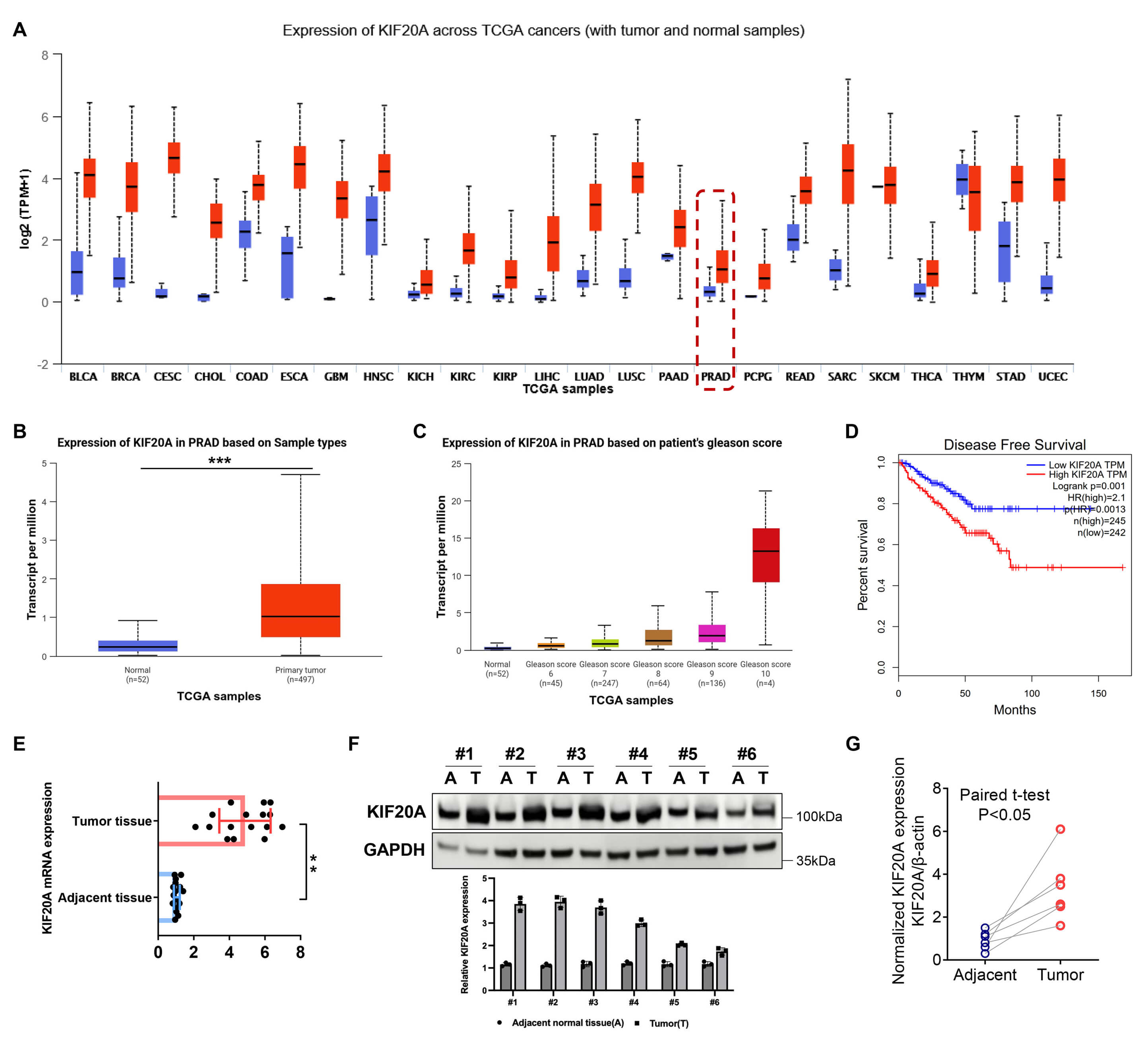
Interrogation of The Cancer Genome Atlas (TCGA) datasets through the UALCAN (University of Alabama at Birmingham CANcer data analysis Portal) and GEPIA (Gene Expression Profiling Interactive Analysis) portals revealed a marked upregulation of KIF20A mRNA expression across the majority of cancer types (Fig. 1A). In prostate cancer specimens specifically, KIF20A expression exhibited a significant elevation when compared with benign prostate tissues (Fig. 1B). Notably, KIF20A expression showed a progressive increase with rising Gleason scores, indicating a positive association with tumor aggressiveness (Fig. 1C). A strong correlation was observed between high KIF20A expression and reduced survival (Fig. 1D).
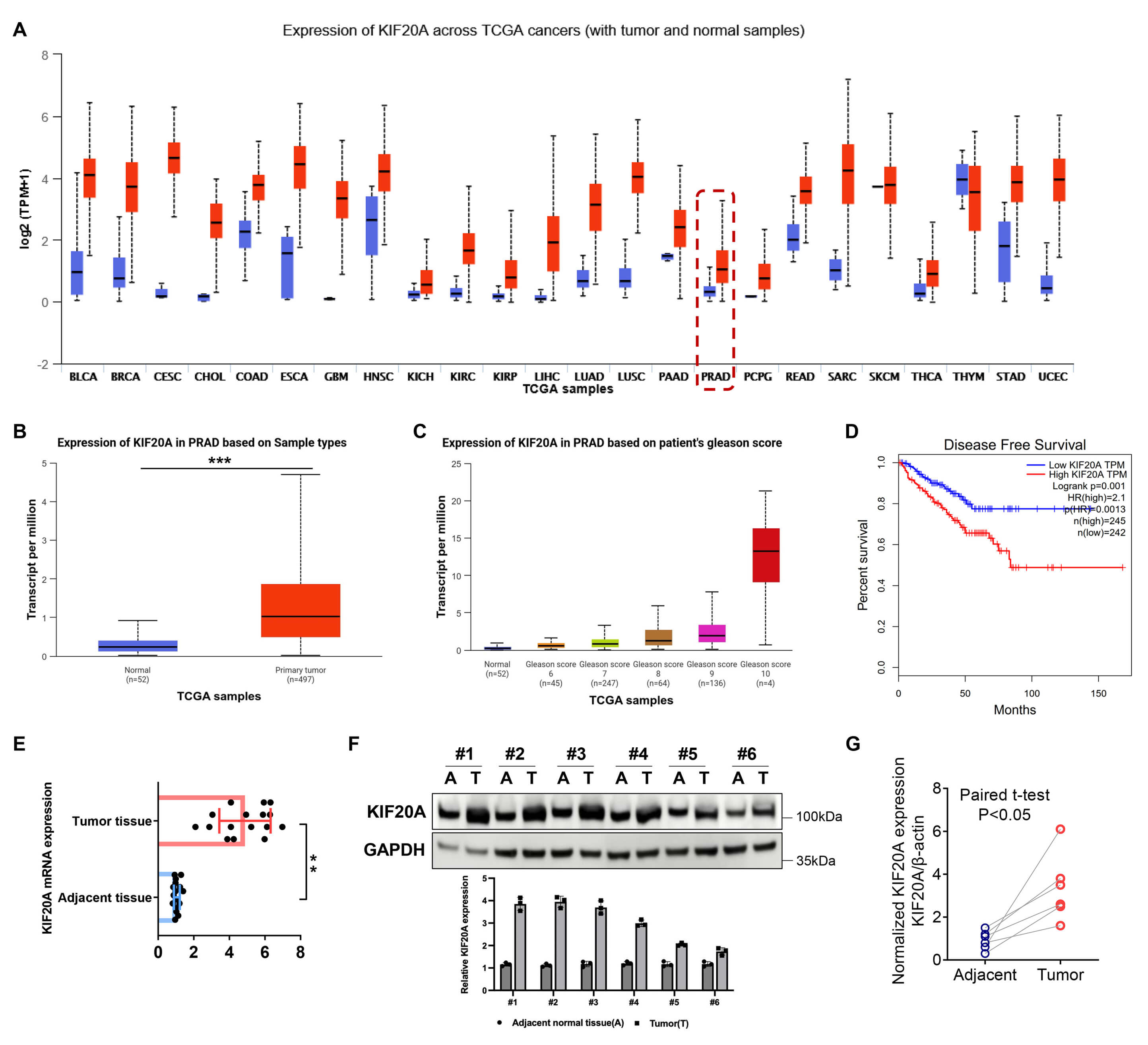 Fig. 1.
KIF20A is overexpressed in prostate cancer tissues. (A–D)
UALCAN (University of Alabama at Birmingham CANcer data analysis Portal) and
GEPIA (Gene Expression Profiling Interactive Analysis) analyses of KIF20A mRNA
levels in pan cancers, normal versus tumor tissues, stratified by Gleason grade
and prognosis. (E,F) Detection of KIF20A expression in paired tumor and adjacent
tissues using quantitative real-time PCR (qRT-PCR) and western blot analyses. (G)
Quantification of protein band intensities by paired t-test. **,
p
To validate these bioinformatics findings, our study utilized surgical specimens from 16 patients, each including prostate tumor tissue and matched adjacent normal tissue. qRT-PCR confirmed the significant upregulation of KIF20A mRNA in tumor tissues compared to their matched adjacent counterparts (Fig. 1E). Consistently, Western blot analysis of eight randomly selected tissue pairs demonstrated elevated KIF20A protein levels in prostate cancer tissues (Fig. 1F,G).
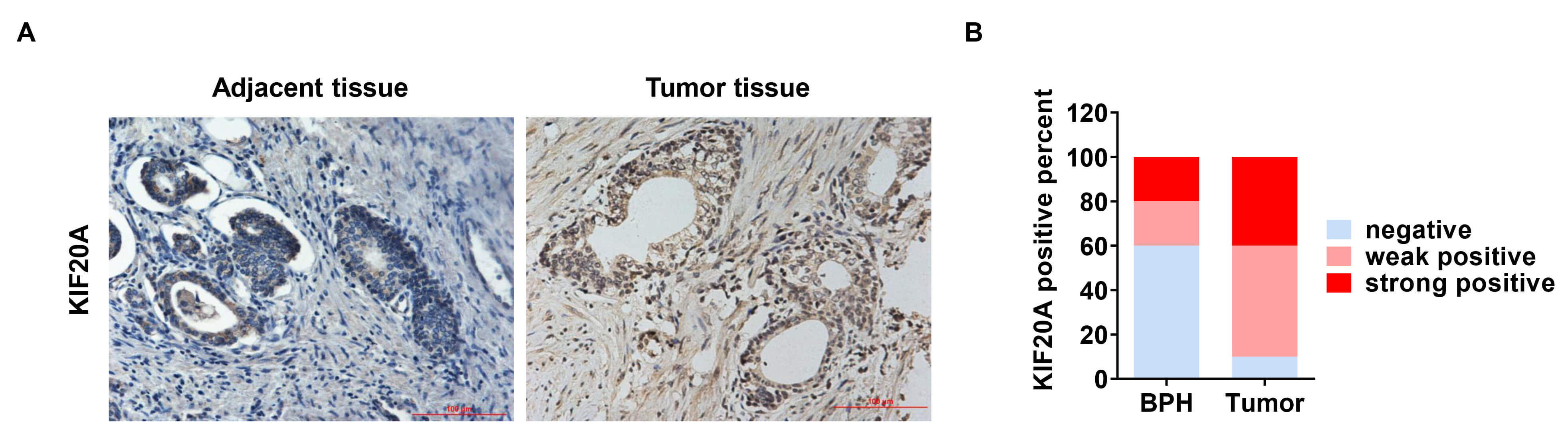
Immunohistochemical analysis was performed on 80 paired tissue samples (prostate cancer and adjacent non-cancerous). In these samples, KIF20A protein exhibited predominant cytoplasmic localization (Fig. 2A). IHC scoring revealed that KIF20A expression was significantly upregulated in tumor tissues: approximately 40% of cases exhibited strong positivity and 50% showed weak positivity. In contrast, in benign prostatic hyperplasia (BPH) tissues, strong and weak KIF20A positivity were observed in only 20% of cases each (Fig. 2B).
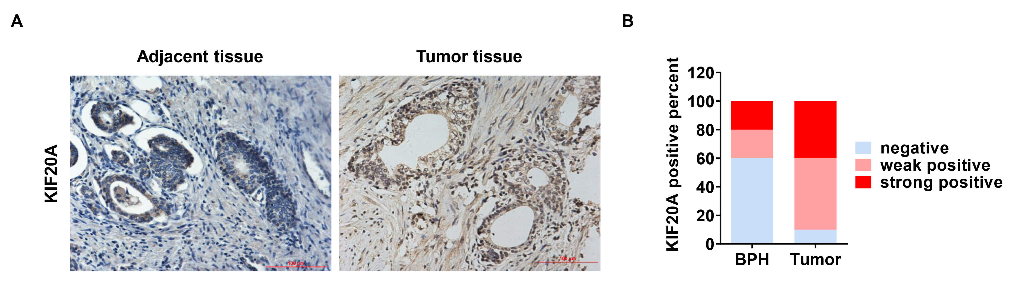 Fig. 2.
Protein levels of KIF20A are significantly upregulated in tumor tissues. (A) Immunohistochemistry (IHC) analysis showing KIF20A expression patterns in representative paired tumor and adjacent tissue sections. Scale bar = 100 µm. (B) The histogram illustrates the comparison of KIF20A-positive staining rates between tumor tissues and matched adjacent tissues.
KIF20A overexpression emerged as a marker of poor prognostic factors in prostate cancer, showing significant associations with advanced pathological stage, high ISUP (international society of urological pathology) grade, elevated serum PSA, and metastatic spread (Table 1). Collectively, these results suggest that elevated KIF20A expression is closely correlated with a more aggressive malignant phenotype and may serve as an indicator of poor prognosis in prostate cancer.
| Variables | All n = 80 | KIF20A | p value# | ||
| Low n = 26 | High n = 54 | ||||
| Age | |||||
| 34 | 10 | 24 | 0.61 | ||
| 46 | 16 | 30 | |||
| iPSA | |||||
| 33 | 15 | 18 | 0.03* | ||
| 47 | 11 | 36 | |||
| Tumor stage | |||||
| T2 | 29 | 14 | 15 | 0.02* | |
| T3/T4 | 51 | 12 | 39 | ||
| ISUP Gleason grading | |||||
| 32 | 16 | 16 | 0.01* | ||
| 48 | 10 | 38 | |||
| Lymph node metastasis | |||||
| No | 28 | 14 | 14 | 0.01* | |
| Yes | 52 | 12 | 40 | ||
| Seminal vesicle metastasis | |||||
| No | 27 | 14 | 13 | 0.01* | |
| Yes | 53 | 12 | 41 | ||
#p value was analyzed by Chi-square test; * indicates p
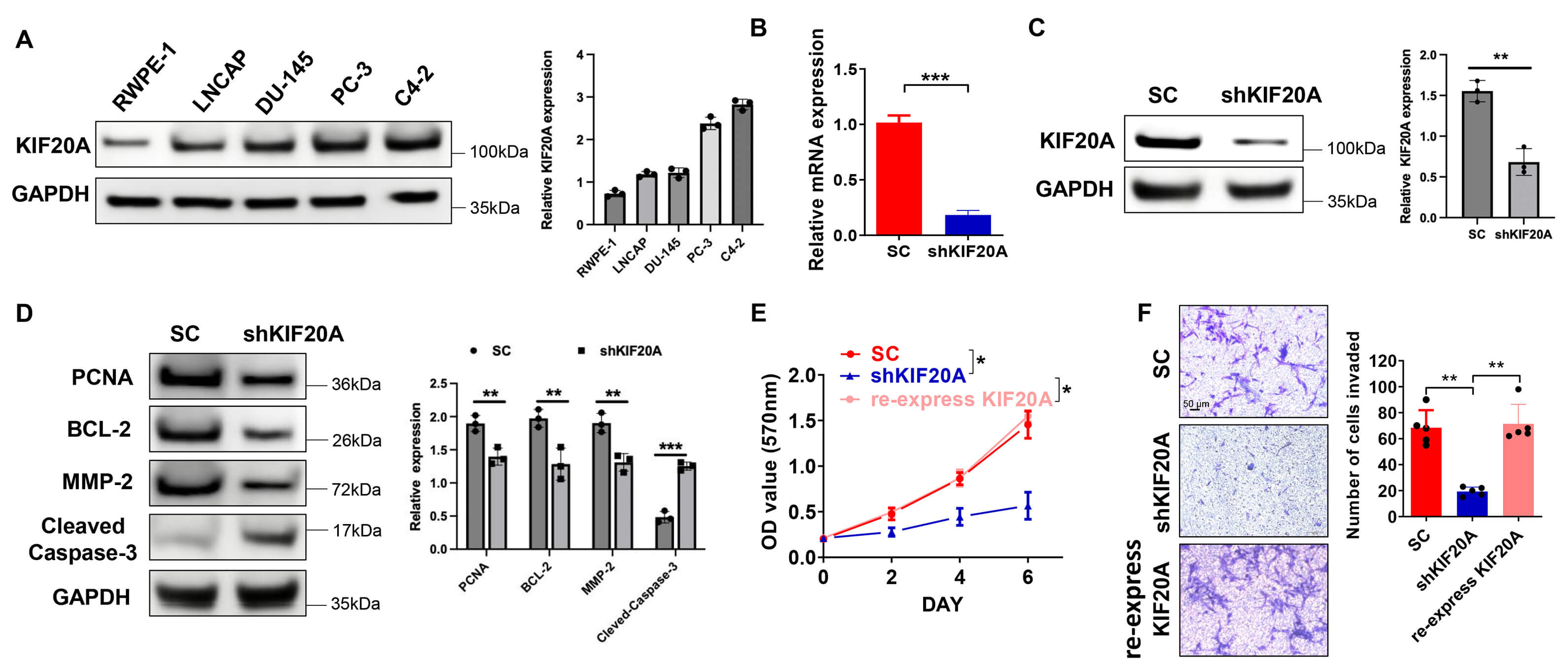
Analysis of a panel of prostate cancer cell lines (LNCaP, DU145, PC-3, C4-2) revealed consistent and marked upregulation of KIF20A, at both transcriptional and translational levels, compared to normal RWPE-1 cells, with the most striking overexpression seen particularly in C4-2 cells (Fig. 3A). Accordingly, C4-2 cells were selected for subsequent functional experiments. To generate a stable KIF20A-knockdown model, C4-2 cells were transduced with KIF20A-targeting lentiviral shRNA. The resulting cell line (shKIF20A) exhibited efficient gene silencing, as verified by qRT-PCR for mRNA and western blotting for protein (Fig. 3B,C).
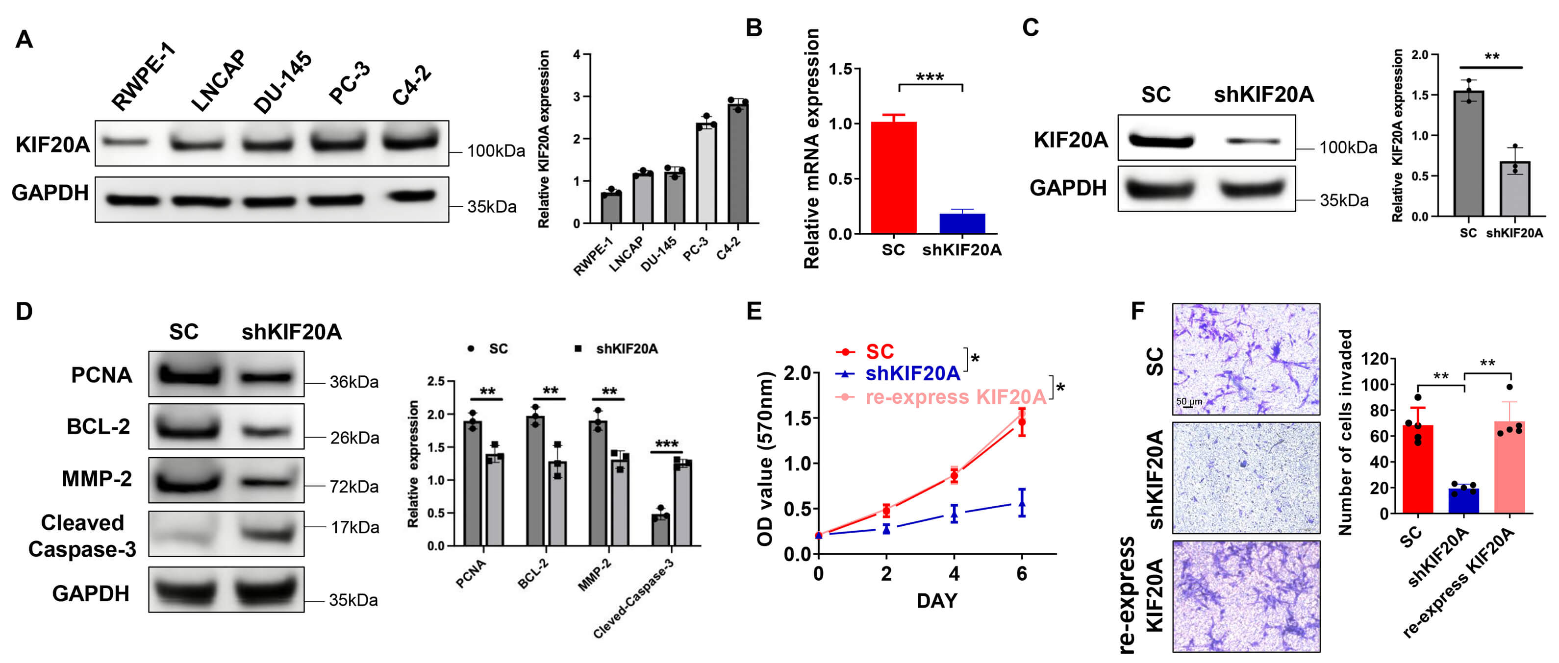 Fig. 3.
KIF20A knockdown impairs prostate cancer cell proliferation and
invasive capacity. (A) Western blot profiling of KIF20A expression across normal
and prostate cancer cell lines. (B,C) Validation of KIF20A knockdown in C4-2
cells using qRT-PCR and western blot analysis. (D) Western blot analysis
assessing the expression levels of PCNA (proliferating cell nuclear antigen),
BCL-2 (B-cell lymphoma 2), MMP-2 (matrix metalloproteinase 2), and cleaved
Caspase-3 after KIF20A knockdown. (E) MTT assay of cell proliferation (Optical
Density 570 nm). (F) Transwell invasion assay quantifying invaded cells. *, p
Corroborating the phenotypic data, western blot analysis revealed that KIF20A knockdown notably reduced the protein levels of key markers associated with proliferation and invasion, including PCNA, and MMP-2, while markedly increasing the level of the apoptosis-related protein cleaved Caspase-3 (Fig. 3D). These findings indicate that KIF20A depletion suppresses prostate cancer cell proliferation and invasion while promoting apoptotic signaling.
Results from the MTT assay quantitatively confirmed that silencing KIF20A significantly inhibited the viability and proliferative capacity of C4-2 cells (Fig. 3E). Furthermore, Transwell invasion assays confirmed that silencing KIF20A markedly attenuated the invasive capacity of prostate cancer cells (Fig. 3F). Notably, when KIF20A was re-expressed, the proliferation and invasive capacity of prostate cancer cells were restored (Fig. 3E,F).
ATF2 functions as a critical transcriptional driver of prostate cancer
progression, in part through its regulation of downstream targets like KIF20A. To
identify potential direct regulation, we performed a JASPAR database analysis,
which identified several conserved ATF2 binding motifs within the KIF20A promoter
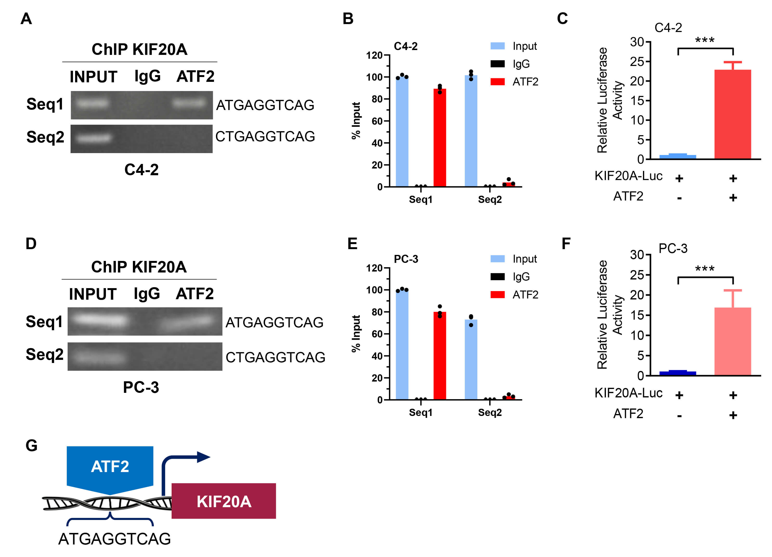
region, motivating further experimental validation (Table 2). Chromatin
immunoprecipitation (ChIP) experiments targeting the two highest-scoring
predicted sites (relative scores
| Matrix ID/Name | Score | Relative score | Sequence ID | Start | End | Strand | Predicted sequence |
| MA1632.2.ATF2 | 14.537009 | 0.9590964 | NC_000005.10:138177112-138179112 | 57 | 66 | + | ATGAGGTCAG |
| MA1632.2.ATF2 | 10.613593 | 0.8971946 | NC_000005.10:138177112-138179112 | 201 | 210 | + | CTGAGGTCAG |
| MA1632.2.ATF2 | 6.9715123 | 0.83973163 | NC_000005.10:138177112-138179112 | 439 | 448 | − | AGGATGTCAG |
| MA1632.2.ATF2 | 6.8458185 | 0.8377485 | NC_000005.10:138177112-138179112 | 57 | 66 | − | CTGACCTCAT |
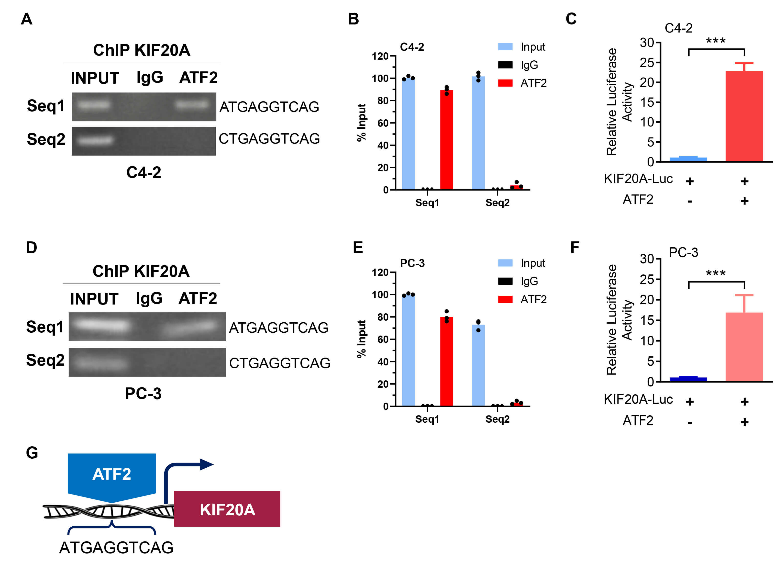 Fig. 4.
Activating transcription factor 2 (ATF2) regulates the
transcriptional activity of KIF20A. Chromatin immunoprecipitation (ChIP) and
luciferase assays were performed on C4-2 and PC-3 cells, respectively. (A,D) ChIP
assay demonstrating ATF2 binding to specific sites in the KIF20A promoter. (B,E)
ChIP-qPCR showing enrichment of ATF2 at two promoter regions (Seq1 and Seq2)
compared to IgG control. Input DNA is shown as reference. (C,F) Dual-luciferase
reporter assay showing increased KIF20A promoter activity following ATF2
overexpression. (G) Schematic illustration of the transcriptional regulatory
mechanism between ATF2 and KIF20A. ***, p
Despite its high global prevalence among men, the molecular pathogenesis of prostate cancer is incompletely understood, necessitating further investigation into its regulatory mechanisms. Recently, there has been increasing interest in integrating imaging-derived information, such as radiomics, into multi-omics frameworks. Radiomics allows quantitative extraction of features from medical images, which can then be correlated with molecular characteristics and clinical outcomes. The imaging landscape for prostate cancer comprises conventional methods (MRI, ultrasound, CT) and molecular techniques, with the emergence of targeted PET/CT tracers like radiolabeled PSMA and 18F-choline enhancing diagnostic precision [19]. Incorporating radiomic features with genomic, transcriptomic, and proteomic data may provide a more comprehensive understanding of tumor heterogeneity and enhance prognostic and therapeutic stratification.
Given its central role in mitosis-regulating spindle assembly and chromosome segregation. We investigated whether the kinesin-6 motor protein KIF20A (MKLP2) contributes to prostate cancer progression. KIF20A localizes to the Golgi apparatus and interacts with RAB6AB-GTP, reflecting its central role in intracellular transport and cytokinesis [11].
Its oncogenic potential was first observed in pancreatic cancer, where overexpression promoted tumor proliferation, while RNA interference-mediated knockdown inhibited growth [8]. Beyond its established role in mitosis, KIF20A is also overexpressed in cancers such as HCC, where its silencing promotes polyploidization while bypassing apoptosis [20, 21]. Beyond proliferation, KIF20A has been explored as an immunotherapeutic target; KIF20A-derived peptides can elicit tumor-specific cytotoxic T lymphocyte (CTL) responses with minimal off-target effects [22]. Emerging evidence positions KIF20A as a promising therapeutic target. Early-phase clinical trials in pancreatic cancer have yielded encouraging results, and its overexpression has been further linked to chemoresistance. For instance, by conferring paclitaxel resistance in breast cancer via FOXM1 regulation. Collectively, these findings highlight KIF20A as a promising molecular target across multiple cancer types.
Although KIF20A’s oncogenic role has been established in multiple cancers, its function in prostate cancer required further clarification. Our analyses demonstrated that KIF20A is upregulated at both mRNA and protein levels in prostate cancer tissues and cell lines. Clinically, high KIF20A expression correlated with elevated Gleason scores, advanced tumor stage, and lymph node metastasis. Consistent with prior studies, we observed a significant correlation between KIF20A upregulation and poor clinicopathological outcomes in prostate cancer. Furthermore, we confirmed that KIF20A knockdown inhibits tumor cell proliferation and invasion, reinforcing its oncogenic role. These functional findings indicate that KIF20A exerts a pro-tumorigenic effect by coordinately stimulating proliferation and invasion while blocking apoptosis, highlighting its multifaceted role in advancing prostate cancer.
ATF2 plays a highly complex and context-dependent role across multiple cancer
types, exhibiting significant tumor type specificity in its function. This
extensive regulatory network confers a dual role to ATF2: in tumors like melanoma
and hepatocellular carcinoma, it primarily acts as an oncogene driving
progression, whereas in settings such as cutaneous squamous cell carcinoma, it
exhibits tumor-suppressing functions [23]. Clinical pathological analysis reveals
that ATF2 exhibits an upward trend in expression and increased nuclear
localization within prostate cancer tissues [14]. Particularly in high-grade
tumors (Gleason score
Consistent with our findings, TCGA dataset analysis indicated that high KIF20A expression predicts poorer disease-free survival, confirming its clinical relevance and supporting its utility as a prognostic biomarker. Given its involvement in mitosis, invasion, and transcriptional regulation, KIF20A represents a promising molecular target for therapeutic intervention. Future studies should investigate upstream signaling pathways that regulate the ATF2–KIF20A interaction and downstream effectors mediating its oncogenic functions. In vivo studies and clinical validation are also warranted to assess whether inhibition of KIF20A expression or blockade of its transcriptional activation could suppress prostate cancer growth and metastasis.
Intriguingly, recent observations suggest that KIF20A may also modulate prostate cancer stem cells (PCSCs), a cell population critical for tumorigenesis, metastasis, and relapse, thereby potentially influencing therapeutic outcomes [24]. The potential regulation of PCSCs by KIF20A suggests a mechanism through which it could fuel tumor heterogeneity and facilitate relapse. Thus, targeting KIF20A may address not only bulk tumor growth but also the resistant cell populations responsible for treatment failure. Considering this potential role in stem-like cell populations provides a broader translational perspective and strengthens the clinical significance of targeting KIF20A in prostate cancer.
In summary, our data converge to establish KIF20A as a master regulator in prostate cancer, integrating control over cell proliferation, invasive capacity, transcriptional networks, and possibly stem cell maintenance. The convergence of clinical correlations and functional evidence reinforces the rationale for targeting KIF20A therapeutically, while also highlighting its potential utility as a predictive biomarker for disease monitoring.
This study has several limitations. The sample size for fresh‑frozen tissue analysis was relatively small, which may limit statistical power and generalizability, and all clinical samples were obtained from a single institution, potentially introducing selection bias. Survival analysis was primarily derived from public databases rather than long‑term follow‑up data from our own cohort. Functional experiments were mainly conducted in two prostate cancer cell lines, and the findings may not fully represent the heterogeneity of human prostate cancer. Additionally, in vivo animal studies are lacking, and future investigations using xenograft models are needed to validate the oncogenic role of the ATF2‑KIF20A axis. Despite these limitations, our results provide new insights into the transcriptional regulation of KIF20A and its functional significance in prostate cancer progression.
KIF20A is transcriptionally regulated by ATF2 in prostate cancer tissue, leading to its abnormally high expression and promoting the progression of prostate cancer. KIF20A may serve as an independent prognostic factor for prostate cancer patients and a therapeutic target.
The data presented in this study are available upon request from the corresponding author. The data are not publicly available due to further development of the work.
MY and XY designed the study, collected clinical information and performed experiments. XL and KW performed data analysis. MY wrote the initial draft. XY, TS and BW designed the study and revised the manuscript. All authors read and approved the final manuscript. All authors have participated sufficiently in the work and agreed to be accountable for all aspects of the work. All authors contributed to editorial changes in the manuscript.
The study was conducted in accordance with the Declaration of Helsinki, and approved by the Tianjin Medical University General Hospital Medical Ethics Committee (protocol code IRB-2021-KY-126 and February 19th, 2021 of approval). Written informed consent was obtained from all participants involved in this study. All participants were informed about the purpose, procedures, potential risks, and benefits of the study, and they voluntarily agreed to participate.
We would like to express our gratitude to all those who helped me during the writing of this manuscript and thanks to all the peer reviewers for their opinions and suggestions.
This work was supported by the Youth Professional and Technical Backbone Project of Tianjin Medical University General Hospital (Grant No. GG-202407) and the Tianjin Municipal Education Commission Research Program Project (Grant No. 2025KJ022).
The authors declare no conflict of interest.
References
Publisher’s Note: IMR Press stays neutral with regard to jurisdictional claims in published maps and institutional affiliations.