


 , Xiaolin Chen 1,*
1 Department of Orthopedics, Xiamen University Affiliated Chenggong Hospital, 361026 Xiamen, Fujian, China
2 Department of Orthopedics, The 920th Hospital of Chinese People’s Liberation Army Joint Logistics Support Force, 650032 Kunming, Yunnan, China
†These authors contributed equally.
Abstract
Intervertebral disc degeneration (IDD) is a major cause of chronic lower back pain, with current treatment options offering limited efficacy. Exosome-loaded hydrogels have emerged as a promising therapeutic approach due to their biocompatibility and regenerative potential, making them a focus of research for IDD treatment. This study systematically evaluates and performs a meta-analysis of the effectiveness of exosome-loaded hydrogels in preclinical models of IDD.
A comprehensive literature search was conducted across four major databases (PubMed, Embase, Cochrane, Web of Science), including animal studies that met predefined criteria. Data extraction and quality assessment were independently performed by two authors. Treatment effects were quantified using standardized mean differences (SMD) with 95% confidence intervals (CI). Outcome measures included disc height index (DHI), magnetic resonance imaging (MRI) grade, histological grade, IDD-related immunohistochemical (IHC) markers (e.g., collagen type II (COL2), matrix metalloproteinase 13 (MMP13)), and aging-related markers (e.g., p16Ink4a-positive cells, p21CIP1A-positive cells).
Treatment with exosome-loaded hydrogels significantly enhanced DHI scores at 4 (p = 0.002) and 8 weeks (p < 0.0001), and decreased MRI scores at 8 (p < 0.00001) and 12 weeks (p < 0.0001), and histological assessments. Furthermore, the treatment group exhibited increased COL2 expression at 8 (p = 0.0002) and 12 weeks (p = 0.002), decreased MMP13 levels at 8 (p = 0.0001) and 12 weeks (p = 0.0009), and a reduction in aging markers (p16Ink4a, p21CIP1A, all p < 0.05), suggesting that exosome-loaded hydrogels facilitate intervertebral disc repair through the modulation of molecular pathways. Sensitivity analysis confirmed the robustness of the findings.
Exosome-loaded hydrogels show potential for improving the structure and function of intervertebral discs in IDD treatment, potentially slowing degeneration by inhibiting matrix degradation and cellular aging. Further investigation is required to elucidate the underlying mechanisms and to assess the safety and efficacy of these hydrogels for clinical application.
CRD420250649970 (https://www.crd.york.ac.uk/PROSPERO/view/CRD420250649970).
Keywords
- exosome
- hydrogel
- intervertebral disc degeneration
- animal studies
- meta-analysis
Intervertebral disc degeneration (IDD) is a primary cause of chronic low back pain and spinal disorders [1]. The intervertebral disc plays a critical role in the spine by acting as a cushion between vertebrae, absorbing and distributing spinal pressure. It consists of the outer annulus fibrosus, inner nucleus pulposus, and transitional zone cells, with the nucleus pulposus containing significant water content to maintain disc elasticity and height [2]. Degeneration leads to reduced hydration of the nucleus pulposus and structural compromise of the annulus fibrosus, ultimately impairing the disc’s cushioning and support functions. This may result in conditions such as disc herniation, spinal instability, and nerve root compression [3, 4]. IDD development is influenced by multiple factors, including aging, genetics, lifestyle, and mechanical injury [5, 6]. As aging progresses, the hydration and metabolic functions of intervertebral discs decline, resulting in structural degeneration [7]. Clinically, IDD manifests as chronic back pain, radiating leg pain, and reduced mobility, severely impacting patients’ quality of life. One study indicates that approximately 80% of adults will experience some form of back pain in their lifetime, with a portion progressing to IDD [8]. Thus, effective treatment options are urgently needed on a global scale.
Current treatments for IDD include conservative management, pharmacological interventions, physical therapy, and surgical procedures. Conservative approaches, such as physical therapy, massage, and rehabilitation exercises, aim to alleviate pain and improve disc function [9]. However, these strategies primarily focus on symptom management and do not address the root cause of disc degeneration [10]. Pharmacological treatments, including non-steroidal anti-inflammatory drugs (NSAIDs) and analgesics, provide temporary pain relief but may lead to side effects with prolonged use, such as gastrointestinal discomfort and kidney dysfunction [11]. In more severe cases, surgical treatments like discectomy and spinal fusion are employed as last-resort options. While these procedures relieve symptoms, they do not restore disc structure or function and carry the risk of postoperative complications, such as spinal instability and degeneration of adjacent segments [12].
In recent years, exosomes and hydrogels have emerged as promising biological therapeutic strategies [13, 14]. Exosomes, small vesicles secreted by cells, are rich in bioactive molecules such as proteins, lipids, and RNA. They exert a range of biological functions in vivo, including promoting cell repair, exerting anti-inflammatory effects, and offering anti-aging properties [15, 16]. Hydrogels, a type of polymeric material, exhibit excellent biocompatibility, tunable mechanical properties, and the ability to mimic the extracellular matrix (ECM). These features make hydrogels highly suitable for drug delivery and tissue repair applications [17, 18]. Combining exosomes with hydrogels leverages the three-dimensional structure and controlled release properties of hydrogels, facilitating efficient exosome delivery to the target area [19]. This combination promotes the repair of intervertebral disc cells and slows degeneration, holding significant potential for clinical application.
While some preliminary studies have investigated the individual effects of exosomes and hydrogels [15, 17], there is a notable lack of evidence-based research on their combined use for treating IDD. This meta-analysis aims to systematically evaluate the effects of exosome-loaded hydrogels in animal models of IDD, with a focus on their potential to improve IDD. Specifically, the analysis assessed the impact of exosome-loaded hydrogels on disc height index (DHI), magnetic resonance imaging (MRI) scores, histological evaluations, and aging-related molecular markers. Additionally, an in-depth risk-of-bias analysis was conducted using quality assessment tools, providing robust scientific evidence to inform future clinical trials and facilitating the clinical translation of this therapeutic approach.
Two independent authors conducted a comprehensive literature search across four major databases (PubMed, Embase, Cochrane, and Web of Science). The search strategy utilized Medical Subject Headings (MeSH) terms and corresponding free-text keywords, as follows: ((“Extracellular Vesicle” OR “Exosomes” OR “Cell-Derived Microparticles” OR “EV” OR “Exovesicle” OR “Apoptotic Body”) OR (“Hydrogel” OR “In Situ Hydrogel” OR “Patterned Hydrogel”)) AND (“Hydrogel” OR “In Situ Hydrogel” OR “Patterned Hydrogel”) AND (“Intervertebral disc degeneration” OR “IDD” OR “Disc Degeneration” OR “Degenerative Disc Disease”). Modifications to the search strategy were made to align with the formatting requirements of each database. No restrictions were placed on the source of exosomes or the composition of hydrogels to ensure a thorough analysis. Following this, the authors systematically reviewed the titles, abstracts, and full texts of the identified studies to exclude irrelevant ones. The references of the full-text articles were carefully examined to ensure comprehensive inclusion of relevant studies in the meta-analysis. The study protocol was developed according to the guidelines of the Preferred Reporting Items for Systematic Reviews and Meta-Analyses (PRISMA) [20] (PRISMA_2020_checklist can be found in Supplementary material) and registered in the PROSPERO database (registration number CRD420250649970).
Inclusion criteria were: (1) Animal models of IDD created through various
methods such as needle puncture, tumour necrosis factor-alpha (TNF-
Exclusion criteria included: (1) Studies involving animal models unrelated to IDD; (2) In vitro studies only; (3) Experimental groups treated with hydrogels loaded with substances other than exosomes, such as nanoparticles; (4) Studies lacking a control group; (5) Studies that did not report primary outcome measures; (6) Non-original studies, including reviews, meta-analyses, commentaries, and letters to the editor.
Data extraction was performed by two independent authors, followed by summarization and verification. Any discrepancies were resolved through discussion with a third author to ensure accuracy. The extracted data included: (1) Study characteristics (first author, publication year, country of origin); (2) Animal baseline characteristics (species, weight, quantity, age, and model creation methods); (3) Exosome and hydrogel characteristics (source and particle size of exosomes, hydrogel composition); (4) Treatment methods (administration routes, dosage, frequency, and duration); (5) Outcome measures (DHI grade, MRI grade, histological grade, IDD-related IHC markers such as COL2 and MMP13, and aging-related markers such as p16Ink4a-positive cells and p21CIP1A-positive cells). For studies presenting results in graphical format, data extraction was performed using Origin 2023 software (OriginLab Corporation, Northampton, MA, USA).
Primary outcomes included direct evidence of IDD progression or alleviation, specifically DHI grade, MRI grade, and histological grade. Secondary outcomes encompassed relevant IHC markers, including COL2, MMP13, p16Ink4a-positive cells, and p21CIP1A-positive cells.
The methodological quality of the included animal studies was assessed using the Systematic Review Center for Laboratory Animal Experimentation (SYRCLE) risk of bias tool [21], which covers 10 assessment criteria: (1) Random sequence generation (selection bias); (2) Baseline characteristics (selection bias); (3) Allocation concealment (selection bias); (4) Random housing (performance bias); (5) Blinding (performance bias); (6) Random outcome assessment (detection bias); (7) Blinding (detection bias); (8) Incomplete outcome data (attrition bias); (9) Selective reporting (reporting bias); (10) Other bias. Two authors independently evaluated each study. Positive responses to these items were classified as low risk, while unsatisfactory responses were marked as high risk. Items without a clear response were categorized as unclear risk. Any discrepancies in evaluation were resolved through discussion with a third author.
For the meta-analysis, mean values and standard deviations of reported
indicators were extracted from each study, and a summary analysis of the overall
effect size was performed. The combined effect size was assessed using the
standardized mean difference (SMD) and 95% confidence interval (CI).
Heterogeneity was evaluated using the I2 statistic and
Fig. 1 illustrates the literature screening process. A total of 1844 potential articles were initially retrieved from four databases using a fixed search strategy. After removing duplicates with Endnote 20 (Thomson Corporation, Stamford, CT, USA), 1336 articles remained. Subsequently, 1275 articles were excluded based on title and abstract reviews. Of the remaining 61 articles, 51 were further excluded according to the inclusion and exclusion criteria. Finally, 10 articles [22, 23, 24, 25, 26, 27, 28, 29, 30, 31] were included in the meta-analysis and quality assessment.
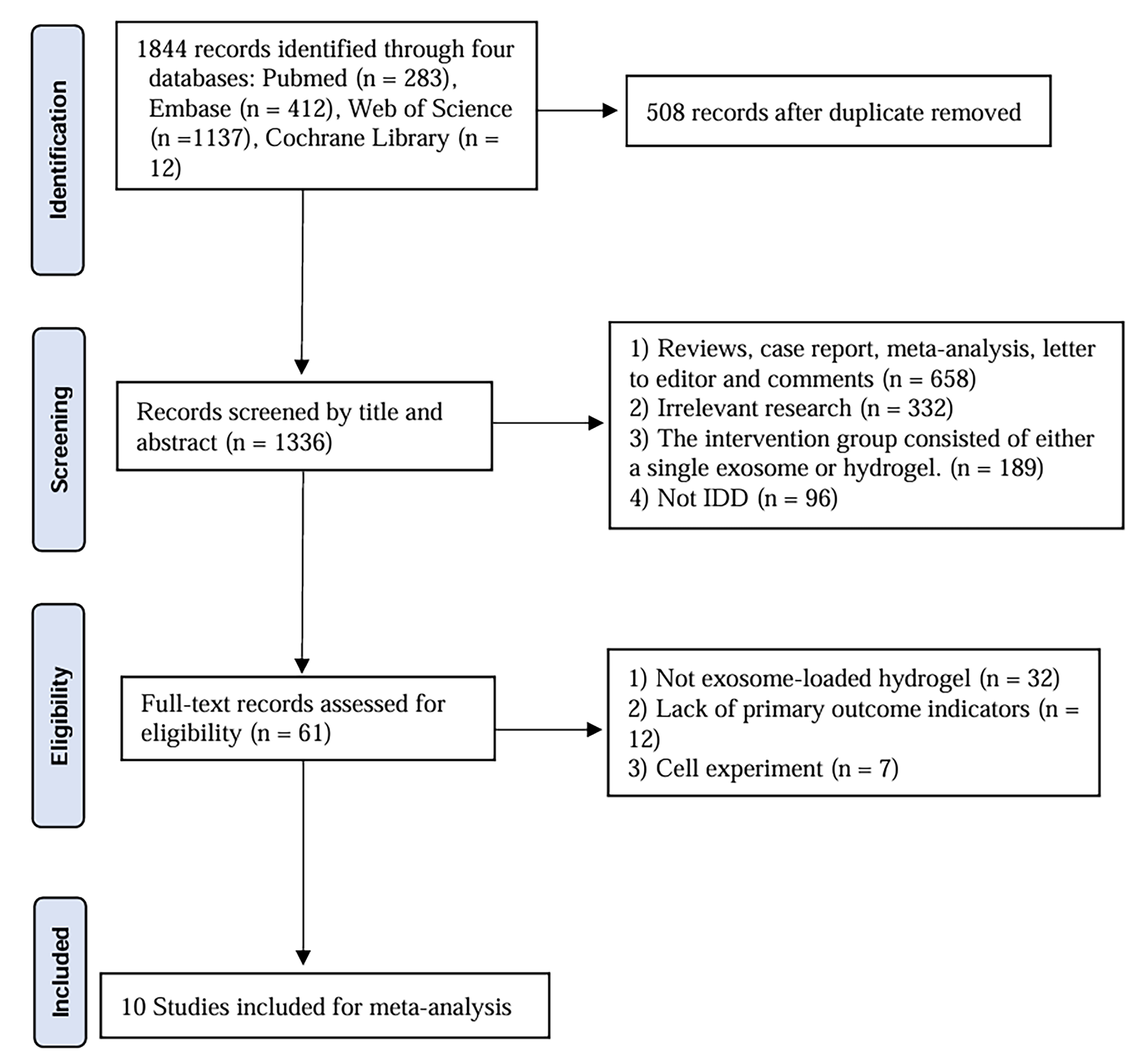 Fig. 1.
Flow diagram of the study selection. IDD, intervertebral disc degeneration.
All included studies were published within the past four years and conducted in
China. The characteristics of the animal models used are detailed in Table 1
(Ref. [22, 23, 24, 25, 26, 27, 28, 29, 30, 31]). Notably, except for one study that did not specify rat species,
all other studies employed Sprague-Dawley (SD) rats. Seven studies used male
rats, with ages ranging from 2 to 12 weeks and weights ranging from 250 to 350 g.
IDD was induced in eight studies via needle puncture, while the remaining two
studies used TNF-
| Author | Year | Country | Specie | Gender | Age | Weight | Number | Model of osteoporosis | Ref. |
| Liao et al. | 2021 | China | SD rats | male | 8-week-old | 300 g | 20 | TNF- |
[22] |
| Liu et al. | 2023 | China | SD rats | NA | NA | 250–300 g | 20 | needle puncture | [23] |
| Liao et al. | 2022 | China | SD rats | male | 8-week-old | NA | 30 | needle puncture | [24] |
| Luo et al. | 2021 | China | Rats | male | 2–3-week-old | NA | 20 | needle puncture | [25] |
| Xing et al. | 2021 | China | SD rats | male | NA | 250–270 g | 48 | needle puncture | [26] |
| Shi et al. | 2024 | China | SD rats | NA | NA | NA | 20 | needle puncture | [27] |
| Zhan et al. | 2025 | China | SD rats | male | 12-week-old | 300–350 g | 30 | subendplate was injected with 20 µL of lipopolysaccharide | [28] |
| Guan et al. | 2023 | China | SD rats | NA | 12-week-old | 250–280 g | NA | needle puncture | [29] |
| Liu et al. | 2023 | China | SD rats | male | 12-week-old | NA | 80 | needle puncture | [30] |
| Peng et al. | 2023 | China | SD rats | male | 6-week-old | NA | NA | needle puncture | [31] |
SD rats, Sprague-Dawley rats; NA, not applicable.
| Author | Year | Characteristics of exosomes | Characteristics of hydrogel | Therapeutic method | Ref. | |||||
| Source | Diameter (nm) | Source | Thickness (nm) | Route | Dose | Time | Duration | |||
| Liao et al. | 2021 | MSCs | NA | 3% w/v solution of alginate, nucleus pulposus cells and 0.1 M calcium chloride | 2 mm | discs | 100 µg/mL, 2 µL | once | once | [22] |
| Liu et al. | 2023 | MSCs | NA | dopamine-functionalized gelatin | NA | discs | 4 µL | once | once | [23] |
| Liao et al. | 2022 | MSCs | 30–150 nm | decellularized ECM | 10–20 nm | intravenously | 2 µL with 10 µg exosomes | one a week | 8 weeks | [24] |
| Luo et al. | 2021 | Cartilage endplate stem cells | NA | costal cartilage ECM | NA | discs | 10 µL | once | once | [25] |
| Xing et al. | 2021 | ADMSCs | 30–150 nm | thermosensitive acellular ECM | NA | discs | 5 mL | once | once | [26] |
| Shi et al. | 2024 | MSCs | 141 nm | gelatin methacrylate | 20–50 µm | intravenously | 2 µL | one a week | 6 weeks | [27] |
| Zhan et al. | 2025 | MSCs | 130 nm | CaCO3/chitosan composite hydrogel | NA | discs | 50 μL | week 1 and week 5 | twice | [28] |
| Guan et al. | 2023 | MSCs | NA | quaternized chitosan and oxidized starch | NA | discs | 3 µL | NA | NA | [29] |
| Liu et al. | 2023 | M2c macrophages | NA | 1 mM hydrochloric acid and hyaluronic acid | NA | discs | 5 µL | once | once | [30] |
| Peng et al. | 2023 | MSCs | 50–200 nm | DNP hydrogels | NA | discs | 2 µL | week 1 and week 4 | twice | [31] |
MSCs, mesenchymal stem cells; ADMSCs, adipose-derived MSCs; NPSCs, nucleus pulposus stem cells; ECM, extracellular matrix; DNP, decellularized nucleus pulposus; NA, not applicable.
The methodological quality of the included studies was assessed using the SYRCLE RoB tool. One study reported allocation concealment for experimental animals, which was deemed to have a low risk of bias. However, no study described the random allocation method for animals, nor did any study report blinding methods for implementation or measurement. One study lacked a description of the baseline characteristics of the experimental animals, which was considered to carry a high risk of bias regarding baseline characteristics. Additionally, two studies did not report the number of experimental animals, leading to a higher risk of bias (Fig. 2A,B). Overall, most studies displayed an uncertain risk of bias.
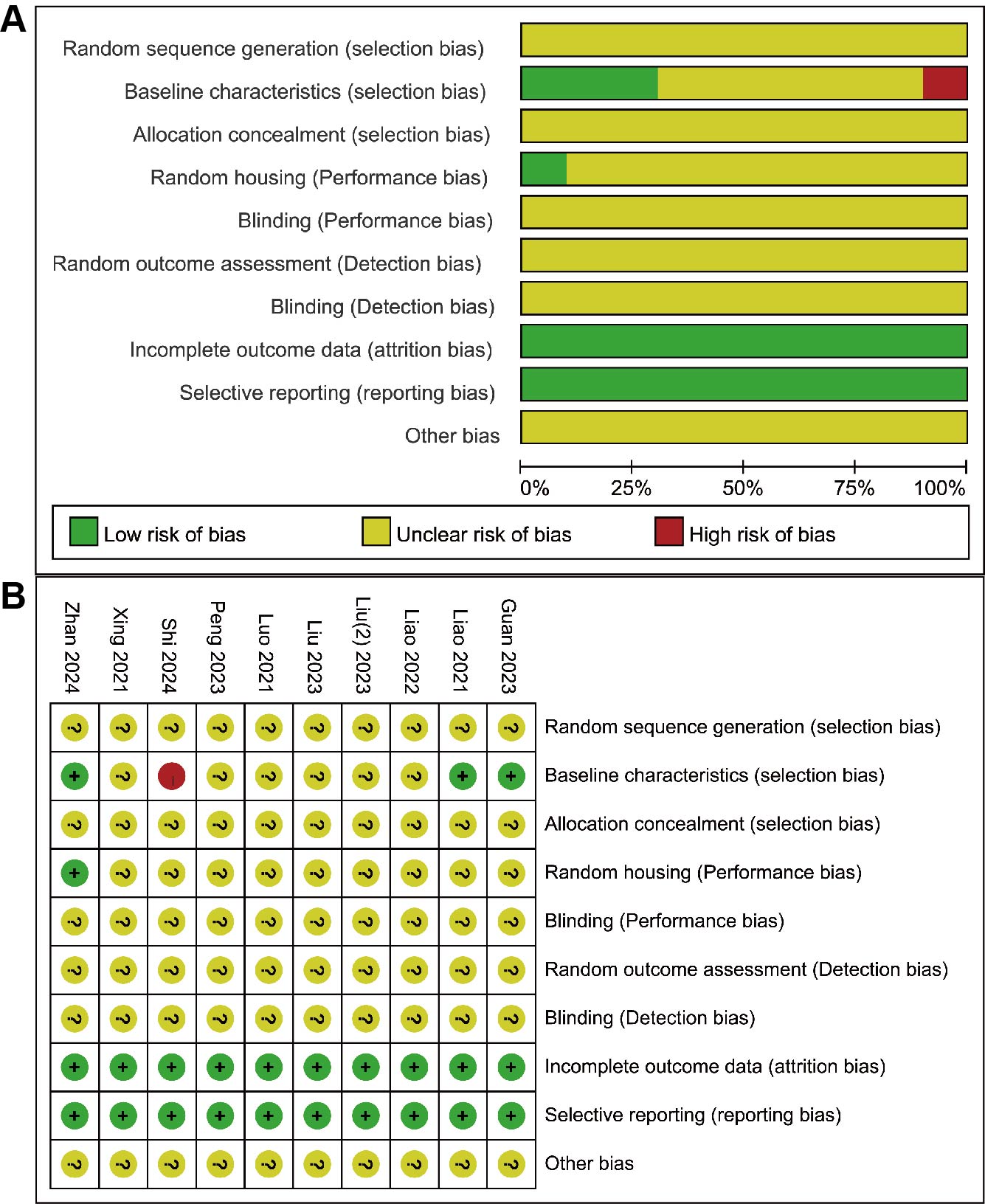 Fig. 2.
Risk of bias assessment based on the SYRCLE’s RoB tool. (A) Risk of bias graph for the included studies. (B) Risk of bias summary for each included study.
The DHI grade, reflecting changes in disc height, is a critical indicator for
assessing recovery following IDD treatment. Given the varying treatment time
points, changes in DHI grade were analyzed separately at 4 weeks and 8 weeks. At
4 weeks, the meta-analysis revealed that the exosome-loaded hydrogel group
significantly increased the DHI grade compared to the control group (SMD = 2.07,
95% CI: 0.76 to 3.38, p = 0.002) (Fig. 3A). Similarly, at 8 weeks, the
exosome-loaded hydrogel group demonstrated a significant increase in the DHI
grade (SMD = 3.15, 95% CI: 1.69 to 4.61, p
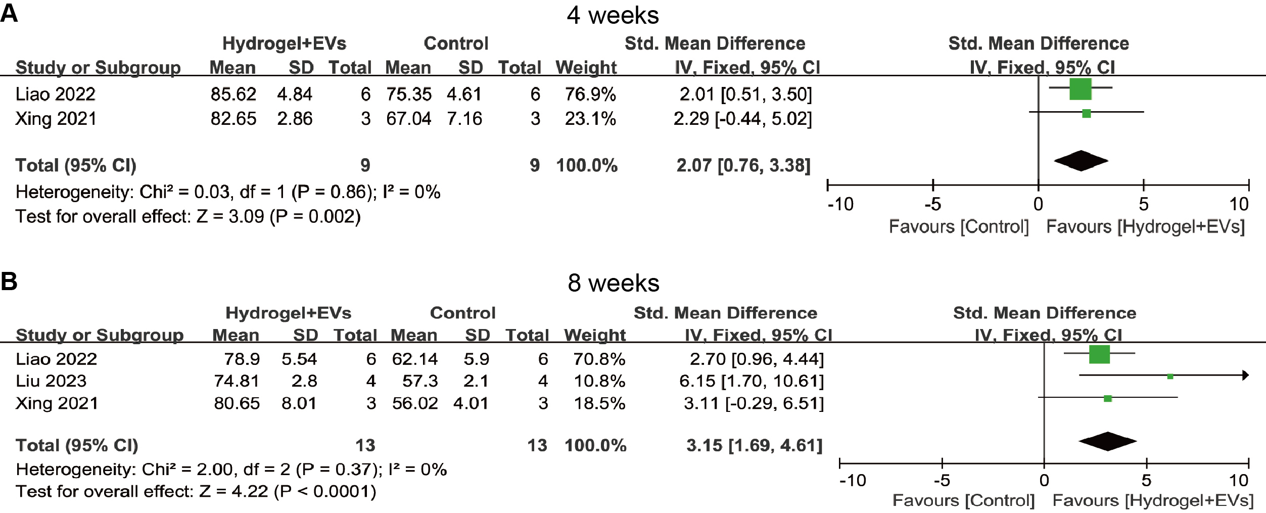 Fig. 3.
Forest plot showing the effect of exosome-loaded hydrogel on disc height index (DHI) grade at 4 weeks (A) and 8 weeks (B). EVs, extracellular vesicles; CI, confidence intervals.
The MRI grade is an essential imaging tool for assessing structural,
morphological, and pathological changes in the intervertebral disc. The
meta-analysis showed that, compared to the control group, the exosome-loaded
hydrogel group significantly reduced the MRI grade at 8 weeks (SMD = –3.16, 95%
CI: –4.40 to –1.92, p
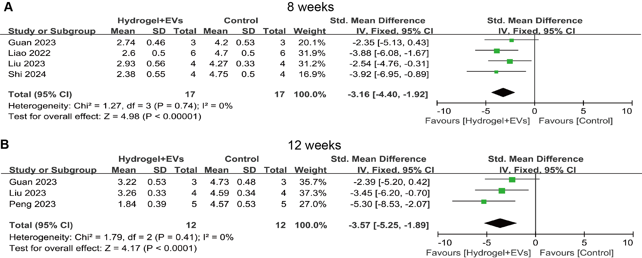 Fig. 4.
Forest plot showing the effect of exosome-loaded hydrogel on magnetic resonance imaging (MRI) grade at 8 weeks (A) and 12 weeks (B).
Additionally, the impact of the treatment on the histological grade of the
intervertebral disc was assessed. Due to significant heterogeneity
(I2 = 61%, p = 0.03), a random-effects model was applied.
The meta-analysis results revealed that the exosome-loaded hydrogel group
significantly reduced the histological grade of the intervertebral disc (SMD =
–4.49, 95% CI: –6.72 to –2.27, p
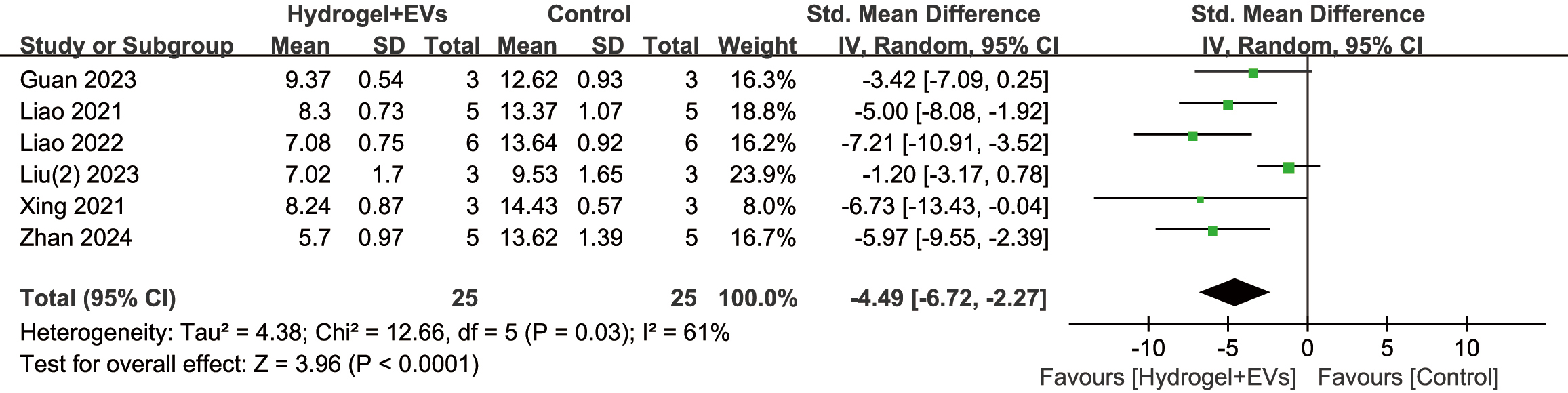 Fig. 5.
Forest plot showing the effect of exosome-loaded hydrogel on histological grade.
COL2 and MMP13 are key biomarkers involved in the degeneration, repair, and regeneration of the intervertebral disc. Meta-analysis results showed that the exosome-loaded hydrogel significantly upregulated the expression of COL2 protein in the IDD model at both 8 weeks (SMD = 4.66, 95% CI: 2.17 to 7.15, p = 0.0002) and 12 weeks (SMD = 5.10, 95% CI: 1.92 to 8.27, p = 0.002) (Fig. 6A). Conversely, the exosome-loaded hydrogel significantly downregulated the expression of MMP13 protein in the IDD model at 8 weeks (SMD = –6.09, 95% CI: –9.23 to –2.95, p = 0.0001) and 12 weeks (SMD = –14.16, 95% CI: –22.48 to –5.84, p = 0.0009) (Fig. 6B). These results suggest that exosome-loaded hydrogels may facilitate intervertebral disc repair and inhibit matrix degradation by upregulating COL2 and downregulating MMP13.
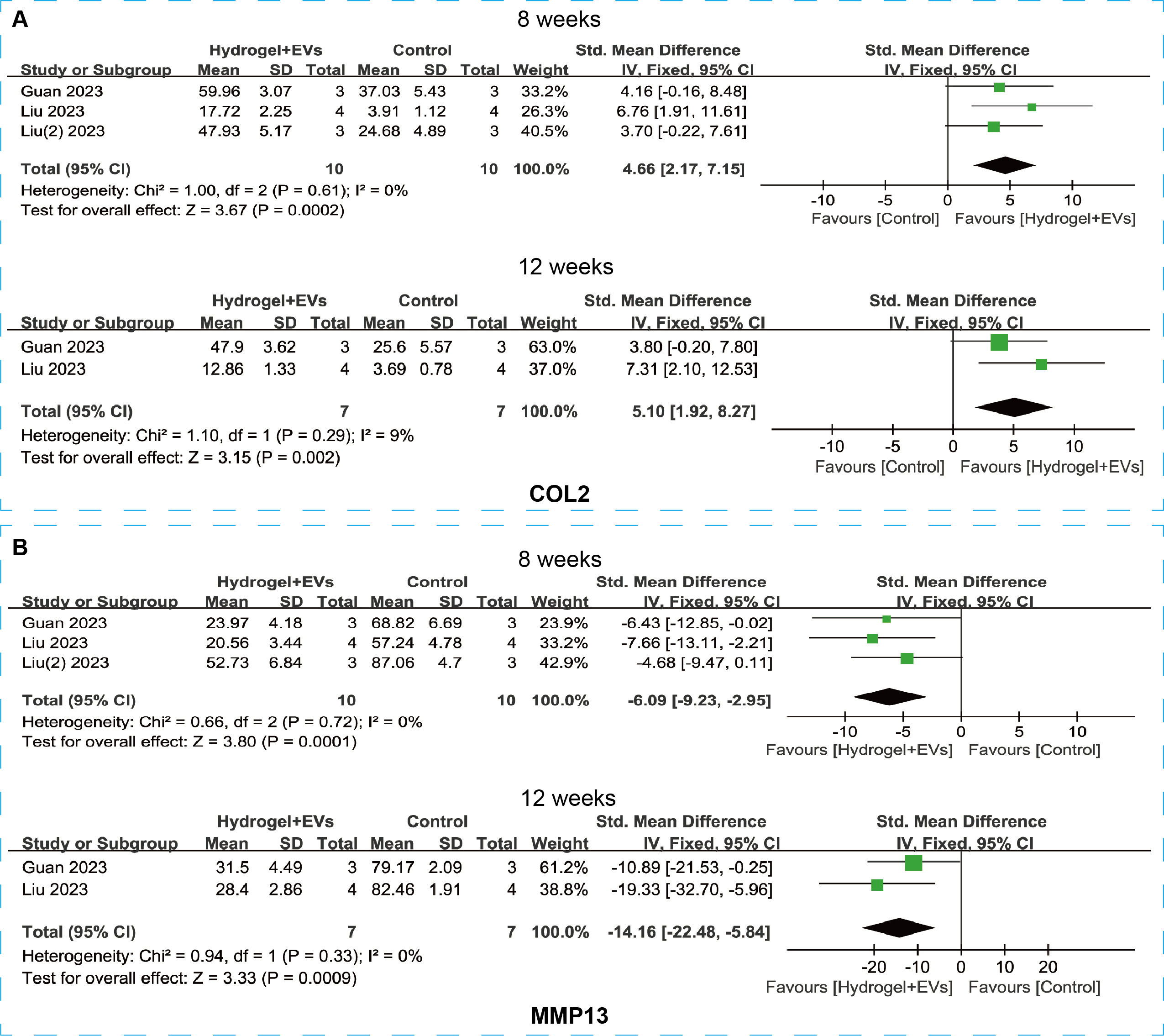 Fig. 6.
Forest plot showing the difference in relative expression of Collagen Type II (COL2) (A) and Matrix Metalloproteinase 13 (MMP13) proteins (B) between the exosome-loaded hydrogel group and the control group at 8 weeks and 12 weeks post-treatment for intervertebral disc degeneration (IDD).
Regarding cellular aging, p16Ink4a and p21CIP1A proteins are critical markers for assessing the aging, proliferation, and repair of intervertebral disc cells. Two studies evaluated the effects of exosome-loaded hydrogels on cellular aging in the IDD model. Meta-analysis results showed that, compared to the control group, the exosome-loaded hydrogel significantly reduced the number of p16Ink4a-positive cells and p21CIP1A-positive cells at both 8 weeks and 12 weeks (Fig. 7A,B). These findings suggest that exosome-loaded hydrogels may promote disc repair by slowing or reversing cellular aging during the later stages of treatment.
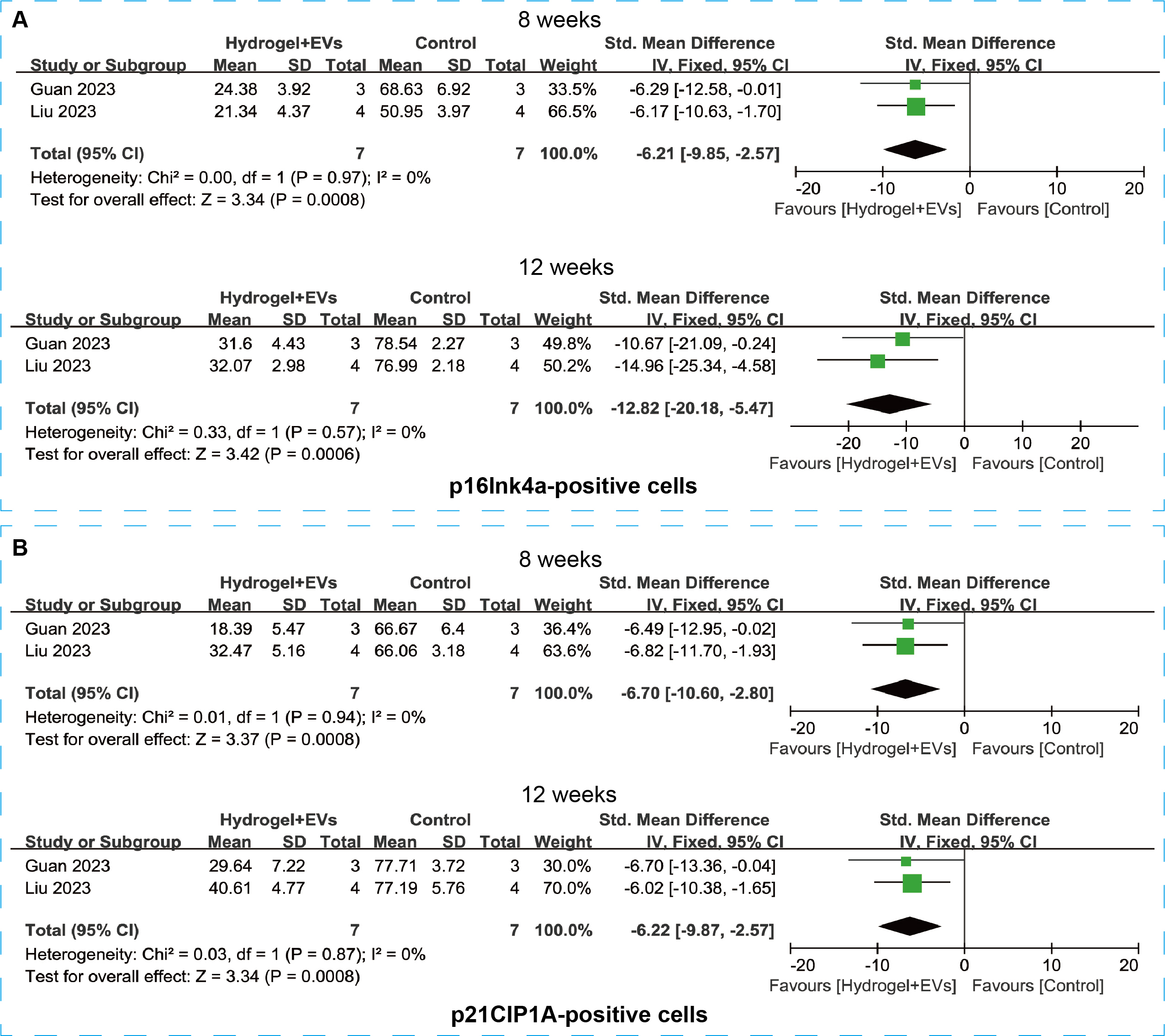 Fig. 7.
Forest plot showing the difference in p16Ink4a-positive cells (A) and p21CIP1A-positive cells (B) between the exosome-loaded hydrogel group and the control group at 8 weeks and 12 weeks post-treatment for IDD.
Four studies reported MRI grading of intervertebral discs at 8 weeks following treatment with exosome-loaded hydrogels, including two studies using gelatin-based hydrogels and two using composite hydrogels. Subgroup analysis revealed that both types of exosome-loaded hydrogels effectively reduced MRI grading of intervertebral discs (Supplementary Fig. 1), with no significant heterogeneity observed (I2 = 0%).
Due to significant heterogeneity in the pooled analysis of histological grade
(I2 = 61%), subgroup analyses were conducted based on hydrogel
type, exosome source, and animal model characteristics to identify potential
sources of heterogeneity. Regarding hydrogel type, studies were classified into
ECM-based hydrogels and composite hydrogels. Both types of exosome-loaded
hydrogels significantly reduced histological grading of intervertebral discs
(Supplementary Fig. 2). However, significant heterogeneity persisted in
the composite hydrogel subgroup (I2 = 60%), suggesting that
hydrogel type might not be the primary source of heterogeneity. When analyzed by
exosome source, subgroups were defined as MSC-derived and M2c macrophage-derived
exosome. The results showed that MSC-derived exosome-loaded hydrogels
significantly reduced histological grade (SMD = –5.45, 95% CI: –7.13 to
–3.77, p
Subgroup analyses based on animal model characteristics also revealed valuable
insights. The modeling method-based subgroup analysis showed that exosome-loaded
hydrogels reduced histological scores in both the needle puncture subgroup and
the LPS/TNF-
Given the significant heterogeneity in the combined effect size for histological grade (I2 = 61%), a sensitivity analysis was conducted to assess the robustness of the results. The sensitivity analysis revealed that, excluding individual studies, the size and direction of the combined effect size remained consistent (Fig. 8), suggesting that the summary analysis results for histological grade are robust.
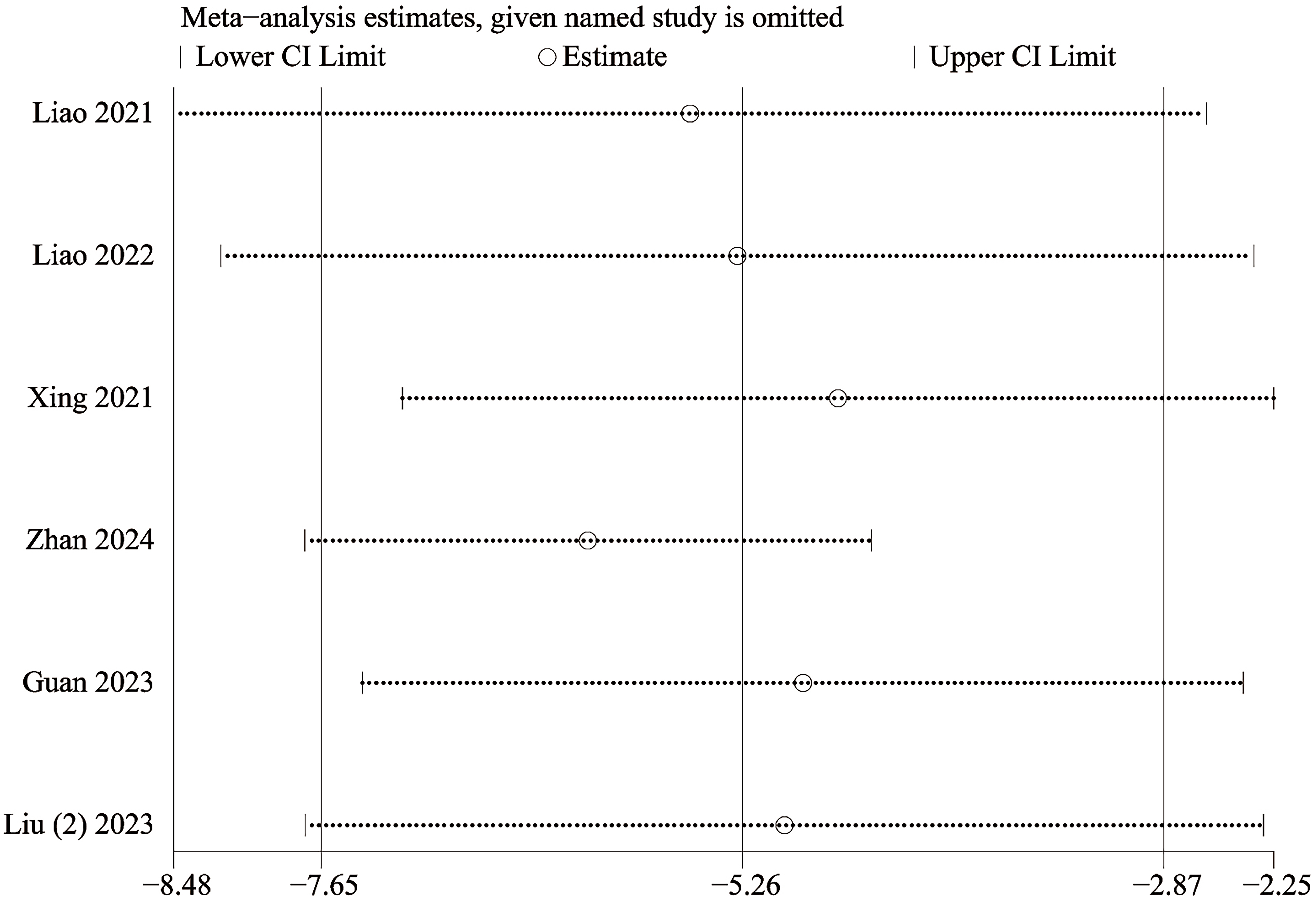 Fig. 8.
Sensitivity analysis of the studies included in histological grade. CI, confidence intervals.
This meta-analysis demonstrated that, compared to the control group, exosome-loaded hydrogels significantly improved the DHI scores, reduced MRI and histological scores, and enhanced the structure of intervertebral discs in IDD models, thereby slowing the degeneration process. Additionally, at the microscopic level, exosome-loaded hydrogels promoted the expression of COL2 protein, while decreasing MMP13 protein levels and reducing the number of p16Ink4a and p21CIP1A-positive cells. These results suggest that exosome-loaded hydrogels may improve IDD by promoting the synthesis of the intervertebral disc matrix, inhibiting its degradation, and slowing or reversing the aging process of disc cells.
The study found that exosome-loaded hydrogels significantly improved the DHI grade at both 4 weeks and 8 weeks, and significantly reduced the MRI grade at 8 weeks and 12 weeks, indicating a consistent positive effect on the recovery of intervertebral disc structure in the early stages of IDD. These findings align with previous research on exosome treatments for IDD. For instance, Liao et al. [32] reported that MSC-derived exosome significantly increased DHI scores and reduced MRI scores in the IDD model at 4 and 8 weeks, demonstrating a beneficial effect in delaying and improving IDD. Similarly, another study showed that hypoxia-preconditioned bone marrow mesenchymal stem cell (BMSC)-derived exosome upregulated DHI scores and downregulated MRI scores at 4 and 8 weeks, alleviating the progression of the rat IDD model [33]. These results further support the notion that exosome-loaded hydrogels can effectively improve disc height and morphology in IDD. However, while exosome-loaded hydrogels exhibit significant efficacy in improving disc degeneration in the short term, their therapeutic effects may be limited in later stages due to the delivery efficiency of exosome and the degradation rate of the hydrogel [34, 35]. This insight offers valuable context for our study, suggesting that precise control over exosome release may be crucial for exploring the long-term efficacy of exosome-loaded hydrogels in treating IDD.
As with previous meta-analyses, a thorough assessment of the potential risk of bias in the included studies is essential. The SYRCLE risk of bias tool, consisting of 10 items, was employed to evaluate the methodological quality of the preclinical studies. The assessment revealed that most studies exhibited an unclear risk of bias. Regarding selection bias, 90% of the studies did not report the method of random sequence generation (e.g., computer-based randomization or sealed envelope methods), and only one study mentioned allocation concealment. The absence of these measures may lead to imbalances in baseline characteristics, such as uncontrolled variations in animal age or weight, affecting the comparability of effect sizes. Three studies provided detailed descriptions of key baseline characteristics, such as animal age and weight, and were thus considered to have a low risk of bias in this domain. Conversely, one study lacked sufficient information on baseline characteristics and was rated as having a high risk of bias.
For selective reporting and incomplete outcome data, all included studies were judged to have a low risk of bias. However, in terms of performance and detection bias, none of the studies reported whether blinding was applied to operators or animal caretakers, which could lead to non-standardized administration of interventions, such as variations in exosome-loaded hydrogel dosage and frequency. Additionally, blinding during outcome assessment was not clearly described, raising concerns about potential bias, especially for subjective endpoints like histological scoring. Despite these uncertainties, the consistency in the direction of key outcome measures across the studies strengthens the overall stability of the meta-analysis results. Moving forward, future animal studies on the therapeutic effects of exosome-loaded hydrogels for IDD should adhere to the ARRIVE 2.0 guidelines [36] for standardized reporting, with particular emphasis on clear descriptions of randomization methods and the implementation of blinding procedures, to enhance the rigor and reproducibility of the evidence base.
Variations in animal models, hydrogel types, and exosome characteristics across
studies may introduce potential biases that could affect the outcomes of this
meta-analysis. The pooled analysis of histological scores for intervertebral
discs revealed significant heterogeneity (I2 = 61%). To address
this, subgroup analyses were conducted based on hydrogel type, exosome source,
administration route, intervention frequency, modeling method, and animal age.
The results showed that ECM-based hydrogels, composite hydrogels, stem
cell-derived exosome-loaded hydrogels, intradiscal administration, single-dose
administration, alternative injection frequency subgroups, needle puncture
models, and LPS/TNF-
In patients with intervertebral disc IDD, the balance between ECM degradation and synthesis is disrupted, often accompanied by abnormal MMP expression and a reduction in COL2 [37, 38]. Restoring the ECM metabolic balance is critical in treating IDD. Recent interest has focused on decellularized ECM, which, due to its reduced immunogenicity, may enhance the therapeutic efficacy of exosomes [39]. Subgroup analyses suggest that ECM-based hydrogels may play a beneficial role in reducing histological scores associated with IDD. exosome derived from various MSC sources are pivotal in slowing IDD progression, including exosome from BMSCs, umbilical cord-derived MSCs (UCMSCs), adipose-derived MSCs (ADSCs), induced pluripotent stem cells (iPSCs), and nucleus pulposus stem cells [40, 41]. Compared to the limitations of intravenous MSC infusion, MSC-derived exosomes overcome this barrier and demonstrate therapeutic efficacy in IDD treatment. Subgroup analysis indicates that MSC-derived exosomes significantly reduce histological scores, highlighting their potential in ameliorating IDD. However, research on exosomes derived from M2c macrophages for IDD treatment remains limited [30], emphasizing the need for further investigation to assess their therapeutic value in this context.
In pharmacological interventions for IDD, local injection at the disc site
offers the advantage of rapidly delivering therapeutic agents directly to the
lesion, maintaining a local therapeutic concentration, and is therefore the most
common delivery route in animal model studies. Previous research has demonstrated
that the intervertebral disc primarily acquires nutrients through diffusion from
blood vessels in adjacent vertebrae via the endplate [42]. Subgroup analysis
based on intervention characteristics revealed that intradiscal administration
significantly reduced histological scores. Whether administered as a single or
multiple injections, exosome-loaded hydrogels consistently led to a reduction in
histological scores post-injection. Further studies incorporating a greater
number of standardized intervention protocols are necessary to validate the
effects of different delivery routes and injection frequencies on therapeutic
outcomes in IDD animal models. Needle puncture is recognized as the standard
method for inducing IDD in animal models due to its minimally invasive nature,
stability, and reliability [43]. Other modeling approaches include chondroitinase
ABC, LPS, and TNF-
Exosome-loaded hydrogels significantly improved histological scores of IDD intervertebral discs and inhibited disc matrix degradation by upregulating COL2 expression and downregulating MMP13 expression. These findings align with the study by Fan et al. [45], who demonstrated that exosomes derived from M1 macrophages restored ECM metabolic balance in both in vitro and in vivo models by promoting COL2 expression and reducing MMP13 expression. Another study showed that exosomes derived from human adipose tissue stem cells alleviated rat IDD by downregulating MMP13 expression [46]. Additionally, this meta-analysis revealed that exosome-loaded hydrogels slowed the aging process of disc cells by reducing the number of p16Ink4a and p21CIP1A-positive cells. Peng et al. [31] further demonstrated that functionalized extracellular vesicles coupled with matrix hydrogels alleviate nucleus pulposus stem cell (NPSC) aging by targeting the Homeodomain-Interacting Protein Kinase 2 (HIPK2)/p53 pathway, promoting intervertebral disc regeneration. In summary, exosome-loaded hydrogels may alleviate IDD by restoring nucleus pulposus (NP) cell aging and ECM metabolic balance, with hydrogels contributing synergistically through their regenerative and anti-aging properties [47].
From a mechanistic perspective, multiple signaling pathways are involved in the
regulation of exosomes in IDD repair. MSC-derived exosomes maintain nucleus
pulposus cell (NPC) homeostasis by modulating autophagy balance. For instance,
Xiao et al. [48] observed that BMSC-exosomes promote autophagy by
inhibiting the Akt-mTOR pathway, reducing NPC apoptosis and alleviating IDD. Luo
et al. [25] developed ECM-modified hydrogels loaded with MSC-exosomes,
which promote autophagy in neural precursor cells by transporting Sphk2 to
activate the Phosphatidylinositol 3-Kinase (PI3K)/p-AKT pathway, improving IDD.
MSC-exosomes also regulate ECM synthesis and degradation. In a study by Liao
et al. [24], a thermo-responsive ECM hydrogel was developed to
continuously release MSC-exosomes, activating the Notch signaling pathway and
maintaining ECM metabolic balance. Additionally, miR-199a carried by
BMSC-exosomes improved IDD by inhibiting the TGF-
This study has several limitations. First, there are variations in the types of hydrogels, characteristics of exosomes, and animal model parameters across studies. Although subgroup analyses were performed to identify potential sources of heterogeneity, no significant sources were found. Second, the included studies exhibit an unclear risk of bias, particularly regarding randomization and blinding, which could impact the stability of the pooled analysis results. Finally, despite comprehensive literature searches, the limited number of studies and small sample sizes may affect the reliability of the summary analysis. Future research should focus on standardized, large-sample controlled trials to validate the preclinical efficacy of exosome-loaded hydrogels for IDD.
Although the results suggest that exosome-loaded hydrogels have a positive therapeutic effect in IDD treatment, their clinical translation faces several challenges. First, the source and preparation methods of exosomes can influence their therapeutic efficacy. While most studies used MSC-derived exosomes, those from different sources might vary in treatment outcomes. Second, the type of hydrogel and delivery method significantly impact therapeutic outcomes. Variations in hydrogel materials, such as gelatin and hyaluronic acid, may lead to differences in biodegradation rates, structural stability, and biocompatibility, directly affecting exosome release and therapeutic effects [51]. Future research should focus on optimizing exosome sources, selecting appropriate hydrogel types, and determining the most effective delivery methods to achieve optimal therapeutic outcomes. Moreover, while preclinical studies offer insights, they may not fully predict clinical outcomes, necessitating more rigorous clinical trials to confirm the efficacy and safety of exosome-loaded hydrogels for clinical applications.
This meta-analysis suggests that exosome-loaded hydrogels improve disc height, MRI scores, and histological grading, potentially alleviating IDD progression by promoting COL2 expression, inhibiting MMP13, and slowing intervertebral disc cell aging. However, the optimization of exosome-loaded hydrogel components and precise delivery methods remains critical to exploring their long-term efficacy in treating IDD. Future research should adhere to standardized protocols to further validate their efficacy and safety in both preclinical and clinical studies.
ADSCs, adipose-derived mesenchymal stem cells; ARRIVE, Animal Research: Reporting of In Vivo Experiments; BMSCs, Bone marrow mesenchymal stem cells; CI, confidence interval; COL2, collagen type II; DHI, disc height index; ECM, extracellular matrix; EV, extracellular vesicle; HIPK2, Homeodomain-Interacting Protein Kinase 2; IDD, intervertebral disc degeneration; IHC, immunohistochemical; iPSCs, induced pluripotent stem cells; LPS, lipopolysaccharide; MSC, mesenchymal stem cell; MMP13, matrix metalloproteinase 13; MRI, magnetic resonance imaging; MeSH, medical subject headings; NSAIDs, non-steroidal anti-inflammatory drugs; NPSCs, nucleus pulposus stem cells; NPCs, nucleus pulposus cells; PRISMA, Preferred Reporting Items for Systematic Reviews and Meta-Analyses; PI3K, Phosphatidylinositol 3-Kinase; SMD, standardized mean difference; SYRCLE, Systematic Review Center for Laboratory Animal Experimentation; TGF-
All data relevant to the study are included in the article. Further supplementary data be available from the first author or corresponding author upon reasonable request.
BW, DX and XC designed the study and wrote the manuscript. JH and ZH conducted a literature analysis search. WW, DH and YZ extracted the data and conducted the data analysis. All authors have read and agreed to the final version of the manuscript. All authors contributed to editorial changes in the manuscript. All authors have participated sufficiently in the work and agreed to be accountable for all aspects of the work.
Not applicable.
Not applicable.
This research was supported by the Natural Science Foundation of Xiamen (project number: 3502Z20227288).
The authors declare no conflict of interest.
Supplementary material associated with this article can be found, in the online version, at https://doi.org/10.31083/FBL38302.
References
Publisher’s Note: IMR Press stays neutral with regard to jurisdictional claims in published maps and institutional affiliations.