


 , Barani Kumar Rajendran 3, Senthilkumar Kaliamoorthy 4, Maddaly Ravi 5, Gomathy Baskar 1, Mugip Rahaman Abdul Wahab 1, Hemapreethi Surendran 1, Mahalakshmi Nannan 6, Manojkumar Govindaraj 7, Asha Sivaji 8, Wahidah H. Al-Qahtani 9, Rashid Ayub 9
1 Department of Biotechnology, Dr. M.G.R Educational and Research Institute, 600 095 Chennai, Tamil Nadu, India
2 ACS-Advanced Medical Research Institute, Dr. M.G.R Educational and Research Institute, Maduravoyal, 600095 Chennai, Tamil Nadu, India
3 Yale School of Medicine, Yale University, New Haven, CT 06510, USA
4 Department of Electronics and Communication Engineering, Dr. M.G.R Educational and Research Institute, 600 095 Chennai, Tamil Nadu, India
5 Department of Human Genetics, Sri Ramachandra Institute of Higher Education and Research, Porur, 600116 Chennai, Tamil Nadu, India
6 Department of Genetic Engineering, School of Bioengineering, SRM Institute of Sciences and Technology, Kattankulathur, 603 203 Chengalpattu, Tamil Nadu, India
7 PG & Research Department of Microbiology and Biotechnology, Presidency College, 600005 Chennai, Tamil Nadu, India
8 Department of Biochemistry, DKM College for Women, 632001 Vellore, Tamil Nadu, India
9 Department of Food Sciences & Nutrition, College of Food & Agriculture Sciences, King Saud University, 11451 Riyadh, Saudi Arabia
Abstract
Plant-mediated iron nanoparticles are increasingly utilized in biomedical and health applications due to their biocompatibility and nontoxicity. The therapeutic characteristics of these nanoparticles are extensively diverse.
In this study, iron nanoparticles synthesized from Tribulus terrestris were characterized using various techniques, including Fourier transform infrared (FTIR) spectroscopy, scanning electron microscopy (SEM), transmission electron microscopy (TEM), UV-visible spectroscopy, vibrating sample magnetometry (VSM), and X-ray diffraction (XRD) analysis. Antioxidant properties were assessed using the hydrogen peroxide (H2O2) and 2, 2-diphenyl-1-picrylhydrazyl (DPPH) assays. Anti-inflammatory activity was evaluated through protein denaturation studies. Antimicrobial activity was tested against wound pathogens. The effects of anticancer and wound healing were investigated using HCT-116 (colon cancer) and MG-63 (osteosarcoma) cells. Molecular docking studies were performed to assess the binding affinity of Tribulus terrestris bioactive compounds with proteins involved in the Adenomatous polyposis coli (APC) pathway of colon cancer.
The Tribulus terrestris-mediated Fe3O4 nanoparticles exhibited a peak at 290 nm using UV-visible spectroscopy. SEM and TEM analyses revealed that the nanoparticles were aggregated with an average size of 29 ± 0.24 nm. XRD analysis indicated a cubic crystalline structure. FTIR spectroscopy identified the biomolecules involved in the synthesis, and VSM confirmed a magnetic saturation of 14.75 emu/g. The antioxidant activity was demonstrated with DPPH (65.5%) and hydrogen peroxide (65.56%) assays at a dosage of 50 μg/mL, demonstrating a significant inhibition. The protein denaturation assay revealed a maximum inhibition of 54.57%. Lactobacillus had the strongest antibacterial activity at a concentration of 100 μg/mL, with an inhibitory zone of 35 mm. The anticancer assays showed IC50 values of 25.95 μg/mL for colon cancer (HCT-116) and 35.36 μg/mL for osteosarcoma (MG-63), indicating significant cytotoxicity, particularly against colon cancer cells. The nanoparticles also demonstrated effective regulation of cell migration at 50 μg/mL. Molecular docking studies revealed strong binding affinities between Tribulus terrestris compounds and APC pathway proteins relevant to colon cancer.
This research underscores the potential of Tribulus terrestris-mediated iron nanoparticles as a sustainable and eco-friendly approach with significant antioxidant and anticancer properties, especially in combating colon cancer. The findings highlight their effectiveness in reducing oxidative stress, inhibiting cancer cell proliferation, and enhancing wound healing.
Keywords
- Tribulus terrestris
- iron nanoparticles
- antibacterial
- antioxidant
- anticancer
- colon cancer
- molecular docking
- osteosarcoma
Cancer is characterized by uncontrolled cell proliferation caused by oncogenic signals that occur at an incorrect time and location [1]. Mutations in the DNA sequence of cancer cell genomes cause all malignancies [2]. Colon and bone cancer is one of the most common cancers, which leads to the highest mortality rate worldwide. Early detection of colon and bone cancer makes them extremely treatable and frequently curable with conventional therapy [3], but at the metastatic stage, it’s not treatable, and also treatments contain major obstacles like balancing treatment toxicity with quality and the high cost of disposing of chemical waste sludge, lack of therapeutic target, the resulting production of toxic metabolites, and the expense of operation. Hence, to get over these obstacles, researchers have studied the use of nanotechnology in cancer detection and therapy [4].
Nanotechnology is one of the most well-known multidisciplinary fields and a new area of technology that has enormous promise to provide ground-breaking discoveries with practical applications including physics, chemistry, biology, medicine, informatics, and engineering [5, 6]. The key characteristic of nanotechnology is its capacity to generate huge structures with essentially novel molecular organization at the molecular level, atom by atom [7]. Nanoparticles are a general term for materials having a minimum size of less than 100 nm, and they are a byproduct of nanotechnology [8]. Their atomic, electronic, and magnetic structures, physical and chemical characteristics, and reactivity about the bulk material are all significantly altered by the small number of atoms in the particles and the significant fraction of atoms that are at or near surfaces [9]. Concerns for the production of nanomaterials include social adaptation, environmental sustainability, and economic feasibility in addition to the availability of local resources [10, 11].
In silico approaches are advantageous for the investigation of the pharmacology of potential therapeutics through the use of computer-simulated models and for the development of biomedicines. The pharmaceutical industry has used in in silico methods for decades to find small molecules that can modulate the function of an identified target protein and thereby modulate the disease phenotype [12]. Iron oxide nanoparticles (Fe3O4-NPs) synthesised using Tribulus terrestris (T. terrestris) were evaluated using in vitro tests to determine their antimicrobial and cytotoxic effects. Compared to the Tribulus terrestris extract, T. terrestris-mediated iron nanoparticles demonstrated superior in vivo and in vitro results. This improvement is attributed to the active phytochemicals in the plant, which contribute significantly to the observed effects, as noted by Kaushik et al. [13].
Fe3O4 nanoparticles (Fe3O4-NPs) vary by their distinctive optical, electrical, antibacterial, and catalytic properties. The synthesis of iron NPs represents a significant advancement in nanomaterial due to their accessibility, relative affordability, and low toxicity [14, 15]. Plant-based pharmaceuticals continue to be a major source of therapeutic molecules. Consequently, Fe3O4-NPs have broad applications across various sectors, including biomedicine, bioremediation, engineering, cosmetics, and clinical materials [16, 17]. They are increasingly recognized as promising options for cancer treatment [18].
Tribulus terrestris exhibits a range of pharmacological activities, including effects on the central nervous system, anti-urolithiasis, aphrodisiac properties, cardiac health, antibacterial action, hepatoprotection, analgesic effects, antispasmodic properties, anticancer activity, anthelmintic properties, larvicidal effects, diuretic action, and anti-cariogenic properties [19, 20]. Plants with anti-cancer properties primarily act by inhibiting cancer-promoting enzymes, repairing DNA, promoting the synthesis of antitumor enzymes, boosting immunity, and providing antioxidant effects without harming healthy human cells [21, 22]. Tribulus terrestris is particularly rich in spirostanol and furostanol saponins, with diosgenin being a key spirostanol sapogenin. Measuring diosgenin is crucial for the quality control of T. terrestris, which also has potent antilithiatic activity demonstrated by in vitro and in vivo studies [23].
When combined with herbal treatments, nanomedicines can deliver drugs specifically to the target site, minimizing effects on healthy cells, stomach acidity, liver metabolism, and blood circulation. Their small size also extends the duration of the drug’s presence in the bloodstream [24]. Iron nanoparticles are increasingly emphasized for their role in diagnostics and drug delivery, particularly in cancer treatment, where they enhance the efficacy of primary irradiation. Magnetic orientation ensures that magnetic particles reach the tumor site, potentially having a significant anticancer effect [12].
This is the first study to demonstrate that iron nanoparticles (Tt-Fe3O4-NPs) produced from the aerial parts of T. terrestris are effective against colon cancer cells. The research investigated Fe3O4-NPs biosynthesized from T. terrestris aerial parts using in vitro and in silico methods. Notably, Tribulus-Fe3O4-NPs were found to be more effective against colon cancer cells than bone cancer cells at lower concentrations.
Tribulus terrestris (Tt) was used in this study. It is a flowering plant with yellow flowers and is included in numerous traditional medicine formulations. The plant was collected from Pudhur and its surrounding areas in Chennai. It was identified as 387.1753 and authenticated by the Siddha Central Research Institute in Chennai.
The aerial parts of the plant were washed and cleaned with distilled water, then cut into small pieces, shade-dried, and finally powdered. This powder was used in the synthesis of nanoparticles [25].
A beaker containing 100 mL of deionized water and 1g of powdered plant material was heated at 60 °C for 30 minutes. The extract was then stored at ambient temperature, cooled, and filtered through Whatman filter paper. Fe3O4-NPs were synthesized from the filtrate. In a conical flask, 50 mL of the extract was mixed with 50 mL of ferric chloride and stirred for 20 minutes using a magnetic stirrer. The reaction mixture was continuously stirred to produce Fe3O4-NPs. The appearance of a black color indicated the formation of nanoparticles [26].
Ascorbic acid (vitamin C), Antibiotic-antimycotic solution, Bovine Serum Albumin
(BSA), FeCl₃ (ferric chloride), Hydrogen peroxide (H2O2),
2,2-Diphenyl-1-picrylhydrazyl (DPPH) were purchased from HiMedia, India. Methyl
Thiazole Tetrazolium (MTT) salt, trypsin-EDTA, Dulbecco’s Modified Eagle Medium
(DMEM), and 100
The optical characteristics of the nanoparticles were examined using UV-visible spectroscopy (Shimadzu, Kyoto, Japan). The synthesised Fe3O4-NPs were analyzed in this work by identifying their highest absorption peak within a particular wavelength range of 200–700 nm [27]. Fourier-transform infrared spectroscopy (FTIR) (IR Prestige 21, Shimadzu, Kyoto, Japan) was used to analyze the functional groups of the iron oxide nanoparticles with scan rates between 400 and 4000 cm-1 were used for this analysis [28]. X-ray diffraction (XRD) (PANalytical, Almelo, Netherlands) patterns were utilized to determine the crystal structure of the nanoparticles [29]. Scanning electron microscopy (SEM) (FEI, Quanta 200, Hillsboro, OR, USA) [30] and transmission electron microscopy (TEM) (JEOL Japan, JEM-2100 plus) [31] provided information on the size and morphology of the nanoparticles. The magnetic properties, including magnitude and direction of the magnetic field, were assessed using a vibrating sample magnetometer (VSM) [32].
The DPPH radical scavenging activity of Tribulus-Fe3O4-NPs and ascorbic acid was evaluated using a standard method. Test samples and standard ascorbic acid at varying concentrations (10–50 µg/mL) were mixed with DPPH (1 mL of 0.1 mM) diluted in 50% ethanol. The reaction mixture was incubated for 30 minutes and then measured spectrophotometrically at 517 nm [33].
Percentage of Inhibition = [(Ab – As) / Ab]
where Ab = absorbance of the blank and As = absorbance of the test samples.
The scavenging activity of vitamin C and Tribulus-Fe3O4-NPs against hydrogen peroxide was tested. Phosphate buffer was added to an aqueous solution of 40 mM hydrogen peroxide (pH 7.4). A 0.6 mL solution of hydrogen peroxide containing standard and test samples at concentrations (10, 20, 30, 40, and 50 µg/mL) was prepared. The mixture was analyzed spectrophotometrically at 230 nm after a 10-minute incubation period [34].
Percentage of Inhibition = [(Ab – As) / Ab]
where Ab = absorbance of the blank and As = absorbance of the test samples.
The protein denaturation assay was conducted using the method outlined by Sultana et al. [35]. The reaction mixture (5 mL) consisted of 0.02 mL of extract, 4.78 mL of phosphate-buffered saline (PBS, pH 6.4), and 0.2 mL of 1% bovine serum albumin. After incubation at 37 °C for 15 minutes, the mixture was heated to 70 °C for 5 minutes and then cooled. The intensity of protein denaturation was measured at 660 nm using a UV-visible spectrometer. The control solution was phosphate buffer. The percentage of inhibition was calculated as follows:
Percentage of Inhibition = [(Ab – As) / Ab]
where Ab = absorbance of the control sample and As = absorbance of the test samples.
The antibacterial activity of Tribulus-Fe3O4-NPs was evaluated using the agar well diffusion technique. Bacterial cultures of Lactobacillus, Vibrio cholerae, Staphylococcus aureus, and Pseudomonas aeruginosa were maintained in nutrient broth. A 24-hour bacterial culture was used to swab Muller Hinton agar plates. Wells (6 mm in diameter) were punched into the agar plates, and 25 µL, 50 µL, and 100 µL of the green-synthesized Fe3O4-NPs were added to each well, along with 30 µL of a control antibiotic. The plates were incubated at 37 °C for 24 hours, and the zone of inhibition around the wells was measured [36].
Anticancer drug testing was conducted using human colorectal cancer (HCT-116) and bone cancer (MG-63) cells. Cell lines were obtained from the National Centre for Cell Science (NCCS) in Pune, India, and cultured in T-25 flasks with DMEM medium. The medium was supplemented with antimitotic solution (1%), fetal bovine serum (10%) and antibiotics. The flasks were maintained in an incubator (37 °C) with 5% carbon dioxide (CO2) and monitored regularly [37]. Cell lines were validated by short tandem repeat (STR) profiling and tested negative for mycoplasma. Cells were all cultured in a humidified incubator at 37 °C and 5% CO2.
The cytotoxicity of Tribulus-Fe3O4-NPs was tested against colorectal
cancer (HCT-116) and bone cancer (MG-63) cells. Cells were plated into 96-well
plates at a density of 4
In a 6-well plate, 7.5
Molecular docking predicts ligand-protein interactions and is widely used in structural biology and drug development. Protein-ligand interactions are crucial for structure-based drug design and protein function prediction, as noted by Naqvi et al. [40]. PyrX virtual screening tool version 0.8 (https://sourceforge.net/projects/pyrx/) was used for virtual docking, and Discovery Studio was used for interaction analysis.
Docking small molecule compounds into receptor binding sites and determining the binding affinity of the complex are essential steps in drug development. Ibrahim et al. [41] conducted a GC/MS study of Tribulus terrestris fractions, identifying bioactive substances in the plant’s methanol extract. These substances were selected as ligands for docking studies. The chosen compounds are stearic acid (CID: 5281), 2-hexanol, 2-methyl (CID: 12240), hexadecanoic acid (CID: 8181), octadecanoic acid (CID: 8201), 2-pentadecanone (CID: 10408), coumaric acid (CID: 323), benzofuran (CID: 10329), arachidic acid (CID: 444899), and 5FU (CID: 3385). The PyMOL program 3.0.4 (Schrödinger, Inc. New York, NY, USA) was used to convert ligand structures into PDB (Protein Data Bank) format for docking studies.
Proteins involved in the APC pathway were selected for computational docking based on their role in colon cancer development. Proteins were obtained from the RCSB PDB collection. The selected proteins are KRT8 (PDB ID: P05787), TFRC (PDB ID: 3KAS), KLK6 (PDB ID: 1LO6), C3AR1 (PDB ID: 81A8), AHSG (PDB ID: P02765), GRP (PDB ID: 7W3Z), APOC1 (PDB ID: 6DVU), and CAV1 (PDB ID: Q03135). Ligands and water molecules were removed, and hydrogen bonds were added to upgrade the structures for docking studies [42].
Active sites of the proteins were predicted using the Castp server. The PDB file was converted, and findings were used to predict active sites. The grid box dimensions for docking studies were set by condensing residues associated with these active sites in PyRx tools [42].
Statistical analysis was performed using Microsoft Excel 2019 (Redmond, Washington, United States) to calculate standard deviation (SD) and compare test and control sample results. Results were considered statistically significant when the p value was less than 0.05 [42].
The synthesis of iron nanoparticles was indicated by an immediate change in solution color and a decrease in pH after 1 hour of incubation when plant extract was added to the aqueous FeCl₃ solution. The reaction between ferric chloride and Tribulus terrestris aerial extract resulted in a color change from pale orange to dark brown (Fig. 1A,B). This color alter indicates that iron oxide nanoparticles (FeO) are being produced in solution as a result of the reduction of ferric ions (Fe3+). Ferric chloride solution addition of effects, such as stabilization of the nanoparticles and a reduction in Fe3+. Fe3O4-NPs synthesized from tribulus, were examined for absorption spectrum in the range of 200–800 nm wavelength. Periodically, the biosynthesized Fe3O4-NPs were initially, not shown any noticeable peaks. The synthesized T. terrestris-mediated iron nanoparticles (Tt-Fe3O4-NPs) showed a clear peak at 290 nm after the incubation of 30 minutes, shown in Fig. 2. Similar results were observed in previous studies, including the synthesis of Fe3O4-NPs from Tribulus terrestris leaves [43] and Phyllanthus niruri plant leaf and fruit parts [44].
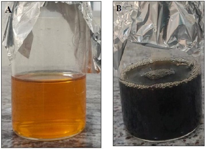 Fig. 1.
Synthesis of T. terrestris-mediated iron nanoparticles. (A) Before colour change, (B) After colour change.
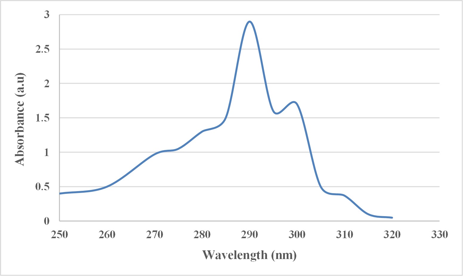 Fig. 2.
UV-visible spectroscopy analysis of T. terrestris-mediated iron nanoparticles.
FTIR spectroscopy was employed to identify the functional groups involved in the reduction of Fe3O4 nanoparticles by Tribulus terrestris and to determine the specific groups responsible for this process. The FTIR spectrum of the T. terrestris extract showed bands at 2970.80 cm⁻1 and 2885.51 cm⁻1, corresponding to the C-H stretching of alkanes, indicating the presence of alkane groups. Fig. 3 depicts additional bands at 2121.70 cm⁻1 (N=N=N stretching, indicating azide groups), 1639.49 cm⁻1 (C=C stretching of alkenes), 1384.89 cm⁻1 (C-H bending of alkanes), 1321.24 cm⁻1 (N-O stretching of nitro compounds), and 1038.99 cm⁻1 (C-O stretching of primary alcohols). The broad and intense peak at 2970.38 cm⁻1 suggests the presence of O-H stretching bonds from both alcohol and phenolic groups in the synthesized T. terrestris-Fe3O4-NPs.
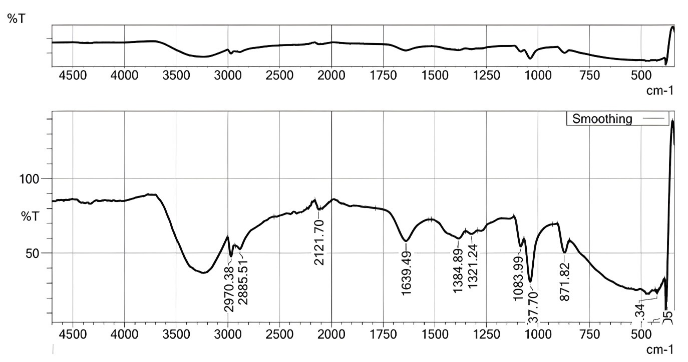 Fig. 3.
FTIR spectra analysis of T. terrestris-mediated iron nanoparticles. FTIR, Fourier-transform infrared spectroscopy.
Phenolic compounds in plant extracts are often attributed as the primary agents responsible for the reduction of Fe3⁺ to Fe0 [45]. Rajendran et al. [46] conducted FTIR analysis on iron oxide nanoparticles synthesized from Sesbania grandiflora (Agati) and observed a significant peak at 1650 cm⁻1, corresponding to N-H bending, with a visible bandwidth spanning 1500–600 cm⁻1. Similarly, Pallela et al. [47] identified functional groups in Sida cordifolia through FTIR spectroscopy, noting strong peaks around 2928 cm⁻1 and 2429 cm⁻1, which correspond to methyl and amine groups, respectively.
X-ray powder diffraction (XRD) was utilized to evaluate the crystalline structure and phase of the nanoparticles. The XRD pattern of iron oxide nanoparticles synthesized from Tribulus terrestris (T. terrestris)-Fe3O4-NPs is shown in Fig. 4. The diffraction peaks observed at 15.96°, 20.67°, 32.31°, and 31.01° correspond to Bragg’s reflections for the (220), (311), (400), (440), and (533), respectively. These peaks are consistent with the Fe3O4 nanoparticle data in the JCPDS database. Dhuper et al. [48] performed XRD analysis on Mangifera indica-mediated Fe3O4-NPs and found that the X-ray diffraction, using a graphite monochromator, revealed a rhombohedral crystalline structure with a wavelength of 1.54060 Å. Similarly, Patil et al. [49] reported that Triadica procumbens-mediated Fe3O4-NPs displayed peaks at 17.24°, 32.5°, and 39.06°, which corresponded to the (311) planes and confirmed the cubic structure of the nanoparticles.
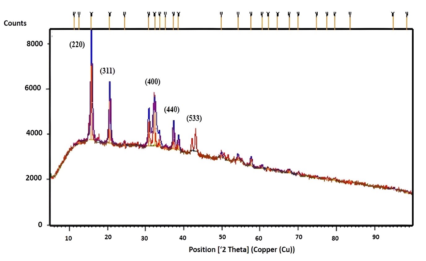 Fig. 4.
XRD analysis of T. terrestris-mediated iron nanoparticles. XRD, X-ray diffraction.
Scanning Electron Microscopy (SEM) provides high-resolution images of
nanoparticles, revealing their size, shape, composition, electrical conductivity,
topography, and surface features [50]. The SEM analysis of Tribulus
terrestris-Fe3O4-NPs showed clusters of nanoparticles with an irregular
spherical shape (Fig. 5A,B). Transmission Electron Microscopy (TEM) further
revealed that the green-synthesized T. terrestris-Fe3O4 nanoparticles
were predominantly spherical and exhibited accumulation. This morphology is
attributed to the hydroxyl groups present in the plant extract. The nanocomposite
was composed of iron and oxygen, with a mean diameter of 29
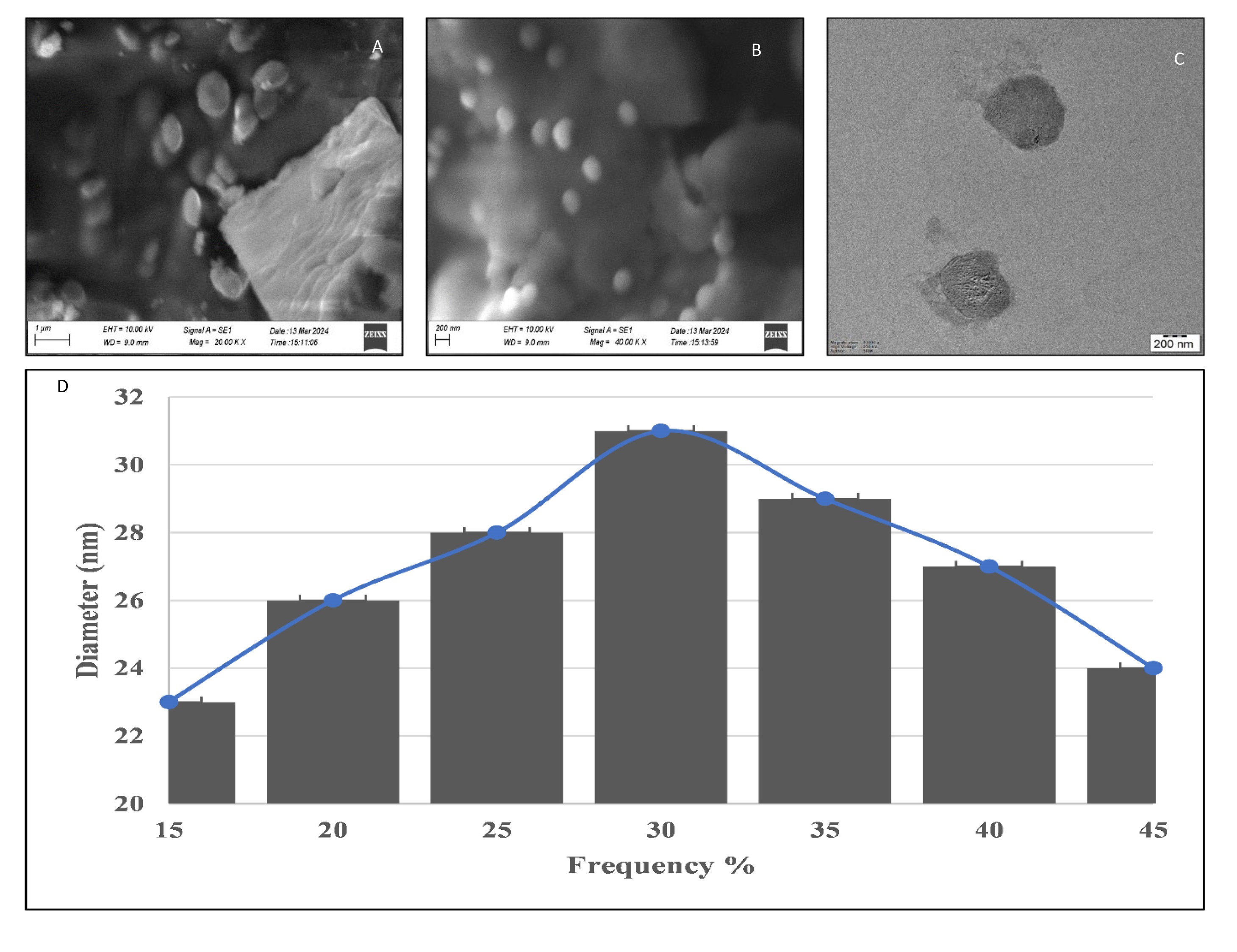 Fig. 5.
SEM and TEM analysis of T. terrestris-mediated iron nanoparticles. (A,B) SEM micrograph images in different magnification. Scale bar: 1 µm. (C) TEM image of T. terrestris-Fe3O4-NPs. Scale bar: 200nm. (D) Size distribution histogram.
A prominent method for determining the magnetic properties of materials is
vibrating sample magnetometry (VSM). This method involves subjecting samples to a
varying electromagnetic field to measure properties such as magnetic retentivity
(Mr), coercivity (Hci), and saturation magnetization (Ms). The magnetic
properties of Tribulus terrestris-mediated Fe3O4-NPs were evaluated
using VSM. Fig. 6 illustrates the magnetic behavior of these nanoparticles. The
Fe3O4-NPs showed a saturation magnetization (Ms) of 14.75 emu/g, a coercivity
(Hci) of 285.28 Oe and a magnetic retentivity (Mr) of 1.0244
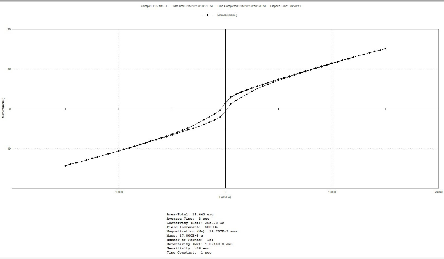 Fig. 6.
Magnetic characterization of T. terrestris-mediated iron nanoparticles by vibrating sample magnetometry(VSM) technique.
Mahdavi et al. [54] studied the magnetic properties of Fe3O4-NPs synthesized from Sargassum muticum extract. These nanoparticles exhibited superparamagnetic behavior with a saturation magnetization of 22.1 emu/g, a coercivity of 82.3 Oe, and no remanence, indicating the presence of a single magnetic domain. Similarly, Izadiyan et al. [55] reported high saturation magnetization in iron oxide nanoparticles obtained from Juglans regia. These nanoparticles displayed a high saturation magnetization of 53.32 emu/g and a low coercivity of 32.50 Oe. The magnetization curves confirmed the high saturation magnetization and low coercivity characteristics of the J. regia-derived iron oxide NPs.
Free radicals are molecules that are extremely reactive and possess unpaired electrons. They have the potential to cause cellular injury during the metabolic process. This oxidative stress is mitigated by antioxidants, which are recognized for their ability to neutralize these free radicals. Secondary metabolites in plants often exhibit antioxidant properties, despite being non-nutritive, and can benefit human health when consumed [56].
The DPPH and hydrogen peroxide radical assays were employed to evaluate the antioxidant activity of Tribulus terrestris-mediated Fe3O4-NPs. The Fe3O4-NPs effectively neutralized free radicals. In the DPPH assay, conducted with doses ranging from 10 to 50 µg/mL, the Fe3O4-NPs demonstrated significant radical-scavenging activity, with a peak inhibition of 65.5% at 50 µg/mL. For comparison, standard ascorbic acid (Vitamin-C) showed an inhibition of 70.4% at the same concentration, as illustrated in Fig. 7. Nandini and Subhashini [57] reported a DPPH radical scavenging activity of about 61.4% in Cuminum cyminum, while Deshmukh et al. [58] observed a maximum radical scavenging activity of 60% in fenugreek seed-mediated Fe-NPs.
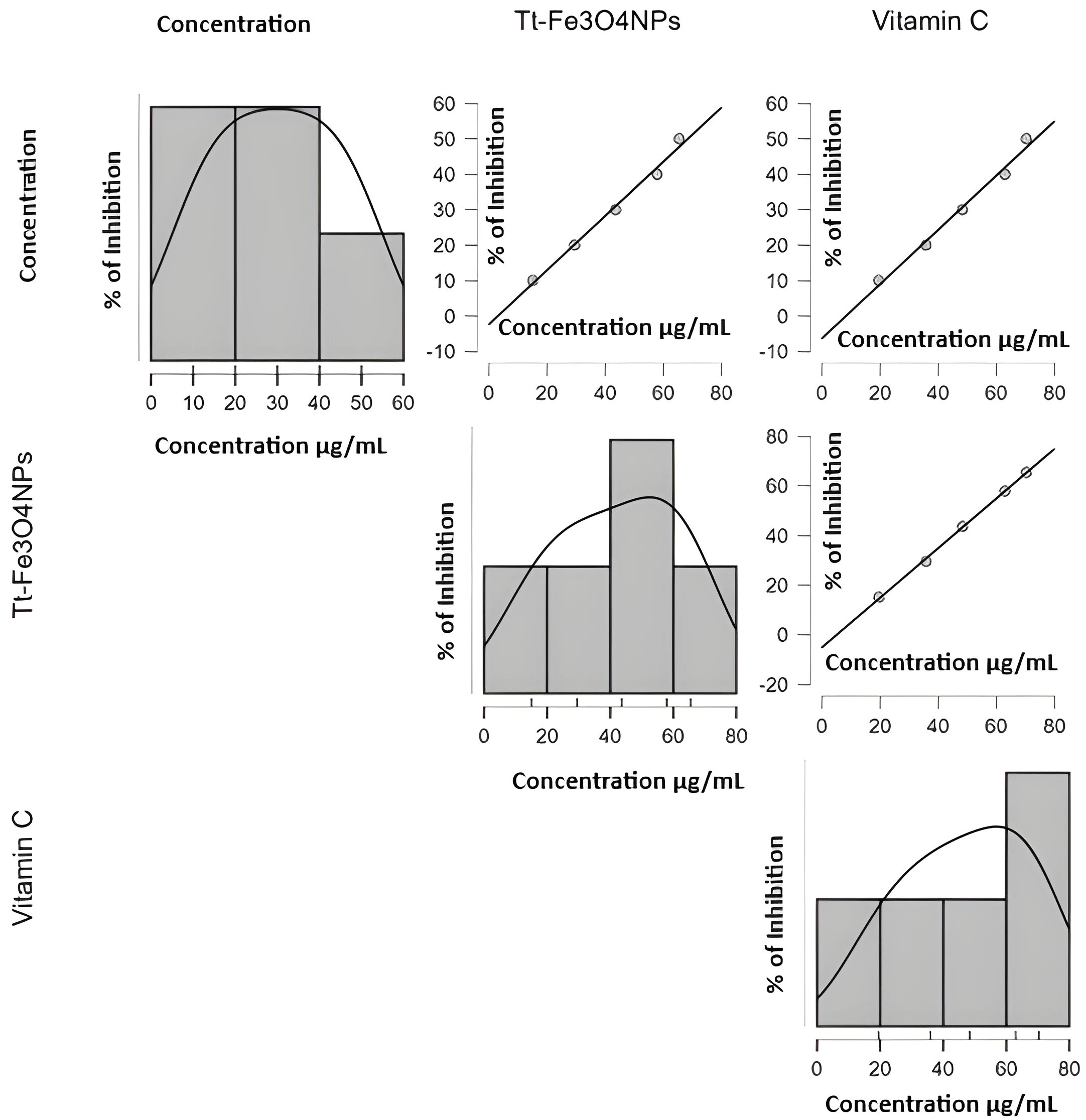 Fig. 7.
DPPH assay of T. terrestris-mediated iron nanoparticles. DPPH, 2,2-Diphenyl-1 picrylhydrazyl.
In the hydrogen peroxide assay, T. terrestris-Fe3O4-NPs exhibited 65.56% inhibition of free radicals at a concentration of 50 µg/mL. Fig. 8 shows that vitamin C suppressed free radicals by 74.23% at the same dose. Essa et al. [59] reported a 42.38% scavenging activity for hydrogen peroxide in Vitis vinifera leaf-mediated Fe-NPs, while Sandhya and Kalaiselvam [60] observed a 44.12% radical scavenging activity in Borassus flabellifer tender seed-synthesized iron nanoparticles. The significant antioxidant activity of Tribulus terrestris is attributed to its numerous phenolic hydroxyl groups [61]. Based on Lushchak [62], antioxidants reduce oxidative damage to biological components and restrict cancer cell development by suppressing free radicals.
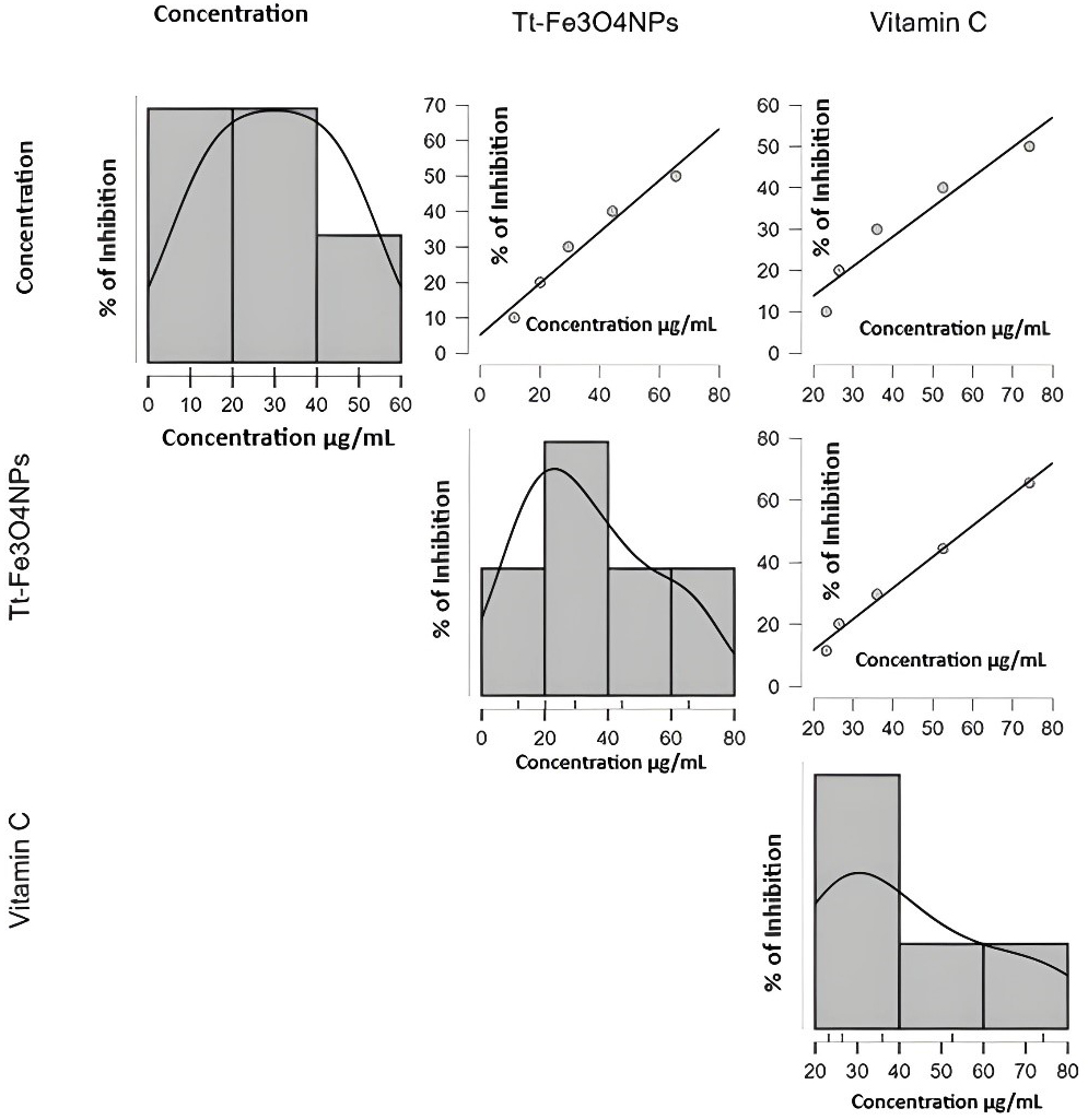 Fig. 8.
Hydrogen peroxide assay of T. terrestris-mediated iron nanoparticles.
Tribulus terrestris-synthesized Fe3O4-NPs demonstrated the capacity to inhibit the denaturation of bovine serum albumin (BSA). At 50 µg/mL, Fe3O4-NPs showed maximum inhibition of 43.28. For comparison, the standard drug diclofenac, at the same concentration of 50 µg/mL, achieved a higher inhibition of 54.57%, as shown in Fig. 9. Khuda et al. [63] investigated the in vitro anti-inflammatory activity of crude extracts from Xanthium strumarium and Achyranthes aspera leaves, reporting a maximum inhibition of 73%. Additionally, Bailey-Shaw et al. [64] studied the anti-inflammatory properties of 99 plant samples. Among these, iron nanoparticles synthesized from Mangifera indica (Julie mango) leaves showed a significant inhibition of 72.60%.
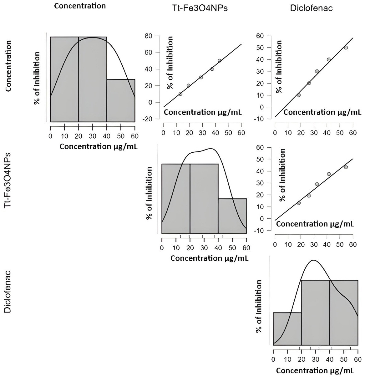 Fig. 9.
Protein denaturation assay of T. terrestris-mediated iron nanoparticles.
The antibacterial efficacy of the synthesized nanoparticles is comparable to that of the original Tribulus terrestris plant extract. This antimicrobial activity arises because the functional groups from the plant extract, which possess inherent herbal qualities, saturate the nanoparticle surfaces. The nanoparticles’ diminutive size enables them to easily pass through the bacterial cell walls, resulting in the demise of the cells [65].
The antibacterial efficacy of the synthesized T. terrestris-Fe3O4-NPs was evaluated, as illustrated in Fig. 10. At a concentration of 100 µL, the maximum zones of inhibition were observed against Lactobacillus (35 mm) and Vibrio cholerae (34 mm). Moderate inhibition was noted for Staphylococcus aureus (19 mm) and Pseudomonas aeruginosa (25 mm). The synthesized nanoparticles demonstrated the highest effectiveness against Lactobacillus, as illustrated in Fig. 11.
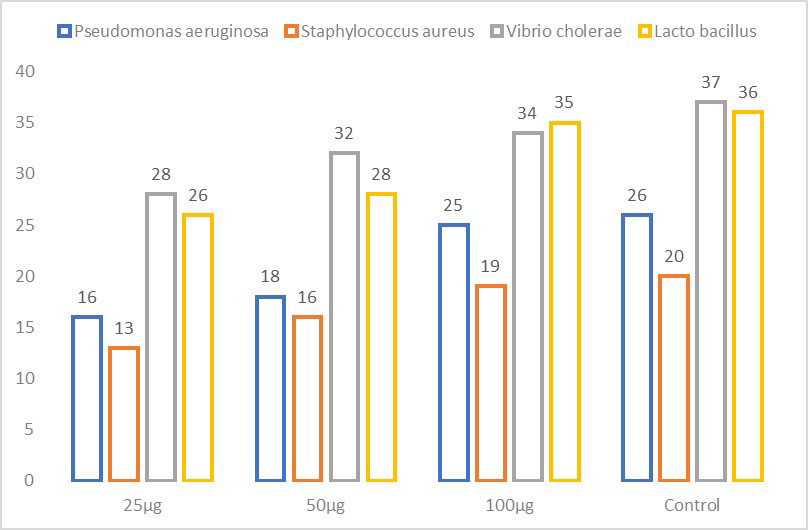 Fig. 10.
Antibacterial activity of T. terrestris-mediated iron nanoparticles against wound pathogens.
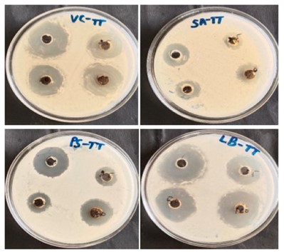 Fig. 11.
Zone of inhibition of T. terrestris-mediated iron nanoparticles.
Kanagasubbulakshmi and Kadirvelu [65] reported similar findings for nanoparticles synthesized from Lagenaria siceraria. Their study showed that these nanoparticles had comparable antibacterial potential to the plant extract, with higher efficacy against Staphylococcus aureus at higher concentrations. At lower concentrations, however, the nanoparticles did not show inhibition, as depicted in Fig. 11. Additionally, Yadav et al. [66] examined the antibacterial efficiency of Aloe vera nanoparticles against Klebsiella pneumoniae and reported a 17-mm maximal inhibitory zone.
The MTT method was used to evaluate the in vitro anticancer effectiveness of T. terrestris-Fe3O4-NPs against the HCT-116 colon cancer cell line. The results indicated that higher concentrations of T. terrestris-Fe3O4-NPs led to a greater percentage of cell inhibition. The half-inhibitory concentration (IC₅₀) for HCT-116 cells was 25.95 µg/mL, as shown in Fig. 12. In contrast, for the MG-63 (osteosarcoma) cell lines, the IC₅₀ value was 35.36 µg/mL, depicted in Fig. 13. Microscopic images of both cell lines reveal that T. terrestris-Fe3O4-NPs are more effective against colon cancer than bone cancer. According to Alkahtane et al. [67] studied the cytotoxicity of the iron oxide nanoparticles on HCT 116 cell lines and it limit proliferation at 55 mg/mL and less toxic for fibroblast cells.
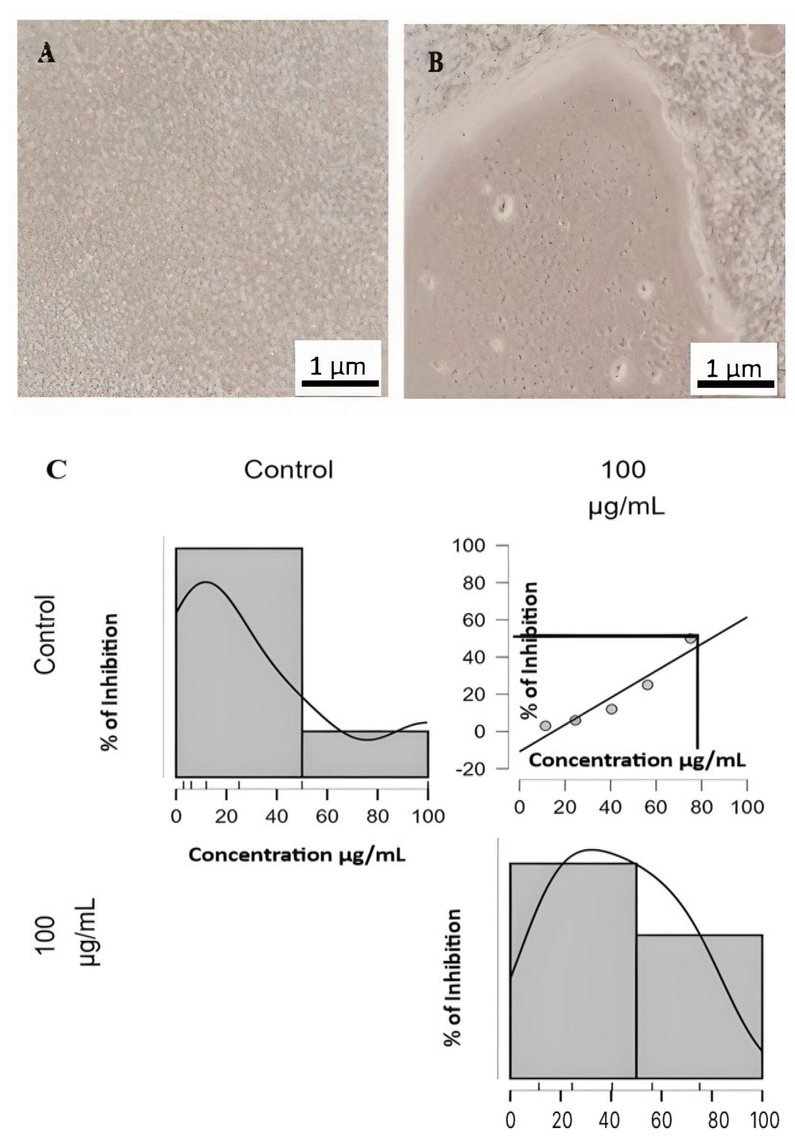 Fig. 12.
Cell Viability (A) Control cells (B) Treated Cells (C) Cytotoxic activity of T. terrestris-mediated iron nanoparticles in HCT-116 cell line. Scale bar: 1 µm.
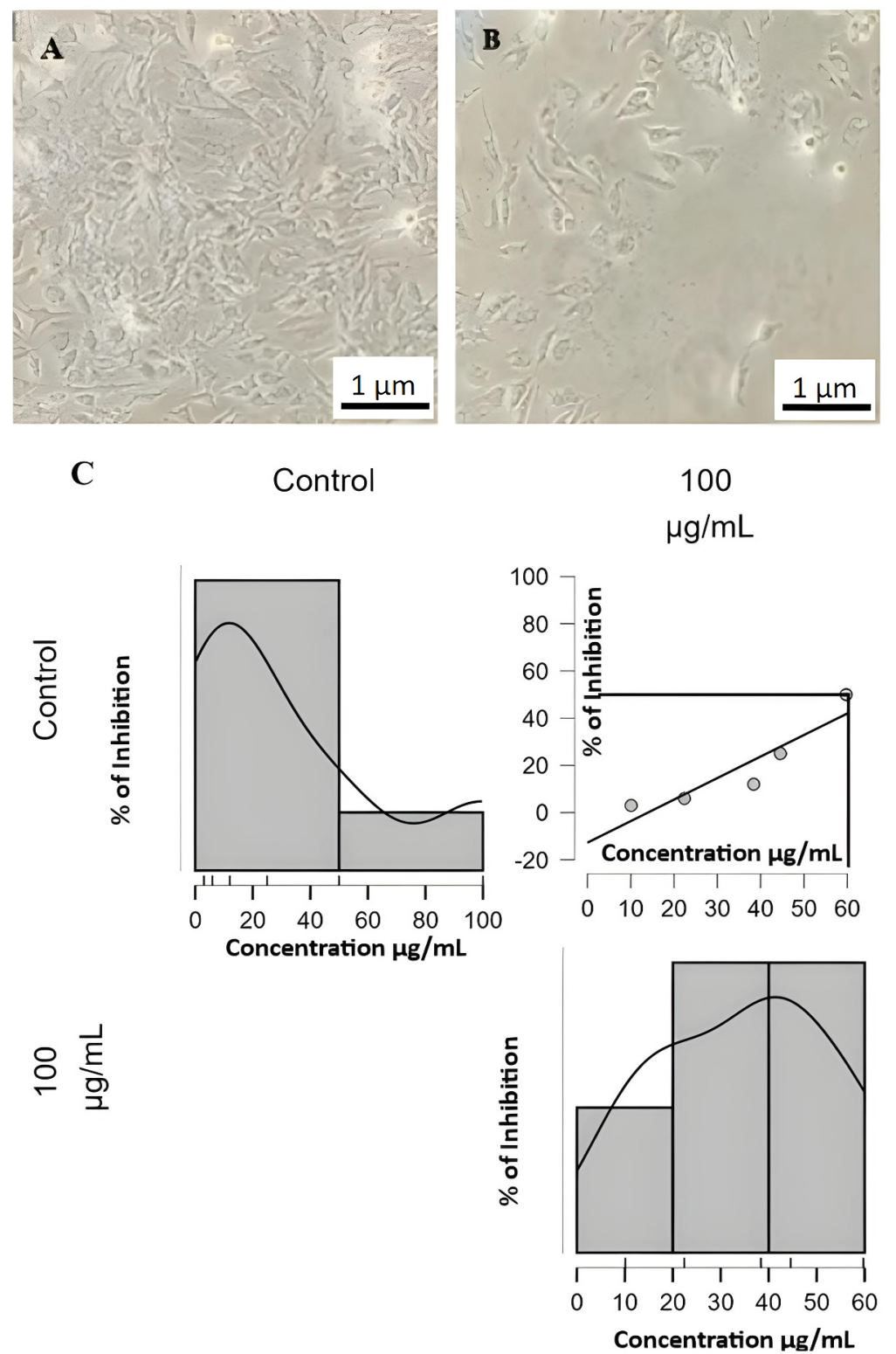 Fig. 13.
Cell Viability (A) Control cells (B) Treated Cells (C) Cytotoxic activity of T. terrestris-mediated iron nanoparticles in MG-63 cell line. Scale bar: 1 µm.
Yusefi et al. [68] investigated the cytotoxic effects of Fe3O4-NPs maintained with Garcinia mangostana fruit peel on HCT-116 and normal CCD112 cell lines. At a dosage of 250 µg/mL, a 68% suppression of colon cancer cells was observed. Chauhan et al. [69] noted that malignant cells, lacking a protective mucus layer, are more susceptible to therapeutic agents like those derived from T. terrestris. Based on these cytotoxicity results, further anticancer screening of T. terrestris-Fe3O4-NPs at concentrations of 50, 25, and 10 µg/mL was deemed necessary. According to Ratan et al. [70], plant extracts include biological components that serve as capping and reducing agents in nanoparticles, effectively inhibiting cancer cell development. The dimensions, and morphology of iron nanoparticles are also contributing factors in inducing apoptosis in cancer cells by stimulating the increase of reactive oxygen species (ROS). As per Ansari and Asiri [71], the anticancer mechanism of iron oxide nanoparticles is caused by releasing Fe ions to eliminate cancer cells. It is conceivable that Fe3O4-NPs are responsible for the development of reactive oxygen species, which eradicate malignant cells.
The impact of green synthesized T. terrestris-Fe3O4-NPs on cancer cell migration was evaluated through a wound healing assay using HCT-116 colon cancer cells. This study showed that T. terrestris-Fe3O4-NPs at 10, 25, and 50 µg/mL substantially reduced wound closure compared to normal control cells. Notably, at the highest concentration of 50 µg/mL, the migration of HCT-116 cells was reduced by 90%, as illustrated in Fig. 14. This result indicates that the nanoparticles effectively hindered cancer cell migration, in contrast to the untreated control cells, which showed continued migration and spread.
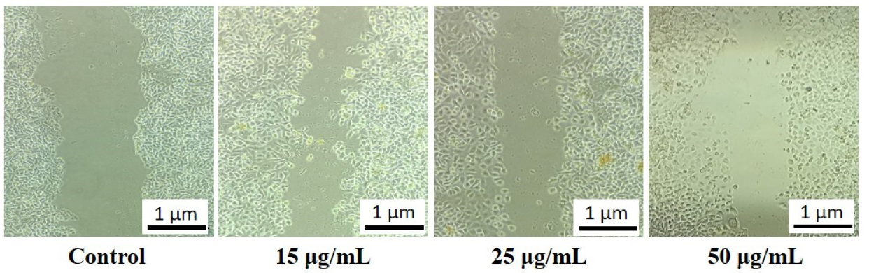 Fig. 14.
Wound healing assay of T. terrestris-mediated iron nanoparticles. Scale bar: 1 µm.
The results underscore the potential of plant-mediated iron nanoparticles in reducing tumor cell migration. Such nanoparticles may enhance wound healing processes by promoting repair mechanisms and collagen synthesis, thereby improving tensile strength [72]. Adnan et al. [73] conducted a similar wound closure assay with HCT-116, and A549 cells were treated with an ethanolic extract of Selaginella repanda, observing dose-dependent inhibition of cell migration.
The command-line software utilized in this study divided the docking results into an output registry in PDB format and a log file in text format. Given the frequent abnormalities in the Adenomatous Polyposis Coli (APC) pathway in colon cancer, which underscores its significance in colon cancer development [74], the docking analysis was centered on proteins associated with the APC pathway. Binding affinity values derived from molecular docking investigations are outlined in Table 1. The results indicated that coumaric acid effectively targets several proteins involved in colon cancer, including KRT8, TFRC, KLK6, C3AR1, AHSG, GRP, APOC1, and CAV1, with 5-Fluorouracil used as a standard drug for comparison. Figs. 15,16 highlight that coumaric acid demonstrated the highest binding affinities with GRP (6.8), TFRC (6.2), KLK6 (6), and C3AR1 (6) proteins. These results suggest that coumaric acid exhibits substantial binding affinity towards key proteins associated with colon cancer, as evidenced by these bioinformatics analyses. Zhang et al. [75] also investigated the binding affinity of ginger compounds with colon cancer proteins using in silico methods. They found that the targeted proteins TP53, HSP90AA1, and JAK2 exhibited stronger interactions with ginger compounds compared to prototype ligands, supporting the efficacy of targeted therapeutic approaches.
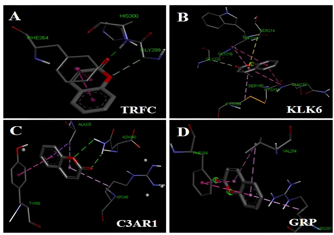 Fig. 15.
Molecular interaction between T. terrestris-mediated iron nanoparticles derived compounds with Adenomatous polyposis coli (APC) pathway proteins (A) TRFC (B) KLK6 (C) C3AR1 and (D) GRP.
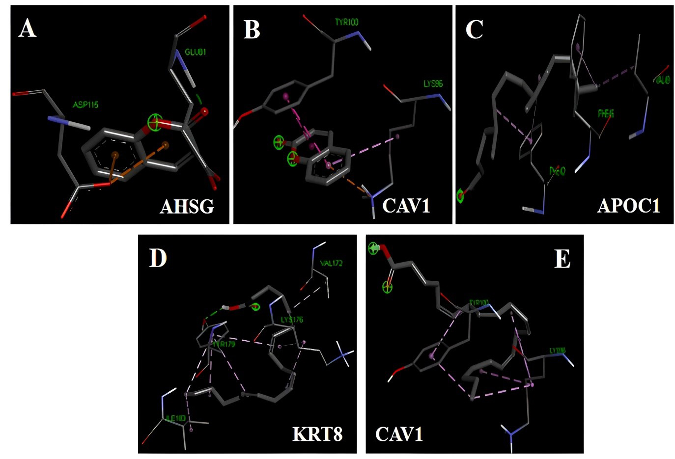 Fig. 16.
Molecular interaction between T. terrestris-derived compounds with Adenomatous polyposis coli (APC) proteins (A) AHSG, (B) CAV1, (C) APOC1, (D) KRT8, (E) CAV1.
| Compounds | KRT8 | TFRC | KLK6 | C3AR1 | AHSG | GRP | APOC1 | CAV1 |
| Stearic acid | –3.5 | –4.8 | –4.6 | –4.5 | –4.4 | –5.5 | –4 | –3.8 |
| 2-Hexanol,2-Methyl | –3.1 | –4.3 | –4.3 | –3.6 | –3.9 | –4.9 | –3.5 | –3.2 |
| Hexa decanoic acid | –3.3 | –5.7 | –4.8 | –4.3 | –4 | –5.6 | –3.9 | –3.7 |
| Octadecanoic acid | –3.4 | –5.1 | –4.2 | –4.4 | –3.1 | –5.6 | –3.9 | –3.8 |
| 2-Pentadecanone | –3.7 | –5.8 | –4.3 | –4.8 | –4.8 | –5.7 | –4.7* | –4.7 |
| Coumaric acid | –3.5 | –6.2* | –6* | –6* | –5.6* | –6.8* | –4.1 | –4.8* |
| Benzofuran | –3.6 | –5.2 | –5 | –5.9 | –5 | –5.6 | –3.5 | –4.3 |
| Archidic acid | –4.1* | –5.8 | –4.8 | –5.3 | –4.1 | –6.7 | –4.4 | –4.8 |
| 5 Fluorouracil** | –3.5 | –5.1 | –5.2 | –4.8 | –4.3 | –5 | –3.7 | –3.6 |
** Standard drug and * Highest binding affinity.
The green synthesized T. terrestris-Fe3O4-NPs are environmentally safe and pose no significant risks. Various characterization techniques were performed, such as UV spectroscopy, FTIR, XRD, SEM, TEM, and VSM. The Tt-Fe3O4-NPs demonstrated significant antioxidants, which is one of the main components required for DNA repair in the etiology of cancer. A lot of anticancer drugs work by inhibiting cancer cells through antioxidants. Many cancer cells are infected by microorganisms in advanced stages so the eradication of microorganisms from cancer cells, clinically is very challenging. Currently, only a few antibiotics are capable of killing microorganisms and cancer cells. The Tt-Fe3O4-NPs were able to kill the microorganism growth, which was confirmed through antimicrobial activity. Some of the toxic chemicals in the body cause inflammation, and normal cells are converted into cancer cells through inflammation. The synthesized nanoparticle has anti-inflammatory activity, as demonstrated by an anti-inflammation assay. Generally, normal cells have inflammation from some of the foreign agents and its converted cancer cells which is followed by microorganism infection. when normal cells have a deficiency of antioxidant capacity in the body to form inflammation, infection, and cancer. T. terrestris also exhibited notable anticancer efficacy against colon cancer cell lines through anticancer activity and scratch assays. The bioactive compound coumaric acid strongly binds to key proteins linked to colon cancer, such as TRFC, KLK6, C3AR1, and GRP, as shown by molecular docking studies. It also contains many secondary metabolites that interact with colon cancer protein markers. As a result, it is suggested that our nanoparticles can modulate the colon cancer signaling pathway. In this study, we confirmed our plant has anti-inflammation, antimicrobial, and anticancer activity, which is necessary for any anticancer drug. So, we believe this drug may be very optimum for colon cancer patients. This drug will be used in clinical trials in our future studies.
The datasets used and/or analyzed during the current study are available from the corresponding author on reasonable request.
Conceptualization: TP, GB, BKR; Methodology: RR, HS, GB; Analysis and interpretation of data: MG, SK; Data curations: MN, MRAW; writing-original draft preparation: RR; Writing-review and editing, interpretation of data: TP, MR; Visualization: TP, AS, RA; Supervision and data curation: TP, BKR, MR, SK, AS, WHA. All authors have participated sufficiently in the work to take public responsibility for appropriate portions of the content and agreed to be accountable for all aspects of the work in ensuring that questions related to its accuracy or integrity. All authors contributed to editorial changes in the manuscript. All authors read and approved the final manuscript.
Tribulus terrestris (Tt) was used in this study. It is a flowering plant with yellow flowers. The plant was collected from Pudhur and its surrounding areas in Chennai. It was identified as 387.1753 and authenticated by the Siddha Central Research Institute in Chennai.
We gratefully acknowledge Er. A.C.S. Arun Kumar, President, Dr. M.G.R. Educational and Research Institute, for providing the necessary facilities, and we acknowledge the Nanotechnology Research Centre (NRC), SRMIST, for providing an instrumentation facility for nanoparticle characterization. We would like to acknowledge the Researchers Supporting Project Number (RSP2025R293), King Saud University, Riyadh, Saudi Arabia.
This work was funded by the Researchers Supporting Project Number (RSP2025R293), King Saud University, Riyadh, Saudi Arabia.
The authors declare no conflict of interest.
References
Publisher’s Note: IMR Press stays neutral with regard to jurisdictional claims in published maps and institutional affiliations.