


 , Anastasia V. Prozorova 1, Iuliia G. Samoilova 1, Dmitry A. Svarovsky 1,2, Kira A. Sidorenkova 1, Liudmila V. Spirina 1,2,*
 , Marina N. Stakheeva 1,2, Oxana S. Timofeeva 1, Ilya A. Petrov 1
1 Division of Biochemistry and Molecular Biology, Federal State Budgetary Educational Institution of Higher Education “Siberian State Medical University” of the Ministry of Health of Russia, 634050 Tomsk, Russia
2 Laboratory of Tumor Biochemistry, Cancer Research Institute, Tomsk National Research Medical Center, 634009 Tomsk, Russia
Abstract
Over the past five years, the pregnancy rate in assisted reproductive technology (ART) programs in Russia has remained relatively stable. The aim of this study was to assess the distribution of monocyte and macrophage subsets in the blood and follicular fluid of infertile women undergoing assisted reproductive technology.
The study involved 45 women with a mean age of 35 ± 4.66 years. Monocytes and macrophages were identified using flow cytometry.
We observed a decrease in the CD68+CD163+CD206+ and the CD68+CD163–CD206+ cells in patients with a body mass index (BMI) >25 by 0.19 times and 6.56 times, respectively, compared to the group with a BMI <25 (p = 0.031). Patients with fair oocyte quality had 3.6 times more oocytes than those with poor quality (p = 0.010). The relative content of CD14+163–206+ monocytes was found to be 24.15 times higher in the follicular fluid of women with poor embryo quality compared to the group with good embryos (p = 0.010). We also noted that the number of oocytes increased in women with male factor infertility (p = 0.020) and those with unspecified infertility when compared to tubal infertility. An increase in the relative content of CD14+163+206+ in the blood was higher in women with other causes of female infertility compared to those with male factor infertility (p = 0.010). The relative content of M2-monocytes (CD14+163+206–) in the blood was 4.38 times higher in women with male factor infertility than in women with unexplained infertility (p = 0.010).
A critical component of the inflammatory reaction in patients undergoing in vitro fertilization (IVF) involves more than just the activation of pro-inflammatory cells in response to ovarian stimulation. Our research shows that changes in the distribution of monocytes and macrophages can influence embryo implantation success and pregnancy outcomes in women. These processes are influenced by various infertility-related factors, including those mentioned above. However, these findings are preliminary and require further investigation.
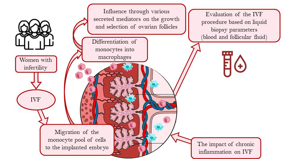
Graphical Abstract
Keywords
- follicular fluid
- macrophages
- monocytes
- oocytes
- assisted reproductive technology
- in vitro fertilization
- embryo competence
Over the past five years, the pregnancy rate in Russia for assisted reproductive technology (ART) has remained largely stable. According to the 2021 report from the ART registry of the Russian Association of Human Reproduction, the pregnancy rate for embryo transfers in in vitro fertilization (IVF) programs was 34.8%. It is clear that enhancing the effectiveness of infertility treatments through IVF is essential.
The quality of embryos is influenced by the properties of reproductive cells [1]. The maximum implantation potential can be achieved by evaluating the quality of oocytes [2]. It is a fact that the microenvironment and composition of follicular fluid have an impact on the completeness of oocytes [3]. Special attention is paid to identifying factors influencing the quality of oocytes and embryos in follicular fluid. The investigating the immune system’s role in the development of infertility holds promise [4]. There is evidence that a decline in ovarian reserve is linked to chronic sterile inflammation of the ovaries, associated with macrophage activity [5].
Research has demonstrated that macrophages participate in folliculogenesis by
secreting mediator substances [6]. Pro-inflammatory cytokines secreted by
macrophages, such as tumor necrosis factor
Monocytes are a type of white blood cell that play a crucial part in the immune system. They can be divided into different groups, each with distinct characteristics (M1 and M2 subpopulations). These groups exhibit different appearances and perform various functions. Understanding these distinctions during pregnancy can shed light on how the mother’s immune system interacts with the developing fetus [11]. The inflammatory process associated with M1 polarization of monocytes can significantly influence the success of ART, particularly in individuals who are overweight or have other immune-compromising factors. Therefore, macrophage and monocyte subsets can be considered as potential predictors of oocyte and embryo viability in in vitro fertilization (IVF) programs [12]. The aim of the study was to assess the distribution characteristics of monocyte and macrophage subpopulations in the blood and follicular fluid of women with infertility undergoing IVF procedures.
The study involved fifty women with an average age of 35
| Clinical signs | Female infertility of tubal origin | Female infertility associated with male factors | Other forms of female infertility | Unspecified female infertility |
| BMI |
14.29% (7) | 9.52% (5) | 21.43% (10) | 11.90% (6) |
| BMI |
13.64% (7) | 4.55% (3) | 18.18% (9) | 4.76% (3) |
| Excellent embryo quality | 14.29% (7) | 14.29% (7) | 23.81% (12) | 4.76% (4) |
| Good embryo quality | 4.76% (2) | 0.00% (0) | 4.76% (2) | 9.52% (4) |
| Fair embryo quality | 4.76% (2) | 0.00% (0) | 0.00% (0) | 4.76% (2) |
| Poor embryo quality | 4.76% (2) | 0.00% (0) | 9.52% (6) | 0.00% (0) |
| The number of embryos, n = 1–5 | 19.05% (10) | 9.52% (4) | 38.10% (19) | 9.52% (4) |
| The number of embryos, n |
9.52% (4) | 4.76% (3) | 4.76% (3) | 4.76% (3) |
BMI, body mass index; n, number of patients.
Furthermore, the participants were divided into four groups according to specific causes of infertility as classified by ICD-10: the first group consisted of 12 individuals with tubal infertility; the second group included 8 individuals with male factor infertility; and the fourth group included 7 individuals with an unknown cause of infertility. On the day of transvaginal follicle puncture, peripheral blood and follicular fluid samples were collected from all patients, following the guidelines of the ethics committee of the Siberian State Medical University, reviewed and approved by the Helsinki Declaration (Protocol of the Ethics Committee No. 9314 dated December 5, 2022).
The blood and follicular fluid aliquots (100 µL) from patients were
incubated with antibodies to CD14 (FITC Anti-Human CD14 Antibody, Elabscience,
Beijing, China, 1 µL/mL), CD163 (PE anti-human CD163 Antibody,
BioLegend, San Diego, CA, USA, 5 µL/mL), and CD206 (APC Anti-Mouse
CD206/MMR Antibody, Elabscience, Beijing, China, 1 µL/mL) in the
dark at room temperature for 20 minutes. Similarly, incubation was conducted with
a combination of antibodies to CD68 (FITC Anti-Human CD68 Antibody, Elabscience,
Beijing, China, 1 µL/mL), CD163 (PE anti-human CD163 Antibody,
BioLegend, San Diego, CA, USA, 5 µL/mL), and CD206 (APC Anti-Mouse
CD206/MMR Antibody, Elabscience, Beijing, China, 1 µL/mL) for whole
blood and follicular fluid. After incubation, 900 µL of red blood
cell lysis buffer (Erythrolyse red blood cell lysing buffer (10
 Fig. 1.
The determination of CD14+ and CD68+ cell subpopulations in blood and follicular fluid. (A) Forward (FSC-A) and side (SSC-A) light scattering, gate cells selected, indicating cells. (B) Singlet signals within all signals of the cells gate in the sample. (C) Histogram distribution of CD14– or CD68–positive cells within singlet signals of the sample. (D) CD163–positive cells within singlets. (E) CD206–positive cells within singlets. (F) Dual staining with antibodies against CD14 or CD68 and CD163 within singlets (quadrants highlight gates reflecting the ratio of cell subpopulations of the sample based on the content of CD14– or CD68–positive cells and CD163). (G) Histogram distribution of CD206–positive cells within CD14+CD163+ or CD68+CD163+. SSCA, strip spectral correlation algorithm; FSC-A, forward and side scatter plot; APC-A, allophycocyanin; FITC-A, fluorescein isothiocyanate; PE-A, phycoerythrin.
The statistical analysis of the data was conducted using Statistica
12.0 (TIBCO Software, Alto, CA, USA) and SPSS Statistics 28.0.1.0 (IBM, Armonk, NY, USA) software
packages. The Kolmogorov-Smirnov test was used to check for normal distribution.
The Mann-Whitney U test was used to assess the significance of differences in
unpaired groups. Correlation analysis using Spearman’s rank correlation
coefficient was performed to identify associations between the studied variables
and features of the reproductive system. Differences were considered significant
at p
Table 2 presents data on the ratio of monocyte and macrophage cell populations
in blood plasma and follicular fluid of infertility patients, categorized by BMI.
We observed a decrease in the cellular subpopulation with the phenotype
CD68+CD163+CD206+ and with the phenotype CD68+CD163–CD206+ in patients with a BMI
| Indicators | BMI |
BMI |
p |
| (Q1; Q3) | (Q1; Q3) | ||
| The number of oocytes, n | 8.00 (3.00; 14.00) | 8.50 (5.00; 14.00) | 0.592 |
| The number of embryos, n | 3.00 (1.50; 3.50) | 2.50 (1.50; 5.50) | 1.000 |
| The number of punctured follicles, n | 8.00 (5.00; 18.00) | 9.50 (6.00; 14.00) | 0.724 |
| CD14+163+206+ (М2-monocytes) blood, % | 81.94 (25.38; 95.98) | 59.38 (29.03; 94.96) | 0.683 |
| CD14+163–206+ (monocytes) blood, % | 14.29 (0.13; 99.47) | 7.05 (1.85; 98.79) | 0.901 |
| CD14–163+206+ blood, % | 15.38 (6.55; 76.27) | 50.23 (2.89; 90.91) | 0.845 |
| CD14+163+206– (М2-monocytes) blood, % | 18.06 (4.02; 74.62) | 40.62 (5.04; 70.97) | 0.683 |
| CD68+CD163+CD206+ blood, % | 16.57 (11.52; 23.00) | 13.45 (0.00; 15.48) | 0.031 |
| CD68+CD163–CD206+ blood, % | 6.56 (4.24; 9.45) | 0.00 (0.00; 5.41) | 0.009 |
| CD68–CD163+CD206+ blood, % | 6.00 (5.00; 11.00) | 8.00 (0.00;13.00) | 0.901 |
| CD68+CD163+CD206– blood, % | 15.05 (6.96; 17.99) | 14.48 (0.00; 22.48) | 1.000 |
| CD14+163+206+ (М2-monocytes) follicular fluid, % | 67.35 (17.03; 85.42) | 61.36 (46.49; 73.75) | 0.958 |
| CD14+163–206+ (monocytes) follicular fluid, % | 3.53 (0.39; 62.71) | 3.83 (0.00; 16.44) | 0.327 |
| CD14–163+206+ follicular fluid, % | 5.27 (0.00; 54.55) | 33.79 (3.93; 64.44) | 0.118 |
| CD14+163+206– (М2-monocytes) follicular fluid, % | 25.89 (11.46; 35.62) | 28.59 (25.62; 31.62) | 0.157 |
| CD68+CD163+CD206+ (М2-macrophages) follicular fluid, % | 69.86 (62.76; 88.57) | 76.66 (71.89; 83.26) | 0.488 |
| CD68+CD163–CD206+ (macrophages) follicular fluid, % | 67.17 (64.87; 80.62) | 68.57 (66,69; 75.86) | 0.958 |
| CD68–CD163+CD206+ follicular fluid, % | 23.85 (16.86; 27.69) | 19.23 (8,13; 26.27) | 0.261 |
| CD68+CD163+CD206– (М2-macrophages) follicular fluid, % | 28.38 (11.43; 43.56) | 26.47 (16,74; 39.56) | 1.000 |
Me, median; p, significance level; n, number.
Table 3 presents the ratios of monocyte-macrophage subpopulations in the blood and follicular fluid of infertility patients. An increased number of oocytes was found in patients with fair quality oocyte compared to poor quality oocytes (p = 0.010). Specifically, there was a 0.68-fold decrease in the number of good quality oocytes (p = 0.004) and a 0.44-fold decrease in excellent quality oocytes (p = 0.011) when compared to fair quality oocytes. Additionally, there were five times more fair quality embryos than poor quality embryos (p = 0.038). The fair quality embryos also showed a 2.57-fold increase compared to the poor quality group (p = 0.010). Patients with excellent quality embryos had 0.39 times more punctured follicles compared to those with fair quality embryos (p = 0.026).
| Indicators | Poor quality of embryos | Fair quality of embryos | Good quality of embryos | Excellent quality of embryos |
| The number of oocytes, n | 5.00 (3.00; 12.00) | 18.00 (18.00; 18.00) | 7.50 (5.50; 11.50)** | 10.00 (4.00; 14.00) |
| The number of embryos, n | 1.00 (1.00; 3.00) | 5.00 (3.00; 7.00) | 2.50 (1.50; 4.50) | 3.00 (1.00; 4.00) |
| The number of punctured follicles, n | 7.00 (5.00; 12.00) | 18.00 (18.00; 18.00) | 8.00 (6.50; 14.00) | 11.00 (4.00; 15.00)*** |
| CD14+163+206+ (М2-monocytes) blood, % | 38.66 (21.84; 99.18) | 88.45 (81.94; 94.96) | 89.65 (88.33; 90.96) | 29.22 (10.83; 89.98) |
| CD14+163–206+ (monocytes) blood, % | 56.23 (0.13; 100.00) | 50.87 (2.94; 98.79) | 49.39 (0.06; 98.72) | 7.05 (2.19; 65.45) |
| CD14–163+206+ blood, % | 40.82 (0.02; 96.99) | 32.64 (6.55; 58.73) | 49.36 (6.82; 91.89) | 13.25 (1.23; 75.86) |
| CD14+163+206– (М2-monocytes) blood, % | 61.34 (0.82; 78.16) | 11.55 (5.04; 18.06) | 13.43 (9.04; 17.81) | 68.09 (10.03; 89.18) |
| CD68+CD163+CD206+ blood, % | 14.50 (8.93; 16.57) | 18.50 (18.24; 18.75) | 20.96 (18.91; 23.00) | 14.42 (5.76; 17.22)*** |
| CD68+CD163–CD206+ blood, % | 6.80 (6.20; 12.06) | 9.11 (6.90; 11.31) | 3.80 (3.35; 4.24)** | 4.46 (0.00; 7.43)*** |
| CD68–CD163+CD206+ blood, % | 7.00 (6.00; 11.00) | 6.50 (6.00; 7.00) | 8.00 (6.00; 10.00) | 8.50 (2.00; 12.50) |
| CD68+CD163+CD206– blood, % | 15.05 (12.42; 17.99) | 12.03 (11.50; 12.56) | 13.86 (12.11; 15.60) | 16.67 (2.31; 22.73) |
| CD14+163+206+ (М2-monocytes) follicular fluid, % | 40.41 (17.03; 85.42) | 70.55 (67.35; 73.75) | 58.34 (46.97; 69.70) | 47.73 (17.01; 74.07) |
| CD14+163–206+ (monocytes) follicular fluid, % | 4.83 (0.55; 82.76) | 9.36 (2.27; 16.44)* | 0.20 (0.00; 0.39) | 6.34 (0.75; 37.65)*** |
| CD14–163+206+ follicular fluid, % | 0.73 (0.00; 29.93) | 33.52 (5.27; 61.76) | 37.60 (20.65; 54.55) | 3.60 (0.00; 61.52) |
| CD14+163+206– (М2-monocytes) follicular fluid, % | 27.21 (0.00; 38.13) | 32.97 (26.25; 39.69) | 28.10 (25.89; 30.30) | 26.82 (17.96; 33.62) |
| CD68+CD163+CD206+ (М2-macrophages) follicular fluid, % | 65.90 (63.29; 86.30) | 79.50 (66.93; 92.06) | 81.32 (73.50; 89.13) | 71.40 (60.90; 80.11) |
| CD68+CD163–CD206+ (macrophages) follicular fluid, % | 64.87 (59.38; 65.44) | 83.20 (66.40; 100.00) | 72.35 (65.74; 78.95) | 68.76 (64.73; 78.24) |
| CD68–CD163+CD206+ follicular fluid, % | 17.21 (0.00; 21.03) | 29.49 (16.86; 42.11) | 11.93 (0.00; 23.85) | 23.99 (17.09; 27.18) |
| CD68+CD163+CD206– (М2-macrophages) follicular fluid, % | 35.17 (13.70; 39.87) | 25.75 (7.94; 43.56) | 16.93 (10.87; 22.98) | 30.29 (21.23; 42.74) |
*, significance of difference between the poor and fair quality of embryos; **, significance of difference between the fair and good quality of embryos; ***, significance of difference between the good and excellent quality of embryos; n, number.
The ratio of the CD68+CD163+CD206+ subpopulation in the blood was significantly different between groups of patients with fair and good embryos compared to those with poor-quality embryos. Women with good-quality embryos exhibited a higher relative content of this subpopulation than those with fair quality embryos. The group of good embryos showed a 0.21-fold increase in CD68+CD163+CD206+ cells compared to the fair embryo group (p = 0.035).
A decrease in CD68+CD163–CD206+ cells was identified in embryos with good and excellent quality (by 0.44 times and 0.34 times, respectively), compared to the group with poor quality embryos. Compared to embryos of fair quality, the groups with good and excellent embryos had a lower relative content of CD68+CD163–CD206+.
The follicular fluid had higher levels of CD14+163–206+ monocytes in the group with poor embryo quality than in those with good quality ones. Fair quality embryos demostrated the increased level of CD14+163–206+ monocytes compared to embryos with good quality. The high CD68+CD163–CD206+ macrophage level in the follicular fluid was detected in the groups with fair, good, and excellent embryo quality by 1.28 times (p = 0.010), 1.12 times (p = 0.010), and 1.06 times (p = 0.038) respectively, compared to the poor embryo quality ones. This was associated with a 1.39-fold increase in the relative level of CD68–CD163+CD206+ cells in the follicular fluid of the poor embryo quality group compared to the good embryo quality group (p = 0.029). CD68+CD163+CD206–macrophages tended to increase by 1.79 times in the group with excellent quality embryos compared to those with good quality embryos (p = 0.035).
Women with male factor infertility experienced a 4.14 increase in oocyte numbers (p = 0.020) compared to the group with tubal infertility (Table 4). In the group with unspecified infertility, the number of oocytes was 4.29 times higher (p = 0.041) than in the tubal infertility group. When compared to the other female infertile group, women with infertility associated with male factors had a 2.42 times increase in the number of oocytes. The number of oocytes in women with male factor infertility increased by 2.42 times compared to other forms of female infertility (p = 0.002).
| Indicators | Tubal female infertility, Me (Q1; Q3) | Female infertility associated with male factors, Me (Q1; Q3) | Other forms of female infertility, Me (Q1; Q3) | Unspecified female infertility, Me (Q1; Q3) |
| The number of oocytes, n | 3.50 (3.00; 13.00) | 14.50 (10.50; 16.50)* | 6.00 (5.00; 9.00)** | 15.00 (8.00; 18.00)***,# |
| The number of embryos, n | 1.00 (1.00; 6.00) | 3.00 (2.00; 8.00)* | 2.50 (1.50; 3.50) | 3.00 (3.00; 6.00)# |
| The number of punctured follicles, n | 5.00 (3.00; 13.00) | 16.50 (11.00; 18.00)* | 7.00 (5.00; 9.00) | 18.00 (8.00; 20.00)**,***,# |
| CD14+163+206+ (М2-monocytes) blood, % | 29.41 (12.28; 38.66) | 22.09 (9.38; 34.79) | 83.97 (25.38; 98.71) | 88.33 (81.94; 90.96)*** |
| CD14+163-206+ (monocytes) blood, % | 2.53 (1.85; 14.29) | 0.23 (0.00; 0.45)* | 56.23 (7.31; 99.47)** | 2.94 (0.06; 98.72),# |
| CD14–163+206+ blood, % | 15.38 (0.00; 40.82) | 26.42 (11.11; 41.73)* | 76.27 (2.09; 91.76) | 6.82 (6.55; 91.89) |
| CD14+163+206– (М2-monocytes) blood, % | 61.34 (52.94; 87.72) | 77.92 (65.21; 90.63) | 16.03 (1.29; 74.62) | 17.81 (9.04; 18.06)*** |
| CD68+CD163+CD206+ blood, % | 11.52 (8.93; 18.24) | 7.57 (0.00; 15.13) | 14.50 (11.76; 15.97) | 18.91 (18.75; 23.00)***,# |
| CD68+CD163–CD206+ blood, % | 6.20 (4.49; 11.31) | 2.71 (0.00; 5.41) | 6.80 (0.00; 9.45) | 4.24 (3.35; 6.90) |
| CD68–CD163+CD206+ blood, % | 7.00 (6.00; 8.00) | 7.50 (0.00; 15.00) | 9.00 (4.00; 12.00) | 6.00 (6.00; 10.00) |
| CD68+CD163+CD206– blood, % | 17.99 (12.56; 18.32) | 13.53 (0.00; 27.06) | 12.42 (4.61; 16.39) | 12.11 (11.50; 15.60) |
| CD14+163+206+ (М2-monocytes) follicular fluid, % | 17.03 (0.00; 67.35) | 33.59 (20.69; 46.49)* | 73.75 (40.41; 85.42) | 67.35 (46.97; 69.70)*** |
| CD14+163-206+ (monocytes) follicular fluid, % | 0.55 (0.00; 16.44) | 18.39 (1.49; 35.29) | 6.17 (3.53; 62.71) | 0.39 (0.00; 2.27)# |
| CD14–163+206+ follicular fluid, % | 0.00 (0.00; 29.93) | 2.91 (0.00; 5.81) | 3.93 (0.73; 64.44) | 20.65 (5.27; 54.55) |
| CD14+163+206– (М2-monocytes) follicular fluid, % | 26.25 (21.97; 27.21) | 55.12 (30.92; 79.31) | 25.62 (11.46; 35.62) | 30.30 (25.89; 39.69) |
| CD68+CD163+CD206+ (М2-macrophages) follicular fluid, % | 63.29 (59.04; 69.86) | 44.28 (12.20; 76.36)* | 76.96 (65.90; 86.30) | 73.50 (66.93; 89.13) |
| CD68+CD163–CD206+ (macrophages) follicular fluid, % | 72.76 (64.87; 80.62) | 58.40 (50.00; 66.79) | 67.17 (65.44; 75.86) | 66.40 (65.74; 78.95) |
| CD68–CD163+CD206+ follicular fluid, % | 24.47 (17.21; 27.69) | 38.43 (26.27; 50.59) | 21.03 (0.00; 23.50) | 16.86 (0.00; 23.85) |
| CD68+CD163+CD206– (М2-macrophages) follicular fluid, % | 35.17 (32.19; 45.91) | 63.68 (39.56; 87.80) | 25.71 (13.70; 28.38) | 22.98 (10.87; 43.56) |
*, significance of difference between the tubal female infertility and the female infertility associated with male factors; **, significance of difference between the tubal female infertility and the other forms of female infertilety; ***, significance of difference between the female infertility associated with male factors and the unexplained female infertility; #, significance of difference between the other forms of female infertility and the unexplained female infertility.
Women with male factor infertility saw an increase of 3.3 times (p = 0.012) in punctured follicles when compared to the tubal infertility group. The number of detected oocytes was 3.6 times higher in the group with unspecified infertility than in the group with tubal infertility. The number of punctured follicles increased by 2.36 times in women with male factor infertility compared to other forms of female infertility, as found. Furthermore, women with other forms of female infertility had a 0.61 times lower number of follicles than those with infertility of unspecified etiology.
In women with other forms of female infertility, the number of CD14+163+206+ cells in the blood increased by 3.8 times when compared to those with male factor infertility. This pattern was also observed in women with tubal infertility. These women had a higher percentage of CD14+63–206+ cells in their blood compared to the male factor infertility group (p = 0.024) compared to the other forms of infertility. The group with other forms of female infertility showed an increase in cell numbers (p = 0.006), but these cells decreased by 0.96 times (p = 0.006).
The study revealed that the number of M2-monocytes in women with male factor infertility decreased by 0.996 times compared to women with unexplained infertility. The relative content of M2-monocytes in the group with male factor infertility was 19.19 times higher than in women with unexplained infertility (p = 0.033). In addition, women with unexplained infertility had 4.96 times more CD14–163+206+ cells in their blood compared to those with tubal infertility. In women with male factor infertility, the proportion of CD14+163+206– cells in their blood increased by 4.38 times compared to those without infertility.
The male factor infertility group showed a 0.6 times reduction in the presence of the CD68+CD163+CD206+ subpopulation in their blood, which was detected compared to the unspecified infertility. This higher occurrence was attributed to women with various types of female infertility rather than those with unspecified causes.
The CD14+163+206+ cells in the blood decreased by 0.77 times in the tubal infertility group compared to the other female infertility groups. These cells decreased by 0.09 times (p = 0.010) when compared to the other forms of female infertility in the unspecified group. A similar trend was observed in the relative content of CD14+163–206+ cells in the follicular fluid. According to the findings, the rate of female infertility increased by 15.82 times among women with other forms of female infertility compared to women with unexplained infertility.
When comparing women with tubal infertility to those with other types of female infertility, it was found that the levels of M2-macrophages in their follicular fluid increased by 1.22 times. The follicular fluid showed a 0.45 times decrease in CD68–CD163+CD206+ cells in women with other forms of female infertility compared to male factor infertility. This indicator was 0.56 times higher in the group with unspecified female infertility than in the group with male factor infertility.
Women with tubal infertility experienced a 0.45 times reduction in the CD68+CD163+CD206– content in follicular fluid, as evidenced by the study. In women with male factor infertility, there was a 2.48 increase in the number of these cells when compared to women experiencing infertility for other reasons.
Fig. 2 presents a heatmap with annotations that illustrates the correlation among various biological variables related to oocytes and embryos, punctured follicles, and specific cellular markers in blood and follicular fluid. This heat map aims to highlight the associations and statistical significance of these variables. Both the X and Y axes of the heat map represent the same set of biological variables, including counts of oocytes, embryos, punctured follicles, as well as cellular markers such as CD14+, CD163+, CD206+, and CD68+ in both blood and follicular fluids.
 Fig. 2.
An annotated heat map of the correlation between the relative
content of monocytes and macrophages in blood and follicular fluid, as well as
in vitro fertilisation (IVF) parameters, for groups of patients with different embryo qualities and
infertility. Note: (A) Group of female infertility due to tubal factors. (B)
Male factor female infertility. (C) Other forms of female infertility. (D)
Unspecified female infertility. * p-value
The colors in the heatmap range from blue to red, with red indicating a strong
positive correlation and blue signifying a strong negative correlation.
White/light colors indicate weak or no correlation. A color legend on the right
side of the heatmap explains this gradient scale. Each cell in the map is
annotated with the corresponding correlation coefficient value. Asterisks denote
the level of statistical significance: one asterisk for p
The data used to create this heatmap provides valuable insights into the biological processes that underlie successful oocyte retrieval and embryonic development. The strong positive correlations between the number of oocytes, embryos, and follicles indicate that these factors are interdependent and influence each other in reproductive outcomes. Additionally, correlations between cell markers in blood and follicular fluid and reproductive factors may point to key biomarkers for reproductive health. This detailed heatmap analysis provides a comprehensive view of the relationships between various reproductive and cellular factors, underscoring the importance of understanding these interactions to enhance reproductive health.
To further explore the correlation between monocyte and macrophage subpopulations and the success of assisted reproductive techniques, we conducted an in-depth analysis to identify the key factors associated with successful implantation, fetal viability, and pregnancy outcomes (Table 5).
| Pregnancy | n | Chi-square | |||
| no | yes | df | |||
| p | |||||
| Embryo implantation | no | 10 | 0 | 10 | 4.861111 df = 1 p = 0.02747 |
| 20.00% | 0.00% | 20.00% | |||
| yes | 26 | 14 | 40 | ||
| 52.00% | 28.00% | 80.00% | |||
| Infertility factor | Tubal female infertility | 14 | 0 | 14 | 19.11111 df = 3 p = 0.00026 |
| 29.17% | 0.00% | 29.17% | |||
| Female infertility associated with male factors | 10 | 2 | 12 | ||
| 20.83% | 4.17% | 25.00% | |||
| Other forms of female infertility | 6 | 12 | 18 | ||
| 12.50% | 20.83% | 33.33% | |||
| Unspecified female infertility | 6 | 0 | 6 | ||
| 12.50% | 0.00% | 12.50% | |||
| Quality of embryos | Excellent quality of embryos | 16 | 14 | 30 | 12.96296 df = 3 p = 0.00472 |
| 32.00% | 28.00% | 60.00% | |||
| Good quality of embryos | 8 | 0 | 8 | ||
| 16.00% | 0.00% | 16.00% | |||
| Fair quality of embryos | 4 | 0 | 4 | ||
| 8.00% | 0.00% | 8.00% | |||
| Poor quality of embryos | 8 | 0 | 8 | ||
| 16.00% | 0.00% | 16.00% | |||
It has been observed that the success rate of embryo transfers resulting in pregnancy is 28% for women undergoing IVF treatment. Additionally, 80% of these women experienced successful transfers. Remarkably, successful transfers were achieved in 28% of cases involving embryos of exceptional quality. Among these cases, 12 individuals (85%) had a history of infertility with unknown causes, while 5% were related to male factor infertility.
The activation of monocytes and macrophages, accompanied by their redistribution in follicular fluid, holds prognostic potential for assessing the success of assisted reproductive technologies (ART). Metabolic disorders, particularly obesity, are associated with a relative reduction in circulating monocyte subpopulations characterized by the CD68+CD163+CD206+ and CD68+CD163–CD206+ phenotypes in individuals with a body mass index (BMI) exceeding 25.
In this study, we observed a significant increase in the relative content of CD14+CD163–CD206+ monocytes in follicular fluid among women with poor embryo quality, specifically by 24.15-fold compared to those with good embryos, which was statistically significant (p = 0.010). We also observed a notable elevation in the level of CD14+CD163–CD206+ cells in women with fair embryo quality, reaching a remarkable 46.8-fold increase compared to those with high-quality embryos (p = 0.029).
Furthermore, we discovered that the number of oocytes was higher in women experiencing infertility attributed to male factors (p = 0.020) compared to those experiencing tubal infertility. A statistically significant increase in the proportion of CD14+163+206+ cells in the bloodstream was observed in the group experiencing infertility from other causes compared to those with male factor infertility (p = 0.010).
Moreover, the relative abundance of M2 monocytes (CD14+163+206–) was found to be 4.38 times higher in women with male factor infertility than in those with unexplained infertility (p = 0.010). We also observed a decrease in the proportion of M2 macrophages (CD68+CD163+CD206–) within the follicular fluid in women with tubal infertility, showing a decrease of 0.45-fold compared to the group with male factor infertility (p = 0.026).
Monocytes are a type of white blood cell that play an important role in the immune system. They can be divided into several different subpopulations, each with distinct characteristics. These subpopulations exhibit different phenotypes and functions, and understanding their roles during pregnancy can provide insights into the complex interactions between a mother’s immune system and her developing fetus (Fig. 3).
 Fig. 3.
The monocytes and macrophages in blood and follicular fluid in
women with obesity. Note: The assessment of inflammatory activity and the
distribution of monocytes and macrophages in blood and follicular fluid has
potential for predicting the success of IVF treatments. High-quality embryos are
more likely to be obtained from women with higher levels of M2-type monocytes and
M2-macrophages in their follicular fluid. We observed an increase in the number
of CD68+, CD163+, and CD206– macrophages in the follicular fluid of women with
tubal infertility, suggesting that these cells may play a role in embryo
development. These findings are preliminary and require further research to
confirm their significance. M-CSF, macrophage colony stimulating factor; GM-CSF,
granulocyte macrophage colony-stimulating factor; LPS, lipopolysaccharide; INF
Classical monocytes (CD14+/CD16–) are vital for establishing immune tolerance during pregnancy by phagocytizing apoptotic cells and debris at the maternal-fetal interface [12, 14]. They also play a key role in regulating inflammation and tissue repair, which are essential for sustaining a healthy pregnancy. During viral infections, the number of classical monocytes can increase in pregnant women, as they contribute to the immune response to pathogens and assist in immune surveillance [15]. Intermediate monocytes, which express both CD14 and CD16, represent a transitional stage between classical and non-classical monocytes. These cells may be involved in immune surveillance and help maintain tissue homeostasis. They activate and promote the proliferation of T helper cells, which are crucial for an effective immune response to infections [16].
There is a growing interest in altering the inflammatory cell pool in women with infertility and obesity who are undergoing IVF procedures. The potential negative outcomes of pregnancy after IVF in obese patients may be associated with chronic inflammation. For instance, a study [17] found that as body mass index (BMI) increased, the rates of implantation and live births decreased. Our research indicated that obese patients experienced a relative reduction in the CD68+CD163+CD206+ and CD68+CD163–CD206+ blood cell subpopulations. These cells are likely to be in the monocyte lineage, as monocytes can migrate into tissues and differentiate into macrophages, acquiring specific macrophage markers, including CD68 [18]. Therefore, the decrease may be associated with a redistribution of the monocyte cell pool in the body due to ongoing chronic inflammation associated with obesity, potentially diminishing the chances of a successful IVF outcome.
Inflammatory processes associated with viral infections, such as SARS-CoV-2, and the reproductive system [19, 20], can impact the quality of IVF procedures. The relationship between embryo quality and the content of follicular fluid M2 monocytes and M2 macrophage is particularly intriguing. Notably, there was a significant increase in the proportion of monocytes with the CD14+163–206+ phenotype in follicular fluid. This increase was observed in groups with poor and moderate embryo quality (by 24.15 times and 46.8 times, respectively) compared to the group with good embryos. This suggests that the immune system plays a vital role in women’s fertility. Possibly, the relative increase in this cell subpopulation is linked to the fact that during IVF, embryos are alloantigenic to the woman’s immune system, leading to a redistribution of this cell pool [21]. The current study indicates that changes in the subpopulation structure of immune-competent cells towards the predominance of inflammatory subpopulations in the acute environment may determine the success of embryo implantation in women [22]. A separate assessment of changes in the surface marker CD163 expression, as a marker of M2 polarization of monocytes and macrophages, may also indicate a positive pregnancy outcome. There is evidence supporting the hypothesis of an increase in the expression of M2 anti-inflammatory macrophages as a compensatory mechanism for dealing with the alloantigenicity of embryos in IVF. In other words, M2 macrophages serve as controlled suppressors of the immune response [21]. Furthermore, M2 polarization may reduce the risks of pregnancy complications. Disruption in the control of CD68+ M1 macrophage expression could lead to pregnancy complications by causing degradation of the vascular-stromal component of chorionic villi, resulting in their involution [23].
The observed decrease in the relative content of M2 macrophages CD68+CD163+CD206– in the follicular fluid in the group of tubal infertility may be associated with the activation of pro-inflammatory macrophages. M2 macrophage polarization could play a key role in triggering, as successfully demonstrated in murine tuberculosis-associated immune reconstitution inflammatory syndrome (TB-IRIS) models, changes in the pro-inflammatory functions of CD4+ T-cells largely contributed to resident macrophages [20]. When discussing CD68+ macrophages, it is worth noting that their placental expression not only has pro-inflammatory effects but also facilitates vertical transmission of viral loads from mother to fetus, as seen in cases involving SARS-CoV-2 [24].
Local inflammation, characterized by the redistribution of cells from the monocyte-macrophage pool in patients undergoing IVF procedures, typically poses a negative impact on pregnancy outcomes. In addition to evaluating immune cell surface markers, molecular-genetic tests that assess mRNA expression of cytokine genes (IL1B, IL8, IL10, IL18, TNFa, TGFB1), transcription factors (TBX21, GATA3, RORC2), and toll-like receptors (TLR2, TLR4) may serve as promising alternative markers [25].
A study by Elisabeth R. Krow-Lucal et al. [26], published in 2014, presents interesting data regarding the JAK/STAT signaling pathway. The study evaluated mononuclear cells and found that IL-6 induces STAT3 phosphorylation in both fetal and mature monocytes [26]. This suggests an enhancement of cytokine signaling and a variation in monocyte function, which could influence cell proliferation, apoptosis, and overall survival. These processes are crucial for embryo development and sensitive to signaling molecules [27].
The migration of monocytes plays a crucial role in the development of the embryo’s vascular supply. Trophoblasts express vasoactive intestinal peptide (VIP), which stimulates the migration of monocytes by increasing the production of chemotactic factors CCL2 and CCL3 in decidual tissues. This has been demonstrated in the study by C. Zhang et al. in 2022 [28]. On the other hand, a decrease in monocyte count can lead to reduced angiogenesis. This was shown in a study by C. Zhang et al. [28] in 2022. A decrease in microsomal count affects angiogenesis and is also accompanied by a reduction in the number of monocytes [28].
An essential aspect for further research is the role of adipose tissue in the interaction between the embryo and monocytes. Adipose tissue is structurally diverse and can contain up to 50% monocytes and macrophages [29]. There is evidence suggesting that obesity in pregnant women enhances the antimicrobial activity of fetal monocytes [12]. One potential mechanism through which monocytes might influence embryo development is through the expression of extracellular vesicles. These vesicles can be produced by both monocytes and adipocytes and may serve as a means of communication between them, sharing crucial signaling molecules that certainly influence embryo development.
The small sample size used in the study is justified by the careful selection of patients in order to eliminate the influence of factors that might be present in a larger group and are highly heterogeneous. This strategy allows us to focus on critical aspects of the relationship between the immune system and immune function. It is important to note that this approach has proven effective. Variability within a population is known to be quantified by its standard deviation. The standard deviation of a sample is a measure of how much the results of the sample might differ from the true results for the entire population. While collecting a larger sample can enhance the accuracy of findings, it may also introduce greater variability, potentially skewing the data.
The quality of oocytes is a crucial factor in assisted reproductive technology (ART), and several key elements influence it. These include the number of oocytes, embryos, and punctured follicles, along with various cellular markers found in both blood and follicular fluid. Our correlation analysis shows a significant relationship that sheds light on how the immune system affects reproductive health issues in women. We found that metabolic factors in biological fluids can influence oocyte quality [30]. Pathologies of the reproductive system from various origins have a typical biological effect due to an immune system response. It is important to note that the age of the woman and the duration of her infertility are the key factors that contribute to treatment failure [31]. Our analysis of monocyte and macrophage distribution in follicular fluid and serum has revealed a common immune response mechanism for assisted reproductive technology (ART) procedures. Interestingly, our findings did not correlate with the cause of infertility but rather influenced the number and quality of oocytes following ART.
While this study has several strengths, it also has some limitations that need to be considered. The small sample size of fifty participants limits the generalizability of the findings, as it may not be representative of the wider population. Furthermore, the stratification of participants based on BMI and infertility factors was not fully explored, potentially limiting the ability to draw conclusions about potential differences in molecular mechanisms between different groups of patients. Another limitation of the study was the use of flow cytometry for sample preparation, which requires volumetric sampling. This can be challenging due to factors such as instrument calibration, the quality of blood and follicular fluid samples, nonspecific binding, and inadequate antibody binding, all of which can affect the accuracy of the results.
Thus, immune tolerance is essential for a successful pregnancy, as it protects the developing fetus from infection. Various immune cells, such as T cells, B cells, natural killer cells (NK cells), monocytes, macrophages, and dendritic cells, play a crucial role in regulating the immune response between the mother and fetus [32]. Understanding the characteristics and functions of these cells in peripheral blood can enhance the effectiveness of assisted reproductive technologies. For example, NK cells may be involved in preeclampsia, while monocytes and tissue macrophages trigger changes in the immune system [33, 34]; however, more data on their roles in infertility is needed. Further research into changes in phagocyte function could help clarify these findings. In addition, data analysis has highlighted the importance of immune response in developing high-quality embryos, achieving successful implantation, and sustaining pregnancy. It is worth noting that observed redistribution of monocytes and macrophages in individuals with male factor infertility and unexplained infertility (other factors) could serve as a valuable tool for predicting the success of assisted reproductive technologies.
The assessment of the inflammatory activity and the distribution of monocytes and macrophages in the blood and follicular fluid holds prognostic potential for evaluating the success of in vitro fertilization (IVF) procedures. Metabolic disorders and obesity are associated with a relative reduction in specific subpopulations of blood monocytes, namely those with the CD68+CD163+CD206+ and CD68+CD163–CD206+ phenotypes, in individuals with a body mass index (BMI) over 25.
Interestingly, women who have higher levels of M2 monocytes and M2 macrophages in their follicular fluid tend to produce high-quality embryos. Our study has found an increase in the concentration of M2 macrophages (CD68+CD163+CD206–) in the follicular fluid among women with tubal infertility. However, the role of M2-polarized cells within the monocyte-macrophage population is still unclear, with evidence pointing to both positive and negative effects on pregnancy outcomes following IVF treatment. A critical factor in the inflammatory activity of individuals undergoing IVF is not only the activation of pro-inflammatory cells in response to oocyte donation but also underlying inflammatory alterations, such as persistent inflammation associated with obesity or metabolic disorders, as well as varying durations of viral exposure from past illnesses. The study has demonstrated that alterations in the distribution of monocytes and macrophages may impact the success of embryonic implantation and subsequent pregnancy in women. These processes are also influenced by various factors related to infertility. It is important to note that these findings are preliminary and require further exploration.
ART, Assisted Reproductive Technologies; IVF, in vitro fertilization.
The data are not publicly available due to privacy or ethical restrictions, but the data presented in this study are available on request from the corresponding author.
EDM and DAS performed the research. IAP, OST, and AVP designed the research study. IGS and KAS provided help and advice on data analysis. LVS and MNS provided laboratory tests and wrote the manuscript. All authors contributed to editorial changes in the manuscript. All authors read and approved the final manuscript. All authors have participated sufficiently in the work and agreed to be accountable for all aspects of the work.
All studies were conducted in accordance with the rules of the Ethics Committee of the Siberian State Medical University and the Helsinki Declaration (protocol of the Ethics Committee No. 9314 dated 05.12.2022). Written informed consent has been obtained from the patients or their families/legal guardians to publish this paper.
Not applicable.
This research received no external funding.
Given her role as the Guest Editor member, Liudmila V. Spirina had no involvement in the peer-review of this article and has no access to information regarding its peer review. Full responsibility for the editorial process for this article was delegated to Graham Pawelec. The authors declare no conflict of interest.
References
Publisher’s Note: IMR Press stays neutral with regard to jurisdictional claims in published maps and institutional affiliations.